
MRCP-rundum: IPMN u.a. (muzinöse) zystische Pankreasläsionen
Kittner T, Klinik für Radiologie Krankenhaus Dresden-Friedrichstadt
Städtisches Klinikum Akademisches Lehrkrankenhaus der TU Dresden
Anforderungen an die radiologische Diagnostik
Detektion (nur 5% aller Pankreas-Tu) Artdiagnose, Dignität Resektabilität T und NM anatomische Varianten (A.hep. dex. in 25% aus AMS)
Methoden
Sonographie (Endosonografie incl. Biopsie) Computertomographie MR-Tomographie incl. MRCP und MRA -Verfügbarkeit Angiographie (DSA) ERCP FDG PET PET CT PET-MRT
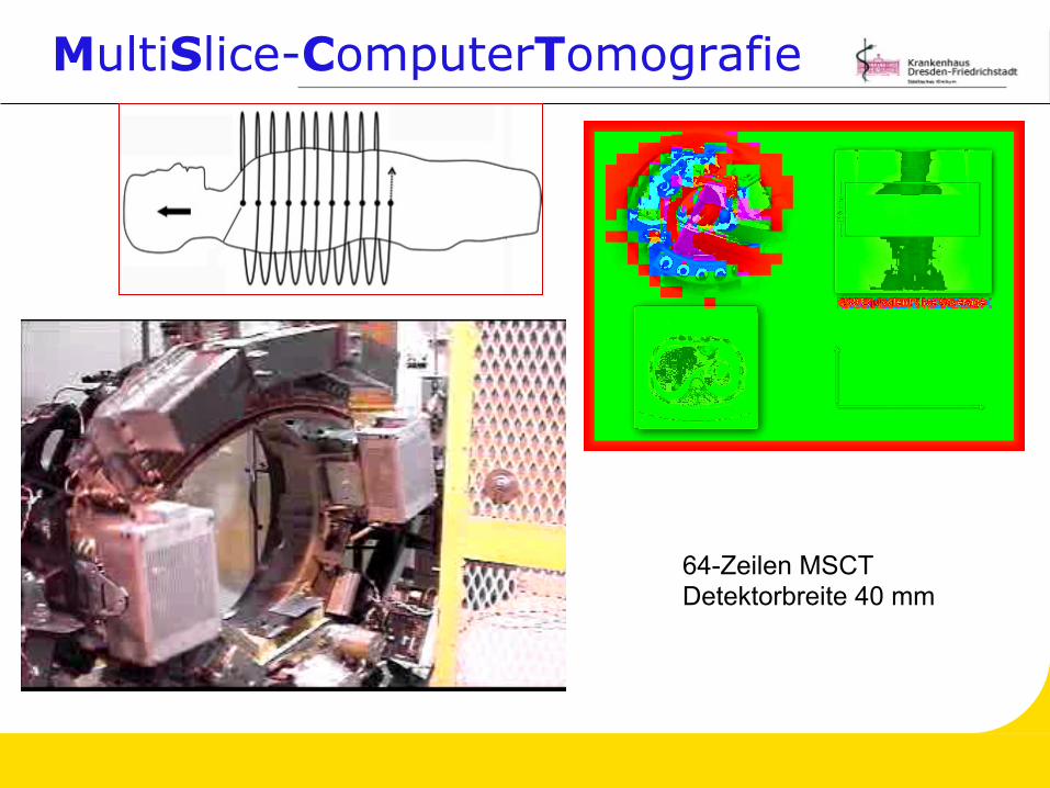
MultiSlice-ComputerTomografie
64-Zeilen MSCT Detektorbreite 40 mm
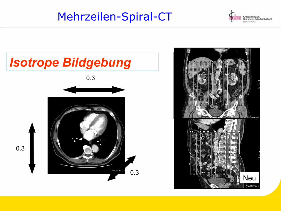
Mehrzeilen-Spiral-CT
0.3
0.3
0.3
Isotrope Bildgebung
Alt
Neu

MR-“one-stop-shopping“
MR-Schnittbilddarstellung in Atemanhaltetechnik i.v.- Kontrastmittel dynamische Untersuchung
(arteriell, portal-venös, Equilibrium) mit Gd-DTPA MR-Cholangio Pankreatikografie MR-Angiografie
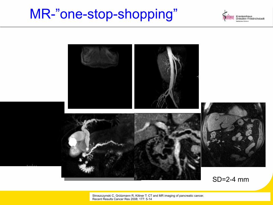
MR-”one-stop-shopping”
Stroszczynski C, Grützmann R, Kittner T: CT and MR imaging of pancreatic cancer. Recent Results Cancer Res 2008; 177: 5-14
SD=2-4 mm
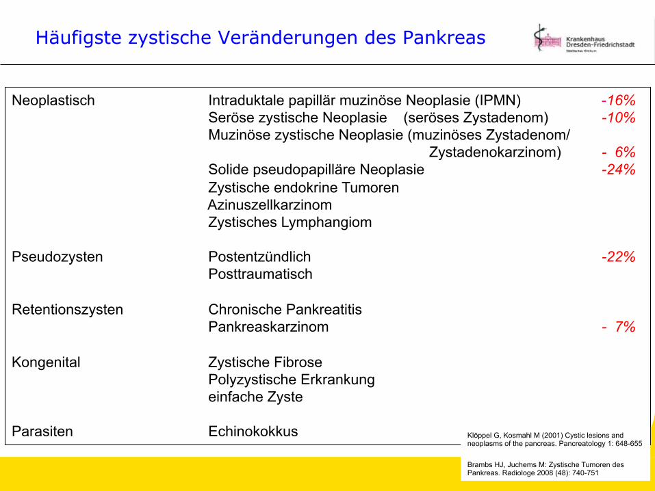
Häufigste zystische Veränderungen des Pankreas
Neoplastisch Intraduktale papillär muzinöse Neoplasie (IPMN) -16% Seröse zystische Neoplasie (seröses Zystadenom) -10% Muzinöse zystische Neoplasie (muzinöses Zystadenom/ Zystadenokarzinom) - 6% Solide pseudopapilläre Neoplasie -24% Zystische endokrine Tumoren Azinuszellkarzinom Zystisches Lymphangiom
Pseudozysten Postentzündlich -22%
Posttraumatisch Retentionszysten Chronische Pankreatitis
Pankreaskarzinom - 7% Kongenital Zystische Fibrose
Polyzystische Erkrankung einfache Zyste
Parasiten Echinokokkus Klöppel G, Kosmahl M (2001) Cystic lesions and
neoplasms of the pancreas. Pancreatology 1: 648-655
Brambs HJ, Juchems M: Zystische Tumoren des Pankreas. Radiologe 2008 (48): 740-751
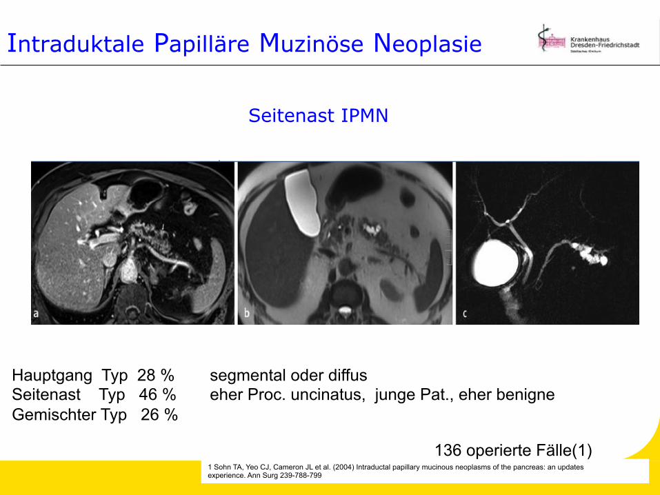
Intraduktale Papilläre Muzinöse Neoplasie
Hauptgang Typ 28 % segmental oder diffus Seitenast Typ 46 % eher Proc. uncinatus, junge Pat., eher benigne Gemischter Typ 26 %
1 Sohn TA, Yeo CJ, Cameron JL et al. (2004) Intraductal papillary mucinous neoplasms of the pancreas: an updates experience. Ann Surg 239-788-799
136 operierte Fälle(1)
Seitenast IPMN
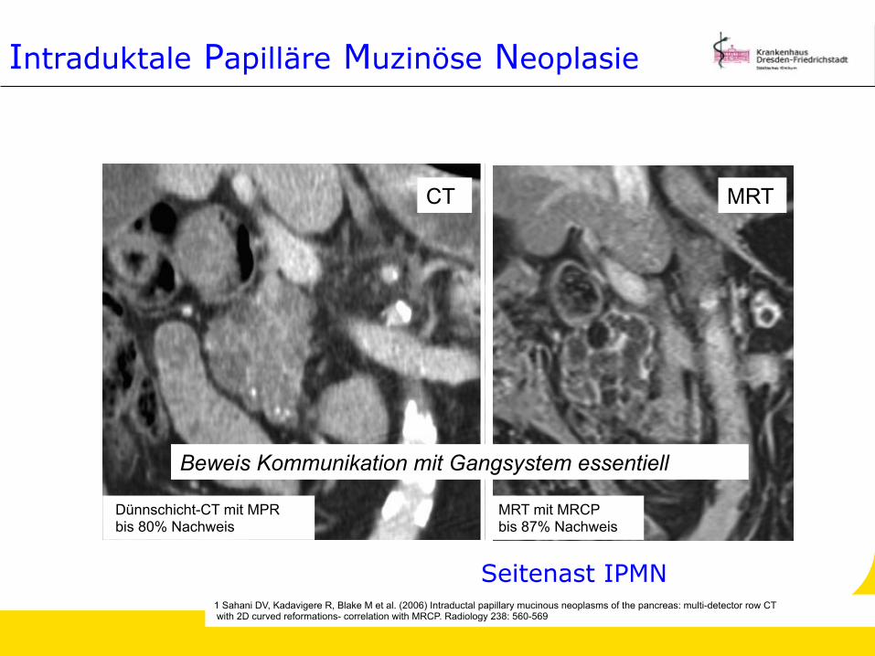
Dünnschicht-CT mit MPR bis 80% Nachweis
1 Sahani DV, Kadavigere R, Blake M et al. (2006) Intraductal papillary mucinous neoplasms of the pancreas: multi-detector row CT with 2D curved reformations- correlation with MRCP. Radiology 238: 560-569
Seitenast IPMN
CT
Intraduktale Papilläre Muzinöse Neoplasie
MRT
MRT mit MRCP bis 87% Nachweis
Beweis Kommunikation mit Gangsystem essentiell
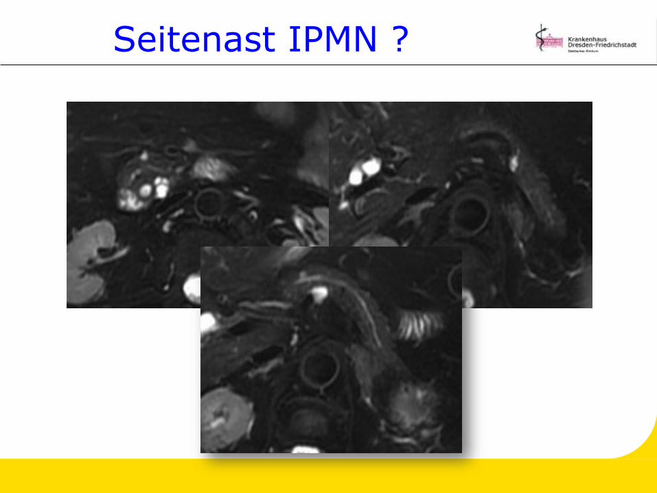
Seitenast IPMN ?
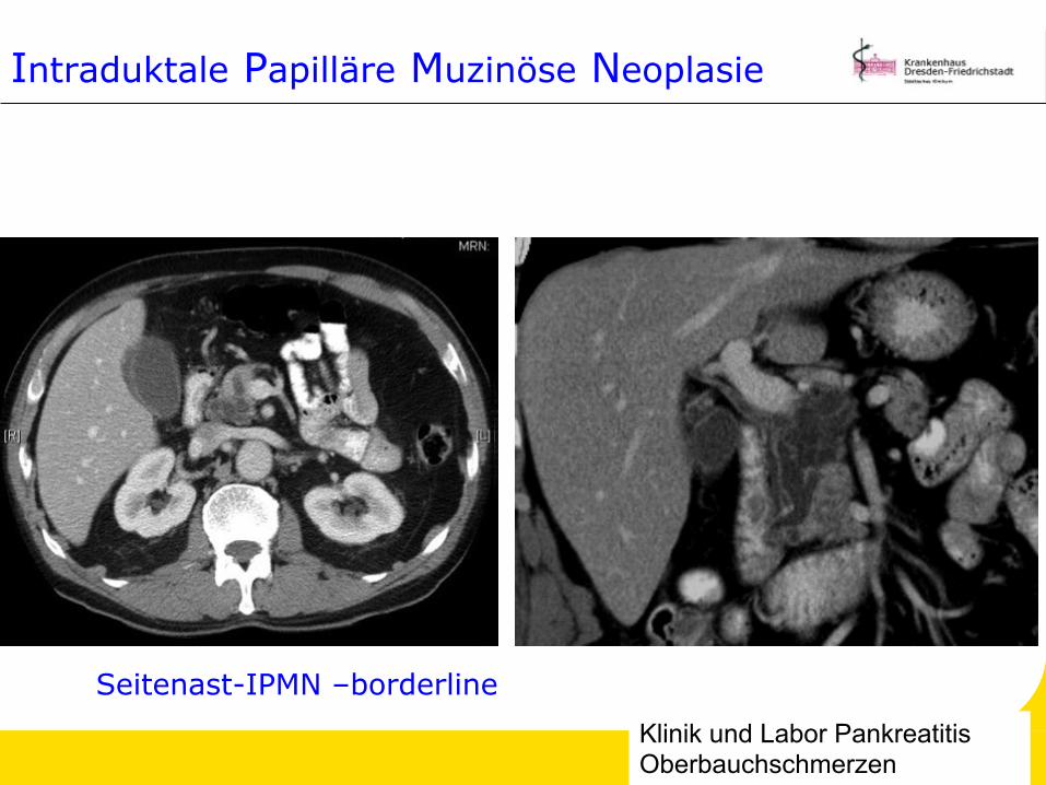
Seitenast-IPMN –borderline Klinik und Labor Pankreatitis Oberbauchschmerzen
Intraduktale Papilläre Muzinöse Neoplasie
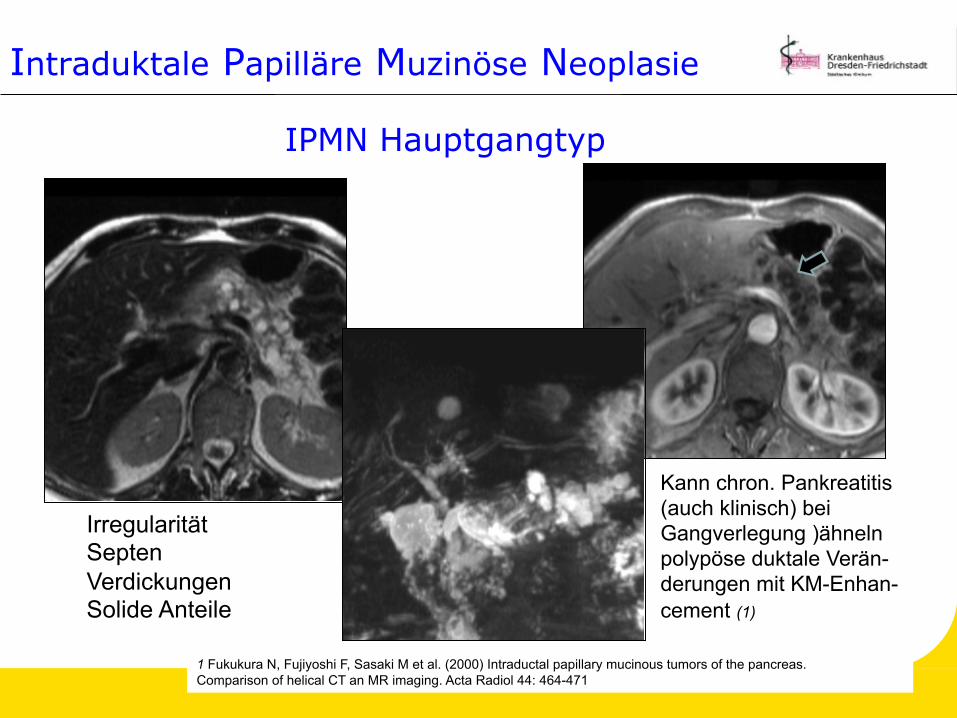
IPMN Hauptgangtyp
Irregularität Septen Verdickungen Solide Anteile
Kann chron. Pankreatitis (auch klinisch) bei Gangverlegung )ähneln polypöse duktale Verän- derungen mit KM-Enhan- cement (1)
1 Fukukura N, Fujiyoshi F, Sasaki M et al. (2000) Intraductal papillary mucinous tumors of the pancreas. Comparison of helical CT an MR imaging. Acta Radiol 44: 464-471
Intraduktale Papilläre Muzinöse Neoplasie
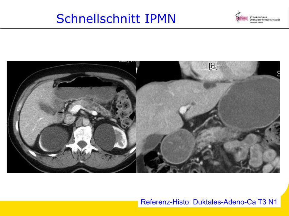
Schnellschnitt IPMN
Referenz-Histo: Duktales-Adeno-Ca T3 N1
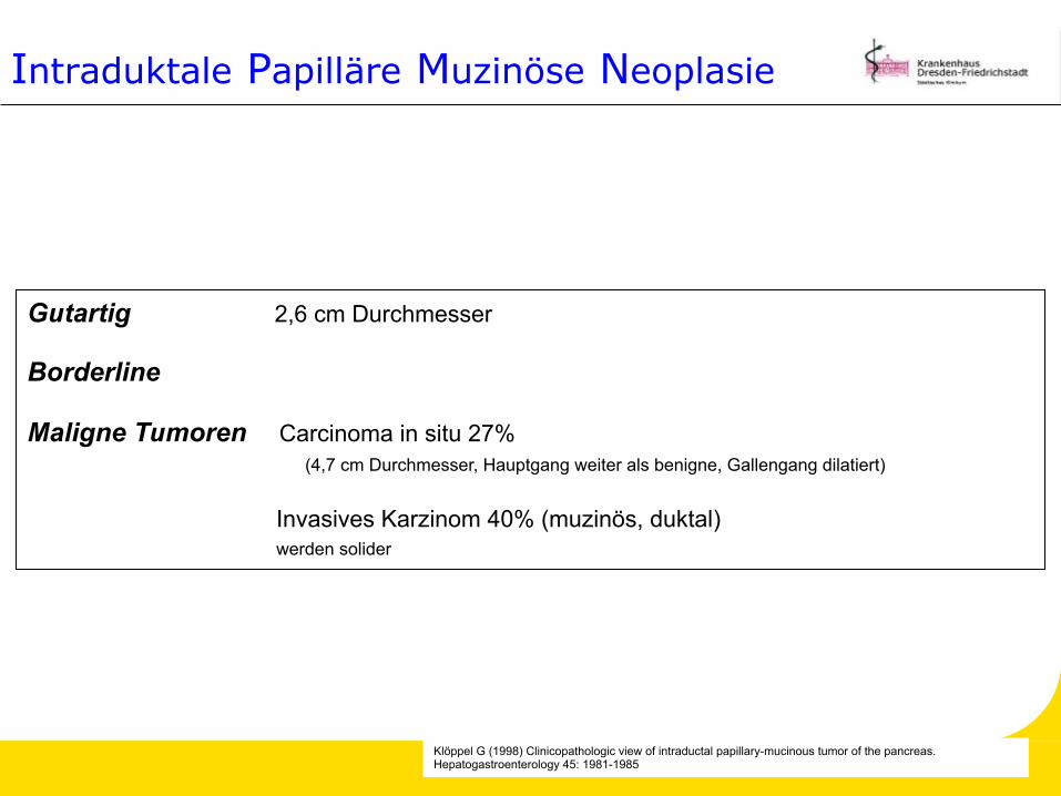
Gutartig 2,6 cm Durchmesser Borderline Maligne Tumoren Carcinoma in situ 27% (4,7 cm Durchmesser, Hauptgang weiter als benigne, Gallengang dilatiert)
Invasives Karzinom 40% (muzinös, duktal) werden solider
Klöppel G (1998) Clinicopathologic view of intraductal papillary-mucinous tumor of the pancreas. Hepatogastroenterology 45: 1981-1985
Intraduktale Papilläre Muzinöse Neoplasie
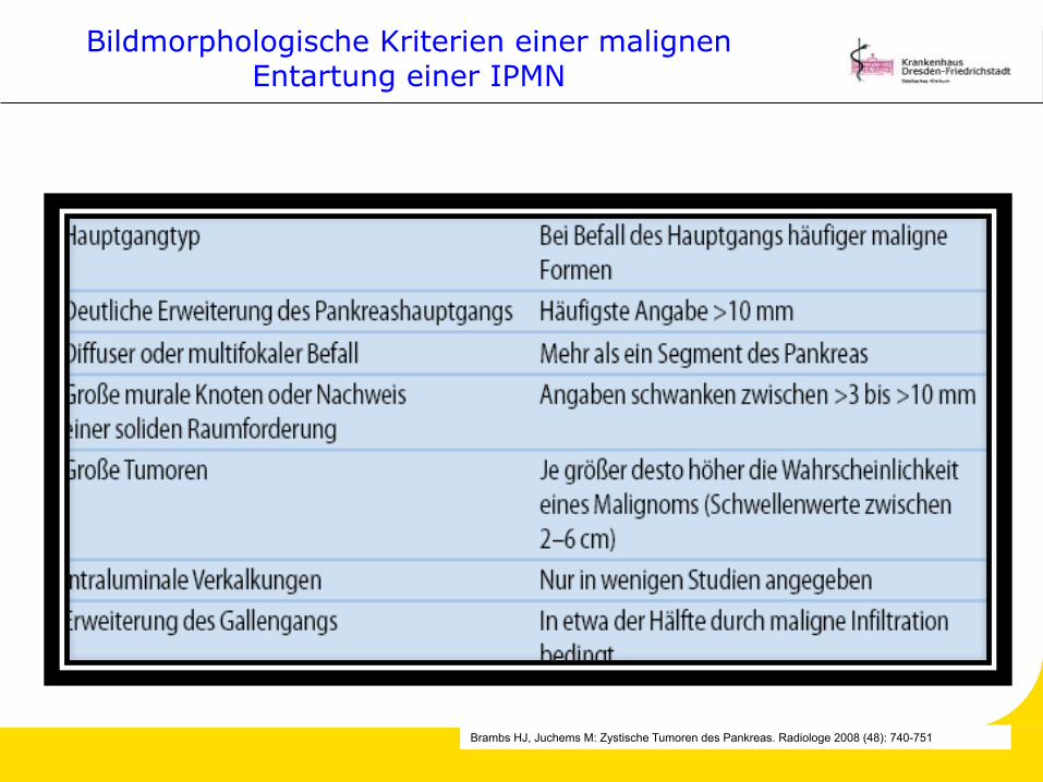
Bildmorphologische Kriterien einer malignen Entartung einer IPMN
Brambs HJ, Juchems M: Zystische Tumoren des Pankreas. Radiologe 2008 (48): 740-751

Muzinöse zystische Neoplasien
Tumoren ohne Verbindung mit dem Gangsystem, die schleimbildendes Epithel und häufig ein ovarienartiges Stroma aufweisen (1) fast ausschließlich weibliche Patienten (M:F=1:9) (2) meist solitär dickwandige bzw. gekammerte Zysten meist größer und Druckschmerzen, nur 25% symptomlos Tumorgröße > 6 cm und Knotennachweis korreliert mit Entartungsrisiko (3)
(17,5% Ca, 10,5% borderline, 70% Adenom. Crippa S et al., Ann Surg, 2008) 95% in Korpus und Schwanz
Hämorrhagie stark hinweisend auf MZN Ddx monozystische Tumore vs. Pseudozysten schwer (4)
4 Brambs HJ, Juchems M: Zystische Tumoren des Pankreas. Radiologe 2008 (48): 740-751
1 Zamboni G, Scarpa A, Bogina G et al. (1999 ) Mucinous cystic tumors of the pancreas: clinicopathological features, prognosis, and relationship to other mucinous cystic tumors. Am J Surg Pathol 23: 410-422
2 Klöppel G, Kosmahl M (2001) Cystic lesions and neoplasms of the pancreas. Pancreatology 1: 648-655
3 Thompson LD, Becker RC, Przygodzky et al. (1999) Mucinous cystic neoplasms (mucinous cystadenocarcinoma of low-grade malignant potential )of the pancreas: a clinicopathological study of 130 casesAm J Surg Pathol 23: 1-16

Muzinöse zystische Neoplasie
MZ Adenom
IPMN:MZN=7:1 Suzuki Y, Atomi y, Sugiyama M et al. (2004) Cystic neoplasms of the pancreas: a japanese multiinstitutional study of intraductal papillary mucinous tumors and mucinous cystic tumors. Pancreas28: 241-246
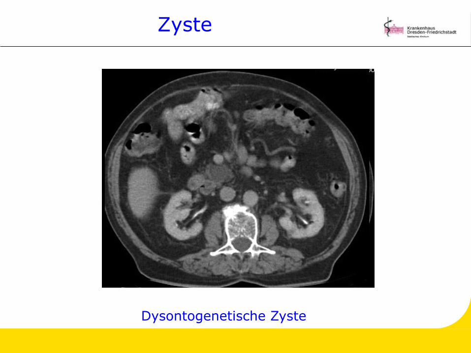
Zyste
Dysontogenetische Zyste
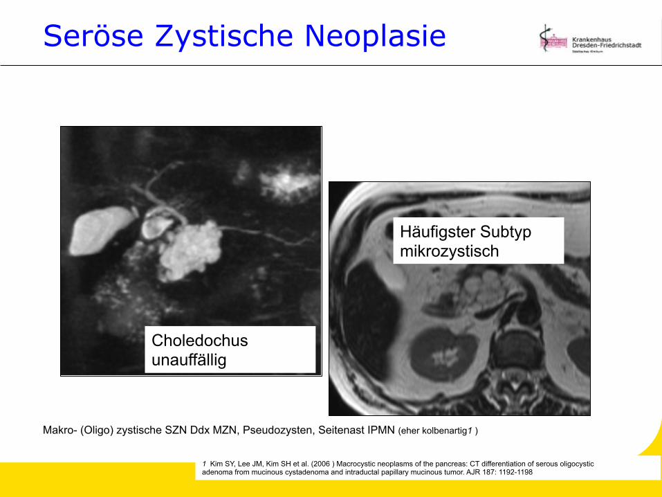
Seröse Zystische Neoplasie
Makro- (Oligo) zystische SZN Ddx MZN, Pseudozysten, Seitenast IPMN (eher kolbenartig1 )
Häufigster Subtyp mikrozystisch
Choledochus unauffällig
1 Kim SY, Lee JM, Kim SH et al. (2006 ) Macrocystic neoplasms of the pancreas: CT differentiation of serous oligocystic adenoma from mucinous cystadenoma and intraductal papillary mucinous tumor. AJR 187: 1192-1198
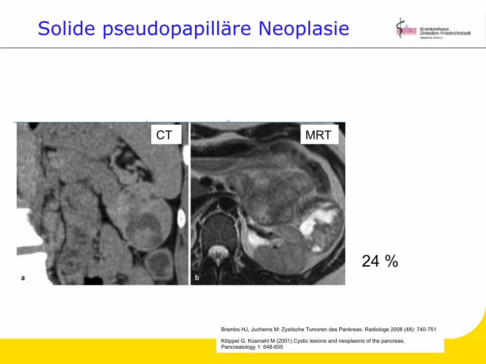
Solide pseudopapilläre Neoplasie
CT MRT
Brambs HJ, Juchems M: Zystische Tumoren des Pankreas. Radiologe 2008 (48): 740-751
24 %
Klöppel G, Kosmahl M (2001) Cystic lesions and neoplasms of the pancreas. Pancreatology 1: 648-655
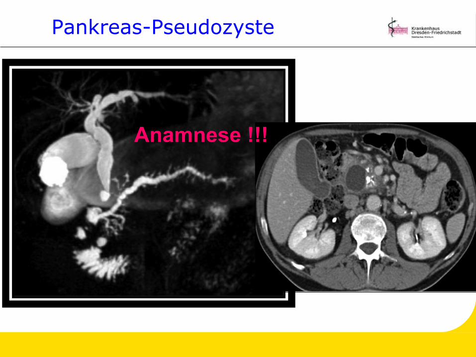
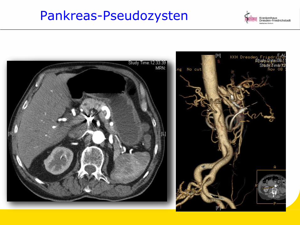
Pankreas-Pseudozyste
Anamnese !!!
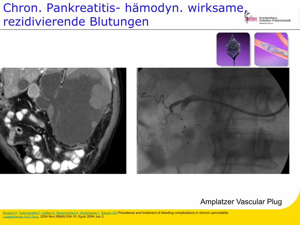
Chron. Pankreatitis- hämodyn. wirksame, rezidivierende Blutungen
Amplatzer Vascular Plug
Display
Setti
ngs:
For
mat
Send
to:
Choose
Destination
Langenbecks
Arch
Surg.
2004
Nov;389(6):504-10.
Epub
2004
Jun
2.
Prevalence
and
treat
ment
of
bleedi
ng
complicati
ons i
n
chronic
pancreatitis.
Bergert
H
,
Dobrowolski
F,
Caffi
er
S,
Bl
oomenthal
A,
Hi
nterseher I,
Saeger
HD
.
Bergert H, Dobrowolski F, Caffier S, Bloomenthal A, Hinterseher I, Saeger HD:Prevalence and treatment of bleeding complications in chronic pancreatitis. Langenbecks Arch Surg. 2004 Nov;389(6):504-10. Epub 2004 Jun 2.
Pankreas-Pseudozysten
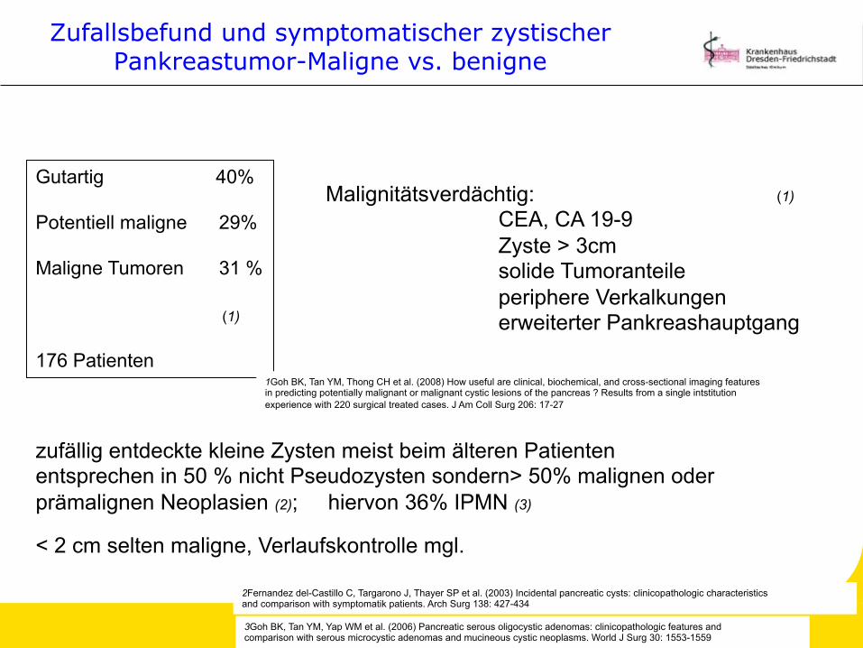
Zufallsbefund und symptomatischer zystischer Pankreastumor-Maligne vs. benigne
Gutartig 40% Potentiell maligne 29% Maligne Tumoren 31 % (1) 176 Patienten
Malignitätsverdächtig: (1) CEA, CA 19-9 Zyste > 3cm solide Tumoranteile periphere Verkalkungen erweiterter Pankreashauptgang
1Goh BK, Tan YM, Thong CH et al. (2008) How useful are clinical, biochemical, and cross-sectional imaging features in predicting potentially malignant or malignant cystic lesions of the pancreas ? Results from a single intstitution experience with 220 surgical treated cases. J Am Coll Surg 206: 17-27
zufällig entdeckte kleine Zysten meist beim älteren Patienten entsprechen in 50 % nicht Pseudozysten sondern> 50% malignen oder prämalignen Neoplasien (2); hiervon 36% IPMN (3)
< 2 cm selten maligne, Verlaufskontrolle mgl.
2Fernandez del-Castillo C, Targarono J, Thayer SP et al. (2003) Incidental pancreatic cysts: clinicopathologic characteristics and comparison with symptomatik patients. Arch Surg 138: 427-434
3Goh BK, Tan YM, Yap WM et al. (2006) Pancreatic serous oligocystic adenomas: clinicopathologic features and comparison with serous microcystic adenomas and mucineous cystic neoplasms. World J Surg 30: 1553-1559
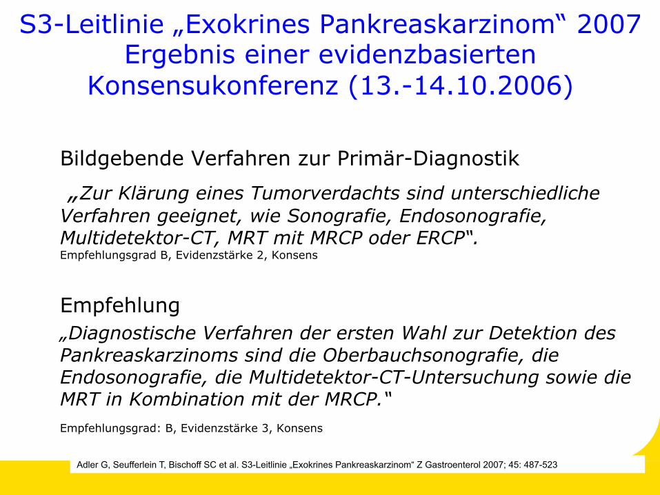
S3-Leitlinie „Exokrines Pankreaskarzinom“ 2007 Ergebnis einer evidenzbasierten
Konsensukonferenz (13.-14.10.2006)
Bildgebende Verfahren zur Primär-Diagnostik
„Zur Klärung eines Tumorverdachts sind unterschiedliche Verfahren geeignet, wie Sonografie, Endosonografie, Multidetektor-CT, MRT mit MRCP oder ERCP“. Empfehlungsgrad B, Evidenzstärke 2, Konsens
Empfehlung „Diagnostische Verfahren der ersten Wahl zur Detektion des Pankreaskarzinoms sind die Oberbauchsonografie, die Endosonografie, die Multidetektor-CT-Untersuchung sowie die MRT in Kombination mit der MRCP.“ Empfehlungsgrad: B, Evidenzstärke 3, Konsens
Adler G, Seufferlein T, Bischoff SC et al. S3-Leitlinie „Exokrines Pankreaskarzinom“ Z Gastroenterol 2007; 45: 487-523
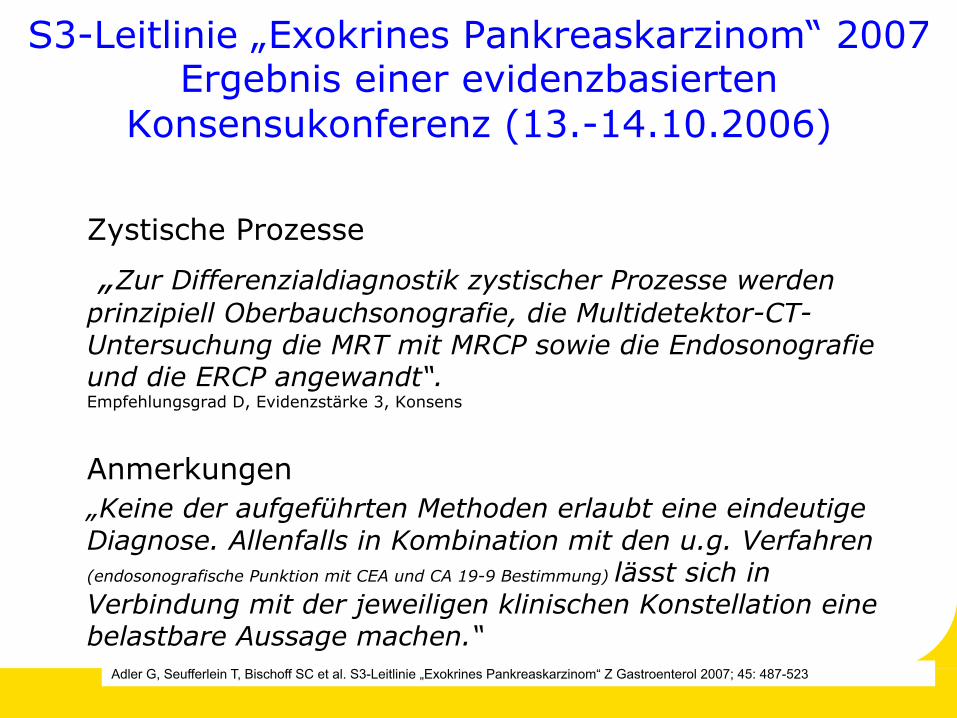
S3-Leitlinie „Exokrines Pankreaskarzinom“ 2007 Ergebnis einer evidenzbasierten
Konsensukonferenz (13.-14.10.2006)
Zystische Prozesse
„Zur Differenzialdiagnostik zystischer Prozesse werden prinzipiell Oberbauchsonografie, die Multidetektor-CT-Untersuchung die MRT mit MRCP sowie die Endosonografie und die ERCP angewandt“. Empfehlungsgrad D, Evidenzstärke 3, Konsens
Anmerkungen „Keine der aufgeführten Methoden erlaubt eine eindeutige Diagnose. Allenfalls in Kombination mit den u.g. Verfahren (endosonografische Punktion mit CEA und CA 19-9 Bestimmung) lässt sich in Verbindung mit der jeweiligen klinischen Konstellation eine belastbare Aussage machen.“
Adler G, Seufferlein T, Bischoff SC et al. S3-Leitlinie „Exokrines Pankreaskarzinom“ Z Gastroenterol 2007; 45: 487-523
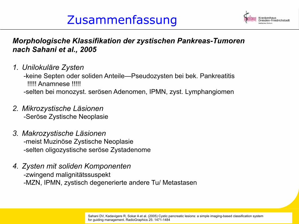
Sahani DV, Kadavigere R, Sokar A et al. (2005) Cystic pancreatic lesions: a simple imaging-based classification system for guiding management. RadioGraphics 25: 1471-1484
Morphologische Klassifikation der zystischen Pankreas-Tumoren nach Sahani et al., 2005 1. Unilokuläre Zysten
-keine Septen oder soliden Anteile—Pseudozysten bei bek. Pankreatitis !!!!! Anamnese !!!!! -selten bei monozyst. serösen Adenomen, IPMN, zyst. Lymphangiomen
2. Mikrozystische Läsionen -Seröse Zystische Neoplasie
3. Makrozystische Läsionen -meist Muzinöse Zystische Neoplasie -selten oligozystische seröse Zystadenome
4. Zysten mit soliden Komponenten -zwingend malignitätssuspekt -MZN, IPMN, zystisch degenerierte andere Tu/ Metastasen
Zusammenfassung
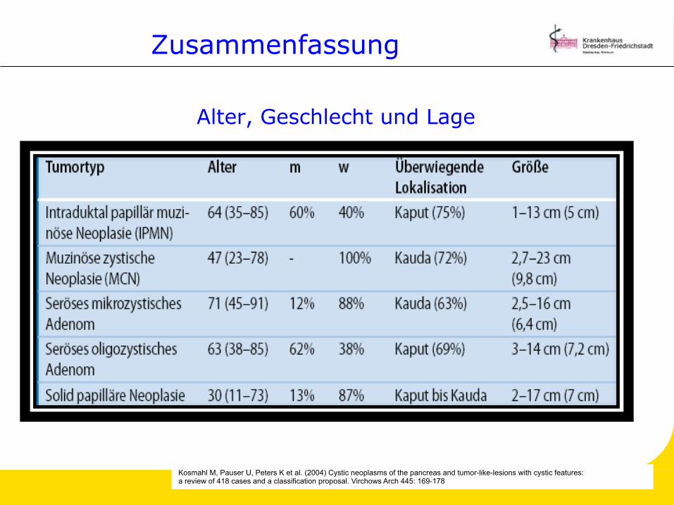
Alter, Geschlecht und Lage
Kosmahl M, Pauser U, Peters K et al. (2004) Cystic neoplasms of the pancreas and tumor-like-lesions with cystic features: a review of 418 cases and a classification proposal. Virchows Arch 445: 169-178
Zusammenfassung
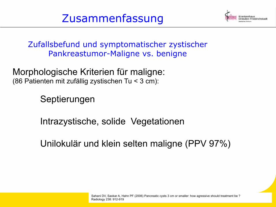
Sahani DV, Saokar A, Hahn PF (2006) Pancreatic cysts 3 cm or smaller: how agressive should treatment be ? Radiology 238: 912-919
Morphologische Kriterien für maligne: (86 Patienten mit zufällig zystischen Tu < 3 cm):
Septierungen
Intrazystische, solide Vegetationen
Unilokulär und klein selten maligne (PPV 97%)
Zufallsbefund und symptomatischer zystischer Pankreastumor-Maligne vs. benigne
Zusammenfassung
Rückblick
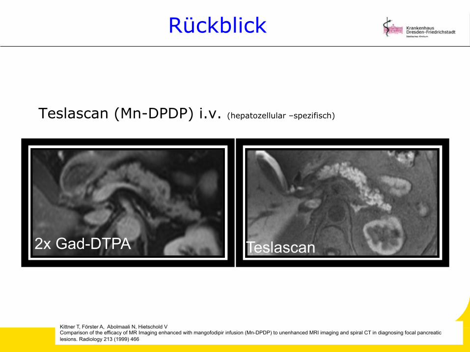
Teslascan (Mn-DPDP) i.v. (hepatozellular –spezifisch)
Kittner T, Förster A, Abolmaali N, Hietschold V Comparison of the efficacy of MR Imaging enhanced with mangofodipir infusion (Mn-DPDP) to unenhanced MRI imaging and spiral CT in diagnosing focal pancreatic lesions. Radiology 213 (1999) 466
2x Gad-DTPA Teslascan
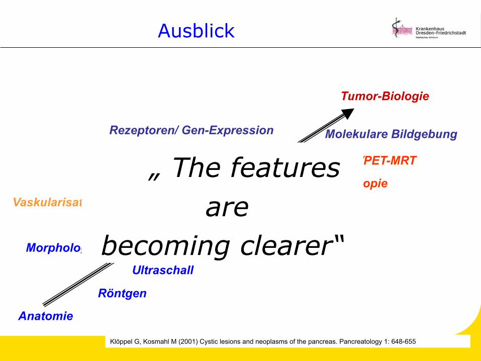
Ausblick
Molekulare Bildgebung
MRT/ MR-Spektroskopie
Mehrzeilen-Spiral-CT
Angiografie
Ultraschall
Röntgen
Anatomie
PET/ PET-CT/PET-MRT
Tumor-Biologie
Morphologie
Metabolismus
Vaskularisation/ Perfusion
Rezeptoren/ Gen-Expression
„ The features are
becoming clearer“
Klöppel G, Kosmahl M (2001) Cystic lesions and neoplasms of the pancreas. Pancreatology 1: 648-655

Panta rhei- alles fließt
„Alles fließt und nichts bleibt; es gibt nur ein ewiges Werden und Wandeln“
Platon (nach Heraklits Lehre)


