
Ferromagnetic properties of barium titanate ceramics
doped with cobalt, iron, and nickel
H. T. Langhammer1,*, T. Muller1, T. Walther1, R. Bottcher2, D. Hesse3, E. Pippel3, and S. G. Ebbinghaus1
1 Institut für Chemie, Martin-Luther-Universität Halle-Wittenberg, Kurt-Mothes-Straße 2, 06120 Halle (Saale), Germany2Fakultät für Physik und Geowissenschaften, Universität Leipzig, Linnéstraße 5, 04103 Leipzig, Germany3Max-Planck-Institut für Mikrostrukturphysik Halle, Weinberg 2, 06120 Halle (Saale), Germany
Received: 8 June 2016
Accepted: 26 July 2016
Published online:
4 August 2016
� Springer Science+Business
Media New York 2016
ABSTRACT
The influence of annealing in strongly reducing atmosphere on the magnetic prop-
erties of hexagonal BaTiO3 ? 0.04 BaO ? x/2 Co2O3 (0.0025 B x B 0.10) ceramics
was investigated. The samples air-sintered at 1673 Kwere subsequently tempered at
1473 K in H2/Ar stream. While the as-sintered samples exclusively exhibit para-
magnetic behavior, the annealed samples show distinct saturation in the field
dependence of the magnetization at 300 K measured in the range between -90 and
90 kOe. Besides, the field dependence of themagnetization is hystereticwith coercive
fields in the order of 100 Oe. Both properties point to ferromagnetic regions which
were identified as precipitations of metallic cobalt by TEM and EDX. The cobalt
precipitationswere exclusively found in tetragonal grainswhichwere completelyCo-
freeoutside theprecipitations.Obviously, these tetragonalgrainswere formedduring
the annealing process when the Co content of the formerly hexagonal grains was
concentrated into metallic Co particles by diffusion processes. Hence, the Co-free
matrixof thegrains transformedinto thecubicphasewhich is theequilibriumphaseof
undoped BaTiO3 at the annealing temperature 1473 K and during cooling to room
temperature into tetragonal phase. The size of themetallic precipitations ranges from
about 20 to 100 nm. A reduction both of the annealing temperature to 1373 K and of
the annealing time from 120 to 30 min did not change theminimumparticle size, but
now, the very rare precipitations only occurred at triple points or grain boundaries.
EPR measurements confirmed the occurrence of the ferromagnetic precipitations.
Whileat300 K, theas-sinteredsamplesdidnot showanyCoEPRsignal, theannealing
in strongly reducing atmosphere causeda strong andbroad linewhich is attributed to
the ferromagnetic resonance signal of the Co precipitations. The investigations were
extended for the dopants, iron or nickel, with a nominal sample composition of
BaTiO3 ? 0.04 BaO ? 0.01 Fe2O3 or 0.02 NiO, respectively. The field dependence of
themagnetization as well as the EPR spectra showed similar results compared to the
case of Co-doped samples. Hence, also in Fe- or Ni-doped BaTiO3 ceramics, ferro-
magnetic properties are caused by annealing in strongly reducing atmosphere.
Address correspondence to E-mail: [email protected]
DOI 10.1007/s10853-016-0263-3
J Mater Sci (2016) 51:10429–10441

Introduction
Multiferroics are a class of solids in which ferroelec-
tric and magnetic orders coexist. These materials
have the potential ability to couple electric polariza-
tion and magnetization and provide an additional
degree of freedom in device design. Therefore, they
are subject of intense investigations. However, multi-
ferroics are quite rare in nature, particularly at room
temperature (RT) and above [1, 2]. Thus, a consider-
able number of studies have been performed on the
fabrication of multiferroic materials in recent years.
Barium titanate (BaTiO3), a ferroelectric oxide with
perovskite structure, has become one of the promis-
ing systems. At present, much interest is focused on
the realization of ferromagnetism in BaTiO3 by dop-
ing with paramagnetic (PM) ions of the 3d group.
Due to their strong resemblance to the titanium ion,
most of the 3d transition metals can be easily doped
into BaTiO3 and act as substitutes for titanium [3, 4].
So far, ferromagnetic (FM) ordering has been
achieved in Co- [5, 6], Mn- [5], and Fe-doped [6–16]
BaTiO3 systems (single crystals [5, 6, 11, 13, 14], thin
films [7–9], and ceramics [10, 12, 15, 16]). While the
vast majority of published works concern Fe-doped
BaTiO3, there are only a few reports on ferromag-
netism with other 3d dopants, but, e.g., no report is
found in which Ni-doping would cause ferromag-
netism. Often, the researchers searched for the so-
called ferromagnetism in diluted magnetic oxides
(DMO) [11, 13–16], but it seems that ferromagnetism
can also be caused by metallic precipitations formed
during the preparation of the samples. Thus, Chak-
raborty et al. [13] reported on dilute magnetism in Fe-
doped BaTiO3 single crystals, but later they proposed
that metallic Fe clusters formed inside the BaTiO3
lattice were responsible for the FM behavior of the
specimens [14]. Among the different preparation
methods for achieving FM material, only two differ-
ent procedures are to be mentioned here. Khalitov
et al. [6] implanted BaTiO3 single crystals by Co and
Fe ions, respectively. Depending on the strength of
the ion fluence, they achieved metallic particles
inside the samples with different particle sizes. These
particles caused either superparamagnetic (SPM) or
FM properties. The critical particle size below which
SPM behavior occurs (and above which FM proper-
ties occur), is found to be 5.0 nm for Co implantation
and 6.5 nm for Fe implantation. Another way to
produce FM behavior is the post-annealing of
ceramic samples either in vacuum [10] or in pure
oxygen at 105 Pa [10, 15, 16]. However, for specimens
produced in this way, it could be also assumed here
that metallic precipitations of the dopant (formed by
the heat treatment) cause the ferromagnetism of the
samples.
During our recent investigations of the PM defect
properties of Co incorporated at Ti sites in hexagonal
BaTiO3 ceramics in the doping range of 0.1–2.0 mol%
Co [17], we tried to vary the Co valence state by
annealing in ambient atmospheres with various
oxygen partial pressures (pO2) from 105 Pa (pure
oxygen) to pO2\ 10-10 Pa (mixtures of H2/Ar).
While the samples annealed under pO2 & 0.1 Pa (Ar,
5 N) remained completely PM, the treatment in H2/
Ar caused a distinct saturation of the magnetization
field dependence pointing to FM or SPM properties
of the samples.
To the best of our knowledge, there are no publi-
cations of systematic investigations on achieving
ferromagnetism or superparamagnetism in Co-doped
BaTiO3 ceramics by heat treatment in highly reducing
atmosphere.
Thus, the aim of this paper is a detailed investi-
gation of the cause and the properties of the FM/SPM
behavior of Co-doped BaTiO3 ceramics in the doping
range up to 10 mol%. These investigations include (1)
X-ray diffraction (XRD) for the macroscopic phase
composition, (2) measurement of the field depen-
dence of the magnetization, (3) the use of transmis-
sion electron microscopy (TEM) partly combined
with energy-dispersive X-ray microanalysis (EDX)
for the localization and chemical identification of the
FM/SPM particles, and (4) electron paramagnetic
resonance (EPR) measurements for the characteriza-
tion of the local defect properties. In addition, we
present investigations of hexagonal BaTiO3 ceramics
doped with 2 mol% Fe or Ni, respectively, treated
under the same reducing conditions as for the Co-
doped samples to verify the FM/SPM properties also
for the other two ferromagnetic 3d transition metals,
Fe and Ni.
Experimental procedure
Ceramic powders with the nominal composition
BaTiO3 ? 0.04 BaO ? x/2 Co2O3 (0.0025 B x B 0.10)
and BaTiO3 ? 0.04 BaO ? 0.01 Fe2O3 or 0.02 NiO
were prepared by the conventional mixed-oxide
10430 J Mater Sci (2016) 51:10429–10441
powder technique.1 After mixing (agate balls, water)
and calcining (1373 K, 2 h) of BaCO3 (Solvay, VL600,
\0.1 mol% Sr) and TiO2 (Merck, No. 808), appropri-
ate amounts of either Co2O3 (Apolda-Chemie, p.a.) or
Fe2O3 (Merck, p.a.) or NiO (Berlin-Chemie, p.a.) were
added to the BaTiO3 powder. Then it was fine-milled
(agate balls, 2-propanole) and densified into disks
with a diameter of 6 mm and a height of nearly
3 mm. The samples were sintered in air at 1673 K for
1 h (heating rate 10 K/min). After the sintering, the
samples were annealed in a stream of H2/Ar (20/
20 ml/min) at 1473 K for 120 min (regime T0). The
Co-doped samples with a nominal Co concentration
of 1.5 mol% were annealed under three different
temperature/time regimes. Apart from regime T0,
some sintered samples were annealed at the same
temperature of 1473 K with reduced soaking time of
30 min (T1) or at reduced temperature/time of
1373 K for 30 min (T2).
The microstructure of the resulting ceramics was
examined by optical microscopy of cut planes
through the sample center. The mean grain sizes
were roughly estimated from the two-dimensional
grain areas in the cut plane. The overall phase com-
position was determined quantitatively by XRD of
pulverized samples (STADI MP diffractometer,
STOE, Germany using Co-Ka1 radiation equipped
with a Ge(111) monochromator and image plate
detector). The percentage of tetragonal phase ctg was
determined using the following equation: ctg ¼ a�b�Rc�R�d
considering two different peak height ratios R
(111)tetragonal/(103)hexagonal and (200)tetragonal/
(103)hexagonal and averaging both results. Parameters
a, b, c, d were determined by calibration with well-
defined mixtures of pure tetragonal and hexagonal
barium titanates. The percentage phase composition
of the samples is given as mass percent throughout
the article.
Magnetic measurements were carried out using the
ACMS magnetometer option of a PPMS 9 (Quantum
Design). The field dependence of the magnetic
moment was measured at 300 K with magnetic field
cycling between 90 and -90 kOe. The obtained
magnetic moments were corrected for the magnetic
moment of the sample holder and with respect to the
magnetic contribution of the BaTiO3 matrix.
Transmission electron microscopy (TEM) investi-
gations were performed using a Philips CM20T
electron microscope with energy of the primary
electrons of 200 keV, after thinning the samples by
standard methods involving mechanical polishing
and dimpling as well as argon ion beam thinning. For
performing scanning transmission electron micro-
scopy (STEM) and energy-dispersive X-ray (EDX)
elemental analyses, a FEI TITAN 80-300 high-per-
formance electron microscope was used, operating at
300 keV and equipped with a probe Cs-corrector and
an EDAX X-ray detector. This allows for the ele-
mental analysis with a spatial resolution in the order
of a few tenths of a nanometer.
EPR measurements of well-crushed ceramic sam-
ples were carried out on an X-band ELEXYS E580
spectrometer equipped with an Oxford Instruments
cryostat ESR 910 and a universal ER4102ST cavity
(TE102 mode) with a grid (50 % transmittance) for
optical excitation. For experiments in the Q-band, we
used a Bruker EMX device with a cylindrical cavity
ER 5106 QT (TE012) and an Oxford Instruments
cryostat CF 935.
Results and discussion
Co-doped samples
XRD and microstructure
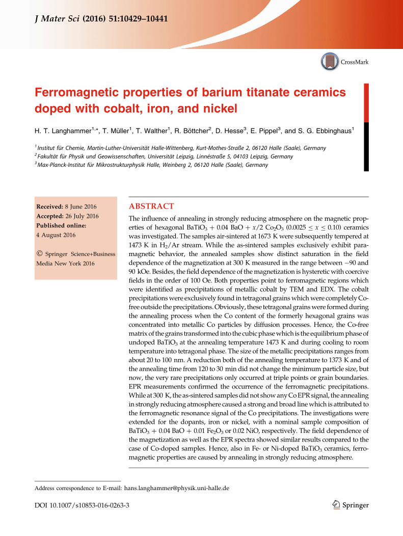
Figure 1 shows the percentage of tetragonal phase at
room temperature as a function of the nominal Co
concentration of air-sintered samples as well as of
samples which were additionally annealed in
strongly reducing atmosphere (H2/Ar stream,
1473 K, 120 min, called annealing T0). In the whole
Co-doping range investigated, exclusively tetragonal
and hexagonal BaTiO3 phases were found within the
detection limit of roughly 5 wt%. Thus, in Fig. 1, the
remaining percentage corresponds to the hexagonal
phase. For the air-sintered samples, the hexagonal
phase starts to occur at about x = 0.001 [17] and for
x[ 0.005 the samples are completely hexagonal.2 The
reducing treatment of the samples causes a small, but
remarkable change of the phase composition.
Whereas for x B 0.005 the reducing annealing
1 The slight Ba-excess supports the preferred incorporation ofCo on Ti sites in the air-sintered samples [17].
2 The influence of Co and other 3d transition elements on thestabilization of the hexagonal phase of BaTiO3 at roomtemperature is not yet fully understood [20–22].
J Mater Sci (2016) 51:10429–10441 10431
increases the percentage of the hexagonal phase, for
larger Co concentrations x C 0.01 it causes a
retransformation into the tetragonal phase with a
maximum percentage of 17 %. Only the specimens
with the highest Co concentration x = 0.10 remain
completely hexagonal.
The microstructure data of the Co-doped samples
are also depicted in Fig. 1. A bimodal grain size
distribution is observed, which is characterized by
small globular grains and rod-like grains with large
aspect ratios which are typical for hexagonal BaTiO3
ceramics with its strongly anisotropic grain growth
[18, 19]. In contrast to Mn-, Cu-, and Cr-doped
h-BaTiO3 [20–22], the amount of these rod-like grains
(of Co-doped samples) does not reflect the portion of
hexagonal phase determined by XRD. Only at
x = 0.10, the majority of the grains exhibit the typical
rod-like shape. The reducing treatment does not
change the microstructure of the ceramics.
Magnetic measurements
The air-sintered Co-doped samples exhibit exclu-
sively PM behavior (superimposed by the weak dia-
magnetism of the BaTiO3 matrix), which is caused by
the PM defect Co2? incorporated at Ti sites. Extensive
investigations of the PM Co-doped BaTiO3 ceramics
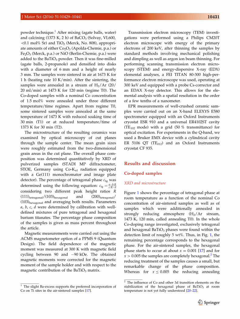
were published recently [17]. The annealing treatment
in strongly reducing atmosphere according to the
regime T0 drastically changes the magnetic properties
of the samples. Figure 2 shows the magnetic field
(H) dependence of the molar magnetization (Mmol,
related to the mole number of BaTiO3) of the Co-
doped samples (0.0025 B x B 0.10) in the field range
of ±90 kOe measured at 300 K. All samples exhibit
distinct saturation behavior of the magnetization
which is superimposed by a PM contribution. The
latter is clearly caused by isolated PM Co2? ions at Ti
sites still occurring in the ceramic grains after the
reducing treatment. It has to be emphasized that the
annealing in Ar atmosphere (oxygen partial pres-
sure & 0.1 Pa) with the same temperature schedule
(which acts as a reducing treatment milder than H2/
Ar) does not cause such a saturation of the magneti-
zation, i.e., the samples remain completely paramag-
netic, which is shown in Fig. 2 for x = 0.10
exemplarily. The S-shaped Mmol(H) curves point to
either FM or SPM properties of metallic particles
which were formed by the reduction of Co2? and
Co3? ions (occurring in the air-sintered samples [17])
to elemental Co atoms. Because of the observed curve
shapes, the Co atoms must have formed metallic
clusters. Whether the particles show FM or SPM
behavior depends on the cluster size. Generally, if the
size of the particles exceeds about 10 nm, they become
ferromagnetic and form different, room-temperature
stable magnetic domains. A distinctive feature for
SPM is the vanishing of a hystereticMmol(H) behavior
Figure 1 Room-temperature fraction of tetragonal phase for Co-
doped BaTiO3 sintered in air at 1673 K as well as subsequently
annealed in H2/Ar stream at 1473 K as a function of the nominal
Co concentration. The accuracy of the percentage data is not better
than 10 %. In addition, the average grain sizes of the air-sintered
samples are shown. Hollow/solid symbols indicate different
fractions of a bimodal microstructure. For the rod-like fraction,
the approximate length of the rods is used. Numbers at the data
points denote the percentage of these grain fractions.
Figure 2 Room-temperature magnetic field dependence of the
molar magnetization of Co-doped BaTiO3 ceramics sintered in air
at 1673 K and subsequently annealed in H2/Ar stream at 1473 K
for nominal Co concentrations x = 0.0025, 0.010, 0.020, and
0.010.
10432 J Mater Sci (2016) 51:10429–10441
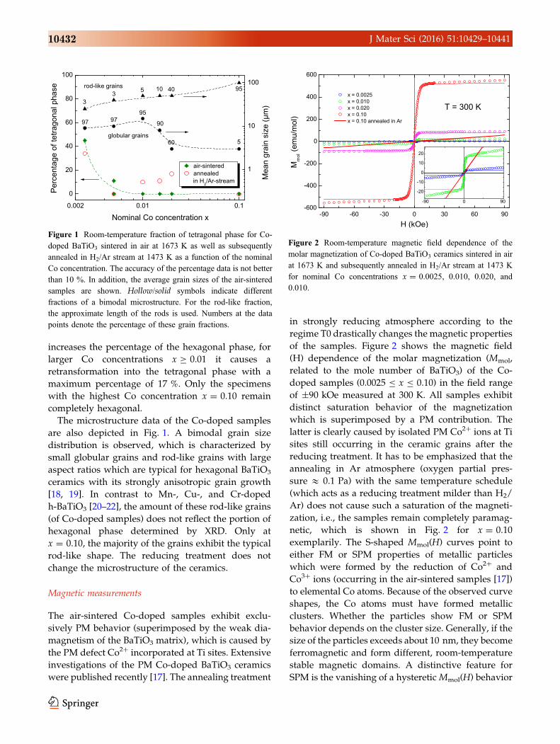
above a certain blocking temperature [23]. The mea-
suring temperature of 300 K is well above typical
blocking temperatures for SPM cobalt, thus, no hys-
teresis should be detectable for samples showing
exclusive SPM. The enlarged field range of ±500 Oe
of Fig. 2 (shown in Fig. 3) clearly reveals the hys-
teretic properties of all samples investigated
(0.0025 B x B 0.10). Hence, all samples exhibit FM
behavior, but coexisting SPM particles cannot be
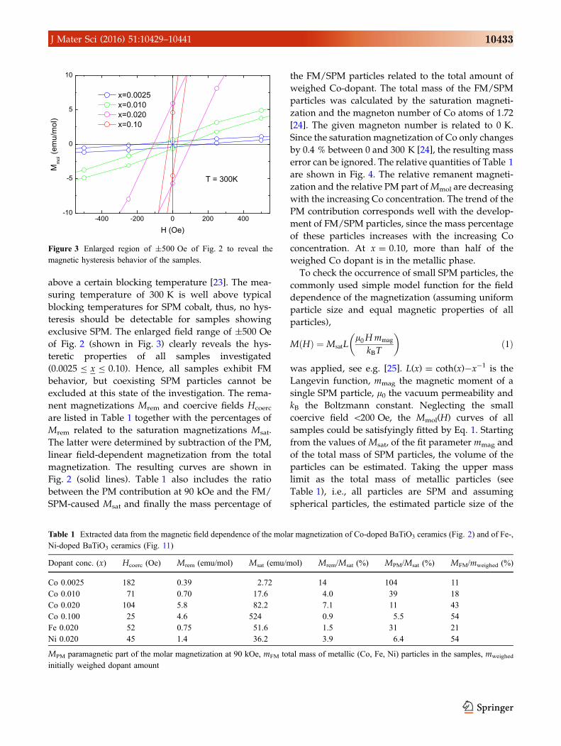
excluded at this state of the investigation. The rema-
nent magnetizations Mrem and coercive fields Hcoerc
are listed in Table 1 together with the percentages of
Mrem related to the saturation magnetizations Msat.
The latter were determined by subtraction of the PM,
linear field-dependent magnetization from the total
magnetization. The resulting curves are shown in
Fig. 2 (solid lines). Table 1 also includes the ratio
between the PM contribution at 90 kOe and the FM/
SPM-caused Msat and finally the mass percentage of
the FM/SPM particles related to the total amount of
weighed Co-dopant. The total mass of the FM/SPM
particles was calculated by the saturation magneti-
zation and the magneton number of Co atoms of 1.72
[24]. The given magneton number is related to 0 K.
Since the saturation magnetization of Co only changes
by 0.4 % between 0 and 300 K [24], the resulting mass
error can be ignored. The relative quantities of Table 1
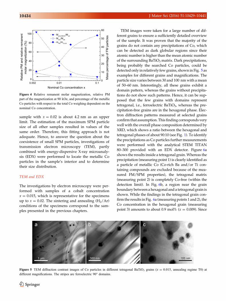
are shown in Fig. 4. The relative remanent magneti-
zation and the relative PM part ofMmol are decreasing
with the increasing Co concentration. The trend of the
PM contribution corresponds well with the develop-
ment of FM/SPM particles, since the mass percentage
of these particles increases with the increasing Co
concentration. At x = 0.10, more than half of the
weighed Co dopant is in the metallic phase.
To check the occurrence of small SPM particles, the
commonly used simple model function for the field
dependence of the magnetization (assuming uniform
particle size and equal magnetic properties of all
particles),
MðHÞ ¼ MsatLl0Hmmag
kBT
� �ð1Þ
was applied, see e.g. [25]. L(x) = coth(x)-x-1 is the
Langevin function, mmag the magnetic moment of a
single SPM particle, l0 the vacuum permeability and
kB the Boltzmann constant. Neglecting the small
coercive field \200 Oe, the Mmol(H) curves of all
samples could be satisfyingly fitted by Eq. 1. Starting
from the values of Msat, of the fit parameter mmag and
of the total mass of SPM particles, the volume of the
particles can be estimated. Taking the upper mass
limit as the total mass of metallic particles (see
Table 1), i.e., all particles are SPM and assuming
spherical particles, the estimated particle size of the
Figure 3 Enlarged region of ±500 Oe of Fig. 2 to reveal the
magnetic hysteresis behavior of the samples.
Table 1 Extracted data from the magnetic field dependence of the molar magnetization of Co-doped BaTiO3 ceramics (Fig. 2) and of Fe-,
Ni-doped BaTiO3 ceramics (Fig. 11)
Dopant conc. (x) Hcoerc (Oe) Mrem (emu/mol) Msat (emu/mol) Mrem/Msat (%) MPM/Msat (%) MFM/mweighed (%)
Co 0.0025 182 0.39 2.72 14 104 11
Co 0.010 71 0.70 17.6 4.0 39 18
Co 0.020 104 5.8 82.2 7.1 11 43
Co 0.100 25 4.6 524 0.9 5.5 54
Fe 0.020 52 0.75 51.6 1.5 31 21
Ni 0.020 45 1.4 36.2 3.9 6.4 54
MPM paramagnetic part of the molar magnetization at 90 kOe, mFM total mass of metallic (Co, Fe, Ni) particles in the samples, mweighed
initially weighed dopant amount
J Mater Sci (2016) 51:10429–10441 10433
sample with x = 0.02 is about 4.2 nm as an upper
limit. The estimation of the maximum SPM particle
size of all other samples resulted in values of the
same order. Therefore, this fitting approach is not
adequate. Hence, to answer the question about the
coexistence of small SPM particles, investigations of
transmission electron microscopy (TEM), partly
combined with energy-dispersive X-ray microanaly-
sis (EDX) were performed to locate the metallic Co
particles in the sample’s interior and to determine
their size distribution.
TEM and EDX
The investigations by electron microscopy were per-
formed with samples of a cobalt concentration
x = 0.015, which is representative for the specimens
up to x = 0.02. The sintering and annealing (H2/Ar)
conditions of the specimens correspond to the sam-
ples presented in the previous chapters.
TEM images were taken for a large number of dif-
ferent grains to ensure a sufficiently detailed overview
of the sample. It was proven that the majority of the
grains do not contain any precipitations of Co, which
can be detected as dark globular regions since their
atomic number is higher than the mean atomic number
of the surrounding BaTiO3 matrix. Dark precipitations,
being probably the searched Co particles, could be
detected only in relatively fewgrains, shown in Fig. 5 as
examples for different grains and magnifications. The
particle size varies between 30 and 100 nmwith amean
of 50–60 nm. Interestingly, all these grains exhibit a
domain pattern, whereas the grains without precipita-
tions do not show such patterns. Hence, it can be sup-
posed that the few grains with domains represent
tetragonal, i.e., ferroelectric BaTiO3, whereas the pre-
cipitation-free grains are in the hexagonal phase. Elec-
tron diffraction patterns measured at selected grains
confirmthat assumption.Thisfindingcorrespondsvery
well with the overall phase composition determined by
XRD, which shows a ratio between the hexagonal and
tetragonal phases of about 90:10 (see Fig. 1). To identify
the precipitations as Co particles furthermeasurements
were performed with the analytical STEM TITAN
80–300 provided with an EDX detector. Figure 6a
shows the results inside a tetragonal grain.Whereas the
precipitation (measuring point 1) is clearly identified as
a particle of metallic Co (Co-rich Ba and/or Ti con-
taining compounds are excluded because of the mea-
sured FM/SPM properties), the tetragonal matrix
(measuring point 2) is completely Co-free (within the
detection limit). In Fig. 6b, a region near the grain
boundarybetweenahexagonal anda tetragonalgrain is
shown. While the findings in the tetragonal grain con-
firm the results inFig. 6a (measuringpoints 1 and2), the
Co concentration in the hexagonal grain (measuring
point 3) amounts to about 0.9 mol% (x = 0.009). Since
Figure 4 Relative remanent molar magnetization, relative PM
part of the magnetization at 90 kOe, and percentage of the metallic
Co particles with respect to the total Co weighing dependent on the
nominal Co concentration.
Figure 5 TEM diffraction contrast images of Co particles in different tetragonal BaTiO3 grains (x = 0.015, annealing regime T0) at
different magnifications. The stripes are ferroelectric 90� domains.
10434 J Mater Sci (2016) 51:10429–10441
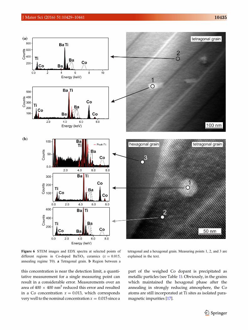
this concentration is near the detection limit, a quanti-
tative measurement for a single measuring point can
result in a considerable error. Measurements over an
area of 400 9 400 nm2 reduced this error and resulted
in a Co concentration x = 0.013, which corresponds
verywell to the nominal concentration x = 0.015 since a
part of the weighed Co dopant is precipitated as
metallic particles (see Table 1). Obviously, in the grains
which maintained the hexagonal phase after the
annealing in strongly reducing atmosphere, the Co
atoms are still incorporated at Ti sites as isolated para-
magnetic impurities [17].
Figure 6 STEM images and EDX spectra at selected points of
different regions in Co-doped BaTiO3 ceramics (x = 0.015,
annealing regime T0). a Tetragonal grain. b Region between a
tetragonal and a hexagonal grain. Measuring points 1, 2, and 3 are
explained in the text.
J Mater Sci (2016) 51:10429–10441 10435

A careful search for Co precipitations smaller than
10 nm with both electron microscope devices brought
no results even with the highest possible resolution of
about 0.5 nm. The size of all detected Co particles is
at least 20 nm. Hence, the existence of SPM particles
in the samples investigated can be excluded. The
saturation magnetization of the specimens is exclu-
sively caused by FM Co particles. To investigate the
kinetics of the formation of the metallic precipita-
tions, which must be a solid-state diffusion process
inside the concerned grains, the temperature sched-
ule of the annealing process was changed. By
reduction of annealing time and/or temperature the
formation of smaller metallic particles is expected,
since the growth of the precipitates is no instanta-
neous process and the growth velocity should
depend on the Co diffusion coefficient in the BaTiO3
matrix. In a first attempt (called regime T1), only the
annealing time was reduced from 120 to 30 min
compared to the standard annealing regime T0 used
for the majority of the samples. For the second
attempt (called regime T2), the annealing tempera-
ture was reduced from 1473 to 1373 K with a soaking
time of 30 min. Figure 7 shows the room-temperature
Mmol(H) curves of specimens for different annealing
regimes with x = 0.01 as an example.3 While the
saturation magnetization of sample T1 is reduced to
about 50 % of the T0 value, but still exhibiting a
distinct saturation effect, the saturation value of
sample T2 is rather small compared to its PM con-
tribution at 90 kOe. Besides, its magnetization curve
is only slightly different to the exclusively paramag-
netic behavior of a sample annealed in pure argon,
which is also depicted in Fig. 7. Compared to the
annealing regime T0 exhibiting a remanent magneti-
zation of 0.7 emu/mol (see Table 1), the remanence
values of the samples annealed under regimes T1 and
T2 are decreased to 0.3 and 0.1 emu/mol, respec-
tively, but still indicating FM properties.
Surprisingly, the lower limit of the size distribution
of the Co precipitates in the samples subject to
annealing regimes T1 and T2 changed only slightly
compared to the sample subject to the annealing
regime T0. Regarding the occurrence of metallic Co
particles, the T1 sample behaves equally to the T0
sample, i.e., the precipitates only occur in tetragonal
grains. The particle size distribution is shifted to
20–80 nm reflecting the reduced amount of metallic
particles. Despite a very extensive search at highest
possible resolution, no Co precipitates were found
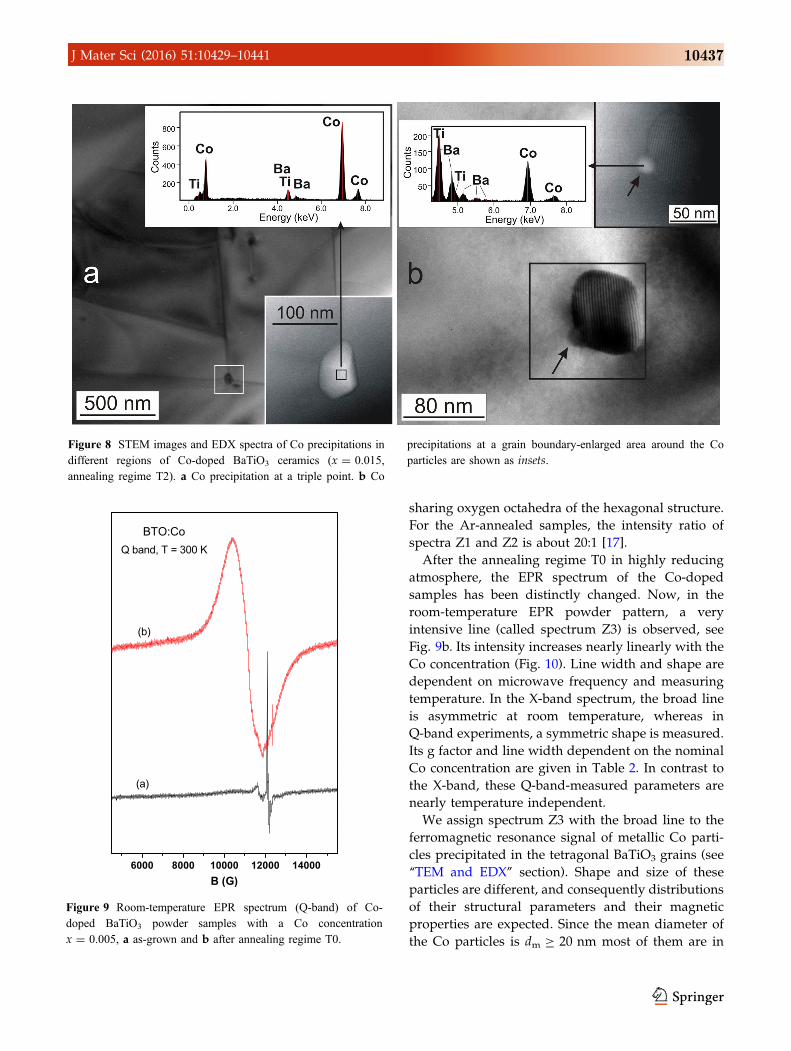
inside the grains of the T2 sample, but very few Co
particles at triple points (Fig. 8a) or at grain bound-
aries (Fig. 8b). The particle sizes amount to 50 and
17 nm, respectively, excluding SPM properties even
for the produced samples with the smallest satura-
tion magnetization.
EPR
Extensive EPR investigations of the PM Co-doped
BaTiO3 ceramics were published recently [17]. In air-
sintered and in subsequently Ar-annealed hexagonal
Co-doped BaTiO3 powder samples, no Co spectra
have been observed at room temperature, but only
spectra with very low intensities of the impurities
Fe3? and Cr3? of the source materials are visible
(Fig. 9a). At a temperature lower than 70 K, spectra
of two different Co2? centers (Z1 and Z2) were
observed. Both centers were assigned to isolated
Co2? ions with an effective electron spin S = �. The
weakly anisotropic center Z1 (�g = 4.33) was assigned
to Co2? ions incorporated at Ti(1) sites in the exclu-
sively corner-sharing oxygen octahedra, whereas the
strongly anisotropic center Z2 (gkc = 7.29, �g?c = 2.18,
c—hexagonal axis) was assigned to Co2? ions incor-
porated at Ti(2) sites in the trigonally distorted face-
Figure 7 Room-temperature magnetic field dependence of the
molar magnetization of Co-doped BaTiO3 ceramics (x = 0.01)
sintered in air at 1673 K and subsequently subjected to different
annealing regimes, T0, T1, and T2 in H2/Ar stream. The result for
a sample annealed in argon stream at 1473 K is shown for
comparison.
3 For a qualitative discussion, the chosen Co concentrationx = 0.01 is sufficiently near the concentration x = 0.015 used forthe TEM/STEM investigations. .
10436 J Mater Sci (2016) 51:10429–10441
sharing oxygen octahedra of the hexagonal structure.
For the Ar-annealed samples, the intensity ratio of
spectra Z1 and Z2 is about 20:1 [17].
After the annealing regime T0 in highly reducing
atmosphere, the EPR spectrum of the Co-doped
samples has been distinctly changed. Now, in the
room-temperature EPR powder pattern, a very
intensive line (called spectrum Z3) is observed, see
Fig. 9b. Its intensity increases nearly linearly with the
Co concentration (Fig. 10). Line width and shape are
dependent on microwave frequency and measuring
temperature. In the X-band spectrum, the broad line
is asymmetric at room temperature, whereas in
Q-band experiments, a symmetric shape is measured.
Its g factor and line width dependent on the nominal
Co concentration are given in Table 2. In contrast to
the X-band, these Q-band-measured parameters are
nearly temperature independent.
We assign spectrum Z3 with the broad line to the
ferromagnetic resonance signal of metallic Co parti-
cles precipitated in the tetragonal BaTiO3 grains (see
‘‘TEM and EDX’’ section). Shape and size of these
particles are different, and consequently distributions
of their structural parameters and their magnetic
properties are expected. Since the mean diameter of
the Co particles is dm C 20 nm most of them are in
Figure 8 STEM images and EDX spectra of Co precipitations in
different regions of Co-doped BaTiO3 ceramics (x = 0.015,
annealing regime T2). a Co precipitation at a triple point. b Co
precipitations at a grain boundary-enlarged area around the Co
particles are shown as insets.
Figure 9 Room-temperature EPR spectrum (Q-band) of Co-
doped BaTiO3 powder samples with a Co concentration
x = 0.005, a as-grown and b after annealing regime T0.
J Mater Sci (2016) 51:10429–10441 10437

the ferromagnetic state. The Larmor precession fre-
quency of the magnetic moment of a particle i in the
presence of the effective magnetic field (Beffi ) is
xiRes ¼ cBi
eff ð2Þ
where c is the gyro-magnetic ratio, and xResi is the
angular frequency. The effective field is the result of
the three main components: the external sweeping
field B0, the demagnetizing field BDi , and the molec-
ular field BMi. 4 The fields BD
i and BMi are dependent on
the shape and size of the particle as well as on the
orientation of the easy magnetic axis with respect to
the external field. The calculation of Beffi is extremely
complicated in our case. Because of the unknown
parameters of the particles in our samples, we
renounce the detailed interpretation of the ferro-
magnetic resonance pattern of the metallic Co parti-
cles embedded into the BaTiO3 grains.
Fe- and Ni-doped BaTiO3
Since the annealing in strongly reducing atmosphere
produces metallic ferromagnetic Co precipitations, it
is obviously useful to check this behavior also with
iron and nickel as doping elements in BaTiO3. Since
the results for Fe- and Ni-doping are qualitatively
similar to the Co-doped material, only the nominal
concentration x = 0.02 for Fe and Ni is presented
here as an example. Figure 11 shows the room-tem-
perature Mmol(H) curves of Fe-, Ni-, and Co-doped
(added for comparison) hexagonal BaTiO3 treated by
the highly reducing annealing regime T0. Contrary to
the Co-doped samples, no partial retransformation
into the tetragonal phase can be observed for
Fe- and Ni-doping (at least for x = 0.02). The com-
plete hexagonal air-sintered samples remain nearly
hexagonal after the annealing regime T0. Also the Fe-
and Ni-doped samples exhibit pronounced satura-
tion behavior of the magnetization (after subtraction
of the PM contribution) and a distinct magnetic
hysteresis (not seen in Fig. 11, Hcoerc and Mrem are
Figure 11 Room-temperature magnetic field dependence of the
molar magnetization of Co-, Fe-, and Ni-doped BaTiO3 ceramics
sintered in air at 1673 K and subsequently annealed in H2/Ar
stream at 1473 K for a nominal doping concentration of
x = 0.020.
Figure 10 Room-temperature EPR-spectrum (Q-band) of Co-
doped BaTiO3 powder samples after applying the annealing
regime T0 dependent on the Co concentration. x = 0.005 (a),
0.010 (b), 0.015 (c), 0.020 (d).
Table 2 g factor and line width values DBpp of spectrum Z3 (Q-
band) dependent on the nominal Co concentration
x g factor DBpp (G)
0.005 2.20 ± 0.01 1600 ± 100
0.010 2.20 ± 0.01 1700 ± 100
0.015 2.20 ± 0.01 1950 ± 100
0.020 2.20 ± 0.01 2250 ± 100
4 Here, molecular field means the summarized contributionsof magnetic dipole–dipole, exchange, electrostatic multipole,and electron–phonon interactions.
10438 J Mater Sci (2016) 51:10429–10441
noted in Table 1). Hence, also in the cases of Fe and
Ni, the treatment in strongly reducing atmosphere
causes remarkable metallic precipitations with a
minimum size of 10–20 nm, which explains the FM
behavior. The analysis of the Mmol(H) curves was
carried out as described in ‘‘Magnetic measurements’’
section. The results are also shown in Table 1. The
different PM contributions can be roughly explained
by the different magnetic moments of the corre-
sponding PM ions. The ions, Fe3? [26], Co2? [17], and
Ni2? [27], which are assumed to represent the major
impurities, possess in the high-spin ground states
total spin values of S = 5/2, 3/2, and 1, respectively.
In the rough spin-only approximation (which is a
good approximation only for d5 systems with van-
ishing total orbital angular momentum like high-spin
Fe3?) the effective magnetic moment is *ffiffiffiffiffiffiffiffiffiffiffiffiffiffiffiffiffiffiSðSþ 1Þ
p.
Hence, a decreasing PM contribution in the order
Fe[Co[Ni is plausible. Comparing the calculated
masses of metallic particles, it is notable that the Fe-
doped sample contains the smallest percentage of
metallic precipitations. Obviously, most of the
weighed Fe remains at Ti sites of the hexagonal
BaTiO3 lattice [26] or is incorporated into paramag-
netic secondary Ba-Ti-Fe-O phases which cannot be
excluded. In contrast, Ni behaves quite differently.
More than half of the weighed Ni is found in metallic
particles, while for Co, the percentage is about 40 %.
These different behaviors could be explained by, e.g.,
different diffusion coefficients of Fe, Co, and Ni in the
BaTiO3 lattice. The three FM doping elements also
exhibit different threshold fields HT to achieve nearly
complete alignment of the magnetic dipoles, i.e., the
saturation state of magnetization. A coarse estimation
of HT was done by the fit of theMmol(H) curves by the
simple arctan function [28].
M Hð Þ ¼ 2
pMsat arctan
H �Hc
HT
� �: ð3Þ
The determined threshold fields of Fe, Co, and Ni
amount to 2460, 1150, and 750 Oe, respectively. Since
HT is somehow correlated to the anisotropy energies
of the FM metals, the observed trend is somewhat
surprising. The anisotropy energy of the lower-sym-
metric, hexagonal Co metal is about one order larger
than the energies of the cubic Fe and Ni metals [29].
Hence, we expected Co to have the highest threshold
field. Moreover, the threshold field also depends on
the volume of the FM particles, size distributions of
which are unknown for Fe and Ni. Finally, it can be
noted that only the magnetization curve of the Ni-
doped sample follows the arctan curve very well.
Both the other curves significantly deviate from the
arctan function in the vicinity of HT pointing to a
distribution of the threshold values of the FM pre-
cipitations, which is caused by the nonuniform size
distribution of the metallic particles of Co and prob-
ably also of Fe.
In the as-prepared samples doped with Fe3? and
Ni2? ions, only EPR spectra of the isolated ions
incorporated into the host lattice on Ti sites were
detected (for details see [3, 4, 26, 30]). No ferromag-
netic resonance absorption could be detected by
measuring the temperature dependence of the spec-
tra in X- and Q-bands. After the samples were sub-
jected to annealing regime T0, the paramagnetic
spectra of isolated ions disappeared, and a very
intensive spectrum consisting of a very broad line
without any structure at room temperature was
observed in X- and Q-bands. The intensity of this line
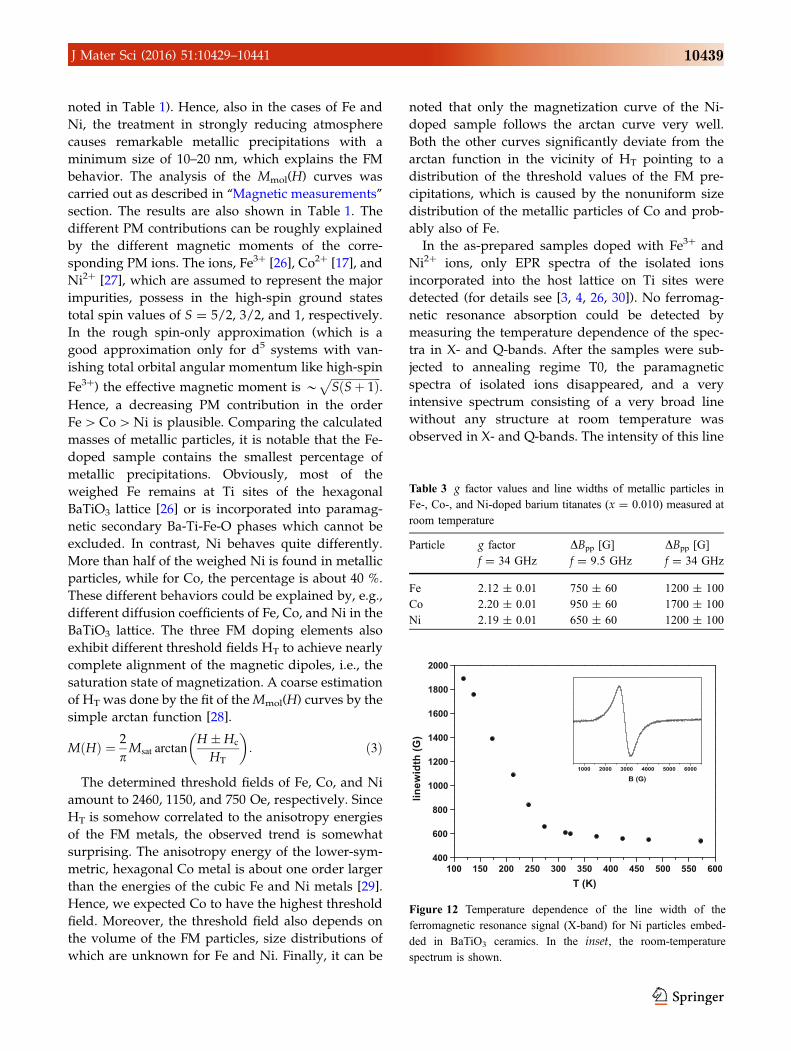
Table 3 g factor values and line widths of metallic particles in
Fe-, Co-, and Ni-doped barium titanates (x = 0.010) measured at
room temperature
Particle g factor
f = 34 GHz
DBpp [G]
f = 9.5 GHz
DBpp [G]
f = 34 GHz
Fe 2.12 ± 0.01 750 ± 60 1200 ± 100
Co 2.20 ± 0.01 950 ± 60 1700 ± 100
Ni 2.19 ± 0.01 650 ± 60 1200 ± 100
Figure 12 Temperature dependence of the line width of the
ferromagnetic resonance signal (X-band) for Ni particles embed-
ded in BaTiO3 ceramics. In the inset, the room-temperature
spectrum is shown.
J Mater Sci (2016) 51:10429–10441 10439
is proportional to the concentration of the doping
materials. Spectroscopic splitting factor and peak-to-
peak line widths DBpp measured at 293 K in X- and
Q-bands are given in Table 3. With the decreasing
temperature, the lines become broader, shifts of the
resonance fields to lower magnetic fields are
observed, and the line shapes become more asym-
metric. Figure 12 shows the temperature dependence
of the width DBpp of the broad line for Ni-doped
samples after the annealing regime T0. The experi-
mentally determined g factor values and room-tem-
perature line widths are characteristic values for
ferromagnetic resonance spectra of metallic Fe and Ni
nanoparticles in solids.
Summary and conclusion
Hexagonal BaTiO3 ceramics, doped with Fe, Co, or
Ni, exhibit a superposition of PM and FM behavior if
the air-sintered samples are post-annealed in strongly
reducing atmosphere (H2/Ar stream) at 1473 K. The
PM properties originate from isolated Fe3?, Co2?, and
Ni2? ions, respectively, which are incorporated at Ti
sites of the hexagonal lattice. The FM properties are
caused by precipitations of the dopants as metallic
particles. Detailed investigations of the Co-doped
samples showed that these precipitations exclusively
occur in grains (max. 20 %) which are retransformed
from the hexagonal 6H- to the 3C-BaTiO3 polytype
(tetragonal at room temperature) during the reducing
treatment, while in the hexagonal grains, no Co pre-
cipitates (detection limit 0.5 nm) can be found. The
size distribution of metallic particles in the tetragonal
grains amounts to about 15–100 nm, thus excluding
partly SPM behavior, since the transition from FM to
SPM behavior only occurs for particle sizes well
below 10 nm. In very rare cases, metallic precipita-
tions ([15 nm) also occur at grain boundaries or tri-
ple points additionally contributing to the FM
properties. The Co concentration in the tetragonal
grains is below the detection limit of the EDX detec-
tor, whereas the Co concentration in the still hexag-
onal grains corresponds more or less to the nominal
Co content of the specimens. Hence, during the
reducing treatment, all incorporated Co ions of the
retransforming, initially hexagonal grains have to
move to the precipitation loci by solid-state diffusion
processes forming the final metallic particles inside
the grains.
Revisiting our former investigations of hexagonal
Mn-doped BaTiO3 where the partial retransformation
into 3C-BaTiO3 after annealing in strongly reducing
atmosphere was already observed [20], we suppose
that also in the case of Mn, the retransformed grains
exhibit precipitations of metallic Mn (similar to that
reported for Co, Fe, and Ni particles), which were not
detected since metallic Mn is not ferromagnetic, and
since we did not seek for such Mn precipitations by
TEM.
Generalizing our findings, we assume that the
annealing in strongly reducing atmosphere of BaTiO3
samples doped with the other 3d dopants like Sc, V,
Cr, Cu, and Zn also produces metallic precipitations
of the dopants. Regarding the observation of FM
behavior of Fe-doped BaTiO3 in the literature [7–16],
we can only speculate whether in some cases the FM
properties were simply caused by metallic Fe pre-
cipitations, at least for samples subjected to a reduc-
ing treatment [10, 13, 14]. The annealing in pure
oxygen also resulted in certain FM properties
[10, 15, 16]. Since those authors applied rather high Fe
concentrations between 10 and 30 mol%, a careful
search for Fe precipitations by TEM or STEM/EDX
should be very useful to exclude metallic particles in
the discussion of the magnetic results. Recent studies
show also that even very small Fe concentrations
«1 mol% in BaTiO3 ceramics can cause weak FM
behavior even if the samples were sintered/annealed
in air [31].
References
[1] Rabe K, Ahn ChH, Triscone JM (2007) Physics of ferro-
electrics: a modern perspective. Springer, Berlin
[2] Kitagawa Y, Hiraoka Y, Honda T, Ishikura T, Nakamura H,
Kimura T (2010) Low-field magnetoelectric effect at room
temperature. Nat Mater 9:797–802
[3] Possenriede E, Jacobs P, Schirmer OF (1992) Paramagnetic
defects in BaTiO3 and their role in light-induced charge
transport: I ESR studies. J Phys 4:4719–4742
[4] Possenriede E (1992) Paramagnetische Storstellen in Bari-
umtitanat und ihre lichtinduzierten Umladungen. Disserta-
tion, University of Osnabruck, Germany
[5] Lee JS, Khim ZG, Park YD, Norton DP, Theodoropoulou
NA, Budai JD, Boatner SJ, Pearton SJ, Wilson RG (2003)
Magnetic properties of Co- and Mn-implanted BaTiO3,
SrTiO3 and KTaO3. Solid State Electron 47:2225–2230
10440 J Mater Sci (2016) 51:10429–10441
[6] Khalitov NI, Khaibullin RI, Valeev VF, Dulov EN, Ivoilov
NG, Tagirov LR, Kazan S, Sale AG, Mikailzade FA (2012)
Structural and magnetic studies of Co and Fe implanted
BaTiO3 crystals. Nucl Instrum Methods Phys Res B
272:104–107
[7] Maier R, Cohn JL, Neumeier JJ, Bendersky LA (2001)
Ferroelectricity and ferrimagnetism in iron-doped BaTiO3.
Appl Phys Lett 78:2536–2538
[8] Rajamani A, Dionne GF, Bono D, Ross J (2005) Faraday
rotation, ferromagnetism, and optical properties in Fe-doped
BaTiO3. J Appl Phys 98:3907
[9] Ramana EV, Yang SM, Jung R, Jung MH, Lee BW, Jung CU
(2013) Ferroelectric and magnetic properties of Fe-doped
BaTiO3 thin films grown by the pulsed laser deposition.
J Appl Phys 113:187219
[10] Lin F, Shi W (2009) Magnetic properties of transition-metal-
codoped BaTiO3 systems. J Alloys Compd 475:64–69
[11] Ray S, Mahadevan P, Mandal S, Krishnakumar SR, Kuroda
CS, Sasaki T, Taniyama T, Itoh M (2008) High temperature
ferromagnetism in single crystalline dilute Fe-doped BaTiO3.
Phys Rev B 77:104416
[12] Du G-P, Hu Z-J, Han Q-F, Qin X-M, Shi W-Z (2010) Effects
of niobium donor doping on the phase structures and mag-
netic properties of Fe-doped BaTiO3 ceramics. J Alloys
Compd 492:L79–L81
[13] Chakraborty T, Ray S, Itoh M (2011) Defect-induced mag-
netism: test of dilute magnetism in Fe-doped hexagonal
BaTiO3 single crystals. Phys Rev B 83:144407
[14] Chakraborty T, Meneghini C, Aquilanti G, Ray S (2014)
Investigating the development of spurious magnetism in
single crystalline BaTi0.95Fe0.05O3-d with high d by local
structural probes. J Phys: Condens Matter 26:196001
[15] Valant M, Arcon I, Mikulska I, Lisjak D (2013) Cation
order–disorder transition in Fe-doped 6h-batio3 for dilute
room-temperature ferromagnetism. Chem Mater 25:3544–
3550
[16] Zorko A, Pregelj M, Gomilsek M, Jaglicic Z, Pajic D,
Telling M, Arcon I, Mikulska I, Valant M (2015) Strain-
induced extrinsic high-temperature ferromagnetism in the
Fe-doped hexagonal barium titanate. Sci Rep 5:7703
[17] Langhammer HT, Bottcher R, Muller T, Walther T,
Ebbinghaus SG (2015) Defect properties of cobalt-doped
hexagonal barium titanate ceramics. J Phys: Condens Matter
27:295901
[18] Recnik A, Kolar D (1996) Exaggerated growth of hexagonal
barium titanate under reducing sintering conditions. J Am
Ceram Soc 79:1015–1018
[19] Kolar D, Kunaver U, Recnik A (1998) Exaggerated aniso-
tropic grain growth in hexagonal barium titanate ceramics.
Phys Status Solidi A 166:219–230
[20] Langhammer HT, Muller T, Felgner K-H, Abicht H-P (2000)
Crystal structure and related properties of manganese-doped
barium titanate ceramics. J Am Ceram Soc 83:605–611
[21] Langhammer HT, Muller T, Bottcher R, Abicht H-P (2003)
Crystal structure and related properties of copper-doped
barium titanate ceramics. Solid State Sci 5:965–971
[22] Langhammer HT, Muller T, Bottcher R, Abicht H-P (2008)
Structural and optical properties of chromium-doped
hexagonal barium titanate ceramics. J Phys: Condens Matter
20:085206
[23] Bean CP, Livingston JD (1959) Superparamagnetism. J Appl
Phys 30:120S–129S
[24] O’Handley RC (2000) Modern magnetic materials: princi-
ples and applications. Wiley, New York
[25] Wang J-Q, Xiao G (1994) Transition-metal granular solids:
microstructure, magnetic properties, and giant magnetore-
sistance. Phys Rev B 49:3982–3996
[26] Bottcher R, Langhammer HT, Muller T, Abicht H-P (2008)
3C–6H phase transition in BaTiO3 induced by Fe ions: an
electron paramagnetic resonance study. J Phys: Condens
Matter 20:505209
[27] Langhammer HT, Muller T, Walther T (to be published)
[28] Geiler AL, Harris VG, Vittoria C, Sun NX (2006) A quan-
titative model for the nonlinear response of fluxgate mag-
netometers. J Appl Phys 99:08B316.
[29] Blundell S (2001) Magnetism in condensed matter. Oxford
University Press, Oxford
[30] Bottcher R, Langhammer HT, Muller T (2011) Paramagnetic
resonance study of nickel ions in hexagonal barium titanate.
J Phys: Condens Matter 23:115903.
[31] Figueras FG, Amorim CO, Amaral J, Agostinho Moreira J,
Tavares PB, Alves E, Amaral VS (2016) Magnetoelectric
effect probe through ppm Fe doping in BaTiO3. J Alloys
Compd 661:495–500
J Mater Sci (2016) 51:10429–10441 10441


