Page 1
Instructions for use
Title Discovery of Mwinilunga alphavirus : A novel alphavirus in Culex mosquitoes in Zambia
Author(s)Torii, Shiho; Orba, Yasuko; Hang'ombe, Bernard M.; Mweene, Aaron S.; Wada, Yuji; Anindita, Paulina D.;Phongphaew, Wallaya; Qiu, Yongjin; Kajihara, Masahiro; Mori-Kajihara, Akina; Eto, Yoshiki; Harima, Hayato; Sasaki,Michihito; Carr, Michael; Hall, William W.; Eshita, Yuki; Abe, Takashi; Sawa, Hirofumi
Citation Virus Research, 250, 31-36https://doi.org/10.1016/j.virusres.2018.04.005
Issue Date 2018-05-02
Doc URL http://hdl.handle.net/2115/73890
Rights © 2018. This manuscript version is made available under the CC-BY-NC-ND 4.0 licensehttp://creativecommons.org/licenses/by-nc-nd/4.0/
Rights(URL) http://creativecommons.org/licenses/by-nc-nd/4.0/
Type article (author version)
File Information Torii_et_al_HUSCUP.pdf
Hokkaido University Collection of Scholarly and Academic Papers : HUSCAP
Page 2
1
Discovery of Mwinilunga alphavirus: a novel alphavirus in Culex mosquitoes in
Zambia
Shiho Torii1, Yasuko Orba1*, Bernard M. Hang’ombe2,10, Aaron S. Mweene3,9,10, Yuji
Wada1, Paulina D. Anindita1, Wallaya Phongphaew1, Yongjin Qiu4, Masahiro Kajihara5,
Akina Mori-Kajihara5, Yoshiki Eto5, Hayato Harima4, Michihito Sasaki1, Michael Carr6,7,
William W. Hall7,8,9,10, Yuki Eshita4, Takashi Abe11, Hirofumi Sawa1,7,9,10*
1Division of Molecular Pathobiology, Research Center for Zoonosis Control, Hokkaido
University, Sapporo, Japan
2Department of Para-clinical Studies, School of Veterinary Medicine, University of
Zambia, Lusaka, Zambia 3Department of Disease Control, School of Veterinary Medicine, University of Zambia,
Lusaka, Zambia 4Hokudai Center for Zoonosis Control in Zambia, Lusaka, Zambia
5Division of Global Epidemiology, Research Center for Zoonosis Control, Hokkaido
University, Sapporo, Japan 6National Virus Reference Laboratory, School of Medicine, University College Dublin,
Dublin, Ireland
7Global Institution for Collaborative Research and Education (GI-CoRE), Hokkaido
University, Sapporo, Japan 8Centre for Research in Infectious Diseases, University College Dublin, Dublin, Ireland 9Global Virus Network, Baltimore, Maryland, USA 10Africa Center of Excellence for Infectious Diseases of Humans and Animals, University
of Zambia, Lusaka, Zambia 11Department of Computer Science, Graduate School of Science and Technology, Niigata
Page 3
2
University, Niigata, Japan
*Corresponding author:
Yasuko Orba
Division of Molecular Pathobiology, Research Center for Zoonosis Control, Hokkaido
University, N20, W10, Kita-ku, Sapporo 001-0020, Japan
Tel: +81-11-706-9508, Fax: +81-11-706-7370,
E-mail: [email protected]
Hirofumi Sawa, M.D., Ph.D.
Division of Molecular Pathobiology, Research Center for Zoonosis Control, Hokkaido
University, N20, W10, Kita-ku, Sapporo 001-0020, Japan
Tel: +81-11-706-5185; Fax: +81-11-706-7370
E-mail: [email protected]
Page 4
3
Abstract
Mosquito-borne alphaviruses are disseminated globally and cause febrile illness in
humans and animals. Since the prevalence and diversity of alphaviruses has not been
previously investigated in Zambia, reverse transcription PCR was employed as a broad-
spectrum approach for the detection of alphaviruses in mosquitoes. From 552 mosquito
pools, a novel alphavirus, tentatively named Mwinilunga alphavirus (MWAV), was
discovered from a single Culex quinquefasciatus mosquito pool. The full genome of
MWAV was subsequently determined, and pairwise comparisons demonstrated that
MWAV represented a new alphavirus species. Phylogenetic analyses and a linear
discriminant analysis based on the dinucleotide ratios in various virus sequences indicated
that MWAV is related to a mosquito-specific alphavirus distinct from other known
mosquito-borne alphaviruses due to its inability to replicate in vertebrate cell lines.
Further analyses of these novel alphaviruses will help to facilitate a greater understanding
of the molecular determinants of host range restriction and the evolutionary relationships
of alphaviruses.
Keywords: alphavirus; mosquito; Culex; Zambia
Page 5
4
Main Text
Alphaviruses, belonging to the genus Alphavirus in the family Togaviridae, can infect
diverse vertebrate hosts, including mammals, birds, reptiles, amphibians and fish (Griffin,
2013). Most alphaviruses are mosquito-borne and are transmitted by mosquitoes from at
least eight genera (Ortiz et al., 2005; Webb et al., 2008). Restricted interactions between
viruses, their invertebrate vector species and vertebrate hosts usually occur in nature;
however, some alphaviruses occasionally can cause widespread epizootics (Weaver and
Reisen, 2010). Humans and/or animals infected by pathogenic alphaviruses exhibit
febrile illnesses that may culminate either in encephalitis or arthritis, depending upon the
viral etiology (Atkins, 2013).
In Africa, human diseases caused by the Old World alphaviruses, such as
chikungunya virus (CHIKV), o'nyong-nyong virus and Sindbis virus (SINV), have been
reported (Lwande et al., 2015). While the Republic of Zambia is located in southern
Africa and vector mosquitoes of alphaviruses have been found widely in the country
(Gaffigan et al.; Kent, 2006), human cases of chikungunya fever, o'nyong-nyong fever or
Sindbis fever have not been documented in prior studies (Lwande et al., 2015; Rezza et
al., 2017; Storm et al., 2014) (https://www.cdc.gov/chikungunya/index.html). Moreover,
comprehensive detection of alphaviruses in mosquitoes has not been conducted in Zambia
to date. Therefore, screening of vector mosquitoes in Zambia is crucial to better predict
future outbreaks of alphaviral diseases.
The alphavirus genome consists of a single-stranded, positive-sense RNA, encoding
nonstructural proteins (nsPs; nsP1-nsP4), which are important for transcription and
replication of viral RNA (Lemm and Rice, 1993), and structural proteins (sPs; Capsid, E3,
E2, 6k and E1), which are the main constituents of virions (Voss et al., 2010). Based on
genetic and serological similarities, around 30 species of alphaviruses are classified into
antigenic complexes (Powers et al., 2012; Powers et al., 2001). In addition, an
Page 6
5
unclassified alphavirus, Eilat virus (EILV), has been discovered in Anopheles coustani
(Nasar et al., 2012) and Taï Forest alphavirus (TALV) has been recently discovered in
Culex decens (Hermanns et al., 2017).
EILV appears to infect only mosquitoes and differs from other known mosquito-
borne alphaviruses which generally maintain their lifecycles between mosquitoes and
vertebrate hosts, although EILV is phylogenetically grouped within the mosquito-borne
clade (Nasar et al., 2014; Nasar et al., 2012). Studies of EILV have contributed to the
identification of the molecular determinants responsible for host range restriction and
provided new insights into the evolutionary history of alphaviruses. However, further
studies on insect-only alphaviruses are required for a better understanding of the ecology
and evolutionary relationships in the genus Alphavirus and the molecular determinants of
host range restriction (Nasar et al., 2015a; Nasar et al., 2015b). In this study, we have
focused on the detection of alphaviruses in mosquitoes collected in Zambia.
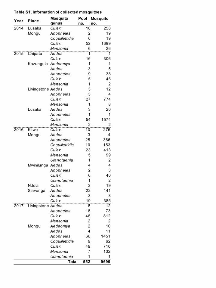
To analyze the prevalence of alphaviruses in mosquitoes in Zambia, a total of 9,699
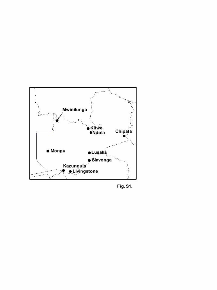
mosquitoes were collected from various locations between 2014 and 2017 (Fig. S1,
available in the online Supplementary Material). Mosquito collection was carried out in
protected areas with the permission of the Zambia Wildlife Authority, now the
Department of National Parks and Wildlife, Ministry of Tourism and Arts of the Republic
of Zambia and the Excellence in Research Ethics and Science (ERES) Converge Ethics
Committee IRB (No: 00005948). Species were at first identified morphologically using
a stereomicroscope, and subsequently some species were genetically confirmed by PCR
and sequencing of the cytochrome-oxidase subunit I gene (Folmer et al., 1994).
Information on the collected mosquitoes is summarized in Table S1 (available in the
online Supplementary Material). The largest numbers of mosquito species were within
the genus Culex. After species identification, mosquito samples were divided into 552
pools containing 1 to 40 individual mosquitoes, belonging to the same species and
Page 7
6
sampling location. Subsequently, the pools were homogenized with Minimum Essential
Medium containing 2% fetal bovine serum (Sigma, St. Louis, MO) using the BioMasher
(Nippi, Tokyo, Japan). Total RNAs were extracted from 100 µl of the homogenates using
the Direct-zol kit (Zymo Research, Irvine, CA) according to the manufacturer’s protocol
and the remaining homogenates were stored at −80 °C until use for virus detection.
We employed a one-step reverse transcription PCR (RT-PCR) assay for detection of
a wide-range of alphaviruses in the extracted mosquito RNAs. Oligonucleotide primers
for the RT-PCR assay were designed based on an alignment of the conserved region of
nsP4 among defined alphavirus species. One-step RT-PCR was performed with the
PrimeScript One Step RT-PCR Kit, Ver. 2 (Takara, Shiga, Japan) with 1 µl of extracted
mosquito RNA and 1 µM (final concentration) of each of the primers, pan-Alpha-nsP4-
6692F (5'-CAYACRYTRTTYGAYATGTCDGC-3') and pan-Alpha-nsP4-7152R (5'-
GCRTCDATKATYTTBACYTCCAT-3') in 15 µl reaction volumes, with the following
thermocycling protocol: 50 °C for 30 minutes; 94 °C for 2 minutes; 43 cycles of 94 °C
for 30 seconds, 52 °C for 30 seconds, and 72 °C for 30 seconds; and 72 ˚C for 5 minutes.
The PCR products (approximately 460 bp) were visualized on a 1% (w/v) agarose gel
stained with ethidium bromide. We confirmed that CHIKV and SINV nsP4 genes were
also detected by our methodology (data not shown).
From 552 pools, one pool, which contained 19 Culex quinquefasciatus mosquitoes
collected in Mwinilunga (North-Western Province of Zambia) in 2016, was found to be
positive by RT-PCR assay. The amplified products were sequenced using the ABI PRISM
3130 Genetic Analyzer (Applied Biosystems, Foster City, CA) with the pan-Alpha-nsP4-
6692F primer and pan-Alpha-nsP4-7152R primer, and the nucleotide sequences were
analyzed using GENETYX, version 13 (GENETYX, Tokyo, Japan). Sequence
comparison of the amplified fragment in the National Center for Biotechnology
Information database employing BLASTN analysis showed 76% nucleotide identity to
Page 8
7
TALV and 75% identity to EILV. The detected alphavirus was tentatively named
Mwinilunga alphavirus (MWAV). Other known alphaviruses were not detected in the
present study.
We sought to isolate MWAV from RT-PCR-positive mosquitoes using Aedes
albopictus cells (C6/36), African green monkey kidney cells (Vero) and baby hamster
kidney cells (BHK-21). Homogenized samples of mosquitoes were diluted five-fold with
DMEM supplemented with 2% fetal bovine serum, penicillin, streptomycin, gentamycin
and 2 mM L-glutamine (Sigma), filtered and used to inoculate onto cells. After
inoculation of the mosquito homogenates, supernatants of the cells were cultured through
three passages for three weeks. Viral production into the supernatant was checked by RT-
PCR with extracted RNA from supernatants of cultured cells and the pan-Alpha-nsP4
primers after three blind passages; however, proliferation of MWAV was not detected in
any of the three cell lines tested.
To determine the full-length MWAV genome sequence, RNA extracted from the
mosquitoes was subjected to next-generation sequencing. A library was constructed by
the Nextera XT DNA Library Prep kit (Illumina, San Diego, CA). Sequencing was
performed with a MiSeq Reagent Kit v3 (600 cycles) and Illumina MiSeq System
(Illumina) following the manufacturer’s protocol. After trimming low-quality reads, the
resulting reads were de novo assembled using a CLC Genomics Workbench 10 (CLC bio,
Qiagen, Hilden, Germany) with default parameters. The sequences were confirmed by
specific PCRs using MWAV-specific primer pairs. The 5'- and the 3'- terminal regions
were amplified using the rapid amplification of cDNA ends (RACE) approach with the
SMARTer RACE cDNA Amplification Kit (Takara) and MWAV-specific primers.
Amplified products were directly sequenced in both directions by Sanger sequencing. The
full-genome sequence of MWAV was determined and the amino acid sequences were
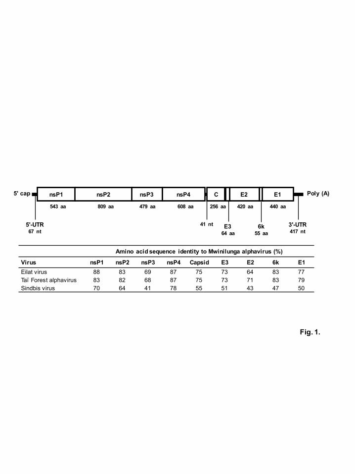
predicted using GENETYX, version 13. The MWAV genome consisted of 11,547 nt,
Page 9
8
coding two open reading frames (ORFs) (2,439 and 1,235 amino acids) in the positive
sense (Fig. 1). The full-genome sequence of MWAV was deposited in the
GenBank/EMBL/DDBJ database under accession number LC361437. The pairwise
nucleotide identities of MWAV were 70% to EILV, 58% to TALV and 52% to SINV.
BLAST analysis indicated that the deduced amino acid sequence of one ORF (2,439
amino acids) was related to that of nsPs from alphaviruses and the highest identity was
83% to EILV. The deduced amino acid sequence of the second ORF (1,235 amino acids)
was related to that of sPs from alphaviruses and the highest identity was 76% to TALV.
The pairwise amino acid identities of MWAV ranged between 64% (E2 protein) and 88%
(nsP1) to EILV for the corresponding proteins, and between 68% (nsP3) and 87% (nsP4)
to TALV for the corresponding proteins (Fig. 1). According to the species designation
criteria of the International Committee on Taxonomy of Viruses (Powers et al., 2012), the
genetic analysis of MWAV suggested that this represented a novel alphavirus species,
closely related to EILV and TALV. Eilat-like viruses, including EILV, TALV and MWAV
were detected from different mosquito species including Anopheles coustani (Nasar et al.,
2012), Culex decens (Hermanns et al., 2017) and Culex quinquefasciatus, respectively.
This supports the view that those viruses have mosquito-restricted host ranges (Nasar et
al., 2014). In addition, the narrow mosquito vector range may potentially explain why
MWAV could not be isolated using Aedes-derived and mammalian-derived cells.
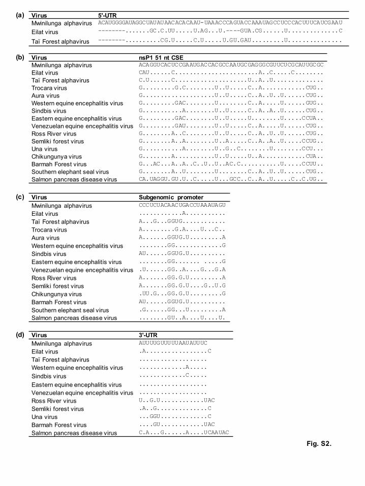
Since conserved sequence elements (CSEs) have important roles in the lifecycle of
alphaviruses (Fayzulin and Frolov, 2004; Hyde et al., 2015; Niesters and Strauss, 1990;
Zhu et al., 2013), we investigated the sequence identities of CSEs with other defined
alphaviruses. The CSEs of MWAV within the nsP1 51 nt, subgenomic promoter sequence
and the 3'-UTR shared more than 90% nt sequence identities with EILV and TALV (Fig.
S2, available in the online Supplementary Material). The identity of the CSE within the
putative 5'-UTR was 80% to that of EILV, and mFold analysis suggested that the putative
Page 10
9
5'-UTR of MWAV might form secondary stem-loop structures, similar to EILV (data not
shown) (Zuker, 2003). In addition, the conserved amino acid motifs, such as the ribosomal
binding site, conserved RNA-dependent RNA polymerase motifs of MWAV were similar
to those of other alphaviruses (data not shown) and the putative protease cleavage sites
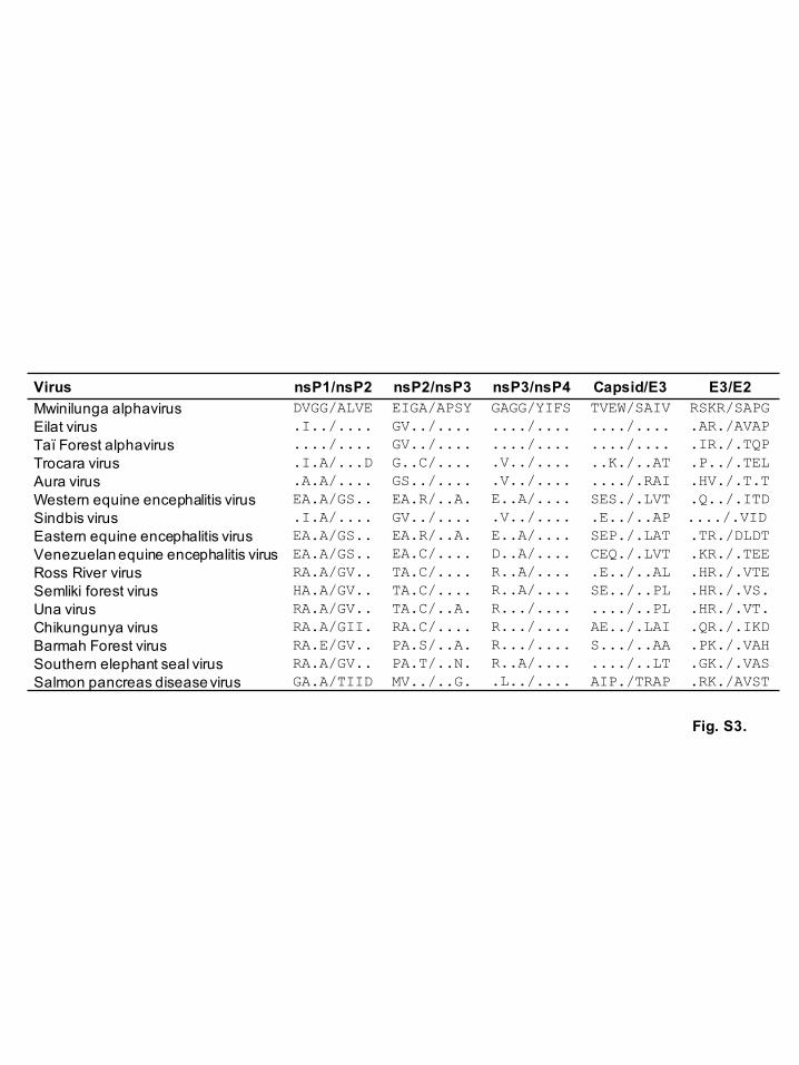
resembled those of EILV (Fig. S3, available in the online Supplementary Material). These
sequence similarities suggest that MWAV may possess similar biological properties to
EILV, which also displays a narrow vector range in mosquitoes (Nasar et al., 2014).
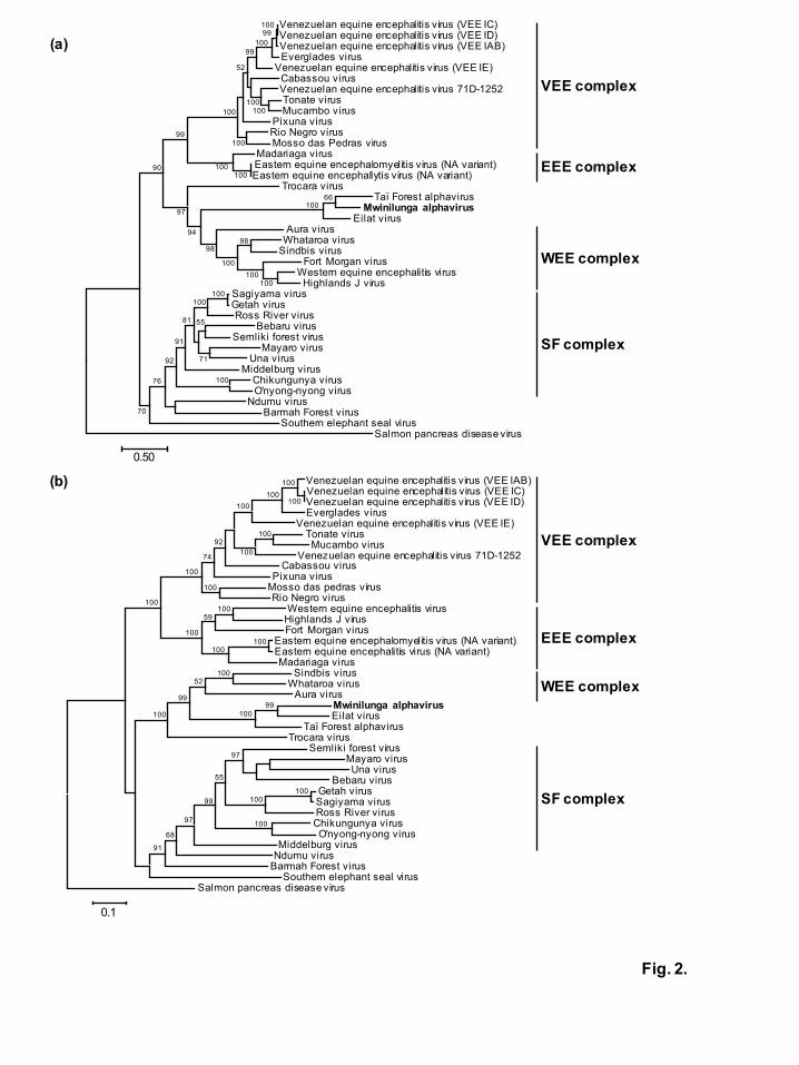
The full-length nucleotide sequences encoding either the nsPs or sPs ORFs of MWAV
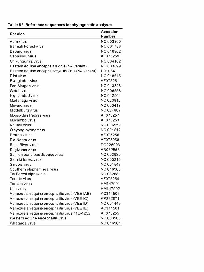
and alphaviruses available from GenBank (Table S2, available in the online
Supplementary Material), were aligned using the ClustalW protocol (Thompson et al.,
1994). For the alignments, the C terminus of nsP3 and the N terminus of capsid sequences
were excluded, since these regions have numerous insertions and deletions and extensive
sequence divergence in previous studies (Forrester et al., 2012; Nasar et al., 2012).
Phylogenetic trees were constructed by maximum-likelihood method using MEGA,
version 6.0 (Tamura et al., 2013). The robustness of the nodes was tested by 1,000
bootstrap replications. In both phylogenies based on nucleotide sequences of nsPs and
sPs, MWAV was classified into the same phylogroup with EILV and TALV (Fig. 2),
suggesting that MWAV belongs to the same antigenic complex with these two viruses in
agreement with the genome analysis (Fig. 1). In both the nsPs and sPs trees, Eilat-like
viruses (EILV, TALV and MWAV) have the same ancestral virus within the Western
equine encephalitis (WEE) complex, placed between Trocara virus (TROV) and Aura
virus (AURAV), as has been reported previously (Hermanns et al., 2017; Nasar et al.,
2012). Although Eilat-like viruses have been detected in the Old World, TROV and
AURAV have also been isolated from mosquitoes in South American countries
(Rümenapf et al., 1995; Travassos da Rosa et al., 2001). Similar to Eilat-like viruses,
vertebrate hosts of both TROV and AURAV have not been identified (Forrester et al.,
Page 11
10
2012).
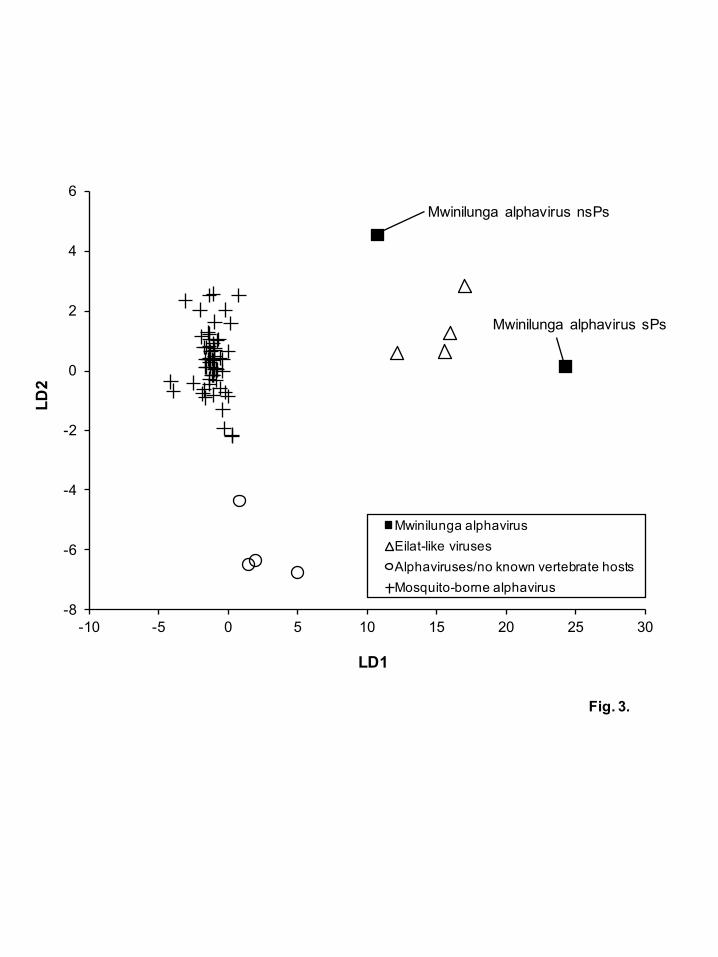
To predict the host range of MWAV, a linear discriminant analysis was performed
based on the dinucleotide ratios in various virus sequences, which has been reported
previously to determine host specificity based on the frequencies of dinucleotide in
flavivirus sequences (Colmant et al., 2017). Complete genome sequences from 53
mosquito-borne alphaviruses with a defined host (bird, horse, donkey, human, hamster
and mosquito) and full length sequences of nsPs and sPs from MWAV, EILV, TALV,
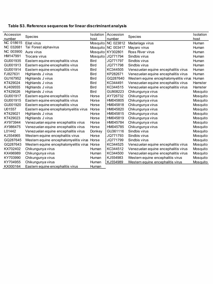
TROV and AURAV were used for the analysis (Table S3, available in the online
Supplementary Material). The dinucleotide ratios were calculated using the formula PXY
= ƒXY/ƒXƒY, in which ƒX and ƒY denote the frequencies of mononucleotides X and Y,
respectively, and ƒXY denotes the frequency of dinucleotide XY (Karlin and Mrázek,
1997; Lobo et al., 2009). The linear discriminant analysis was performed in the R package
(Colmant et al., 2017; Di Giallonardo et al., 2017) (https://www.R-project.org/). The
analysis showed correlations of dinucleotide usage patterns between alphaviruses and
their host ranges (Fig. 3). Mosquito-borne alphaviruses showed similar dinucleotide
usage patterns regardless of their phylogenetic relationships and hosts. On the other hands,
the dinucleotide usage pattern of Eilat-like viruses were also similar to each other,
indicating that MWAV may be a mosquito-specific alphavirus. Interestingly, TROV and
AURAV, who have no known vertebrate hosts showed a different pattern from those of
mosquito-bone alphaviruses and Eilat-like viruses.
If Eilat-like viruses and phylogenetically related viruses have adapted to specific
mosquito hosts and relied on vertical and/or venereal transmission, it is possible that these
viruses have not been spread via vertebrate hosts to other mosquito species. Therefore, 1)
Eilat-like viruses, and phylogenetically related viruses, may exist in different parts of the
world and have lifecycles involving vertical and/or venereal transmission exclusively in
invertebrate mosquito hosts, or 2) Eilat-like viruses might be sporadically transported
Page 12
11
from other areas and based on a previous report, geographic introductions of alphaviruses
may have repeatedly occurred (Forrester et al., 2012). It is possible that Eilat-like viruses
and/or their ancestral viruses might have lost their ability to infect vertebrates after being
spread to other mosquito species in the Old World. Analyses of the biological properties
of MWAV and TALV after isolation will greatly help to clarify the factors that determine
the host range and the evolutionary histories of the Eilat-like alphaviruses.
In summary, we have identified a novel alphavirus, tentatively named MWAV from
Culex quinquefasciatus mosquitoes in Zambia. Genetic analyses revealed that MWAV is
closely related to, but distinct from, Eilat virus, a mosquito-specific virus and key to the
better understanding of the evolution of alphaviruses. Further epidemiological and
functional studies investigating arthropod and animal host species will help to better
characterize future alphaviral disease risk and also provide new insights into their ecology.
Page 13
12
Funding information
This study was supported by grants for Scientific Research (C), (B) from the Ministry of
Education, Culture, Sports, Science and Technology, Japan (MEXT) /Japan Society for
the Promotion of Science (JSPS) KAKENHI (JP15K07715, JP17K00401, JP16H05805);
and grants for Scientific Research on Innovative Areas and International Group from the
MEXT/JSPS KAKENHI (JP16H06431, JP16H06429, JP16K21723), and the Japan
Initiative for Global Research Network on Infectious Diseases, from the Japan Agency
for Medical Research and Development.
Acknowledgements
We appreciate the cooperation of Veterinary officers of the Department of Veterinary
Services, Ministry of Fisheries and Livestock of the Republic of Zambia in each district
for mosquito sampling.
Page 14
13
References
Atkins, G.J., 2013. The Pathogenesis of Alphaviruses. Vol. 2018. ISRN Virology.
Colmant, A.M.G., Hobson-Peters, J., Bielefeldt-Ohmann, H., van den Hurk, A.F., Hall-
Mendelin, S., Chow, W.K., Johansen, C.A., Fros, J., Simmonds, P., Watterson, D.,
Cazier, C., Etebari, K., Asgari, S., Schulz, B.L., Beebe, N., Vet, L.J., Piyasena,
T.B.H., Nguyen, H.D., Barnard, R.T., Hall, R.A., 2017. A New Clade of Insect-
Specific Flaviviruses from Australian Anopheles Mosquitoes Displays Species-
Specific Host Restriction. mSphere 2(4).
Di Giallonardo, F., Schlub, T.E., Shi, M., Holmes, E.C., 2017. Dinucleotide Composition
in Animal RNA Viruses Is Shaped More by Virus Family than by Host Species. J
Virol 91(8).
Fayzulin, R., Frolov, I., 2004. Changes of the secondary structure of the 5' end of the
Sindbis virus genome inhibit virus growth in mosquito cells and lead to
accumulation of adaptive mutations. J Virol 78(10), 4953-4964.
Folmer, O., Black, M., Hoeh, W., Lutz, R., Vrijenhoek, R., 1994. DNA primers for
amplification of mitochondrial cytochrome c oxidase subunit I from diverse
metazoan invertebrates. Mol Mar Biol Biotechnol 3(5), 294-299.
Forrester, N.L., Palacios, G., Tesh, R.B., Savji, N., Guzman, H., Sherman, M., Weaver,
S.C., Lipkin, W.I., 2012. Genome-scale phylogeny of the alphavirus genus
suggests a marine origin. J Virol 86(5), 2729-2738.
Gaffigan, T.V., Wilkerson, R.C., Pecor, J.E., Stoffer, J.A., Anderson, T., Systematic
Catalog of Culicidae. Vol. 2018. The Walter Reed Biosystematics Unit
Griffin, D.E., 2013. Alphaviruses. In: David M. Knipe, P.M.H., Jeffrey I. Cohen, Diane
E. Griffin, Robert A. Lamb, Malcolm A. Martin, Vincent R. Racaniello, Bernard
Roizman (Ed.), Fields Virology 6th edition, 6th ed. Vol. 1. Wolters
Page 15
14
Kluwer/Lippincott Williams & Wilkins, Philadelphia, United States, pp. 651-686.
Hermanns, K., Zirkel, F., Kopp, A., Marklewitz, M., Rwego, I.B., Estrada, A., Gillespie,
T.R., Drosten, C., Junglen, S., 2017. Discovery of a novel alphavirus related to
Eilat virus. J Gen Virol 98(1), 43-49.
Hyde, J.L., Chen, R., Trobaugh, D.W., Diamond, M.S., Weaver, S.C., Klimstra, W.B.,
Wilusz, J., 2015. The 5' and 3' ends of alphavirus RNAs--Non-coding is not non-
functional. Virus Res 206, 99-107.
Karlin, S., Mrázek, J., 1997. Compositional differences within and between eukaryotic
genomes. Proc Natl Acad Sci U S A 94(19), 10227-10232.
Kent, R.J., 2006. The Mosquitoes of Macha, Zambia. Vol. 2018.
Lemm, J.A., Rice, C.M., 1993. Roles of nonstructural polyproteins and cleavage products
in regulating Sindbis virus RNA replication and transcription. J Virol 67(4), 1916-
1926.
Lobo, F.P., Mota, B.E., Pena, S.D., Azevedo, V., Macedo, A.M., Tauch, A., Machado,
C.R., Franco, G.R., 2009. Virus-host coevolution: common patterns of nucleotide
motif usage in Flaviviridae and their hosts. PLoS One 4(7), e6282.
Lwande, O.W., Obanda, V., Bucht, G., Mosomtai, G., Otieno, V., Ahlm, C., Evander, M.,
2015. Global emergence of Alphaviruses that cause arthritis in humans. Infect
Ecol Epidemiol 5, 29853.
Nasar, F., Erasmus, J.H., Haddow, A.D., Tesh, R.B., Weaver, S.C., 2015a. Eilat virus
induces both homologous and heterologous interference. Virology 484, 51-58.
Nasar, F., Gorchakov, R.V., Tesh, R.B., Weaver, S.C., 2015b. Eilat virus host range
restriction is present at multiple levels of the virus life cycle. J Virol 89(2), 1404-
1418.
Nasar, F., Haddow, A.D., Tesh, R.B., Weaver, S.C., 2014. Eilat virus displays a narrow
mosquito vector range. Parasit Vectors 7, 595.
Page 16
15
Nasar, F., Palacios, G., Gorchakov, R.V., Guzman, H., Da Rosa, A.P., Savji, N., Popov,
V.L., Sherman, M.B., Lipkin, W.I., Tesh, R.B., Weaver, S.C., 2012. Eilat virus, a
unique alphavirus with host range restricted to insects by RNA replication. Proc
Natl Acad Sci U S A 109(36), 14622-14627.
Niesters, H.G., Strauss, J.H., 1990. Mutagenesis of the conserved 51-nucleotide region of
Sindbis virus. J Virol 64(4), 1639-1647.
Ortiz, D.I., Anishchenko, M., Weaver, S.C., 2005. Susceptibility of Psorophora confinnis
(Diptera: Culicidae) to infection with epizootic (subtype IC) and enzootic
(subtype ID) Venezuelan Equine encephalitis viruses. J Med Entomol 42(5), 857-
863.
Powers, A., Huang, H., Roehrig, J., Strauss, E., Weaver, S., 2012. Family Togaviridae. In:
King, A.M.Q., Adams, M.J., Carstens, E.B., Lefkowitz, E. (Eds.), Virus
Taxonomy: Ninth Report of the International Committee on Taxonomy of
Viruses. Elsevier Academic Press, Oxford, UK, pp. 1103-1110.
Powers, A.M., Brault, A.C., Shirako, Y., Strauss, E.G., Kang, W., Strauss, J.H., Weaver,
S.C., 2001. Evolutionary relationships and systematics of the alphaviruses. J Virol
75(21), 10118-10131.
Rezza, G., Chen, R., Weaver, S.C., 2017. O'nyong-nyong fever: a neglected mosquito-
borne viral disease. Pathog Glob Health 111(6), 271-275.
Rümenapf, T., Strauss, E.G., Strauss, J.H., 1995. Aura virus is a New World
representative of Sindbis-like viruses. Virology 208(2), 621-633.
Storm, N., Weyer, J., Markotter, W., Kemp, A., Leman, P.A., Dermaux-Msimang, V., Nel,
L.H., Paweska, J.T., 2014. Human cases of Sindbis fever in South Africa, 2006-
2010. Epidemiol Infect 142(2), 234-238.
Tamura, K., Stecher, G., Peterson, D., Filipski, A., Kumar, S., 2013. MEGA6: Molecular
Evolutionary Genetics Analysis version 6.0. Mol Biol Evol 30(12), 2725-2729.
Page 17
16
Thompson, J.D., Higgins, D.G., Gibson, T.J., 1994. CLUSTAL W: improving the
sensitivity of progressive multiple sequence alignment through sequence
weighting, position-specific gap penalties and weight matrix choice. Nucleic
Acids Res 22(22), 4673-4680.
Travassos da Rosa, A.P., Turell, M.J., Watts, D.M., Powers, A.M., Vasconcelos, P.F.,
Jones, J.W., Klein, T.A., Dohm, D.J., Shope, R.E., Degallier, N., Popov, V.L.,
Russell, K.L., Weaver, S.C., Guzman, H., Calampa, C., Brault, A.C., Lemon, A.P.,
Tesh, R.B., 2001. Trocara virus: a newly recognized Alphavirus (Togaviridae)
isolated from mosquitoes in the Amazon Basin. Am J Trop Med Hyg 64(1-2), 93-
97.
Voss, J.E., Vaney, M.C., Duquerroy, S., Vonrhein, C., Girard-Blanc, C., Crublet, E.,
Thompson, A., Bricogne, G., Rey, F.A., 2010. Glycoprotein organization of
Chikungunya virus particles revealed by X-ray crystallography. Nature 468(7324),
709-712.
Weaver, S.C., Reisen, W.K., 2010. Present and future arboviral threats. Antiviral Res
85(2), 328-345.
Webb, C.E., Doggett, S.L., Ritchie, S.A., Russell, R.C., 2008. Vector competence of three
Australian mosquitoes, Verrallina carmenti, Verraullina lineata, and Mansonia
septempunctata (Diptera: Culicidae), for Ross River virus. J Med Entomol 45(4),
737-740.
Zhu, W.Y., Zhao, T., Liang, G.D., 2013. Specific nucleotide changes in the subgenomic
promoter region influence infectivity of the sindbis virus. Intervirology 56(5),
337-342.
Zuker, M., 2003. Mfold web server for nucleic acid folding and hybridization prediction.
Nucleic Acids Res 31(13), 3406-3415.
Chikungunya virus. Centers for Disease Control and Prevention, Atlanta, GA, United
Page 18
17
States. https://www.cdc.gov/chikungunya/index.html (accessed January 2018).
The R project for statistical computing. R Foundation for Statistical Computing, Vienna,
Austria. https://www.R-project.org/ (accessed April 2018).
Page 19
18
Figure Legends
Fig. 1. Genome organization of Mwinilunga alphavirus
The nucleotide lengths of the UTRs and amino acid sizes of each protein are displayed
under the genome. Amino acid sequence identities of each protein between Mwinilunga
alphavirus and closely related alphaviruses are indicated.
Fig. 2. Phylogenetic analysis of Mwinilunga alphavirus with previously described
alphaviruses
Phylogenetic trees were constructed using the maximum-likelihood method in MEGA,
version 6.0 with 1,000 bootstrap replicates based on multiple alignment of nucleotide
sequences of the (a) structural proteins and (b) nonstructural proteins. Bootstrap values
greater than 50 are shown near the branch nodes and the scale bar indicates the number
of substitutions per site.
Fig. 3. Score plot of the linear discriminant analysis
The figure shows a scatterplot of the two discriminant scores that explain the largest
amount of the components from the linear discriminant analysis (72.3% and 16.2% for
LD1 and LD2, respectively). Markers represent values for individual sequences as
follows: complete genome sequences from mosquito-borne alphaviruses (crosses), full
length of nsPs and sPs sequences from Mwinilunga alphavirus (black squares), Eilat-like
viruses (triangles) or alphaviruses with no known vertebrate hosts (circles).
Page 20
41 nt
E1nsP1 nsP2 nsP3 nsP4 E2C
5'-UTR67 nt
3'-UTR417 nt
5' cap Poly (A)
E364 aa
6k55 aa
543 aa 809 aa 256 aa 420 aa479 aa 608 aa 440 aa
Fig. 1.
Amino acid sequence identity to Mwinilunga alphavirus (%)
Virus nsP1 nsP2 nsP3 nsP4 Capsid E3 E2 6k E1Eilat virus 88 83 69 87 75 73 64 83 77Taï Forest alphavirus 83 82 68 87 75 73 71 83 79Sindbis virus 70 64 41 78 55 51 43 47 50
Page 21
Fig. 2.
Venezuelan equine encephalitis virus (VEE IC)Venezuelan equine encephalitis virus (VEE ID)Venezuelan equine encephalitis virus (VEE IAB)Everglades virus
Venezuelan equine encephalitis virus (VEE IE)Cabassou virusVenezuelan equine encephalitis virus 71D-1252Tonate virusMucambo virus
Pixuna virusRio Negro virusMosso das Pedras virus
Madariaga virusEastern equine encephalomyelitis virus (NA variant)Eastern equine encephallytis virus (NA variant)
Trocara virusTaï Forest alphavirus
Mwinilunga alphavirusEilat virus
Aura virusWhataroa virus
Sindbis virusFort Morgan virus
Western equine encephalitis virusHighlands J virus
Sagiyama virusGetah virusRoss River virus
Bebaru virusSemliki forest virus
Mayaro virusUna virus
Middelburg virusChikungunya virusO'nyong-nyong virus
Ndumu virusBarmah Forest virus
Southern elephant seal virusSalmon pancreas disease virus
10066
100
100
10099
100
100
100
98
100
100
100
100
99
100
52
100
100
98
99
94
97
90
100
70
76
92
91
71
5581
0.50
(a)
Venezuelan equine encephalitis virus (VEE IAB)Venezuelan equine encephalitis virus (VEE IC)Venezuelan equine encephalitis virus (VEE ID)Everglades virus
Venezuelan equine encephalitis virus (VEE IE)Tonate virusMucambo virus
Venezuelan equine encephalitis virus 71D-1252Cabassou virus
Pixuna virusMosso das pedras virusRio Negro virus
Western equine encephalitis virusHighlands J virusFort Morgan virus
Eastern equine encephalomyelitis virus (NA variant)Eastern equine encephalitis virus (NA variant)Madariaga virus
Sindbis virusWhataroa virus
Aura virusMwinilunga alphavirusEilat virus
Taï Forest alphavirusTrocara virus
Semliki forest virusMayaro virusUna virus
Bebaru virusGetah virusSagiyama virusRoss River virusChikungunya virusO'nyong-nyong virus
Middelburg virusNdumu virus
Barmah Forest virusSouthern elephant seal virus
Salmon pancreas disease virus
100
100
99100
100
100
100
100
100
100
100
100
100
100
100
100
59
100
92
74
100
100
52
99
100
91
68
97
99
55
97
0.1
(b)
VEE complex
EEE complex
WEE complex
SF complex
VEE complex
EEE complex
WEE complex
SF complex
Page 22
Fig. 3.
Mwinilunga alphavirus nsPs
-8
-6
-4
-2
0
2
4
6
-10 -5 0 5 10 15 20 25 30
LD2
LD1
Mwinilunga alphavirusEilat-like virusesAlphaviruses/no known vertebrate hosts Mosquito-borne alphavirus
Mwinilunga alphavirus sPs
Page 23
Year Place Mosquito genus
Pool no.
Mosquito no.
2014 Lusaka Culex 10 258Mongu Anopheles 2 19
Coquillettidia 6 19Culex 52 1399Mansonia 6 26
2015 Chipata Aedes 1 1Culex 16 306
Kazungula Aedeomya 1 1Aedes 3 5Anopheles 9 38Culex 5 45Mansonia 1 2
Livingstone Aedes 3 12Anopheles 3 4Culex 27 774Mansonia 1 8
Lusaka Aedes 3 20Anopheles 1 1Culex 54 1574Mansonia 2 2
2016 Kitwe Culex 10 275Mongu Aedes 3 4
Anopheles 25 366Coquillettidia 10 153Culex 23 413Mansonia 5 99Uranotaenia 1 2
Mwinilunga Aedes 4 4Anopheles 2 3Culex 6 40Uranotaenia 1 2
Ndola Culex 2 19Siavonga Aedes 22 141
Anopheles 3 3Culex 19 385
2017 Livingstone Aedes 8 12Anopheles 16 73Culex 46 812Mansonia 2 2
Mongu Aedeomya 2 10Aedes 4 11Anopheles 66 1451Coquillettidia 9 62Culex 49 710Mansonia 7 132Uranotaenia 1 1
Total 552 9699
Table S1. Information of collected mosquitoes
Page 24
Species Acession Number
Aura virus NC 003900 Barmah Forest virus NC 001786 Bebaru virus NC 016962 Cabassou virus AF075259Chikungunya virus NC 004162 Eastern equine encephalitis virus (NA variant) NC 003899 Eastern equine encephalomyelitis virus (NA variant) U01034Eilat virus NC 018615 Everglades virus AF075251Fort Morgan virus NC 013528 Getah virus NC 006558 Highlands J virus NC 012561 Madariaga virus NC 023812 Mayaro virus NC 003417 Middelburg virus NC 024887 Mosso das Pedras virus AF075257Mucambo virus AF075253Ndumu virus NC 016959 O'nyong-nyong virus NC 001512 Pixuna virus AF075256Rio Negro virus AF075258Ross River virus DQ226993Sagiyama virus AB032553Salmon pancreas disease virus NC 003930 Semliki forest virus NC 003215 Sindbis virus NC 001547 Southern elephant seal virus NC 016960 Taï Forest alphavirus NC 032681 Tonate virus AF075254Trocara virus HM147991Una virus HM147992Venezuelan equine encephalitis virus (VEE IAB) KC344505Venezuelan equine encephalitis virus (VEE IC) KP282671Venezuelan equine encephalitis virus (VEE ID) NC 001449 Venezuelan equine encephalitis virus (VEE IE) KC344501Venezuelan equine encephalitis virus 71D-1252 AF075255Western equine encephalitis virus NC 003908 Whataroa virus NC 016961
Table S2. Reference sequences for phylogenetic analyses
Page 25
Accession number Species Isolation
hostAccession number Species Isolation
hostNC 018615 Eilat virus Mosquito NC 023812 Madariaga virus HumanNC 032681 Taï Forest alphavirus Mosquito NC 003417 Mayaro virus HumanNC 003900 Aura virus Mosquito KY302801 Ross River virus HumanHM147991 Trocara virus Mosquito JQ771794 Sindbis virus HumanGU001935 Eastern equine encephalitis virus Bird JQ771797 Sindbis virus HumanGU001913 Eastern equine encephalitis virus Bird JQ771798 Sindbis virus HumanGU001914 Eastern equine encephalitis virus Bird KC344505 Venezuelan equine encephalitis virus HumanFJ827631 Highlands J virus Bird KP282671 Venezuelan equine encephalitis virus HumanGU167952 Highlands J virus Bird GQ287640 Western equine encephalomyelitis virus HumanKT429024 Highlands J virus Bird KC344491 Venezuelan equine encephalitis virus HamsterKJ409555 Highlands J virus Bird KC344515 Venezuelan equine encephalitis virus HamsterKT429026 Highlands J virus Bird GU908223 Chikungunya virus MosquitoGU001917 Eastern equine encephalitis virus Horse AY726732 Chikungunya virus MosquitoGU001915 Eastern equine encephalitis virus Horse HM045805 Chikungunya virus MosquitoGU001920 Eastern equine encephalitis virus Horse HM045818 Chikungunya virus MosquitoU01557 Eastern equine encephalomyelitis virus Horse HM045820 Chikungunya virus MosquitoKT429021 Highlands J virus Horse HM045815 Chikungunya virus MosquitoKT429023 Highlands J virus Horse HM045819 Chikungunya virus MosquitoAY973944 Venezuelan equine encephalitis virus Horse HM045784 Chikungunya virus MosquitoAY986475 Venezuelan equine encephalitis virus Horse HM045785 Chikungunya virus MosquitoL01442 Venezuelan equine encephalitis virus Donkey GU361116 Sindbis virus MosquitoKJ554965 Western equine encephalitis virus Horse JQ771793 Sindbis virus MosquitoGQ287645 Western equine encephalomyelitis virus Horse JQ771799 Sindbis virus MosquitoGQ287643 Western equine encephalomyelitis virus Horse KC344525 Venezuelan equine encephalitis virus MosquitoKX702402 Chikungunya virus Human KC344512 Venezuelan equine encephalitis virus MosquitoKX496989 Chikungunya virus Human KC344500 Venezuelan equine encephalitis virus MosquitoKY703990 Chikungunya virus Human KJ554983 Western equine encephalitis virus MosquitoKY704955 Chikungunya virus Human KJ554989 Western equine encephalitis virus MosquitoKX000164 Eastern equine encephalitis virus Human
Table S3. Reference sequences for linear discriminant analysis
Page 26
Fig. S1.
Mwinilunga
LusakaMongu
LivingstoneKazungula
Chipata
Siavonga
KitweNdola
Page 27
Virus nsP1 51 nt CSEMwinilunga alphavirus ACAGGUCACUCCGAAUGACCACGCCAAUGCGAGGGCGUUCUCGCAUUGCGCEilat virus CAU......C.......................A..C.....C........Taï Forest alphavirus C.U......C....................U..A..U..............Trocara virus G.........G.C........U..U.....C..A............CUG..Aura virus G....................U..U.....C..A..U..U......CUG..Western equine encephalitis virus G.........GAC........U........C..A.....U......GUG..Sindbis virus G...........A........U..U.....C..A..A..U......CUG..Eastern equine encephalitis virus G.........GAC........U..U.....U........U.....CCUA..Venezuelan equine encephalitis virus G.........GAU........U..U.....C..A.....U......CUG..Ross River virus G........A..C........U..U.....C..A..U..U......CUG..Semliki forest virus G........A..A........U..A.....C..A..A..U.....CCUG..Una virus G...........A........U..G..C........U........CCU...Chikungunya virus G........A...........U..U.....U..A............CUA..Barmah Forest virus G...AC...A..A..C..U..U..AC.C...........U.....CCUU..Southern elephant seal virus G........A..U........U........C..A..U..U......CUG..Salmon pancreas disease virus CA.UAGGU.GU.U..C.....U...GCC..C..A..U.....C..C.UG..
Virus 3'-UTRMwinilunga alphavirus AUUUUGUUUUUAAUAUUUC
Eilat virus .A.................C
Taï Forest alphavirus ...................
Western equine encephalitis virus .............A.....
Sindbis virus .............C.....
Eastern equine encephalitis virus ...................
Venezuelan equine encephalitis virus ...................
Ross River virus U..G.U............UAC
Semliki forest virus .A..G..............C
Una virus ...GGU.............C
Barmah Forest virus ....GU............UAC
Salmon pancreas disease virus C.A...G......A....UCAAUAC
Virus Subgenomic promoterMwinilunga alphavirus CCCUCUACAACUGACCUAAAUAGU
Eilat virus ............A...........
Taï Forest alphavirus A...G...GGUG............
Trocara virus A.........G.A....U...C..
Aura virus A.......GGUG.U.........A
Western equine encephalitis virus ........GG.............G
Sindbis virus AU......GGUG.U..........
Eastern equine encephalitis virus ........GG....... .....G
Venezuelan equine encephalitis virus .U......GG..A....G...G.A
Ross River virus A.......GG.G.U.........A
Semliki forest virus A.......GG.G.U....G..U.G
Chikungunya virus .UU.G...GG.G.U.........G
Barmah Forest virus AU......GGUG.U..........
Southern elephant seal virus .G......GG...U.........A
Salmon pancreas disease virus ........GU..A....U....U.
Fig. S2.
(b)
(d)
(c)
Virus 5'-UTRMwinilunga alphavirus ACAUGGGGAUAGGCUAUAUAACACACAAU-UAAACCCAGUACCAAAUAGCCUCCCACUUUCAUCGAAU
Eilat virus --------.......GC.C.UU.....U.AG...U.----GUA.CG......U..............C
Taï Forest alphavirus --------..........CG.U.....C.U.....U.GU.GAU.........U...............
(a)
Page 28
Virus nsP1/nsP2 nsP2/nsP3 nsP3/nsP4 Capsid/E3 E3/E2Mwinilunga alphavirus DVGG/ALVE EIGA/APSY GAGG/YIFS TVEW/SAIV RSKR/SAPGEilat virus .I../.... GV../.... ..../.... ..../.... .AR./AVAPTaï Forest alphavirus ..../.... GV../.... ..../.... ..../.... .IR./.TQPTrocara virus .I.A/...D G..C/.... .V../.... ..K./..AT .P../.TELAura virus .A.A/.... GS../.... .V../.... ..../.RAI .HV./.T.TWestern equine encephalitis virus EA.A/GS.. EA.R/..A. E..A/.... SES./.LVT .Q../.ITDSindbis virus .I.A/.... GV../.... .V../.... .E../..AP ..../.VIDEastern equine encephalitis virus EA.A/GS.. EA.R/..A. E..A/.... SEP./.LAT .TR./DLDTVenezuelan equine encephalitis virus EA.A/GS.. EA.C/.... D..A/.... CEQ./.LVT .KR./.TEERoss River virus RA.A/GV.. TA.C/.... R..A/.... .E../..AL .HR./.VTESemliki forest virus HA.A/GV.. TA.C/.... R..A/.... SE../..PL .HR./.VS.Una virus RA.A/GV.. TA.C/..A. R.../.... ..../..PL .HR./.VT.Chikungunya virus RA.A/GII. RA.C/.... R.../.... AE../.LAI .QR./.IKDBarmah Forest virus RA.E/GV.. PA.S/..A. R.../.... S.../..AA .PK./.VAHSouthern elephant seal virus RA.A/GV.. PA.T/..N. R..A/.... ..../..LT .GK./.VASSalmon pancreas disease virus GA.A/TIID MV../..G. .L../.... AIP./TRAP .RK./AVST
Fig. S3.
Page 29
Figure Legends
Fig. S1. The map of study sites in Zambia
Geographic areas where mosquitoes were collected from 2014 to 2017 (black circles) are
shown. The district where the alphavirus-positive mosquito pool was collected in North-
Western Province is indicated by a black star.
Fig. S2. Comparison of deduced conserved sequence elements (CSEs) of
alphaviruses
Alignment of the deduced nucleotide sequences of CSEs were analyzed with the
Mwinilunga alphavirus and selected alphavirus sequences using MEGA, version 6.0.
Alignment of CSE (a) in the 5'-UTR region, (b) in nsP1 51 nt ORF, (c) in subgenomic
promoter and (d) in the 3'-UTR region. Nucleotides identical to Mwinilunga alphavirus
sequence are shown with black dots.
Fig. S3. Comparison of the protease cleavage sites of alphaviruses
Alignment of the amino acid sequences of protease cleavage sites were analyzed with
Mwinilunga alphavirus and selected alphavirus sequences using MEGA, version 6.0.
Conserved residues are indicated with black dots.