
Dissimilatory sulfur metabolism coupled to anaerobic oxidation of methane
Dissertation
zur Erlangung des Grades eines Doktors der Naturwissenschaften – Dr. rer. nat. –
dem Fachbereich Biologie/Chemie der Universität Bremen
vorgelegt von
Jana Milu ká
Bremen 2011
i
Diese Arbeit wurde im Rahmen des Programms „The International Max Planck Research School of Marine Microbiology (MarMic)“ von September 2006 bis Februar 2011 in der Abteilung Mikrobiologie am Max-Planck-Institut für Marine Mikrobiologie in Bremen angefertigt. 1. Gutachter: Prof. Dr. Rudolf Amann
University of Bremen, Bremen Max Planck Institute for Marine Microbiology, Bremen
2. Gutachter: Prof. Dr. Ir. Marc Strous
Radboud University, Nijmegen University of Bielefeld, Bielefeld Max Planck Institute for Marine Microbiology, Bremen
Tag des Promotionskolloquiums: 18. Februar 2011
ii
TABLE of CONTENTS Abstract 1
Zusammenfassung 3
Abbreviations 5
Chapter 1 7 General Introduction 7 Aims and Objectives 23
Chapter 2 25 Bacterial enzymes for dissimilatory sulfate reduction in a marine microbial mat (Black Sea) mediating anaerobic oxidation of methane
Chapter 3 49 Immunological detection of enzymes for sulfate reduction in bacterial cells of anaerobic methane-oxidizing microbial consortia
Chapter 4 69 Sulfur cycling between the archaea and bacteria involved in anaerobic oxidation of methane
Chapter 5 95 Conclusions and Discussion 92 Perspectives 102
References 104
List of Publications 124
Acknowledgements 125

Abstract
1
ABSTRACT
”Something unknown is doing we don't know what.”
Sir Arthur Stanley Eddington (1882–1944)
The seafloor and its microbial inhabitants play an important role in the
biogeochemical cycling of elements. These environments are generally anoxic but
contain high concentrations of sulfate penetrating from the overlying seawater. The
main carbon mineralization processes – such as the anaerobic oxidation of methane
(AOM; Eq. 1) – are therefore generally coupled to sulfate reduction.
CH4 + SO42–
HCO3– + HS
– + H2O (Eq. 1)
AOM plays a crucial role in both carbon and sulfur cycling. It oxidizes the majority of
the methane – a potent greenhouse gas – diffusing from the seafloor and prevents its
escape to the atmosphere. Methane oxidation also returns the carbon ‘trapped’ in the
form of recalcitrant methane back to the carbon cycle as carbon dioxide. The AOM-
coupled sulfate reduction consumes a large portion of the downwards sulfate flux and
forms sulfide, which diffuses upwards towards the seafloor where it supports free-
living sulfide- and sulfur-oxidizers but also gutless worms, clams and mussels that
rely for their nutrition on the thiotrophic symbionts. Despite the pronounced effect of
AOM on the sediment geochemistry little is known about its biology. The organisms
responsible for AOM – a consortium of methanotrophic archaea and
Deltaproteobacteria – have been identified in situ but their slow metabolism
complicates growing them in pure cultures and renders the physiological
investigations challenging. So far, AOM research has predominantly focused on the
C1 metabolism of the methanotrophic archaea. The investigations presented in this
thesis address the dissimilatory sulfur metabolism of the organisms involved in AOM
and the mechanisms of its coupling to methane oxidation.
Chapters 2 and 3 describe the purification and characterization of the three
known enzymes involved in dissimilatory sulfate reduction (SR enzymes; ATP
sulfurylase, APS reductase, sulfite reductase). The enzymes were purified from a
naturally enriched microbial mat using liquid chromatography. The identity of the SR
Abstract
2
enzymes was confirmed by N-terminal amino acid sequencing and their activity – in
total cell extracts as well as in individual chromatography fractions – was quantified
by corresponding enzyme essays. Our aim was to assign these enzymes to a
particular organism in the mat sample. For this purpose, polyclonal antibodies
against the purified ATP sulfurylase and sulfite reductase were used – APS
reductase could not be sufficiently purified for antibody generation – in situ in the
original environmental sample as well as in our other enrichment cultures. This
combination of “environmental proteomics” and immunolocalization allowed us to
unambiguously assign the isolated SR enzymes exclusively to the bacterial partner.
The archaea did not express detectable amounts of the identified SR enzymes
themselves and therefore likely depend on their bacterial partners to perform the
sulfate reduction. These results are presented as manuscripts in revision (Manuscript
1) and in preparation (Manuscript 2).
The following Chapter 4 introduces experiments that were performed in order
to elucidate sulfur transfer and speciation in AOM consortia. We used stable and
radioactive sulfur isotopes to follow sulfur exchange between the medium and
biomass and on a single cell level among individual cells. Based on our results and
thermodynamic consideration we propose a model, in which DSS bacteria reduce
sulfate to a zerovalent sulfur compound (probably polysulfide) that might be utilized
by ANME as an electron acceptor for methane oxidation. Thus, unexpectedly, ANME
participate in the dissimilatory sulfur metabolism coupled to AOM. Our combined data
suggest that ANME obtain this compound from the associated bacteria. Such sulfur
shuttling between two organisms not only represents a unique mechanism for a
syntrophic relationship but also has significant implications for our understanding of
sulfur transformations in the AOM zones in marine sediments. These results are
presented as a manuscript in preparation (Manuscript 3).
Zusammenfassung
3
ZUSAMMENFASSUNG
Der Meeresboden und dessen Bewohner spielen eine wichtige Rolle im
biogeochemischen Kreislauf der Elemente. Diese Umgebungen sind im Generellen
anoxisch jedoch enthalten sie hohe Konzentrationen von Sulfat welches aus dem
Meerwasser in das Sediment diffundiert. Die hauptsächlichen Prozesse der
Kohlenstoffmineralisierung, wie u.a die anaerobe Oxidation von Methan (anaerobic
oxidation of methane, AOM), sind daher an die Reduktion von Sulfat gekoppelt.
AOM spielt eine entscheidende Rolle sowohl im Kohlenstoff- als auch im
Schwefelstoffkreislauf. Dabei wird der größte Teil des aus dem Meeresboden
aufsteigenden Methans, ein relevantes Treibhausgas, oxidiert und ein Entweichen in
die Atmosphäre verhindert. Die Methanoxidation ist auch daher von Bedeutung, da
der stabile Kohlenwasserstoff (Methan) zu Kohlendioxid umgewandelt und so erneut
dem Kohlenstoffkreislauf zugeführt wird. Als Konsequenz der AOM-abhängigen
Sulfatreduktion werden große Mengen Sulfid gebildet, welches in Richtung
Sedimentoberfläche diffundiert und dort Substrat für freilebende und symbiotische
Sulfid- und Schwefeloxidierer ist. Trotz der wichtigen Bedeutung der AOM als
geochemischer Prozess ist wenig über dessen Biologie bekannt. Die für die AOM
verantwortlichen Organismen – ein Konsortium aus methanotrophen Archaeen
(ANME) und Deltaproteobakterien – wurden in situ identifiziert aber aufgrund ihres
langsamen Wachstums nicht als Reinkultur gewonnen, welches physiologische
Untersuchungen erschwert. Bisher haben sich Studien zur AOM überwiegend auf
den C1-Metabolismus der beteiligten Archaeen fokussiert. Daher sollte in der
vorliegenden Arbeit der Metabolismus der dissimilatorischen Sulfatreduktion, die
daran beteiligten Organismen und der Mechanismus der Kopplung von
Sulfatreduktion an die Methanoxidation untersucht werden.
Kapitel 2 und 3 dieser Arbeit beschreiben die Aufreinigung und
Charakterisierung der für die dissimilatorische Sulfatreduktion verantwortlichen
Enzyme (SR Enzyme; ATP-Sulfurylase, APS-Reduktase, Sulfitreduktase). Die
Enzyme wurden aus einer natürlich angereicherten mikrobiellen Matte durch
Verwendung chromatographischer Methoden aufgereinigt. Die Identität der Enzyme
Zusammenfassung
4
wurde durch N-terminale Aminosäuresequenzierung bestätigt und deren Aktivität,
sowohl im gesamten Zellextrakt als auch in individuellen chromatographisch
getrennten Fraktionen wurde durch Enzymessays quantifiziert. Als nächstes sollten
die Enzyme einzelnen Organismen der mikrobiellen Matte zugeordnet werden. Dazu
wurden polyklonale Antikörper gegen die aufgereinigte ATP-Sulfurylase und
Sulfitreduktase gewonnen und in situ in der mikrobiellen Matte und anderen AOM-
Anreicherungen eingesetzt. Für die APS-Reduktase konnten keine Antikörper
gewonnen werden, da sich das Enzym nicht genügend aufreinigen ließ. Die
Kombination von „Umwelt-Proteomik“ und Immunolokalisation erlaubte die eindeutige
Zuordnung der Enzyme ausschließlich zu dem bakteriellen Partner in den AOM-
Konsortien. In den Archaeen konnten die identifizierten SR-Enzyme nicht
nachgewiesen werden was darauf hindeutet, dass diese von ihren bakteriellen
sulfatreduzierenden Partnern abhängig sind. Diese Ergebnisse sind in den
Manuskripten in Revision (Manuskript 1) und in Vorbereitung (Manuskript 2)
zusammengefasst.
Im Kapitel 4 werden Experimente vorgestellt, die den Schwefeltransport
innerhalb der AOM-Konsortien aufklären sollten. Durch Verwendung von stabilen und
radioaktiven Schwefelisotopen wurde der Austausch des Schwefels zwischen
Medium und Biomasse, bis hin zu individuellen Zellen untersucht. Basierend an
unseren Ergebnissen und thermodynamischen Betrachtungen schlagen wir ein
Modell vor, wo die DSS Bakterien Sulfat zu Schwefel der Oxidationsstufe 0
(vermutlich Polysulfid) reduzieren, welcher von den ANME-Archaeen als Elektronen-
Akzeptor für die Methanoxidation genutzt werden kann. Als ein unerwarteter Befund,
scheinen die ANME an dem Prozess der dissimilatorischen Sulfatreduktion gekoppelt
an AOM teilzuhaben. Unsere Daten lassen uns vermuten dass die Verbindungen von
dem bakteriellen Partner bereitgestellt werden. Ein solcher Austausch von
Schwefelverbindungen wurde hier das erste Mal für eine syntrophe Partnerschaft
gezeigt und hat auch signifikante Auswirkung für das Verständnis der Umwandlung
von Schwefelspezies in AOM aktiven marinen Sedimenten. Diese Ergebnisse sind im
Manuskript in Vorbereitung (Manuskript 3) dargestellt.
Abbreviations
5
ABBREVIATIONS
Acd acyl-CoA dehydrogenase
Acs acetyl-CoA-synthase
ANME anaerobic methanotrophic Archaea
AOM anaerobic oxidation of methane
APS adenosine-5’-phosphosulfate
ATP (ADP, AMP) adenosine-5’-tri(di, mono)phosphate
CoA coenzyme A
CoB coenzyme B (7-mercaptoheptanoylthreoninephosphate)
CODH carbon monoxide dehydrogenase
CoM coenzyme M (2-mercaptoethanesulfonate)
Cy (2,3,5) cyanine fluorescent dyes
H4MPT tetrahydromethanopterin
Da dalton
DIC dissolved inorganic carbon
Dsr dissimilatory sulfite reductase
E.coli Escherichia coli
Eq. equation
Fig. figure
FITC fluorescein isothiocyanate
Fmd formyl-MFR dehydrogenase
Fpo H2F420:phenazine oxidoreductase
Fqo H2F420:quinone oxidoreductase
Fsr coenzyme F420-dependent sulfite reductase
Ftr formyl-MFR:H4MPT formyltransferase
GC-MS gas chromatography mass spectrometry
Hdr heterodisulfide reductase
HPLC high performance liquid chromatography
IgG immunoglobulin G
kJ kilojoule
Abbreviations
6
Mch methenyl-H4MPT cyclohydrolase
Mcr methyl-CoM-reductase
Mer methylene H4MPT-reductase
MeSH methylsulfide
MFR methanofuran
Mtd F420-dependent methylene-H4MPT dehydrogenase
Mtr methyl-H4MPT:CoM methyl-transferase
mV millivolt
MV mud volcano
OM organic matter
PDB Pee Dee Belemnite
pers. comm. personal communication
(r)DNA (ribosomal) deoxyribonucleic acid
(r)TCA (reverse) tricarboxylic acid cycle
SIMS secondary ion mass spectrometry
SMTZ sulfate methane transition zone
SR sulfate reduction (evtl. sulfate reducing)
Tg teragram
TRITC rhodamine isothiocyanate
XANES X-ray absorption near-edge structure
General Introduction Chapter 1
7
CHAPTER 1 GENERAL INTRODUCTION
”Happy are they who are starting now.”
Martinus Willem Beijerinck (1851-1931)
The flux of organic matter (OM) in the ocean is mainly
vertical; from the primary producers in the photic zone to the
terminal consumers in the sediment. The (micro)organisms in the
water column and in the sediment-overlying benthic boundary layer
consume most of the OM formed in the sunlit surface waters and
therefore only a part of the sinking OM reaches the seafloor and
enters the (predominantly) anoxic world in it. The OM
mineralization in these anoxic environments proceeds through
several stages that involve fermenting, acetogenic, denitrifying,
sulfate-reducing and methanogenic microorganisms. The ‘left-over’
sedimentary OM is thermogenically converted to petroleum and
natural gas. The cycle of OM mineralization gets completed when
these hydrocarbons seep back to the above-lying sediment and
become oxidized to CO2.
The OM is subjected to many rounds of alteration and
degradation during sedimentation and burial and might therefore be
refractive to further mineralization. However, substrates that are not
degradable by individual microorganisms can still be degraded
through combined activity of metabolically different microorganisms.
Such an unique lifestyle has been termed obligate syntrophy.
Generally, the substrate is partially degraded by one organism to
an intermediate which is scavenged by the second organism. The
intermediates are small molecules capable of rapid diffusion like –
in most instances – hydrogen. Occasionally, also formate and
1. Microbial
degradation of organic matter
1.1
Syntrophic
metabolism
Chapter 1 General Introduction
8
acetate have been reported to serve as syntrophic intermediates; in
methanogenic consortia (Bleicher and Winter, 1994) and syntrophic
acetone and benzoate degradation, respectively (Platen & Schink,
1987; Warikoo et al., 1996). Concentrations of these intermediates
are kept low through their consumption by the second organism.
Low intermediate concentrations prevent thermodynamic end-
product inhibition and increase the energy gain of the first organism
which allows syntrophs to perform reactions that are under
standard conditions endergonic. The syntrophic cooperation
requires that the free energy obtained from the substrate oxidation
is shared among involved metabolic partners. Each of the two
partners has to gain at least an equivalent of the biological energy
quantum – the minimal metabolically conservable amount of energy
(i.e. –20 kJ per mol ATP for E. coli; Thauer et al., 1977). However,
syntrophs can catalyze also reactions which provide less free
energy than –40 kJ (i.e. 2 x –20 kJ) per mol substrate. This is due
to the fact that the increment energy required for ATP synthesis for
syntrophic organisms is probably much lower than for growing E.
coli. For example methanogenic archaea require under
physiological conditions a minimal free energy change of only –10
kJ (mol CH4)-1 (Hoehler et al., 2001). Methanogens have a limited
range of relatively simple substrates which makes them dependent
on other anaerobic organisms and, consequently, very common
syntrophic partners. Through methanogenic activity a large part of
the energy locked in decaying biomass is stored as methane,
forming vast reservoirs in the seafloor (107 Tg carbon; Dickens,
2003).
Of all the methane released from the reservoirs only around
10% successfully reaches oxic waters and, eventually, the
atmosphere because most of the methane is efficiently oxidized to
CO2 in anoxic marine sediments.
General Introduction Chapter 1
9
Dep
th
SMTZ
CH4
SO4
Concentration
2
oxic
anoxic
Dep
th
SMTZ
CH4
SO4
Concentration
2
oxic
anoxic
CH4 + SO42– HCO3
– + HS– + H2O (Eq.1)
G°’= –16.6 kJ.mol–1
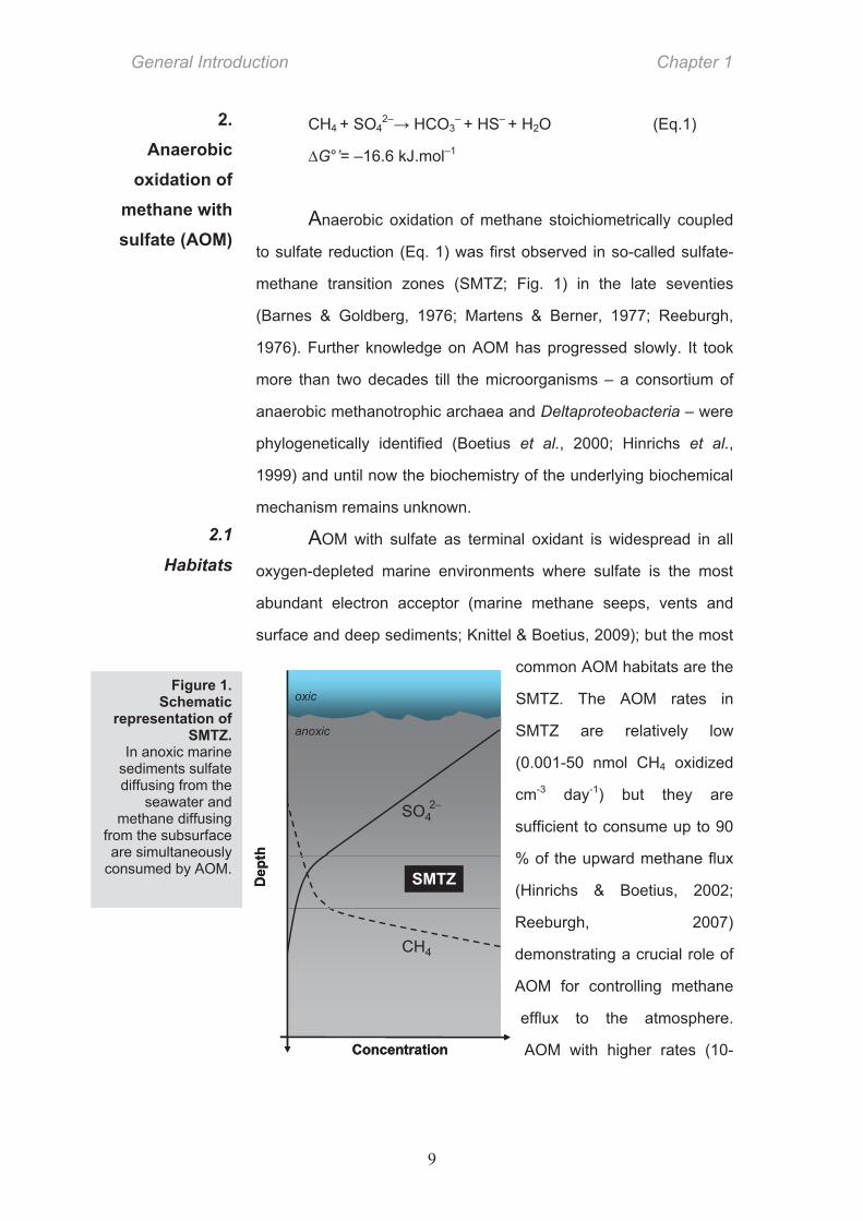
Anaerobic oxidation of methane stoichiometrically coupled
to sulfate reduction (Eq. 1) was first observed in so-called sulfate-
methane transition zones (SMTZ; Fig. 1) in the late seventies
(Barnes & Goldberg, 1976; Martens & Berner, 1977; Reeburgh,
1976). Further knowledge on AOM has progressed slowly. It took
more than two decades till the microorganisms – a consortium of
anaerobic methanotrophic archaea and Deltaproteobacteria – were
phylogenetically identified (Boetius et al., 2000; Hinrichs et al.,
1999) and until now the biochemistry of the underlying biochemical
mechanism remains unknown.
AOM with sulfate as terminal oxidant is widespread in all
oxygen-depleted marine environments where sulfate is the most
abundant electron acceptor (marine methane seeps, vents and
surface and deep sediments; Knittel & Boetius, 2009); but the most
common AOM habitats are the
SMTZ. The AOM rates in
SMTZ are relatively low
(0.001-50 nmol CH4 oxidized
cm-3 day-1) but they are
sufficient to consume up to 90
% of the upward methane flux
(Hinrichs & Boetius, 2002;
Reeburgh, 2007)
demonstrating a crucial role of
AOM for controlling methane
efflux to the atmosphere.
AOM with higher rates (10-
2. Anaerobic
oxidation of methane with sulfate (AOM)
2.1
Habitats
Figure 1. Schematic
representation of SMTZ.
In anoxic marine sediments sulfate diffusing from the
seawater and methane diffusing
from the subsurface are simultaneously
consumed by AOM.
Chapter 1 General Introduction
10
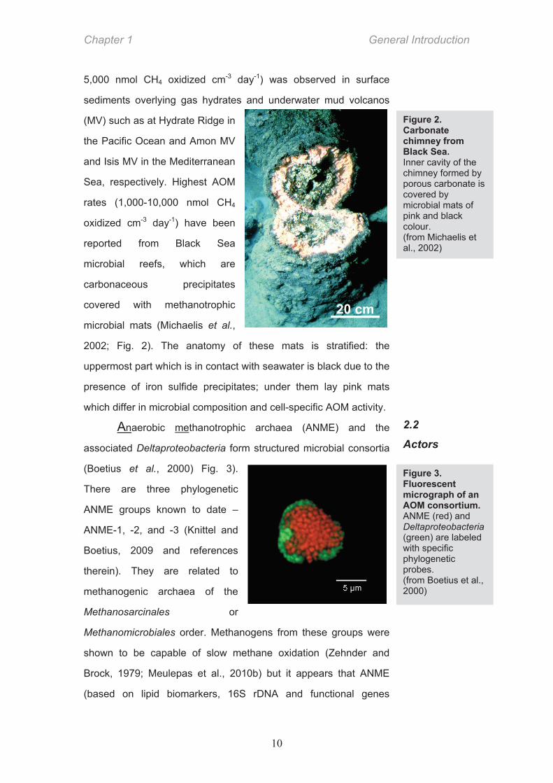
5,000 nmol CH4 oxidized cm-3 day-1) was observed in surface
sediments overlying gas hydrates and underwater mud volcanos
(MV) such as at Hydrate Ridge in
the Pacific Ocean and Amon MV
and Isis MV in the Mediterranean
Sea, respectively. Highest AOM
rates (1,000-10,000 nmol CH4
oxidized cm-3 day-1) have been
reported from Black Sea
microbial reefs, which are
carbonaceous precipitates
covered with methanotrophic
microbial mats (Michaelis et al.,
2002; Fig. 2). The anatomy of these mats is stratified: the
uppermost part which is in contact with seawater is black due to the
presence of iron sulfide precipitates; under them lay pink mats
which differ in microbial composition and cell-specific AOM activity.
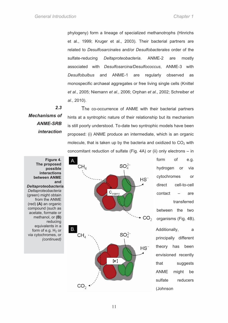
Anaerobic methanotrophic archaea (ANME) and the
associated Deltaproteobacteria form structured microbial consortia
(Boetius et al., 2000) Fig. 3).
There are three phylogenetic
ANME groups known to date –
ANME-1, -2, and -3 (Knittel and
Boetius, 2009 and references
therein). They are related to
methanogenic archaea of the
Methanosarcinales or
Methanomicrobiales order. Methanogens from these groups were
shown to be capable of slow methane oxidation (Zehnder and
Brock, 1979; Meulepas et al., 2010b) but it appears that ANME
(based on lipid biomarkers, 16S rDNA and functional genes
2.2
Actors
Figure 3. Fluorescent micrograph of an AOM consortium. ANME (red) and Deltaproteobacteria (green) are labeled with specific phylogenetic probes. (from Boetius et al., 2000)
Figure 2. Carbonate chimney from Black Sea. Inner cavity of the chimney formed by porous carbonate is covered by microbial mats of pink and black colour. (from Michaelis et al., 2002)
General Introduction Chapter 1
11
phylogeny) form a lineage of specialized methanotrophs (Hinrichs
et al., 1999; Kruger et al., 2003). Their bacterial partners are
related to Desulfosarcinales and/or Desulfobacterales order of the
sulfate-reducing Deltaproteobacteria. ANME-2 are mostly
associated with Desulfosarcina/Desulfococcus, ANME-3 with
Desulfobulbus and ANME-1 are regularly observed as
monospecific archaeal aggregates or free living single cells (Knittel
et al., 2005; Niemann et al., 2006; Orphan et al., 2002; Schreiber et
al., 2010).
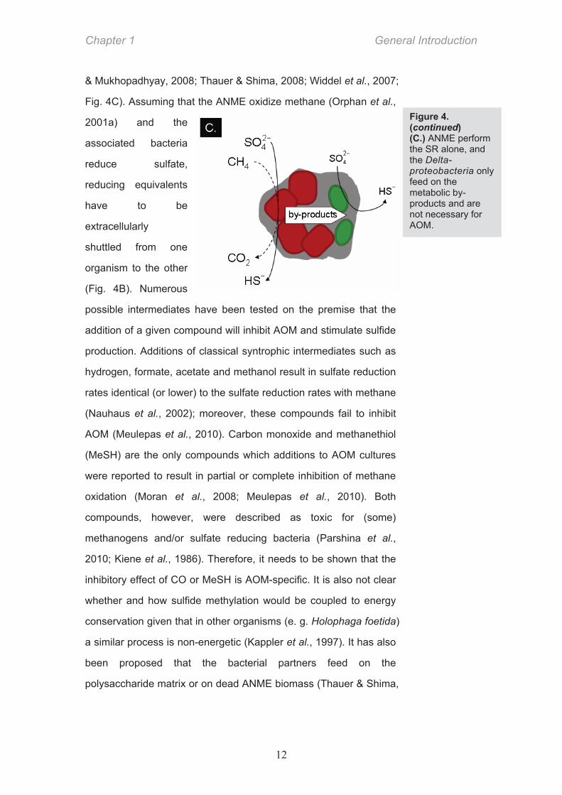
The co-occurrence of ANME with their bacterial partners
hints at a syntrophic nature of their relationship but its mechanism
is still poorly understood. To-date two syntrophic models have been
proposed: (i) ANME produce an intermediate, which is an organic
molecule, that is taken up by the bacteria and oxidized to CO2 with
concomitant reduction of sulfate (Fig. 4A) or (ii) only electrons – in
form of e.g.
hydrogen or via
cytochromes or
direct cell-to-cell
contact – are
transferred
between the two
organisms (Fig. 4B).
Additionally, a
principally different
theory has been
envisioned recently
that suggests
ANME might be
sulfate reducers
(Johnson
2.3
Mechanisms of
ANME-SRB
interaction
Figure 4. The proposed
possible interactions
between ANME and
Deltaproteobacteria.Deltaproteobacteria (green) might obtain
from the ANME (red) (A) an organic compound (such as acetate, formate or
methanol, or (B) reducing
equivalents in a form of e.g. H2 or
via cytochromes, or (continued)
Chapter 1 General Introduction
12
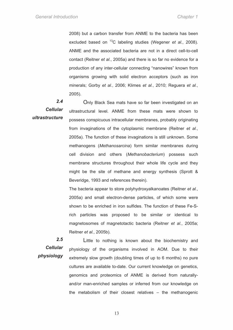
& Mukhopadhyay, 2008; Thauer & Shima, 2008; Widdel et al., 2007;
Fig. 4C). Assuming that the ANME oxidize methane (Orphan et al.,
2001a) and the
associated bacteria
reduce sulfate,
reducing equivalents
have to be
extracellularly
shuttled from one
organism to the other
(Fig. 4B). Numerous
possible intermediates have been tested on the premise that the
addition of a given compound will inhibit AOM and stimulate sulfide
production. Additions of classical syntrophic intermediates such as
hydrogen, formate, acetate and methanol result in sulfate reduction
rates identical (or lower) to the sulfate reduction rates with methane
(Nauhaus et al., 2002); moreover, these compounds fail to inhibit
AOM (Meulepas et al., 2010). Carbon monoxide and methanethiol
(MeSH) are the only compounds which additions to AOM cultures
were reported to result in partial or complete inhibition of methane
oxidation (Moran et al., 2008; Meulepas et al., 2010). Both
compounds, however, were described as toxic for (some)
methanogens and/or sulfate reducing bacteria (Parshina et al.,
2010; Kiene et al., 1986). Therefore, it needs to be shown that the
inhibitory effect of CO or MeSH is AOM-specific. It is also not clear
whether and how sulfide methylation would be coupled to energy
conservation given that in other organisms (e. g. Holophaga foetida)
a similar process is non-energetic (Kappler et al., 1997). It has also
been proposed that the bacterial partners feed on the
polysaccharide matrix or on dead ANME biomass (Thauer & Shima,
Figure 4. (continued) (C.) ANME perform the SR alone, and the Delta- proteobacteria only feed on the metabolic by-products and are not necessary for AOM.
General Introduction Chapter 1
13
2008) but a carbon transfer from ANME to the bacteria has been
excluded based on 13C labeling studies (Wegener et al., 2008).
ANME and the associated bacteria are not in a direct cell-to-cell
contact (Reitner et al., 2005a) and there is so far no evidence for a
production of any inter-cellular connecting “nanowires” known from
organisms growing with solid electron acceptors (such as iron
minerals; Gorby et al., 2006; Klimes et al., 2010; Reguera et al.,
2005).
Only Black Sea mats have so far been investigated on an
ultrastructural level. ANME from these mats were shown to
possess conspicuous intracellular membranes, probably originating
from invaginations of the cytoplasmic membrane (Reitner et al.,
2005a). The function of these invaginations is still unknown. Some
methanogens (Methanosarcina) form similar membranes during
cell division and others (Methanobacterium) possess such
membrane structures throughout their whole life cycle and they
might be the site of methane and energy synthesis (Sprott &
Beveridge, 1993 and references therein).
The bacteria appear to store polyhydroxyalkanoates (Reitner et al.,
2005a) and small electron-dense particles, of which some were
shown to be enriched in iron sulfides. The function of these Fe-S-
rich particles was proposed to be similar or identical to
magnetosomes of magnetotactic bacteria (Reitner et al., 2005a;
Reitner et al., 2005b).
Little to nothing is known about the biochemistry and
physiology of the organisms involved in AOM. Due to their
extremely slow growth (doubling times of up to 6 months) no pure
cultures are available to-date. Our current knowledge on genetics,
genomics and proteomics of ANME is derived from naturally-
and/or man-enriched samples or inferred from our knowledge on
the metabolism of their closest relatives – the methanogenic
2.5
Cellular
physiology
2.4
Cellular
ultrastructure
Chapter 1 General Introduction
14
archaea. Our knowledge on the physiology of the ANME-
associated Deltaproteobacteria is also very scarce.
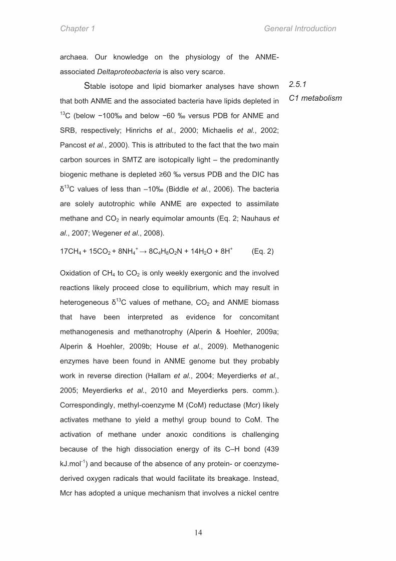
Stable isotope and lipid biomarker analyses have shown
that both ANME and the associated bacteria have lipids depleted in 13C (below 100‰ and below 60 ‰ versus PDB for ANME and
SRB, respectively; Hinrichs et al., 2000; Michaelis et al., 2002;
Pancost et al., 2000). This is attributed to the fact that the two main
carbon sources in SMTZ are isotopically light – the predominantly
biogenic methane is depleted 60 ‰ versus PDB and the DIC has 13C values of less than –10‰ (Biddle et al., 2006). The bacteria
are solely autotrophic while ANME are expected to assimilate
methane and CO2 in nearly equimolar amounts (Eq. 2; Nauhaus et
al., 2007; Wegener et al., 2008).
17CH4 + 15CO2 + 8NH4+ 8C4H8O2N + 14H2O + 8H+ (Eq. 2)
Oxidation of CH4 to CO2 is only weekly exergonic and the involved
reactions likely proceed close to equilibrium, which may result in
heterogeneous 13C values of methane, CO2 and ANME biomass
that have been interpreted as evidence for concomitant
methanogenesis and methanotrophy (Alperin & Hoehler, 2009a;
Alperin & Hoehler, 2009b; House et al., 2009). Methanogenic
enzymes have been found in ANME genome but they probably
work in reverse direction (Hallam et al., 2004; Meyerdierks et al.,
2005; Meyerdierks et al., 2010 and Meyerdierks pers. comm.).
Correspondingly, methyl-coenzyme M (CoM) reductase (Mcr) likely
activates methane to yield a methyl group bound to CoM. The
activation of methane under anoxic conditions is challenging
because of the high dissociation energy of its C–H bond (439
kJ.mol-1) and because of the absence of any protein- or coenzyme-
derived oxygen radicals that would facilitate its breakage. Instead,
Mcr has adopted a unique mechanism that involves a nickel centre
2.5.1
C1 metabolism
General Introduction Chapter 1
15
CHO–MFR
methyl transferase
methyl-CoM reductase
formyldehydrogenase
CO2
formyl transferase
CH4
CH3–S–CoM
CH2=H4MPT
CHO–H4MPT
CH3–H4MPT
CH≡H4MPT
pyruvate
cellularbiomass
CO dehydrogenase/acetyl-CoA synthase
pyruvate dehydrogenase
methylene reductase
methylene dehydrogenase
methenylcyclohydrolase
CO2
CO2
acetyl–CoA
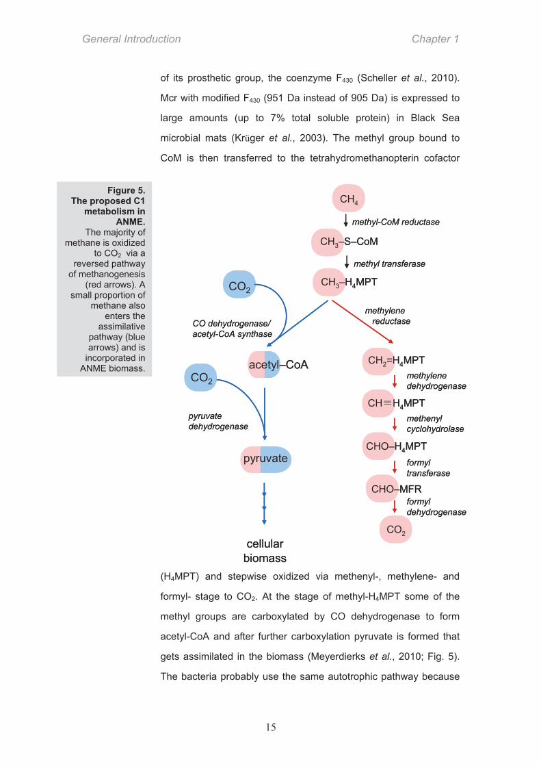
of its prosthetic group, the coenzyme F430 (Scheller et al., 2010).
Mcr with modified F430 (951 Da instead of 905 Da) is expressed to
large amounts (up to 7% total soluble protein) in Black Sea
microbial mats (Krüger et al., 2003). The methyl group bound to
CoM is then transferred to the tetrahydromethanopterin cofactor
(H4MPT) and stepwise oxidized via methenyl-, methylene- and
formyl- stage to CO2. At the stage of methyl-H4MPT some of the
methyl groups are carboxylated by CO dehydrogenase to form
acetyl-CoA and after further carboxylation pyruvate is formed that
gets assimilated in the biomass (Meyerdierks et al., 2010; Fig. 5).
The bacteria probably use the same autotrophic pathway because
Figure 5. The proposed C1
metabolism in ANME.
The majority of methane is oxidized
to CO2 via a reversed pathway
of methanogenesis (red arrows). A
small proportion of methane also
enters the assimilative
pathway (blue arrows) and is
incorporated in ANME biomass.
Chapter 1 General Introduction
16
assimilatorysulfite reductase
dissimilatory sulfite reductase
SO4
AMP SO4
HSO3
2
2
ATP
PPi
HS
out
inmembrane
SO42
HSO3
O3P AMP SO42
HS
ATPsulfurylase
APS kinase
PAPS reductase
APS reductase
sulfate transporter
cysteine
2
assimilatorysulfite reductase
dissimilatory sulfite reductase
SO4
AMP SO4
HSO3
2
2
ATP
PPi
HS
out
inmembrane
SO42
HSO3
O3P AMP SO42
HS
ATPsulfurylase
APS kinase
PAPS reductase
APS reductase
sulfate transporter
cysteine
2
the observed isotopic offset between their biomass and ambient
DIC (~30-40‰; House et al., 2009) is in agreement with the isotope
fractionation observed to be generated by the CODH/Acd pathway
in other bacteria (House et al., 2000; Sirevag et al., 1977).
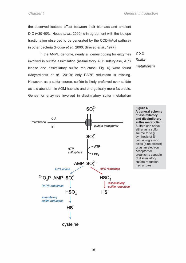
In the ANME genome, nearly all genes coding for enzymes
involved in sulfate assimilation (assimilatory ATP sulfurylase, APS
kinase and assimilatory sulfite reductase; Fig. 6) were found
(Meyerdierks et al., 2010); only PAPS reductase is missing.
However, as a sulfur source, sulfide is likely preferred over sulfate
as it is abundant in AOM habitats and energetically more favorable.
Genes for enzymes involved in dissimilatory sulfur metabolism
2.5.2
Sulfur
metabolism
Figure 6. A general scheme of assimilatory and dissimilatory sulfur metabolism. Sulfate can serve either as a sulfur source for e.g. synthesis of S-containing amino acids (blue arrows) or as an electron acceptor for organisms capable of dissimilatory sulfate reduction (red arrows).
General Introduction Chapter 1
17
(dissimilatory ATP sulfurylase, APS reductase and dissimilatory
sulfite reductase; Fig. 6) were not identified in ANME genomes
(Meyerdierks et al., 2010) and the capacity of ANME or the
associated bacteria to reduce sulfate has not yet been confirmed
either.
Interestingly, other genes with a putative role in
dissimilatory sulfur metabolism have been identified in the ANME
metagenome. There are two heterodisulfide reductases (Hdr)
encoded in the ANME-1 metagenome (Meyerdierks et al., 2010).
The canonical (CoM-CoB-specific) Hdr is a disulfide reductase with
a key function in the energy conservation in methanogenic Archaea.
The other, non-canonical Hdr, is lacking CoM and CoB binding
motifs and has thus an unknown function. Hdr-related genes have
also been identified in the sulfate reducers Desulfovibrio vulgaris
and Archaeoglobus profundus and sulfide oxidizers Chlorobium
tepidum and Allochromatium vinosum (Dahl et al., 1999; Eisen et
al., 2002; Mander et al., 2004; Rossi et al., 1993; Valente et al.,
2001). These Hdr share high similarities with Qmo and DsrJ, the
likely physiological electron donors for APS reductase and sulfite
reductase (Mander et al., 2002; Rossi et al., 1993). Presence of
such non-canonical Hdr in ANME genome is surprising as
dissimilatory sulfur metabolism in ANME has not been anticipated.
In the ANME-2 genome a homologue of the F420-dependent sulfite
reductase (Fsr) was found (Meyerdierks et al., 2005). Fsr is a
fusion between two enzymes: its N-terminus represents the F420H2
dehydrogenase (FpoF or FqoF), and the C-terminus is a
homologue of a siroheme-containing dissimilatory sulfite reductase
(DsrA or DsrB; Johnson & Mukhopadhyay, 2005). All investigated
methanogens encode the Fsr subunit homologous to sulfite-
reductase and its function was proposed to be detoxification of
sulfite as this oxyanion inhibits Mcr. The methanogens
Chapter 1 General Introduction
18
Methanocaldococcus jannaschii and Methanococcus maripaludis
are nevertheless able to grow on sulfite as a sole sulfur source. In
AOM cultures, added sulfite disappears without inhibiting AOM
(Basen et al., unpublished data) but it remains to be resolved
whether Fsr is involved in this process.
The “AOM organisms” most likely use ammonium as a
major nitrogen source but organic compounds such as glycine and
leucine are taken up as well (Orphan et al., 2009). In the ANME
genome no genes for nitrite or nitrate reduction have been
identified in accordance with the fact that both nitrate and nitrite are
generally absent from sulfidic environments. Surprisingly, genes
encoding group III nitrogenases were found (Meyerdierks et al.,
2010; Pernthaler et al., 2008). SIMS studies suggest that 15N-
labeled N2 gets incorporated in the ANME but so slowly that fixing
molecular nitrogen cannot be a major nitrogen gaining process
(Dekas et al., 2009; Pernthaler et al., 2008). It is, however,
intriguing that these energy-limited organisms fix molecular
nitrogen at all given the high energy requirements for nitrogen
fixation (16 mol ATP per mol fixed N2).
In the ANME-1 metagenome, an operon containing genes
coding for a molybdopterin oxidoreductase and multiple multi-heme
c-type cytochromes was found (Meyerdierks et al., 2010).
Cytochromes c are small proteins with covalently bound hemes that
mostly serve as electron transferring agents in aerobic respiratory
chains as well as in ammonium and sulfur oxidation. Cytochromes
c are generally uncommon for archaea although they are present in
the closest relatives of ANME, Methanosarcina spp.. The function
of the cytochromes in ANME has been speculated to be in
extracellular electron transport by formation of nanowires
(Meyerdierks et al., 2010; see Chapter 2.3).
2.5.3.
Nitrogen
metabolism
2.5.4.
c-type
cytochromes
General Introduction Chapter 1
19
Recently, also other, thermodynamicaly more favourable,
oxidants than sulfate were described to serve as electron acceptors
for anaerobic methane oxidation. In the following paragraphs AOM
coupled to denitrification (Raghoebarsing et al., 2006) and to
reduction of iron and manganese oxides (Beal et al., 2009) are
briefly introduced. 5CH4 + 8NO3
– + 8H+ 5CO2 + 4N2 + 14H2O (Eq. 3)
G°’ = –765 kJ.mol–1
3CH4 + 8NO2
– + 8H+ 3CO2 + 4N2 + 10H2O (Eq. 4)
G°’ = –928 kJ.mol–1
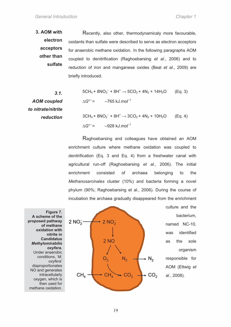
Raghoebarsing and colleagues have obtained an AOM
enrichment culture where methane oxidation was coupled to
denitrification (Eq. 3 and Eq. 4) from a freshwater canal with
agricultural run-off (Raghoebarsing et al., 2006). The initial
enrichment consisted of archaea belonging to the
Methanosarcinales cluster (10%) and bacteria forming a novel
phylum (90%; Raghoebarsing et al., 2006). During the course of
incubation the archaea gradually disappeared from the enrichment
culture and the
bacterium,
named NC-10,
was identified
as the sole
organism
responsible for
AOM (Ettwig et
al., 2008).
3.1.
AOM coupled
to nitrate/nitrite
reduction
3. AOM with electron
acceptors other than
sulfate
CH4 CO2
2 NO2
2 NO
O2 N2
2 NO2
N2
CH4 CO2CH4 CO2
2 NO2
2 NO
O2 N2
2 NO2
N2
CH4 CO2
Figure 7. A scheme of the
proposed pathway of methane
oxidation with nitrite in
Candidatus Methylomirabilis
oxyfera. Under anaerobic
conditions, ‘M. oxyfera’
disproportionates NO and generates
intracellularly oxygen, which is
then used for methane oxidation.
Chapter 1 General Introduction
20
Surprisingly, NC-10 (Candidatus Methylomirabilis oxyfera)
did not possess any of the two types of enzymes known to be
capable of C–H bond activation under anaerobic conditions (glycyl
radical enzymes and nickel-containing methyl-CoM reductase).
This enigma was solved by the discovery that this anaerobically-
growing bacterium generates intracellularly oxygen, which is used
for methane activation via aerobic methane monooxygenase
(Ettwig et al., 2010). The oxygen is produced from NO via a
putative NO dismutase which represents a novel mechanism
capable of generating molecular oxygen (Fig. 7)
The unique metabolism of ‘M. oxyfera’ suggests that even before
the oxygenation of the atmosphere organisms might have existed
that had evolved aerobic enzymatic mechanisms for the utilization
of the abundant atmospheric methane and possibly also of other
substrates. The contribution of nitrite-coupled AOM to present-day
methane cycling has not yet been quantified.
CH4 + 4MnO2 + 7H+ HCO3– + 4Mn2+ + 5H2O (Eq. 5)
G°’= –790 kJ.mol–1
CH4 + 8Fe(OH)3 +15H+ HCO3
– + 8Fe2+ + 21H2O (Eq. 6)
G°’= –572 kJ.mol–1
From an energetic point of view AOM coupled to
manganese- or iron-reduction can provide up to 30 times more
energy per mol methane (Eq. 5 and Eq. 6) than AOM with sulfate.
Recently, Beal and colleagues reported that birnessite (a MnO2
mineral) or ferrihydrite (a Fe(OH)3 mineral) are used as electron
acceptors for anaerobic methane oxidation in the anoxic marine
sediments off California (Beal et al., 2009). Methane oxidation was
observed in incubations supplemented with birnessite or ferrihydrite
as sole added electron acceptors but the rates (<0.3 M/day) were
3.2.
AOM coupled
to iron (III) and
manganese (IV)
reduction
General Introduction Chapter 1
21
3 to 10 times lower than in the sulfate-supplemented controls. A
direct link between the observed AOM and metal oxides reduction
has not been shown. Based on the reported results it cannot be
excluded that sulfide gets oxidized (a)biotically with the metal
oxides to sulfate, which could then fuel “conventional” sulfate-
coupled AOM through a cryptic S-cycle.

Aims and Objectives Chapter 1
23
AIMS and OBJECTIVES
”The task is not to see what has never been seen before but to think what has
never been thought before about what you see everyday. ”
Erwin Schrödinger (1887-1961)
Given that AOM plays a crucial role in sulfur cycling it is intriguing how little is
known about the dissimilatory sulfur metabolism coupled to it. From the beginning it
has been accepted that the Deltaproteobacteria perform sulfate reduction although
there was no experimental evidence to support this assumption apart from the
phylogenetic affiliation of the bacteria to a clade of known sulfate reducing bacteria. It
has been hypothesized later that ANME could perform sulfate reduction as well,
which, if true, would dramatically change our understanding of the functioning of
AOM. The possibility that methane oxidation and SR are intracellularly coupled in
ANME would imply that AOM is not mediated in syntrophy and that the associated
bacteria are merely commensals – if not parasites – of the methanotrophs. This study
was initiated in order to clarify which enzymes are responsible for the dissimilatory
sulfate reduction coupled to AOM, to which organism(s) these enzymes belong and
how the process of sulfate reduction is linked to methane oxidation.
We used liquid chromatography, polyacrylamide gel electrophoresis and
enzyme assays to search for the canonical SR enzymes and quantify their
abundance and activity. We wanted to assign these proteins to a particular organism
in the sample by using specific antibodies custom-generated against the purified
proteins. Such immunolabeling approach for uncultivated microorganisms was not
available at the time of the study and, correspondingly, a large part of my work was
dedicated to antibody generation and methodological establishment of an
immunolabeling protocol for environmental samples. The results of the
immunolableling study indicated that the ANME-associated bacteria perform the
AOM-coupled SR and, are therefore an active part of AOM consortia. Further
investigations have focused on how these two organisms interact in their syntrophic
relationship. We focused on analyzing whether any reduced sulfur compounds
Chapter 1 Aims and Objectives
24
derived from the dissimilatory sulfate metabolism could be involved in the electron
transfer from ANME to bacterial partner. For this we used an integrated approach of
bulk chemical extractions, HPLC and GC-MS analyses, single-cell secondary ion
mass spectrometry, microRaman spectroscopy, and transmission electron
microscopy coupled to energy-dispersive X-ray spectroscopy.
CHAPTER 2 Bacterial enzymes for dissimilatory sulfate
reduction in a marine microbial mat (Black Sea) mediating anaerobic oxidation of methane
Mirko Basen, Martin Krüger, Jana Milucka, Jan Kuever, Jörg Kahnt, Olav Grundmann, Anke Meyerdierks, Friedrich Widdel, and Seigo Shima
Contributions to the manuscript:
M.B., J.M., F.W., and S.S. designed research and project outline, M.B. and J.M.
performed FISH analysis, M.B., M.K., J.M., O.G. and S.S. performed protein purification,
M.B., J.M, J.Ka performed enzyme analyses, M.B. performed enzyme activities
measurements and apr gene amplification,, M.B., J.Ku., and A.M performed
phylogenetic analyses, M.B., J.M., F.W., S.S conceived, wrote and edited the
manuscript.
Chapter is in revision in Environmental Microbiology
Chapter 2 Summary
26
Summary
Anaerobic oxidation of methane (AOM) with sulfate is catalyzed by microbial
consortia of archaea and bacteria affiliating with methanogens and sulfate-
reducing Deltaproteobacteria, respectively. There is evidence that methane
oxidation is catalyzed by enzymes related to those in methanogenesis, but the
enzymes for sulfate reduction coupled to AOM have not been examined. We
collected microbial mats with high AOM activity from a methane seep in the Black
Sea. The mats consisted mainly of archaea of the ANME-2 group and bacteria of
the Desulfosarcina-Desulfococcus group. Cell-free mat extract contained activities
of enzymes involved in sulfate reduction to sulfide: ATP sulfurylase
(adenylyl:sulfate transferase; Sat), APS reductase (Apr), and dissimilatory sulfite
reductase (Dsr). We partially purified the enzymes by anion-exchange
chromatography. The amounts obtained indicated that the enzymes are abundant
in the mat, with Sat accounting for 2% of the mat protein. N-terminal amino acid
sequences of purified proteins suggested similarities to the corresponding
enzymes of known species of sulfate-reducing bacteria. The deduced amino acid
sequence of PCR-amplified genes of the Apr subunits is similar to that of Apr of
the Desulfosarcina/Desulfococcus group. These results indicate that the major
enzymes involved in sulfate reduction in the Back Sea microbial mats are of
bacterial origin, most likely originating from the bacterial partner in the
consortium.
Introduction Chapter 2
27
Introduction
The anaerobic oxidation of methane (AOM) with sulfate according to
CH4 (g) + SO42 HCO3 + HS + H2O (1)
( G° = 16.6 kJ mol 1) is the major sink for methane produced in deep anoxic marine
sediments and is thus of global importance (Reeburgh, 2007; Knittel & Boetius, 2009).
The anaerobic oxidation of methane rather than the aerobic oxidation, which occurs in
terrestrial habitats, is due to the much higher concentration of sulfate compared to that of
oxygen in seawater (28 mM vs. approximately 0.3 mM); sulfate therefore penetrates
deeper into sediments than oxygen.
AOM is often catalyzed by compact aggregates of archaea closely related to
methanogens and bacteria clustering within the Deltaproteobacteria, most commonly
within the Desulfosarcina-Desulfococcus clade (Boetius et al., 2000; Orphan et al.,
2001a; Michaelis et al., 2002; Knittel et al., 2005, Schreiber et al., 2010). Consortia with
Desulfobulbus-related Deltaproteobacteria have recently been identified (Niemann et al.,
2006; Lösekann et al., 2007; Pernthaler et al, 2008). A common view is that the archaea
are anaerobic methanotrophs (ANME) that activate methane and process methane
carbon to CO2 via reverse reactions of methanogenesis, and that the bacterial partner
scavenges reducing equivalents to reduce sulfate (Zehnder & Brock, 1979; Hoehler et
al., 1994;; Boetius et al., 2000; Valentine et al., 2000; Nauhaus et al., 2002; Thauer &
Shima, 2008;).
Consortia anaerobically oxidizing methane have been propagated in vitro (Nauhaus
et al. 2007, Meulepas et al. 2009), but axenic cultures have not been isolated. Insights
into the pathway of methane during AOM have come from metagenomic and protein
analyses using natural samples, and from analogies to the well-established pathway of
methanogenesis (Thauer 1998). The hypothesis of “reverse methanogenesis” was
supported by the finding of orthologs of almost all genes of the methanogenic pathway in
the metagenome of natural samples with AOM activity and of naturally enriched ANME
groups (Hallam et al., 2004; Meyerdierks et al., 2005; Meyerdierks et al. 2010). The
Chapter 2 Results and discussion
28
putative methane-activating enzyme, a dominant nickel protein closely related to methyl-
coenzyme M reductase (Mcr) of methanogens, was purified from microbial mats from the
Black Sea (Krüger et al., 2003). Furthermore, an Mcr-related protein was localized by
immuno-labeling and electron microscopy of the archaeal cells of the mat community
(Heller et al., 2008). Recent NMR experiments have shown that purified MCR from
methonogenic archaea can catalyze the endergonic back reaction (Scheller et al., 2010).
Comparable insights into key genes and enzymes involved in sulfate reduction
linked to AOM are lacking, and the assumption that the deltaproteobacterial cells
associated with the archaea are genuine sulfate reducers has not been verified. Even at
some marine sites with AOM activity, the archaeal cells detected were not associated
with bacteria (Orphan et al., 2002; Knittel et al., 2005; Lösekann et al., 2007; Treude et
al., 2007). Hence, it cannot be excluded that at least some archaea in AOM habitats are
responsible for both methane oxidation and sulfate reduction. This is feasible since
dissimilatory sulfate reduction is a well-established trait in Euryarchaeota (genus
Archaeoglobus; Stetter, 1988; Dahl et al., 1994). Genes for this pathway could have
been acquired via lateral transfer from sulfate-reducing Firmicutes (Wagner et al., 1998;
Friedrich, 2002; Meyer & Kuever, 2007).
Here we show that Black Sea microbial mats with AOM activity contain substantial
amounts of the three enzymes catalyzing sulfate reduction to sulfide: ATP sulfurylase
(sulfate:adenylyl transferase; Sat), adenosine-5'-phosphosulfate (APS) reductase (Apr),
and dissimilatory sulfite reductase (Dsr). We partially purified Sat, the small subunit of
Apr (AprB), and the DsrAB directly from the natural mat biomass and showed that their
N-terminal amino acid sequences were similar to those of deltaproteobacterial enzymes.
Moreover, we show that the deduced products of the Apr-encoding genes (aprBA) are
closely related to enzymes of known sulfate-reducing Deltaproteobacteria of the
Desulfosarcina-Desulfococcus clade.
Results and discussion
Microbiological characterization of the intact microbial mat
Results and discussion Chapter 2
29
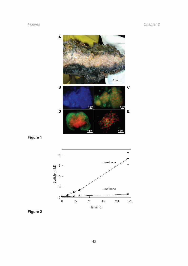
Methane seepage in the northwestern Black Sea at 210–230 m depth sustains AOM,
leading to deposition of chimney-like carbonate structures up to 4 m in height and 1 m in
diameter (Pimenov et al., 1997; Michaelis et al., 2002). The in situ temperature is around
8 °C. The outside and interstitials of the chimneys are populated by soft, somewhat slimy
microbial mats of up to 10 cm thickness (Fig. 1A). The exterior part of the mat directly
exposed to the methane-rich fluid is black and harbors consortia of the ANME-2 and
Desulfosarcina-Desulfococcus phylotypes (Blumenberg et al., 2004; Reitner et al., 2005;
Krüger et al., 2008). The interior part of the mat is pink and often dominated by ANME-1
cells with their characteristic cylindrical shape and also by Desulfosarcina-
Desulfococcus-related cells. The latter do not form compact aggregates but are
distributed within the mat matrix (Michaelis et al., 2002).
Fig. 1
We collected mat samples in the Black Sea and selected a part of the mat with high
AOM activity. The highest rates of methane-dependent sulfate reduction (Fig. 2) were
measured in the black mat at approximately 16 °C; the rate per dry mass was 3.2 (± 0.55)
nkat gdm1 (1 nkat = 6 × 10 2 μmol min 1 = 86.4 μmol d 1). The presence of consortia in
the selected sample was checked by microscopy. Almost all of the cells detected by
general (DAPI) staining formed densely packed aggregates (Fig. 1B). Use of specific
16S-rRNA-targeting fluorescent oligonucleotide probes showed that consortia consisted
either of an archaeal core surrounded by bacterial cells or of intermixed archaeal and
bacterial cells (Fig. 1C E). The selected black mat therefore appeared suitable for
studying enzymes involved in sulfate reduction linked to AOM. We prepared a soluble
cell-free extract of the mat.
Fig. 2
Activity of enzymes involved in sulfate reduction to sulfide
The pathway of dissimilatory sulfate reduction hitherto described in bacteria and archaea
involves a canonical sequence of three enzymatic reactions (Thauer et al., 1977; LeGall
& Fauque, 1988;). ATP sulfurylase (adenylyl:sulfate transferase; Sat; EC 2.7.7.4)
Chapter 2 Results and discussion
30
activates sulfate with ATP to yield adenosine 5'-phosphosulfate (APS) and
pyrophosphate. The latter is hydrolyzed to phosphate by pyrophosphatase. APS is
reduced by APS reductase (Apr; EC 1.8.99.2), yielding inorganic sulfite (or bisulfite) and
AMP. Dissimilatory sulfite reductase (Dsr; EC 1.8.99.1) finally reduces sulfite to sulfide.
Extract preparation was rendered difficult by carbonate grains and the matrix that
formed voluminous bottom layers upon centrifugation of crude extract. Therefore, the
obtained amount of soluble cell-free extract (supernatant) had to be used sparingly.
Activities of Sat and Apr were detected in mat extract (Table 1). The specific activity
of Dsr was very low and variable in the different mat extracts. For comparison, we
measured specific activities of the three enzymes in cell extracts of pure cultures of
Desulfococcus multivorans and Desulfosarcina variabilis; the specific activities were in
the range of those reported for sulfate-reducing prokaryotes (Krämer & Cypionka, 1989;
Dahl et al., 1994; Sperling et al., 1998; Fritz et al., 2000). The cell-type-related specific
activities of Sat and Apr in mat extract did not differ much from activities in extracts of
the pure cultures, especially when only one of two cell types in the consortium contained
the enzymes.
Table 1
Fractionation of enzymes involved in sulfate reduction to sulfide
The soluble mat extract contained substances that interfered with chromatography.
These substances were precipitated by 20% saturated ammonium sulfate. We then
partially purified the enzymes in the desalted supernatant by anion-exchange
chromatography, measured their activities, and analyzed them by denaturing SDS-
PAGE.
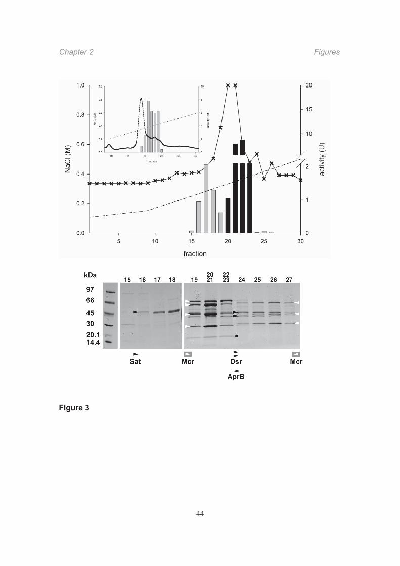
Sat eluted as a single peak of activity at 0.25 0.32 M NaCl (fractions 16–18) that
comprised 90% of the total activity eluted from the column. Fifty percent of the Sat
activity was recovered in this purification step. SDS-PAGE of the fractions revealed a
protein band of ca. 50 kDa (Fig. 3), similar to the monomer size of the known
Results and Discussion Chapter 2
31
homotrimeric Sat (Sperling et al., 1998). The collected active fractions contained 2% of
the total soluble mat protein. The N-terminal amino acid sequence of the 50-kDa protein
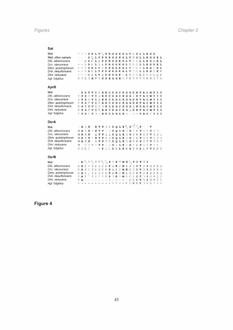
band from the polyacrylamide gel was characteristic of Sat (Fig. 4). Attempts to clarify an
ambiguous position by analyzing another mat sample in the same manner led to a
similar sequence harboring another ambiguous position (Fig. 4).
Fig. 3 and 4
Total Apr activity was significantly decreased by ammonium sulfate precipitation,
leaving 20% of the initial activity in the supernatant. Since there was no detectable Apr
activity in the precipitate and no measurable protein loss by ammonium sulfate treatment,
the observed loss must have been caused by the salt addition and desalting.
Subsequent anion-exchange chromatography eluted more than 80% of the Apr activity
applied to the column at 0.33 0.38 M NaCl (fractions 19–23). These fractions contained
a ca. 20-kDa protein (Fig. 3). Native Apr from sulfate-reducing bacteria and
Archaeoglobus is a heterodimer composed of two subunits, AprA (ca. 70 kDa) and AprB
(ca. 20 kDa) (Fritz et al., 2000; Fritz et al., 2002). However, in our denaturing gels, only
the small subunit was visible because the large AprA subunit probably overlapped with
the highly abundant subunit of “reverse” methyl-coenzyme M reductase (Mcr) from
archaea. The N-terminal amino acid sequence of the small protein had a high similarity
to that of bacterial AprB (Fig. 4).
Dsr activity eluted at 0.35 0.44 M NaCl (fractions 24–26) and thus overlapped partly
with the peak containing Mcr and Apr activity. SDS-PAGE of fractions harboring Dsr
activity showed protein bands of ca. 43 and 47 kDa; these masses are similar to those of
the two subunits of the described Dsr (Arendsen et al., 1993). The intensity of these
bands indicated that Dsr is a relatively abundant protein. A third small subunit
corresponding to DsrC (~11 kDa) (Arendsen et al., 1993; Oliveira et al., 2008) was
negligible in our Dsr fraction. SDS-PAGE of higher polyacrylamide concentrations
revealed that the Dsr fraction containes several minor proteins at 10~15 kDa but the
intensity of these bands was too weak to analyze the N-terminal sequence. N-terminal
sequence analysis of the DsrA and DsrB candidate proteins yielded only relatively short
Chapter 2 Conclusions
32
sequences. Nonetheless, they exhibited similarity to Dsr from sulfate-reducing bacteria
(Fig. 4). The N-terminal sequences included some ambiguous positions, which indicated
that Dsr preparations from mat extract were probably not homogenous and might have
originated from related sulfate-reducing bacteria. The Dsr-containing fraction was
greenish. Its UV-VIS absorption spectrum showed a distinct maximum at 410 nm, a
weak maximum at 540 nm, and a weak shoulder at 580 nm (Fig. S1). This indicates that
the absorption spectrum represented the combined spectra of the siroheme-containing
dissimilatory sulfite reductase (Arendsen et al., 1993) and the co-eluted Mcr harboring a
nickel cofactor (Krüger et al., 2003).
Full-length sequence of APS reductase from the microbial mat
We used the obtained N-terminal amino acid sequences to design degenerate primers to
retrieve full-length sequences of the corresponding genes. Only clones of genes
encoding AprA and AprB were obtained. Their N-terminal sequences were identical with
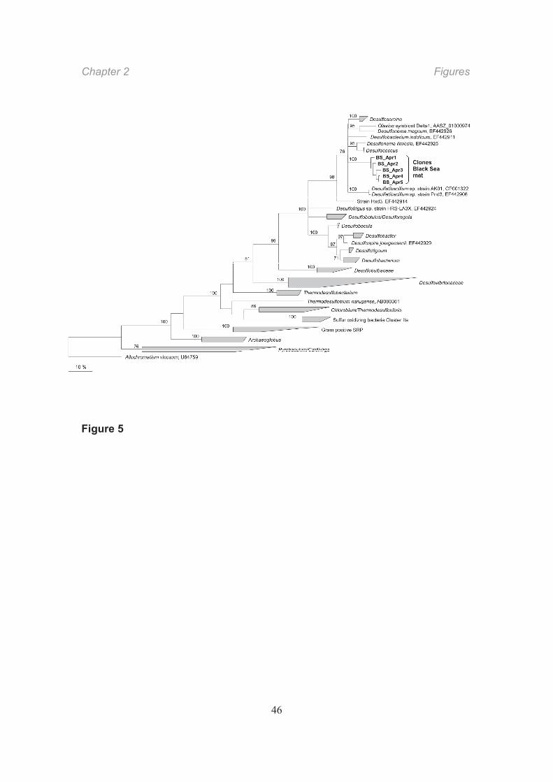
that of five Apr proteins. The full-length sequences were most closely related to those of
AprBA from the Desulfosarcina-Desulfococcus cluster and constituted a sixth lineage
distinct from the hitherto known lineages from Desulfosarcina spp., Desulfonema
magnum/Olavius symbiont Delta-1, Desulfobacterium indolicum, Desulfonema
limicola/Desulfococcus spp., and Desulfatibacillum spp. (Fig. 5; Friedrich, 2002; Meyer &
Kuever, 2007). The AprBA sequence tree mirrors that of 16S rRNA, including sequences
from bacteria associated with ANME from seep areas (Orphan et al., 2001b; Knittel et al.,
2003), which suggests that the Apr partially purified from the mat originates from a
member of the Desulfosarcina/Desulfococcus group.
Fig. 5
Conclusions
If the analyzed enzymes are involved in AOM with sulfate, their activities should in
principle be sufficient to account for the in vivo AOM rate (see above). The measured
protein-to-dry-mass ratio of 0.28 yields a protein-related AOM activity in the intact mat of
Conclusions Chapter 2
33
11 nkat gpr1. This activity was measured at 16 °C, which is similar to the temperature
optimum of the viable mat. Enzyme activities in optical assays were generally measured
at 28 °C, which is the growth temperature of the pure cultures used for comparison and
the appropriate temperature for the auxiliary enzymes in the coupled Sat assay.
Assuming that enzyme activity increases two- to fourfold per temperature increase of T
= 10 K, activities of Sat and Apr (Table 1) are still more than sufficient to account for
sulfate reduction to sulfide with methane as electron donor. In contrast, the measured
Dsr activity would be barely sufficient to explain the observed in vivo rate, even though
the abundance of the enzyme was relevant according to the band intensity in the gel (Fig.
3). It is possible that Dsr is a particularly sensitive enzyme that easily loses activity upon
cell disruption or that our assay, which used an artificial electron donor, may be
insufficient to reveal its in vivo function. Structural studies of the enzyme from
Desulfovibrio vulgaris (Oliveira et al., 2008) indicate that DsrC is highly important for the
catalytic function of Dsr and may be involved in sulfite reduction. The very low Dsr
activity in the mat extract and the anion-exchange column fractions might be attributed to
the instability of the DsrC in the samples.
The similarity of the deduced Apr sequence to Apr sequences from the
Desulfosarcina-Desulfococcus group indicates that the retrieved enzyme is from the
bacterial partner in the methane-oxidizing consortium, which is a member of this group
(Fig. 5). The N-terminal amino acid sequence of Sat and Dsr from the mat showed the
highest similarity to that of the respective proteins of sulfate-reducing
Deltaproteobacteria. Thus, Sat, Dsr, and Apr likely arise from the bacterial partner in the
mat.
Our study supports the assumption that enzymes of the canonical sulfate reduction
pathway are important in AOM in Black Sea microbial mats and arise from the
associated bacterial partner. The study is one of the few examples of the direct
characterization of an enzyme from a natural microbial habitat. Other examples include
the study of Mcr (most likely involved in AOM) in the same habitat (Krüger et al., 2003)
and of Sat in aerobic sulfur bacteria in the trophosome of a tube worm from a
Chapter 2 Experimental procedures
34
hydrothermal vent (Renosto et al., 1991). Such combined enzyme and gene analyses
are a promising approach towards understanding the in situ role of particular
microorganisms, if these microorganisms are highly enriched in their habitats, such as in
microbial mats.
Experimental procedures
Origin of microbial mats and cultivation of strains
Microbial mats were collected by means of a submersible in methane seep areas in the
northwestern Black Sea at 220 m water depth during RV Meteor cruise M72/2 in
February/March 2007 (Project MUMM, Max Planck Institute for Marine Microbiology,
Bremen) at 44° 46' N, 31° 59' E. A mat sample maintained from RV Poseidon cruise
POS 317-2 in August 2004 (Project GHOSTDABS, University of Hamburg) at 44° 46' N,
31° 60' E was used for the preliminary experiments; in principle, the same results were
obtained. Collected samples were maintained active in artificial sea water medium
(Widdel & Bak, 1992) in anoxic 1-l bottles under a headspace (ca. one-third of the bottle
volume) of CH4 and CO2 (5:1) at 8 °C. If more than ca. 10 mM sulfide accumulated, the
supernatant was replaced with fresh anoxic seawater. Strains of sulfate-reducing
bacteria were obtained from the German Collection of Microorganisms and Cell Cultures
(DSMZ, Braunschweig). Desulfococcus multivorans (DSM 2059) and Desulfosarcina
variabilis (DSM 2060) were grown at 28 °C in synthetic brackish medium and seawater
medium (Widdel & Bak, 1992), respectively, with 5 mM benzoate. All subsequent
manipulations were done in anoxic glove boxes with an N2-CO2 (9/1, v/v) atmosphere for
manipuration of the mat and with an N2-H2 (95/5, v/v) atmosphere for the enzyme
purification.
Incubation experiments with intact mats
Microbial mats were gently homogenized using a tissue grinder and suspended in anoxic
artificial seawater medium at a dry mass content of 2.9 mg ml 1. Suspensions of 10 ml
were incubated in 20-ml glass tubes with a headspace of CH4 or N2 (controls) and CO2.
Experimental procedures Chapter 2
35
Tubes were sealed with butyl rubber stoppers and during the experiment horizontally
shaken (40 rpm) to facilitate gas transport. Aliquots were withdrawn with syringes
flushed with N2.
Chemical and other analyses
Sulfide production was determined colorimetrically as brown colloidal CuS (Cord-
Ruwisch, 1985) and via the methylene blue formation reaction (Cline, 1969) in a
miniaturized assay (4 ml). Methane was quantified using a GC14B gas chromatograph
(Shimadzu, Kyoto, Japan) equipped with a Supel-Q Plot column (30 m 0.53 mm;
Supelco) and a flame ionization detector. The carrier gas was N2 at a flow rate of 3 ml
min 1. The column temperature was 110 °C. Dry mass was measured after drying at 80
°C for 48 h. Protein content was determined according to Bradford (1976).
Fluorescence in situ hybridization
Homogenized mat samples (see above) were fixed with 2% formaldehyde in phosphate-
buffered saline (PBS; 7 mM Na2HPO4, 3 mM NaH2PO4, 130 mM NaCl; pH 7.2) for 12 h,
washed with 1 PBS and stored in PBS/ethanol (1:1) at 20 °C. Small proportions were
collected on GTTP polycarbonate filters of 0.2 μm pore size (Millipore, Eschborn,
Germany). Staining with 4 ,6 -diamidino-2-phenylindole (DAPI), hybridization, and
microscopy were carried out as described (Amann et al., 1995). Fluorescent
oligonucleotide probes and formamide concentrations (v/v) were as follows: negative
control, NON338, 10% (Wallner et al., 1993); archaea, Arch915, 35% (Amann et al.,
1990); bacteria, EUBI-III, 35% (Daims et al., 1999); Desulfosarcina-Desulfococcus group,
DSS658, 50% (Manz et al., 1998); ANME-1, ANME-1 350, 40% (Boetius et al., 2000);
and ANME-2, ANME-2 538, 50% (Treude et al., 2005). Probes labeled with Cy3 or
carboxyfluorescein (FLUOS) were purchased from ThermoHybaid (Ulm, Germany).
Chapter 2 Experimental procedures
36
Preparation of extract and protein fractionation
Pieces of the microbial mats (15 g) were cut into smaller (2 mm) pieces and suspended
in 30 ml of 50 mM MOPS-KOH pH 7.0. The cells were disrupted by ultrasonication three
times for 8 min at 160 W (sonication tip MS 72; pulse duration 0.5 s). The resulting crude
extract was centrifuged at 150,000 g for 1 h. The membrane-free supernatant was
fractionated by ammonium sulfate precipitation (20% saturation), followed by
centrifugation at 18,000 g for 20 min. The supernatant (40 ml) was concentrated by
ultrafiltration to 5 ml (10-kDa cut off). The concentrate was diluted 50-fold with 50 mM
MOPS-KOH, pH 7.0. From this, 125 ml were applied to a 5-ml Q-Sepharose anion-
exchange chromatography column (HiTrap Q HP; GE Healthcare) equilibrated with 50
mM MOPS-KOH, pH 7.0. Proteins were eluted with a linear NaCl gradient from 0 to 0.6
M. Enzyme activity in the eluted fractions was measured (see below), and proteins were
analyzed by 4-15% gradient SDS-PAGE (mini-format), followed by staining with
Coomassie Blue G-250. For N-terminal amino acid sequence analysis, protein bands
were blotted onto a PVDF membrane using a wet blot device (BioRad) according to the
manufacturer’s instructions. Protein bands were excised and analyzed by Protein
Analytics (Giessen, Germany).
Enzyme assays
ATP sulfurylase activity was determined in the reverse direction by coupling the following
reactions and photometric (340 nm) determination of NADPH (Dahl and Trüper, 1994) in
50 mM Tris-HCl buffer, pH 7.5, supplemented with 20 mM MgCl2: APS2 + PPi3 ATP4
+ SO42 + H+; ATP4 + glucose ADP3 + G-6-P2 + H+; G-6-P2 + NADP+ + H2O
6-phosphogluconate3 + NADPH + 2 H+ (APS, adenyl-5’-phosphosulfate; PPi,
pyrophosphate; G-6-P, glucose 6-phosphate). APS reductase activity was determined in
the reverse direction according to SO32 + AMP2 + 2 [Fe(CN)6]3 APS2 +
2 [Fe(CN)6]4 with photometric (420 nm) determination of [Fe(CN)6]3 (Kobayashi et al.,
1975) in 50 mM Tris-HCl buffer, pH 7.5. The rate of abiotic [Fe(CN)6]3 reduction was
subtracted. Dissimilatory sulfite reductase activity was determined with reduced
Experimental procedures Chapter 2
37
methylviologen (MV+) according to SO32 + 6 MV+ + 7 H+ HS + 6 MV2+ + 3 H2O with
photometric (578 nm) determination of MV+ consumption (Dahl and Trüper, 2001) in
anoxic 50 mM Tris-HCl buffer in cuvettes under N2. Directly before the assay, MV2+ was
reduced with 0.2 mM titanium (III) citrate. We also performed the assay with sodium
dithionite as reducing agent. Enzyme activities (1 nkat = 10 9 mol s 1 = 6 10 2 μmol
min 1) refer to ATP, APS, or SO32 .
DNA manipulation and analysis
Genomic DNA of Black Sea microbial mats and of pure cultures of sulfate-reducing
bacteria was extracted according to the genomic tip protocol (Genomic DNA Handbook,
Qiagen, Hilden, Germany). The degenerated primer BS-AprB-1-F (5'-ATG CCD AGT
TAT GTH ATH AC-3') was newly designed based on the N-terminal amino acid
sequence of the purified AprB protein. aprBA was amplified using primer combinations
BS-AprB-1-F/AprA-5-RV and AprA-1-FW/AprA-10RV (Meyer & Kuever, 2007), as well as
BS-AprB-1-FW/AprA-10-RV; the latter yielded almost full-length aprBA sequences.
Primer annealing was optimal at 53 °C. PCR products were purified using the Qiaquick
PCR purification kit (Qiagen). Clone libraries were constructed using the TOPO pCR4
vector (TOPO TA cloning kit, Invitrogen, Karlsruhe, Germany) following the
manufacturer’s instructions. PCR products were sequenced using the ABI BigDye
terminator cycle sequencing kit (Applied Biosystems). Sequences were analyzed with
the Lasergene software package (DNAstar, GATC Biotech, Konstanz, Germany) or the
Bioedit sequence alignment editor version 7.0.9.0 (Hall, 1999).
Sequences used for comparison of N-terminal amino acids (Fig. 4) were from the
following microorganisms. Sat: Desulfatibacillum alkenivorans strain AK-01
(ZP_02131628.1); Desulfococcus oleovorans strain Hxd3 (ABW66812.1);
Desulfobacterium autotrophicum strain HRM2 (YP_002604365); Desulfovibrio
desulfuricans strain G20 (YP_388757.1); Desulfotomaculum reducens strain MI-1
(ABO49175.1); Desulfotomaculum acetoxidans strain 1ac2 (YP_003192914.1); and
Archaeoglobus fulgidus strain VC-16 (AAB89581.1). AprB: Desulfatibacillum
Chapter 2 Acknowledgements
38
alkenivorans (YP_002430736); Desulfococcus oleovorans (YP_001528884);
Desulfobacterium autotrophicum (YP_002601731), Desulfovibrio
desulfuricans .(YP_387605); Desulfotomaculum reducens .(YP_001112001);
Desulfotomaculum acetoxidans (YP_003192913.1); and A. fulgidus (NP_070497). DsrA:
Desulfatibacillum alkenivorans (YP_002433449.1); Desulfococcus oleovorans
(ABW68472.1); Desulfobacterium autotrophicum (YP_002605460.1); Desulfovibrio
desulfuricans (ABB37327.1); Desulfotomaculum reducens (YP_001114514.1);
Desulfotomaculum acetoxidans (YP_003189665.1); A. fulgidus (NP_069259.1). DsrB:
Desulfatibacillum alkenivorans (YP_002433448.1); Desulfococcus oleovorans
(YP_001530550.1); Desulfobacterium autotrophicum (ACN17295.1); Desulfovibrio
desulfuricans (ABB37328.1); Desulfotomaculum reducens (YP_001114513.1);
Desulfotomaculum acetoxidans (YP_003189666.1); and A. fulgidus (NP_069260.1).
Contigs of sequences of AprB and AprA were analyzed using ClustalW alignment
and tree construction with the PhyML program (maximum-likelihood method,
http://atgc.lirmm.fr/phyml, 100 bootstraps) as described (Meyer & Kuever, 2007).
Nucleotide sequence accession numbers
The aprBA nucleotide sequence data are deposited in the EMBL, GenBank, and DDBJ
sequence databases under the accession numbers HQ188925 to HQ188929 for AprBA
1 5 from Black Sea microbial mats, HQ188924 for AprBA Desulfosarcina ovata strain
oxyS1, and HQ188930 for AprBA Desulfosarcina cetonica.
Acknowledgements
We are indebted to Rolf Thauer for continuous advice and support, and to Walter
Michaelis, Richard Seifert, and Antje Boetius for access to samples. We thank the crews
of RV Poseidon with JAGO, and RV Meteor with ROV QUEST (MARUM) for help during
field work, Thomas Holler for sample collection, and Daniela Lange and Alexander
Galushko for providing strains. We also thank Chris Hopkins, Jens Harder, and Rudolf
Amann for helpful discussions and critical reading of the manuscript. This work was
Acknowledgements Chapter 2
39
supported by the Max Planck Society, the Fonds der Chemischen Industrie, the Marie
Curie Early Stage Training Site in Marine Microbiology (MEST-CT-2004-007776), and
the projects MUMM, GHOSTDABS (program GEOTECHNOLOGIEN of the BMBF and
DFG), and BEBOP (University of Hamburg). Seigo Shima received support from an
emeritus grant (Max Planck Society) given to Rolf Thauer. This is publication number
xxxx (will be provided) in the framework of MUMM II (program GEOTECHNOLOGIEN).
Chapter 2 Figure legends
40
Figure legends
Fig. 1. Images of microbial mats from the methane seepage area in the northwestern
Black Sea.
A) Piece of a chimney-like structure showing the outer black and inner pink microbial
layers (courtesy of Walter Michaelis, Hamburg). B) “Super” consortium of archaeal-
bacterial consortia from a black mat exhibiting fluorescence after DAPI staining. C) The
same consortium as in B exhibiting fluorescence of labeled oligonucleotide probes
targeting 16S rRNA. The probes were specific for ANME-2 (Cy3, red signal) and the
Desulfosarcina-Desulfococcus group (FITC, green signal). D) Consortium with an
archaeal core and surrounding bacteria stained as in C. E) Consortium with intermixed
archaeal and bacterial cells stained as in C.
Fig. 2. Methane-dependent sulfate reduction in the black microbial mat used for
enzymatic analysis.
Fig. 3. Upper panel: Elution profile of Sat Apr activities in anion- exchange
chromatography fractions. Shown are: activity of Sat (grey bars) and Apr (black bars),
relative absorption at 280 nm (solid line with crosses) and NaCl concentration (dashed
line). One unit (U) of activity refers to one μmol substrate turnover per minute. Inset:
Elution profile of Dsr in a separate run of anion-exchange chromatography.
Lower panel: Denaturing gel electrophoresis (SDS-PAGE) of fractions from anion-
exchange chromatography of supernatants from the microbial mat. Fractions (loaded on
two gels) eluting from 0.23 M (fraction 15) to 0.43 M (fraction 27) NaCl are shown. The
protein bands were assigned to ATP sulfurylase (Sat, ca. 50 kDa), APS reductase (small
subunit, AprB, ca. 20 kDa), and (dissimilatory) sulfite reductase (Dsr, ca. 47 and 43 kDa
bands of DsrA and DsrB) based on N-terminal sequences (Fig. 4); sizes were in
accordance with those of orthologous enzymes from sulfate-reducing bacteria.
Assignment of AprB was further corroborated by cloning and sequencing of the encoding
Figure legends Chapter 2
41
gene (see text; Fig. 5). The large subunit, AprA (ca. 70 kDa), is probably masked by a
subunit of one of the methyl-coenzyme M reductases (Mcr). N-terminal sequencing of
the Mcr subunits in the fractions did not reveal the Apr-like sequence, probably because
Mcr was in excess. Mcr is highly abundant in the mat (Krüger et al., 2003) and is
assumed to catalyze methane activation. The spreading of Mcr from fractions 19 to 27
indicates variant forms of Mcr (see also Krüger et al., 2003) consisting of three subunits
(ca. 66, 48, and 30 37 kDa according to published sizes). The -subunit (66 kDa) of one
Mcr protein (fractions 19–23) is partly present as a second, somewhat lighter protein, as
revealed by an additional band.
Fig. 4. Analyzed N-terminal sequences of ATP sulfurylase (Sat), the small APS
reductase subunit (AprB), and both subunits of (dissimilatory) sulfite reductase (DsrA
and DsrB) from the Black Sea mat and alignment with orthologs from known sulfate-
reducing microorganisms. Species: Desulfatibacillum alkenivorans strain AK-01,
Desulfococcus oleovorans strain Hxd3, Desulfobacterium autotrophicum strain HRM2,
Desulfovibrio desulfuricans strain G20, Desulfotomaculum reducens strain MI-1,
Archaeoglobus fulgidus strain VC-16.
Fig. 5. Phylogenetic tree of AprBA sequences, including five clones from the Black Sea
mat (bold). The tree is based on maximum likelihood. Numbers represent bootstrap
values (100 trials). Bar, 10% estimated sequence divergence.
Chapter 2 Table
42
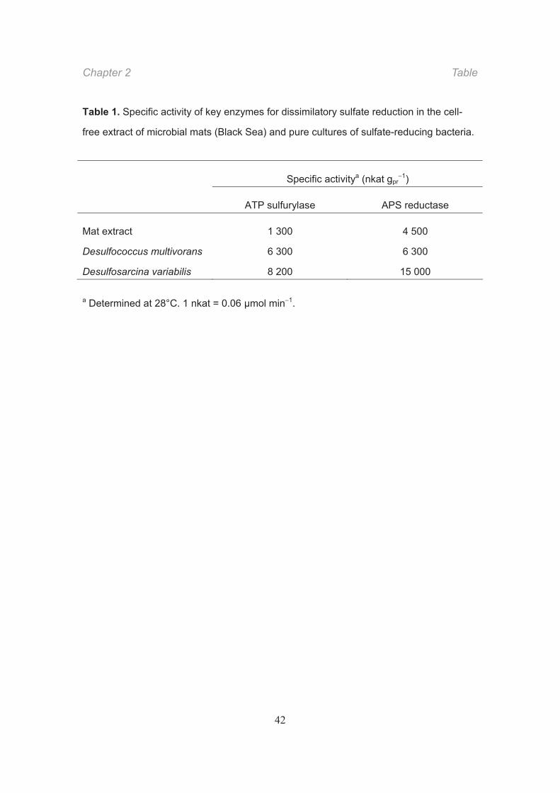
Table 1. Specific activity of key enzymes for dissimilatory sulfate reduction in the cell-
free extract of microbial mats (Black Sea) and pure cultures of sulfate-reducing bacteria.
a Determined at 28°C. 1 nkat = 0.06 μmol min 1.
Specific activitya (nkat gpr1)
ATP sulfurylase APS reductase
Mat extract 1 300 4 500
Desulfococcus multivorans 6 300 6 300
Desulfosarcina variabilis 8 200 15 000
Figures Chapter 2
43
Figure 1
Figure 2
Chapter 2 Figures
44
Figure 3
Figures Chapter 2
45
Figure 4
Chapter 2 Figures
46
Figure 5
Supporting information Chapter 2
47
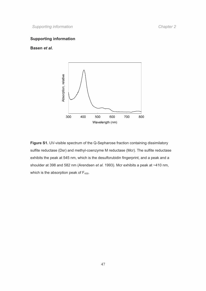
Supporting information
Basen et al.
Figure S1. UV-visible spectrum of the Q-Sepharose fraction containing dissimilatory
sulfite reductase (Dsr) and methyl-coenzyme M reductase (Mcr). The sulfite reductase
exhibits the peak at 545 nm, which is the desulforubidin fingerprint, and a peak and a
shoulder at 398 and 582 nm (Arendsen et al. 1993). Mcr exhibits a peak at ~410 nm,
which is the absorption peak of F430.

CHAPTER 3 Immunological detection of enzymes for sulfate
reduction in bacterial cells of anaerobic methane-oxidizing microbial consortia
Jana Milucka, Friedrich Widdel and Seigo Shima
Contributions to the manuscript:
J.M. and F.W. designed research and project outline, J.M. and S.S. performed protein
purification, J.M. tested the antibodies and established the immunolabeling protocol,
J.M., F.W. and S.S conceived, wrote and edited the manuscript.
Chapter is in preparation for ISME Journal
Chapter 3 Summary
50
Summary
Microbial consortia mediating anaerobic oxidation of methane coupled to sulfate
reduction (AOM) are formed by methane-oxidizing archaea (ANME) and bacteria
belonging mostly to the Desulfosarcina/ Desulfococcus (DSS) clade of
Deltaproteobacteria. Whereas it is generally accepted that methane oxidation is
catalyzed by ANME it has not yet been resolved which organism(s) perform the
AOM-coupled sulfate reduction. Here we demonstrate distribution of enzymes of
the methane oxidation pathway and dissimilatory sulfate-reducing pathway in situ
in the ANME-DSS consortia from Black Sea microbial mats. Using antibodies
raised against ATP sulfurylase and dissimilatory sulfite reductase purified directly
from the active mats we show that these sulfate reducing enzymes were detected
exclusively in one abundant microbial morphotype (~ 50% of all cells) reminiscent
of the DSS bacteria. In contrast, none of the cells labeled with antibodies against
methyl-coenzyme M reductase, an archaeal enzyme catalyzing methane oxidation,
contained the sulfate-reducing enzymes. Our findings show that the abundant
enzymes for methane oxidation and sulfate reduction belong to two dominant but
different microbial morphotypes and suggest that the (major) sulfate-reducing
activity associated with AOM is catalyzed by bacteria.
Introduction Chapter 3
51
Introduction
The anaerobic oxidation of methane (AOM) with sulfate is an environmentally relevant
process controlling both carbon and sulfur cycling in anoxic marine environments
(Valentine & Reeburgh, 2000; Knittel & Boetius, 2009). AOM is presumed to be
catalyzed in a syntrophic manner by a consortium of anaerobic methane-oxidizing
archaea (ANME) and Deltaproteobacteria (Boetius et al., 2000). There are three groups
of ANME described to date – ANME-1, -2, -3. Their bacterial partners mostly belong to
the clades of Desulfosarcina/Desulfococcus (ANME-1 and -2; Boetius et al., 2000;
Orphan et al., 2002a; Knittel et al., 2005; Schreiber et al., 2010) and Desulfobulbus
(ANME-3; Niemann et al., 2006). Mechanism of the ANME-Deltaproteobacteria
interaction remains, despite their common co-occurrence at AOM sites, enigmatic. It is
generally accepted that ANME catalyze the anaerobic methane oxidation. The enzyme
likely responsible for the activation of methane is a ’reverse’ methyl-CoM-reductase (Mcr)
known from methanogenic archaea (Krüger et al., 2003; Scheller et al., 2010). Further
oxidation of the bound methyl moiety probably follows a reversed pathway of
methanogenesis because corresponding genes have been found in the ANME
(meta)genome (Krüger et al., 2003; Hallam et al., 2004; Chistoserdova et al., 2005;
Meyerdierks et al., 2010; A. Meyerdierks, pers. comm.). A presumed function of the
ANME-associated bacteria is utilization of reducing equivalents originating from methane
oxidation for the reduction of sulfate. This would imply that the reducing equivalents are
extracellularly shuttled from ANME to the DSS bacteria but the chemical nature of the
shuttled compound is not yet elucidated. In nature ANME-2 also occur as single cells
and/or monospecies aggregates (Orphan et al., 2002b; Chistoserdova et al., 2005;
Orphan et al., 2009) which provided support for a recent alternative AOM theory
suggesting that (at least some) ANME archaea perform both methane oxidation and
sulfate reduction alone (Widdel et al., 2007; Thauer & Shima, 2008). So far, no genes
encoding for enzymes of the pathway of dissimilatory sulfate reduction could be
retrieved from ANME metagenomes (Hallam et al., 2004; Meyerdierks et al., 2005;
Meyerdierks et al., 2010). Interestingly, the sulfate reducing capability of the bacterial
Chapter 3 Material and Methods
52
partners has never been directly shown either and is merely based on their 16S rDNA
phylogenetic affiliation to a clade of known sulfate reducers.
The biochemical pathway for dissimilatory sulfate reduction is identical in all
currently known sulfate-reducing microorganisms and produces activated sulfate (APS;
adenosine-5’-phosphosulfate) and sulfite as intermediates (Rabus et al., 2006). The
responsible enzymes – adenosine triphosphate (ATP) sulfurylase (Sat), APS reductase
(Apr), and dissimilatory sulfite reductase (Dsr) – have only recently been identified in the
Black Sea mats (Basen et al., in revision). Analysis of the corresponding full-length gene
sequence for APS reductase suggested a bacterial origin of this protein (Basen et al., in
revision). However, a clear assignment of the sulfate reducing enzymes to the DSS
bacteria based on gene or short N-terminal protein sequences might be ambiguous due
to the fact that the genes coding for SR enzymes have been often subjected to
horizontal gene transfer (Klein et al., 2001; Stahl et al., 2002).
In this work we investigate which organism(s) possess the enzymes for sulfate
reduction that are found in large quantities in the cell extracts from AOM samples and
thus which organism(s) are capable of performing AOM-associated sulfate reduction.
We raised specific antibodies against partially purified Sat and Dsr from the Black Sea
microbial mat. The same mat sample was then used for preparing semi-thin (300 – 400
nm) cryosections, on which immunolabeling was performed. This new immunolabeling
approach allowed us for the first time to localize the abundant sulfate-reducing enzymes
from the Black Sea mats and assign them to specific cells in the AOM consortia.
Materials and methods
Microbial cultures
Strains of methanogenic archaea and sulfate-reducing bacteria were obtained from the
German Collection of Microorganisms and Cell Cultures (DSMZ, Braunschweig).
Methanosarcina barkeri strain Fusaro (DSM 804) was grown anaerobically at 37°C in
freshwater medium supplemented with 2 mM acetate under an H2:CO2 (80:20)
atmosphere (Widdel & Bak, 1992). Desulfococcus multivorans (DSM 2059) and
Material and Methods Chapter 3
53
Desulfosarcina variabilis (DSM 2060) were grown at 28°C in synthetic brackish or
seawater medium, (Widdel & Bak, 1992), respectively, supplemented with 4 mM
benzoate. The sulfate-reducing archaeon Archaeoglobus fulgidus (DSM 4304) was
grown in DSM 399 medium with an H2:CO2 (80:20) headspace at 85°C.
Samples of microbial mats were obtained during the cruise of RV Meteor M72-2
to the northwestern Black Sea in February-March 2007 (GHOSTDABS station no. 328;
water depth 220 m; 44° 46' 31'' N, 31° 59' 25''E). The mat pieces were incubated
anoxically in artificial seawater medium (Widdel & Bak, 1992) with pure methane
headspace at 12°C in dark. Maintenance and handling of the samples was performed in
an anoxic glove box (Mecaplex) under N2:CO2 (90:10) atmosphere. At the time of
experiment, the AOM rates were 230 μmol gram (dry weight)-1 day-1.
Fluorescence in situ hybridization
Slurry of homogenized Black Sea mats (black part) was fixed for 12 h in 2%
formaldehyde in phosphate-buffered saline (PBS; 7 mM Na2HPO4, 3 mM NaH2PO4, 130
mM NaCl; pH 7.2), washed with 1 PBS and an aliquot was filtered onto 0.2 μm GTTP
polycarbonate filters (Millipore). In situ hybridizations were performed at 46°C for 120
min in a hybridization buffer containing 0.9 M NaCl, 50% formamide, 20 mM Tris-HCl,
pH 7.4, and 0.01% SDS (sodium dodecyl sulfate). Probe concentrations were 5 ng/ml.
Hybridization mixtures were removed by washing filters for 15 min in a buffer containing
20 mM Tris-HCl, pH 7.4, 35 mM NaCl, and 0.01% SDS at 48°C. The washing buffer was
removed by washing with distilled water and the filters were air dried and mounted onto
glass slides. Oligonucleotide probes were purchased from Biomers (Germany) or
ThermoHybaid (Germany). For double hybridization experiments ANME2-538 (Treude et
al., 2005) labeled with Cy3 and DSS-658 (Manz et al., 1998) labeled with
carboxyfluorescein were used.
Purification of enzymes
Chapter 3 Material and Methods
54
The cell fractionation and protein purification steps were performed in an anaerobic
chamber under an N2:H2 (95:5) atmosphere on ice or at 18ºC. Methyl-coenzyme M
reductase (Mcr) was isolated from cells of Methanosarcina barkeri according to
Grabarse et al. (2000), omitting the hydroxyapatite column purification step. Sat and Dsr
were isolated from the black microbial mats as described by Basen et al. (in revision).
The mats were cut in pieces and suspended in 50 mM MOPS-KOH buffer (pH 7.0). The
cells were disrupted by ultrasonication for 3 times of 8 min at 120 W (puls duration 0.5 s).
The resulting crude extract was subjected to ultracentrifugation at 150,000 g for 1 h.
The membrane-free supernatant was fractionated by ammonium sulfate precipitation
(20% saturation) followed by centrifugation at 18,000 g for 20 min. After desalting by
ultrafiltration, the supernatant was applied on a Q-Sepharose column (HiTrap Q HP; 5 ml;
GE Healthcare), which was equilibrated with 50 mM MOPS/KOH buffer (pH 7.0). The
proteins were eluted with a linear-increasing gradient of NaCl from 0 to 0.6 M. The
eluted fractions (5 ml) were analyzed by small gradient (4-15%) SDS-PAGE (mini-format)
and by Sat-, Apr- and Dsr-specific activity assays. The fractions containing an Sat-
specific activity peak and an abundant ca. 50 kDa protein (0.25 – 0.32 M NaCl) were
pooled and stored at 4°C until immunization. Fractions containing a double band of ca.
43 and 47 kDa (0.35 – 0.44 M NaCl) exhibited a peak of Dsr activity. These fractions
were pooled and applied on a gel filtration column (Superdex 200; 1 x 30 cm; GE
Healthcare) for further purification. Fractions (5 ml in Tris buffer, pH 8) eluting from the
gel filtration column were analyzed by SDS-PAGE and the ones containing the 43 and
47 kDa double bands were pooled and stored at 4°C until immunization. Identity of the
immunizing proteins was ultimately confirmed by N-terminal amino acid sequencing of
the peptides blotted on polyvinylidene difluoride (PVDF) membrane (for details see
Basen et al., in revision).
Antibody production and validation
All primary antibodies used in this study were polyclonal, raised against purified proteins
in rabbits (Mcr) or guinea pigs (Sat, Dsr). Immunizations (ca. 500 g protein for rabbit
Material and Methods Chapter 3
55
and ca. 100 g protein for guinea pig), animal maintenance and antisera preparations
were done by Eurogentec S.A., Seraing, Belgium. IgG concentrations of final bleeds of
anti-Mcr, anti-Sat and anti-Dsr antisera were 25.7, 23.6, and 20.5 mg/ml, respectively;
the working dilutions are listed in the text. The following commercially available
secondary antibodies were used: goat anti-rabbit IgG FITC conjugate (Invitrogen; cat. nb.
81-6111), goat anti-guinea pig IgG TRITC conjugate (Santa Cruz Biotechnology; cat. nb.
sc-2442), goat anti-rabbit IgG peroxidase conjugate (Pierce Biotechnology; cat. nb.
31460), pig anti-guinea pig IgG peroxidase conjugate (Sigma; A5545). The
concentrations of secondary antibodies were adjusted according to manufacturers’
instructions.
SDS PAGE and Western Blotting
Lysates of Black Sea mats and three control strains - Methanosarcina barkeri as
methanogenic archaeon, Desulfosarcina variabilis (or Desulfococcus multivorans) as
sufate-reducing bacterium, and Archeoglobus fulgidus as sulfate-reducing archaeon -
were used for testing the antibody specificity. Protein concentrations were determined
using bicinchoninic acid (BCA) Protein Assay (Pierce Biotechnology). Proteins were
resolved on 10% or 12% denaturing polyacrylamide gels (mini-format), according to
Laemmli (1970). The separated peptides were blotted on a nitrocellulose membrane
using wet blot (BioRad) according to manufacturer’s instructions. The membranes were
blocked overnight with non-fat 5% milk in PBS. After immunolabeling (60 min, room
temperature), the membranes were developed (exposition time 1 min) using
SuperSignal Chemiluminescent Substrate (West Pico; Pierce Biotechnology) and the
signal was recorded on CL-Xposure Films (Pierce Biotechnology).
Preparation of cryosections
Small pieces of the mats were fixed in an anoxic chamber by addition of 8%
formaldehyde in 100 mM phosphate buffer (PB; pH 7.4) in a 1:1 ratio to the sample. The
fixation was performed for 30 minutes at 12ºC. The sample was briefly washed with cold
Chapter 3 Material and Methods
56
PB and fixed again with fresh 4% formaldehyde in 100 mM PB. The samples were
stored in 2% formaldehyde in 100 mM PB at 4ºC until processed further. Pieces of mats
were embedded in 10% gelatine (in water) or 2% low melting point agarose (in water)
and cut to cubes of a volume of ~1 mm3. Afterwards, the sample was infiltrated overnight
at 4°C with 2.3 M sucrose (in PB) on a rotational shaker. Semi-thin cryosections (300-
400 nm) were prepared with an ultramicrotome according to Tokuyasu (1973, 1980),
transferred on poly-L-lysine-coated coverslips in a 1:1 mixture of 2.3 M
sucrose:methylcellulose and dried.
Immunofluorescent labeling on cryosections
All washing and incubation steps were performed “section-side down” on drops of 300 l,
and 30 l, respectively. The coverslips with attached sections were washed in PBS to
dissolve the sucrose:methylcellulose support. After washing, the sections were
incubated with primary and secondary antibodies and each incubation was followed by
three washing steps in PBS with 0.01% Tween. Ultimately, the coverslips were
counterstained and mounted in Mowiol/DABCO (1,4-diazobicyclo-[2,2,2]-octane; Sigma;
2% w/v)/DAPI (4’,6’-diamidino-2-phenylindole; Sigma; final concentration, 1 g ml-1)
mixture. Double labeling, e.g. anti-Mcr + anti-Sat, anti-Mcr + anti-Dsr, and anti-Sat +
anti-Dsr, was performed in a single step. Negative controls without primary antibodies or
with pre-immune sera instead of primary antibodies did not yield any detectable signals.
Blocking step with 5% non-fat milk did not affect the specificity of the signal and was
routinely omitted from the immunolabeling procedure.
Image acquisition and processing
The sections were examined with a Zeiss AxioCam MRc attached to a Zeiss Axioplan
Microscope (Zeiss, Jena, Germany) equipped with oil immersion planapochromat lens
(100x1.3NA; Zeiss, Jena, Germany).
The obtained images were processed with Adobe Photoshop CS4 (version 11.0.2). The
fluorescent signals of TRITC and FITC were changed to green and red color,
Results Chapter 3
57
respectively, to facilitate comparison of the immunolabeling images with the FISH
images where the archaea are probed with red fluorescent probes.
Results
Characterization of antisera
Purified Mcr, Sat and Dsr were used for polyclonal antibody generation. Pre-immune
sera from each immunization were checked for unspecific binding. Final antisera were
analyzed by Western blotting for their ability (i) to discriminate their target proteins
among others in the soluble extract from Black Sea mats and (ii) to cross-react with
homologous proteins from related species.
The anti-Mcr antibody raised against Mcr purified from Methanosarcina barkeri
bound to three proteins in the extract from Black Sea mats. Their sizes corresponded to
the sizes of , , and subunits of Mcr (Ni-protein II; Figure 1A) isolated from Black Sea
mats as reported by Krüger et al. (2003). The methanogenic and methanotrophic Mcr
proteins thus appear to share enough structural similarities to be reciprocally recognized
by our antibody. This is in agreement with the high sequence similarity (64%) between
Mcr-encoding genes from methanogenic archaea and ANME (Hallam et al., 2003;
Krüger et al., 2003).
The anti-Sat and anti-Dsr antisera were raised against proteins that were purified
from the mats and identified via N-terminal amino acid sequencing, activity assays and
polyacrylamide gel electrophoresis. These proteins were the only Sat and Dsr enzymes
recovered from the cell extracts. The antibodies selectively bound peptides with sizes of
their immunizing proteins (ca. 50 kDa Sat and ca. 43 kDa and 47 kDa Dsr; Figure 1B, C)
in the soluble extract from the mat. The antisera were also tested against crude extracts
from Desulfosarcina variabilis where they bound to a ca. 50 kDa (anti-Sat; Figure 1B)
and ca. 43 and 50 kDa (anti-Dsr; Figure 1C) proteins. These sizes agree with previous
reports of known dissimilatory Sat (Baliga et al., 1961; Akagi & Campbell, 1962; Skyring
et al., 1972; Sperling et al., 1998) and Dsr (Arendsen et al., 1993). However, it appears
Chapter 3 Results
58
that the Dsr subunits of the mat protein are closer in size to the Dsr from Desulfococcus
multivorans, a different relative from the DSS clade (Supplementary Figure 1).
Figure 1
Our anti-Sat and anti-Dsr antisera did not bind to their respective target proteins in the
extracts of A. fulgidus (Figure 1B, C), even though the sulfate reducing archaeon
harbors dissimilatory Sat and Dsr (Sperling et al., 1998; Dahl & Trüper, 2001). This
suggests that the archaeal proteins and the proteins isolated from the mats (immunizing
proteins) do not share enough structural or sequence similarities to cause the antibodies
to cross-react. Importantly, neither of the two antisera against SR enzymes bound to any
proteins in the extracts from methanogenic archaea (Figures 1B, C).
In situ localization of Mcr
Our immunofluorescent labeling shows that the cells labeled with the anti-Mcr antibody
constitute a dominant morphotype in the sample. The cells are large and irregularly
shaped (Figure 2) and form small aggregates or are mixed as single cells among other,
mostly rod-shaped, cells in the sample.
Figure 2
The fluorescent signal within the cells appears diffuse suggesting cytoplasmic
localization of the enzyme. A similar “diffuse” labeling pattern of Mcr was observed in
(presumable) ANME cells with immunogold labeling for TEM (Heller et al., 2008). The
Mcr from methanogenic archaea is also localized in the cytoplasm (Ossmer et al., 1986;
Aldrich et al., 1987; Hoppert & Mayer, 1999).
In situ localization of Sat and Dsr
Both sulfate reducing enzymes (Sat and Dsr) co-occur in one cell morphotype, which is
rod-shaped and constitutes a large fraction of cells in the sample (~ 50% of all cells;
Figure 3).
Figure 3
The diffuse fluorescent signal suggests cytoplasmic distribution of the proteins as
reported earlier for Sat and Dsr from Desulfovibrio (Odom & Peck, 1981). The labeled
Discussion Chapter 3
59
cells are rod- or vibrio-shaped and clearly morphologically different from the Mcr-labeled
cells. Indeed, simultaneous double labeling of Mcr and Dsr confirmed that the cells
targeted with these two antibodies form two different cell populations (Figure 2).
Importantly, none of the investigated cells labeled with anti-Mcr antibody contained
detectable amounts of Dsr. Simultaneous labeling of Mcr and Sat showed that the anti-
Mcr labeled cells did not contain detectable amounts of Sat either (data not shown).
Discussion
The view of AOM as an obligately syntrophic process has been challenged by the recent
hypothesis, which proposed that the methane oxidizing archaea might also be capable
of performing AOM-coupled sulfate reduction (Widdel et al., 2007; Thauer & Shima,
2008). To answer this question we located two of the sulfate reducing enzymes in situ in
single cells of the AOM consortia with specific antibodies. The immunolabeling was
performed on thin cryosections of the microbial mat sample. Compared to whole-cell
labeling the sectioning approach allowed full accessibility of both cell types for antibody
binding without any permeabilization steps because the cells were ‘open’ on the surface
of each section. Low thickness of the sections reduced the depth of focus of the
objectives and allowed clear single-cell resolution in these tight microbial aggregates
also with epifluorescence microscopy. The cryosectioning is (currently) incompatible with
FISH and we therefore did not have a direct way of correlating the protein expression of
a given cell with its 16S rDNA-based identity. We used Mcr, the enzyme responsible for
methane activation, as a ‘marker enzyme’ for ANME. In the mat, our anti-Mcr antibodies
bound to cells, which were abundant (Figure 2) and which had size and shape
characteristic of ANME-2 visualized by FISH (Knittel et al., 2003) and with transmission
electron microscopy in AOM consortia (TEM; Reitner et al., 2005; Heller et al., 2008).
The antibody was raised against Mcr purified from a methanogen and it could therefore
also target methanogenic archaea in the mat. However, all genes coding for Mcr in AOM
samples have been so far attributed to ANME (Hallam et al., 2004; Meyerdierks et al.,
2005) and the abundant Ni protein (‘reverse’ Mcr) purified from the Black Sea mats has
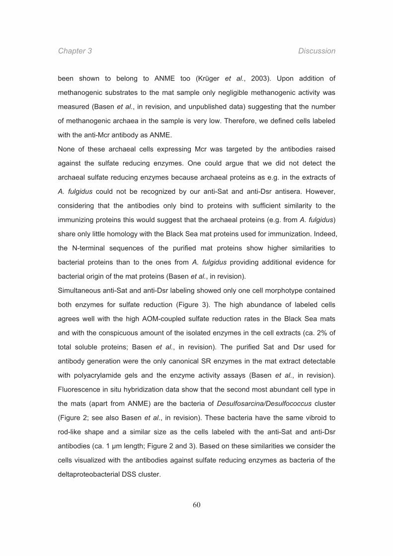
Chapter 3 Discussion
60
been shown to belong to ANME too (Krüger et al., 2003). Upon addition of
methanogenic substrates to the mat sample only negligible methanogenic activity was
measured (Basen et al., in revision, and unpublished data) suggesting that the number
of methanogenic archaea in the sample is very low. Therefore, we defined cells labeled
with the anti-Mcr antibody as ANME.
None of these archaeal cells expressing Mcr was targeted by the antibodies raised
against the sulfate reducing enzymes. One could argue that we did not detect the
archaeal sulfate reducing enzymes because archaeal proteins as e.g. in the extracts of
A. fulgidus could not be recognized by our anti-Sat and anti-Dsr antisera. However,
considering that the antibodies only bind to proteins with sufficient similarity to the
immunizing proteins this would suggest that the archaeal proteins (e.g. from A. fulgidus)
share only little homology with the Black Sea mat proteins used for immunization. Indeed,
the N-terminal sequences of the purified mat proteins show higher similarities to
bacterial proteins than to the ones from A. fulgidus providing additional evidence for
bacterial origin of the mat proteins (Basen et al., in revision).
Simultaneous anti-Sat and anti-Dsr labeling showed only one cell morphotype contained
both enzymes for sulfate reduction (Figure 3). The high abundance of labeled cells
agrees well with the high AOM-coupled sulfate reduction rates in the Black Sea mats
and with the conspicuous amount of the isolated enzymes in the cell extracts (ca. 2% of
total soluble proteins; Basen et al., in revision). The purified Sat and Dsr used for
antibody generation were the only canonical SR enzymes in the mat extract detectable
with polyacrylamide gels and the enzyme activity assays (Basen et al., in revision).
Fluorescence in situ hybridization data show that the second most abundant cell type in
the mats (apart from ANME) are the bacteria of Desulfosarcina/Desulfococcus cluster
(Figure 2; see also Basen et al., in revision). These bacteria have the same vibroid to
rod-like shape and a similar size as the cells labeled with the anti-Sat and anti-Dsr
antibodies (ca. 1 m length; Figure 2 and 3). Based on these similarities we consider the
cells visualized with the antibodies against sulfate reducing enzymes as bacteria of the
deltaproteobacterial DSS cluster.
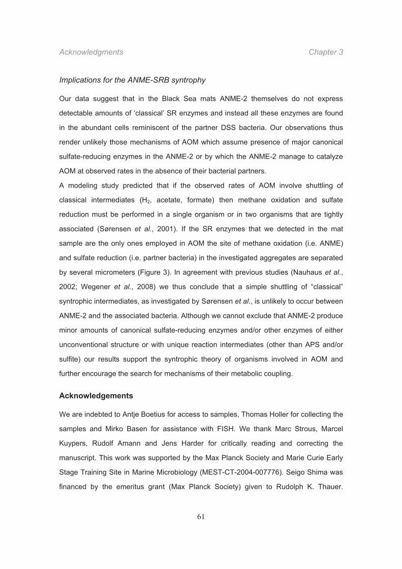
Acknowledgments Chapter 3
61
Implications for the ANME-SRB syntrophy
Our data suggest that in the Black Sea mats ANME-2 themselves do not express
detectable amounts of ‘classical’ SR enzymes and instead all these enzymes are found
in the abundant cells reminiscent of the partner DSS bacteria. Our observations thus
render unlikely those mechanisms of AOM which assume presence of major canonical
sulfate-reducing enzymes in the ANME-2 or by which the ANME-2 manage to catalyze
AOM at observed rates in the absence of their bacterial partners.
A modeling study predicted that if the observed rates of AOM involve shuttling of
classical intermediates (H2, acetate, formate) then methane oxidation and sulfate
reduction must be performed in a single organism or in two organisms that are tightly
associated (Sørensen et al., 2001). If the SR enzymes that we detected in the mat
sample are the only ones employed in AOM the site of methane oxidation (i.e. ANME)
and sulfate reduction (i.e. partner bacteria) in the investigated aggregates are separated
by several micrometers (Figure 3). In agreement with previous studies (Nauhaus et al.,
2002; Wegener et al., 2008) we thus conclude that a simple shuttling of “classical”
syntrophic intermediates, as investigated by Sørensen et al., is unlikely to occur between
ANME-2 and the associated bacteria. Although we cannot exclude that ANME-2 produce
minor amounts of canonical sulfate-reducing enzymes and/or other enzymes of either
unconventional structure or with unique reaction intermediates (other than APS and/or
sulfite) our results support the syntrophic theory of organisms involved in AOM and
further encourage the search for mechanisms of their metabolic coupling.
Acknowledgements
We are indebted to Antje Boetius for access to samples, Thomas Holler for collecting the
samples and Mirko Basen for assistance with FISH. We thank Marc Strous, Marcel
Kuypers, Rudolf Amann and Jens Harder for critically reading and correcting the
manuscript. This work was supported by the Max Planck Society and Marie Curie Early
Stage Training Site in Marine Microbiology (MEST-CT-2004-007776). Seigo Shima was
financed by the emeritus grant (Max Planck Society) given to Rudolph K. Thauer.
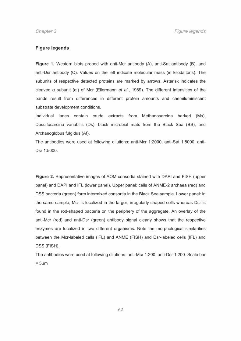
Chapter 3 Figure legends
62
Figure legends
Figure 1. Western blots probed with anti-Mcr antibody (A), anti-Sat antibody (B), and
anti-Dsr antibody (C). Values on the left indicate molecular mass (in kilodaltons). The
subunits of respective detected proteins are marked by arrows. Asterisk indicates the
cleaved subunit ( ‘) of Mcr (Ellermann et al., 1989). The different intensities of the
bands result from differences in different protein amounts and chemiluminiscent
substrate development conditions.
Individual lanes contain crude extracts from Methanosarcina barkeri (Ms),
Desulfosarcina variabilis (Ds), black microbial mats from the Black Sea (BS), and
Archaeoglobus fulgidus (Af).
The antibodies were used at following dilutions: anti-Mcr 1:2000, anti-Sat 1:5000, anti-
Dsr 1:5000.
Figure 2. Representative images of AOM consortia stained with DAPI and FISH (upper
panel) and DAPI and IFL (lower panel). Upper panel: cells of ANME-2 archaea (red) and
DSS bacteria (green) form intermixed consortia in the Black Sea sample. Lower panel: in
the same sample, Mcr is localized in the larger, irregularly shaped cells whereas Dsr is
found in the rod-shaped bacteria on the periphery of the aggregate. An overlay of the
anti-Mcr (red) and anti-Dsr (green) antibody signal clearly shows that the respective
enzymes are localized in two different organisms. Note the morphological similarities
between the Mcr-labeled cells (IFL) and ANME (FISH) and Dsr-labeled cells (IFL) and
DSS (FISH).
The antibodies were used at following dilutions: anti-Mcr 1:200, anti-Dsr 1:200. Scale bar
= 5 m
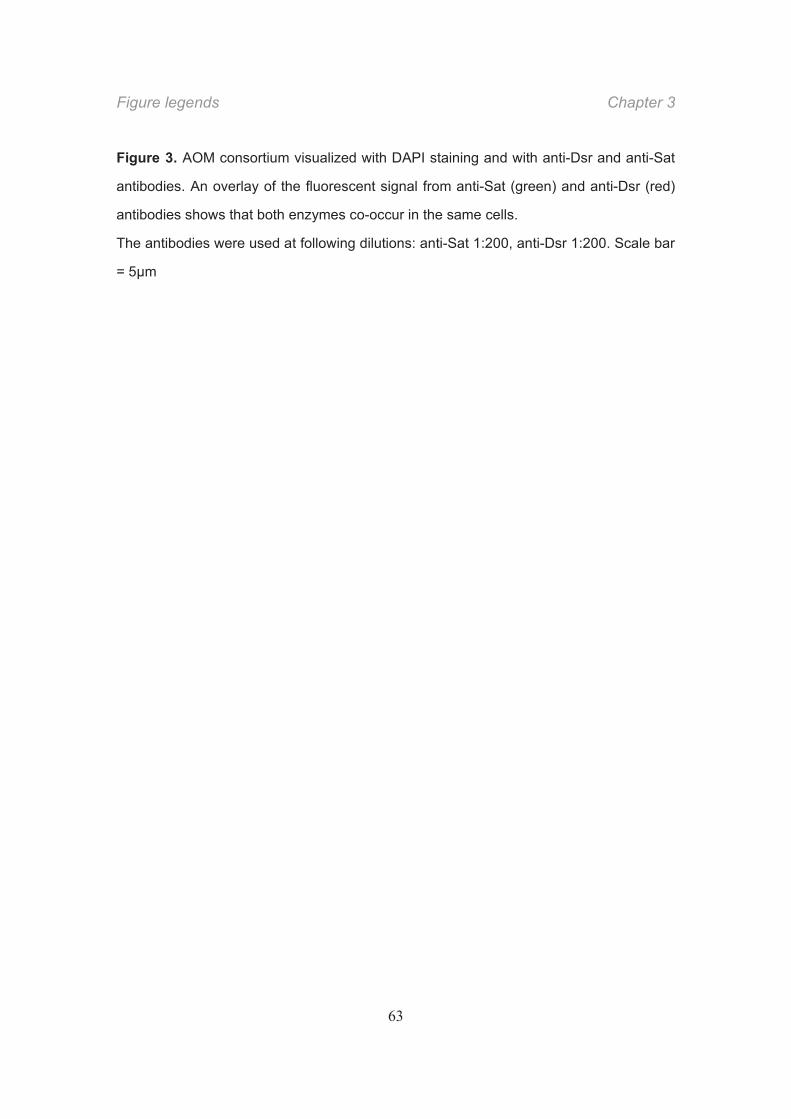
Figure legends Chapter 3
63
Figure 3. AOM consortium visualized with DAPI staining and with anti-Dsr and anti-Sat
antibodies. An overlay of the fluorescent signal from anti-Sat (green) and anti-Dsr (red)
antibodies shows that both enzymes co-occur in the same cells.
The antibodies were used at following dilutions: anti-Sat 1:200, anti-Dsr 1:200. Scale bar
= 5 m
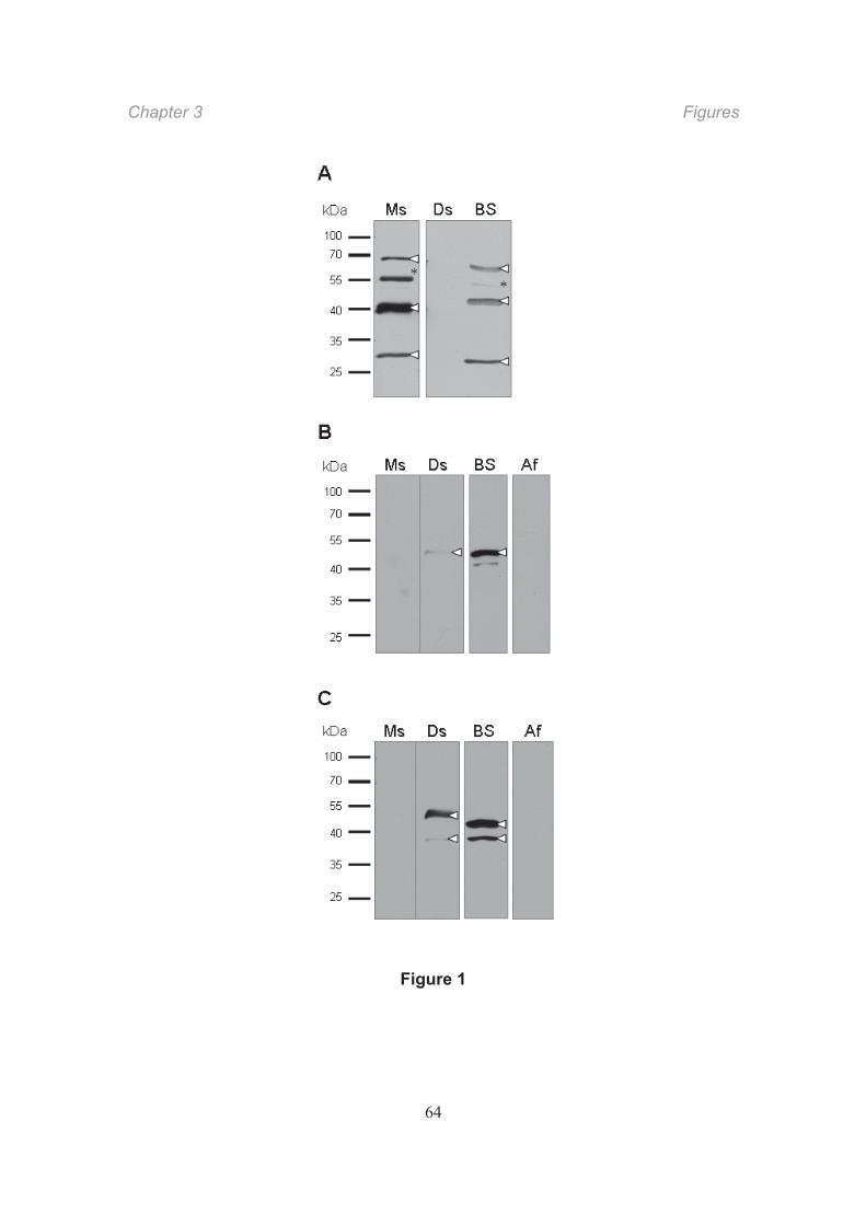
Chapter 3 Figures
64
Figure 1
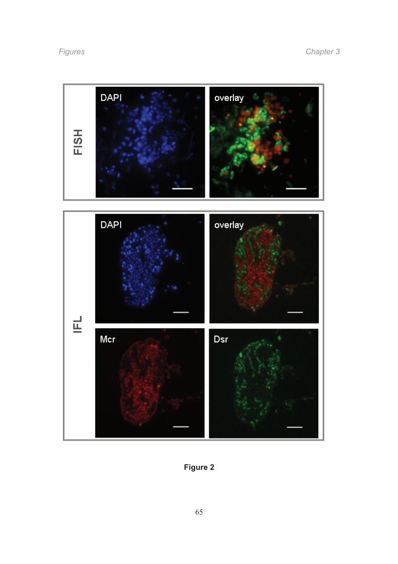
Figures Chapter 3
65
Figure 2
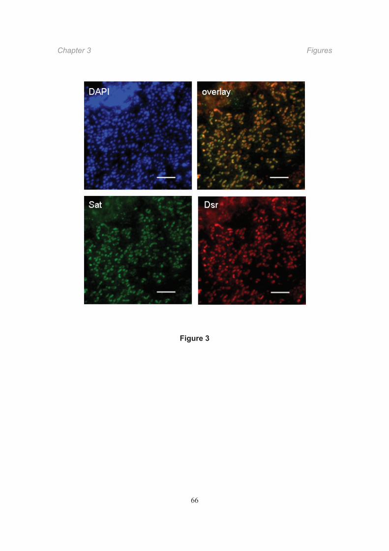
Chapter 3 Figures
66
Figure 3
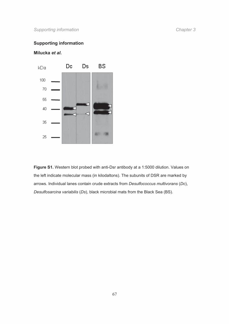
Supporting information Chapter 3
67
Supporting information
Milucka et al.
Figure S1. Western blot probed with anti-Dsr antibody at a 1:5000 dilution. Values on
the left indicate molecular mass (in kilodaltons). The subunits of DSR are marked by
arrows. Individual lanes contain crude extracts from Desulfococcus multivorans (Dc),
Desulfosarcina variabilis (Ds), black microbial mats from the Black Sea (BS).

CHAPTER 4 Sulfur cycling between archaea and bacteria is
involved in anaerobic oxidation of methane
Jana Milucka, Timothy G. Ferdelman, Marcel M. M. Kuypers
Contributors and potential co-authors: Gabriele Klockgether, Lubos Polerecky, Tomas
Vagner, Michael Formolo, Ingo Lieberwirth, Gunter Wegener, Friedrich Widdel, Markus
Schmid, Michael Wagner
Contributions to the manuscript:
J.M., F.W., T.G.F. and M.M.M.K. designed research and project outline, J.M. provided
enrichment cultures and performed isotope labeling experiments, J.M. performed FISH,
IFL and IGL analysis, J.M. and I.L. performed TEM/EDX analysis, J.M., M.M.M.K., M.S.
and M.W. performed Raman spectroscopy and data analysis, J.M., T.V., L.P. and
M.M.M.K. performed nanoSIMS analysis and data evaluation, G.K. and T.F. performed
analysis of sulfur compounds, M.F. performed sulfur extractions, G.W. and T.F.
contributed new analytical technique, T.F. performed thermodynamic calculations, J.M.,
T.F. and M.M.M.K. conceived, wrote and edited the manuscript.
Chapter is in preparation
Chapter 4 Summary
70
Summary
The anaerobic oxidation of methane (AOM) coupled to sulfate reduction plays a
crucial role in biogeochemical carbon and sulfur cycling in marine sediments.
AOM is most likely mediated by a consortium of methanotrophic Archaea (ANME)
and sulfate-reducing Deltaproteobacteria via a so far unknown mechanism. Here
we propose that this mechanism might involve a transfer of zerovalent sulfur (S0)
between the bacteria and the ANME. We show that ANME intracellularly
accumulate large amounts of S0, which is at least partly derived from sulfate. We
provide evidence for sulfur turnover in ANME being directly linked to methane
oxidation. We cannot unambiguously conclude the directionality of sulfur
transport between the ANME and the DSS but based on our observations and
thermodynamic considerations a following model for sulfur cycling between
ANME and SRB involved in AOM can be envisioned. The SRB reduce sulfate with
sulfide to polysulfide, which is released from the cells. The disulfides will likely
react with reduced sulfur outside the cells to form longer-chain polysulfides. This
S0 is taken up by the ANME and used to oxidize methane to CO2. During this
process sulfide is released, which is partly re-used by the SRB to reduce sulfate.
Such cooperation maintained through extracellular sulfur cycling not only
represents a unique syntrophic mechanism but also has significant implications
for our understanding of biogeochemical sulfur cycling and its transformations in
marine sediments.
Introduction Chapter 4
71
Introduction
Anaerobic oxidation of methane coupled to sulfate reduction (AOM; Eq. 1) is widespread
in all oxygen-depleted marine environments where sulfate is the most abundant electron
acceptor (methane seeps, vents, and surface and deep sediments (Knittel & Boetius,
2009 and references therein).
OHHCOHSSOCH 23244 (Eq. 1)
AOM plays a crucial role in biogeochemical carbon and sulfur cycling. It oxidizes the
majority of methane – a potent greenhouse gas – diffusing from the seafloor and
prevents it from escaping to the atmosphere (Hinrichs & Boetius, 2002; Reeburgh, 2007).
The AOM-coupled sulfate reduction consumes a large portion of the downwards sulfate
flux (Devol & Ahmet, 1981) and forms sulfide, which diffuses upwards towards the
seafloor and supports free-living and symbiotic sulfide- and sulfur-oxidizers.
Despite the pronounced effect of AOM on sediment biogeochemistry the biological basis
for AOM has persistently eluded clarification and remains a geomicrobiological puzzle to
this day (Alperin & Hoehler, 2010). The process has been proposed to be syntrophically
mediated by a consortium of methanotrophic Archaea (ANME) and Deltaproteobacteria
(Boetius et al., 2000; Orphan et al., 2001a) because of their co-occurrence at all so far
investigated AOM sites and in all enrichment cultures. However, the syntrophic nature of
their relationship is still a matter of debate because the necessary intermediate that
would shuttle reducing equivalents originating from methane oxidation in the ANME to
the associated bacteria has never been conclusively identified. Moreover, in case of a
syntrophy, the extremely low energy yield of the net reaction (ca. 20 kJ mol–1 CH4) would
have to be shared between two partners. Therefore, it has been suggested (Widdel et al.,
2007; Thauer & Shima, 2008; Johnson & Mukhopadhyay, 2008) that (at least some)
ANME might perform sulfate reduction on their own. Currently, there is no direct
evidence for this hypothesis, in fact, recent biochemical studies, which show the only
detectable enzymes for sulfate reduction are expressed by the associated bacteria
provide evidence for an active involvement of both organisms in AOM (Milucka et al., in
preparation).
Chapter 4 Results and Discussion
72
Several alternative hypotheses have been put forward, all of which involve a flow of
electrons, or intermediates, from the ANME to the bacteria, to explain the syntrophic
mechanism. None of the plausible syntrophic intermediates (i.e. hydrogen, formate,
acetate, or methanol) was conclusively shown to be involved in AOM (Nauhaus et al.,
2002; Meulepas et al., 2010) and a shuttling of a carbon-containing intermediate in
general has been excluded (Wegener et al., 2008). Furthermore, there is no
experimental evidence for pure electron transfer via extracellular nanowires (Shima &
Thauer, 2005; Meyerdierks et al., 2010) or a direct cell-to-cell contact.
Alternatively, it is feasible that reduced sulfur compounds e.g. formed during SR may
serve as intermediates in AOM. Based on energetic and thermodynamic considerations,
zerovalent sulfur (S0) might be used as an electron acceptor for methane oxidation
(Shima & Thauer, 2005) because its redox potential is sufficiently low to be compatible
with the enzyme likely responsible for methane activation in AOM, ‘reverse’ methyl Co-M
reductase (Mcr; Krüger et al., 2003; Scheller et al., 2010). Zerovalent sulfur in the form
of elemental sulfur and dissolved polysulfides (up to 200 M; Kamyshny & Ferdelman,
unpubl. res.) have been observed in the AOM active zone of Black Sea sediments. Here
we investigate the potential role of zerovalent sulfur in AOM using a combination of
microbiological, imaging and biogeochemical techniques. The analyses were performed
on microbial cultures highly enriched in AOM biomass (>95% of all cells).
Results and Discussion
Microbial biomass of enriched AOM cultures contains conspicuous amounts of
zerovalent sulfur
Wet biomass from AOM enrichment cultures was anaerobically extracted with methanol
and subsequently analyzed by HPLC UV spectrometry to examine the potential
presence of S0. The analysis revealed high abundance of elemental sulfur of up to ~ 2
mmol S0 per gram dry biomass (~6 weight %). Other organisms that are known to store
high amounts of sulfur (up to 40% cell dry weight) are chemoheterotrophic and
chemophototrophic sulfur and sulfide oxidizers. These organisms store S0 as cyclic S8
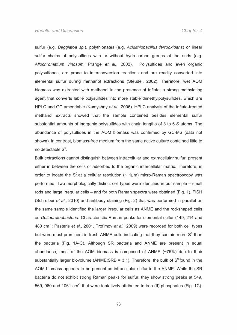
Results and Discussion Chapter 4
73
sulfur (e.g. Beggiatoa sp.), polythionates (e.g. Acidithiobacillus ferrooxidans) or linear
sulfur chains of polysulfides with or without hydrocarbon groups at the ends (e.g.
Allochromatium vinosum; Prange et al., 2002). Polysulfides and even organic
polysulfanes, are prone to interconversion reactions and are readily converted into
elemental sulfur during methanol extractions (Steudel, 2002). Therefore, wet AOM
biomass was extracted with methanol in the presence of triflate, a strong methylating
agent that converts labile polysulfides into more stable dimethylpolysulfides, which are
HPLC and GC amendable (Kamyshny et al., 2006). HPLC analysis of the triflate-treated
methanol extracts showed that the sample contained besides elemental sulfur
substantial amounts of inorganic polysulfides with chain lengths of 3 to 6 S atoms. The
abundance of polysulfides in the AOM biomass was confirmed by GC-MS (data not
shown). In contrast, biomass-free medium from the same active culture contained little to
no detectable S0.
Bulk extractions cannot distinguish between intracellular and extracellular sulfur, present
either in between the cells or adsorbed to the organic intercellular matrix. Therefore, in
order to locate the S0 at a cellular resolution (~ 1μm) micro-Raman spectroscopy was
performed. Two morphologically distinct cell types were identified in our sample – small
rods and large irregular cells – and for both Raman spectra were obtained (Fig. 1). FISH
(Schreiber et al., 2010) and antibody staining (Fig. 2) that was performed in parallel on
the same sample identified the larger irregular cells as ANME and the rod-shaped cells
as Deltaproteobacteria. Characteristic Raman peaks for elemental sulfur (149, 214 and
480 cm-1; Pasteris et al., 2001, Trofimov et al., 2009) were recorded for both cell types
but were most prominent in fresh ANME cells indicating that they contain more S0 than
the bacteria (Fig. 1A-C). Although SR bacteria and ANME are present in equal
abundance, most of the AOM biomass is composed of ANME (~75%) due to their
substantially larger biovolume (ANME:SRB = 3:1). Therefore, the bulk of S0 found in the
AOM biomass appears to be present as intracellular sulfur in the ANME. While the SR
bacteria do not exhibit strong Raman peaks for sulfur, they show strong peaks at 549,
569, 960 and 1061 cm-1 that were tentatively attributed to iron (II) phosphates (Fig. 1C).
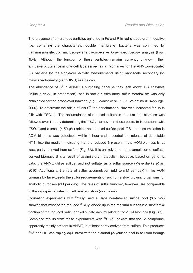
Chapter 4 Results and Discussion
74
The presence of amorphous particles enriched in Fe and P in rod-shaped gram-negative
(i.e. contaning the characteristic double membrane) bacteria was confirmed by
transmission electron microscopy/energy-dispersive X-ray spectroscopy analysis (Figs.
1D-E). Although the function of these particles remains currently unknown, their
exclusive occurrence in one cell type served as a biomarker for the ANME-associated
SR bacteria for the single-cell activity measurements using nanoscale secondary ion
mass spectrometry (nanoSIMS; see below).
The abundance of S0 in ANME is surprising because they lack known SR enzymes
(Milucka et al., in preparation), and in fact a dissimilatory sulfur metabolism was only
anticipated for the associated bacteria (e.g. Hoehler et al., 1994; Valentine & Reeburgh,
2000). To determine the origin of this S0, the enrichment culture was incubated for up to
24h with 35SO42-. The accumulation of reduced sulfate in medium and biomass was
followed over time by determining the 35SO42- turnover in these pools. In incubations with
35SO42- and a small (< 50 μM) added non-labeled sulfide pool, 35S-label accumulation in
AOM biomass was detectable within 1 hour and preceded the release of detectable
H35S– into the medium indicating that the reduced S present in the AOM biomass is, at
least partly, derived from sulfate (Fig. 3A). It is unlikely that the accumulation of sulfate-
derived biomass S is a result of assimilatory metabolism because, based on genomic
data, the ANME utilize sulfide, and not sulfate, as a sulfur source (Meyerdierks et al.,
2010). Additionally, the rate of sulfur accumulation (μM to mM per day) in the AOM
biomass by far exceeds the sulfur requirements of such ultra-slow growing organisms for
anabolic purposes (nM per day). The rates of sulfur turnover, however, are comparable
to the cell-specific rates of methane oxidation (see below).
Incubation experiments with 35SO42- and a large non-labeled sulfide pool (3.5 mM)
showed that most of the reduced 35SO42- ended up in the medium but again a substantial
fraction of the reduced radio-labeled sulfate accumulated in the AOM biomass (Fig. 3B).
Combined results from these experiments with 35SO42- indicate that the S0 compound,
apparently mainly present in ANME, is at least partly derived from sulfate. This produced 35S0 and HS– can rapidly equilibrate with the external polysulfide pool in solution through
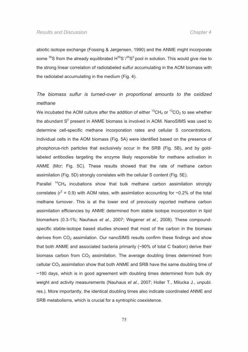
Results and Discussion Chapter 4
75
abiotic isotope exchange (Fossing & Jørgensen, 1990) and the ANME might incorporate
some 35S from the already equilibrated H35S–/35S0 pool in solution. This would give rise to
the strong linear correlation of radiolabeled sulfur accumulating in the AOM biomass with
the radiolabel accumulating in the medium (Fig. 4).
The biomass sulfur is turned-over in proportional amounts to the oxidized
methane
We incubated the AOM culture after the addition of either 13CH4 or 13CO2 to see whether
the abundant S0 present in ANME biomass is involved in AOM. NanoSIMS was used to
determine cell-specific methane incorporation rates and cellular S concentrations.
Individual cells in the AOM biomass (Fig. 5A) were identified based on the presence of
phosphorus-rich particles that exclusively occur in the SRB (Fig. 5B), and by gold-
labeled antibodies targeting the enzyme likely responsible for methane activation in
ANME (Mcr; Fig. 5C). These results showed that the rate of methane carbon
assimilation (Fig. 5D) strongly correlates with the cellular S content (Fig. 5E).
Parallel 14CH4 incubations show that bulk methane carbon assimilation strongly
correlates (r2 = 0.9) with AOM rates, with assimilation accounting for ~0.2% of the total
methane turnover. This is at the lower end of previously reported methane carbon
assimilation efficiencies by ANME determined from stable isotope incorporation in lipid
biomarkers (0.3-1%; Nauhaus et al., 2007; Wegener et al., 2008). These compound-
specific stable-isotope based studies showed that most of the carbon in the biomass
derives from CO2 assimilation. Our nanoSIMS results confirm these findings and show
that both ANME and associated bacteria primarily (~90% of total C fixation) derive their
biomass carbon from CO2 assimilation. The average doubling times determined from
cellular CO2 assimilation show that both ANME and SRB have the same doubling time of
~180 days, which is in good agreement with doubling times determined from bulk dry
weight and activity measurements (Nauhaus et al., 2007; Holler T., Milucka J., unpubl.
res.). More importantly, the identical doubling times also indicate coordinated ANME and
SRB metabolisms, which is crucial for a syntrophic coexistence.
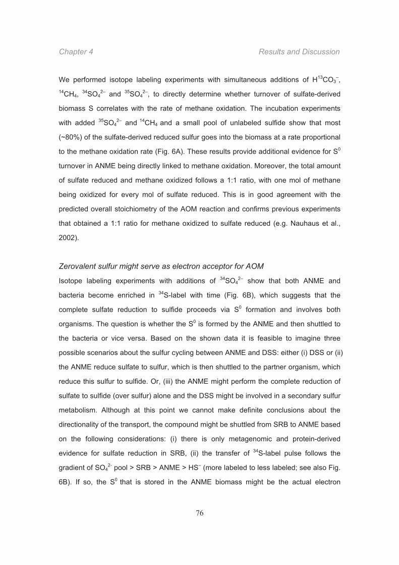
Chapter 4 Results and Discussion
76
We performed isotope labeling experiments with simultaneous additions of H13CO3–,
14CH4, 34SO42– and 35SO4
2–, to directly determine whether turnover of sulfate-derived
biomass S correlates with the rate of methane oxidation. The incubation experiments
with added 35SO42– and 14CH4 and a small pool of unlabeled sulfide show that most
(~80%) of the sulfate-derived reduced sulfur goes into the biomass at a rate proportional
to the methane oxidation rate (Fig. 6A). These results provide additional evidence for S0
turnover in ANME being directly linked to methane oxidation. Moreover, the total amount
of sulfate reduced and methane oxidized follows a 1:1 ratio, with one mol of methane
being oxidized for every mol of sulfate reduced. This is in good agreement with the
predicted overall stoichiometry of the AOM reaction and confirms previous experiments
that obtained a 1:1 ratio for methane oxidized to sulfate reduced (e.g. Nauhaus et al.,
2002).
Zerovalent sulfur might serve as electron acceptor for AOM
Isotope labeling experiments with additions of 34SO42– show that both ANME and
bacteria become enriched in 34S-label with time (Fig. 6B), which suggests that the
complete sulfate reduction to sulfide proceeds via S0 formation and involves both
organisms. The question is whether the S0 is formed by the ANME and then shuttled to
the bacteria or vice versa. Based on the shown data it is feasible to imagine three
possible scenarios about the sulfur cycling between ANME and DSS: either (i) DSS or (ii)
the ANME reduce sulfate to sulfur, which is then shuttled to the partner organism, which
reduce this sulfur to sulfide. Or, (iii) the ANME might perform the complete reduction of
sulfate to sulfide (over sulfur) alone and the DSS might be involved in a secondary sulfur
metabolism. Although at this point we cannot make definite conclusions about the
directionality of the transport, the compound might be shuttled from SRB to ANME based
on the following considerations: (i) there is only metagenomic and protein-derived
evidence for sulfate reduction in SRB, (ii) the transfer of 34S-label pulse follows the
gradient of SO42- pool > SRB > ANME > HS– (more labeled to less labeled; see also Fig.
6B). If so, the S0 that is stored in the ANME biomass might be the actual electron
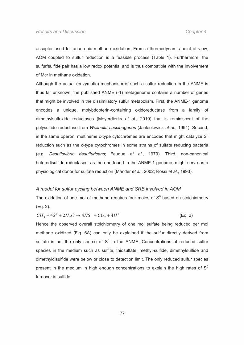
Results and Discussion Chapter 4
77
acceptor used for anaerobic methane oxidation. From a thermodynamic point of view,
AOM coupled to sulfur reduction is a feasible process (Table 1). Furthermore, the
sulfur/sulfide pair has a low redox potential and is thus compatible with the involvement
of Mcr in methane oxidation.
Although the actual (enzymatic) mechanism of such a sulfur reduction in the ANME is
thus far unknown, the published ANME (-1) metagenome contains a number of genes
that might be involved in the dissimilatory sulfur metabolism. First, the ANME-1 genome
encodes a unique, molybdopterin-containing oxidoreductase from a family of
dimethylsulfoxide reductases (Meyerdierks et al., 2010) that is reminiscent of the
polysulfide reductase from Wolinella succinogenes (Jankielewicz et al., 1994). Second,
in the same operon, multiheme c-type cytochromes are encoded that might catalyze S0
reduction such as the c-type cytochromes in some strains of sulfate reducing bacteria
(e.g. Desulfovibrio desulfuricans; Fauque et al., 1979). Third, non-canonical
heterodisulfide reductases, as the one found in the ANME-1 genome, might serve as a
physiological donor for sulfate reduction (Mander et al., 2002; Rossi et al., 1993).
A model for sulfur cycling between ANME and SRB involved in AOM
The oxidation of one mol of methane requires four moles of S0 based on stoichiometry
(Eq. 2).
HCOHSOHSCH 4424 220
4 (Eq. 2)
Hence the observed overall stoichiometry of one mol sulfate being reduced per mol
methane oxidized (Fig. 6A) can only be explained if the sulfur directly derived from
sulfate is not the only source of S0 in the ANME. Concentrations of reduced sulfur
species in the medium such as sulfite, thiosulfate, methyl-sulfide, dimethylsulfide and
dimethyldisulfide were below or close to detection limit. The only reduced sulfur species
present in the medium in high enough concentrations to explain the high rates of S0
turnover is sulfide.
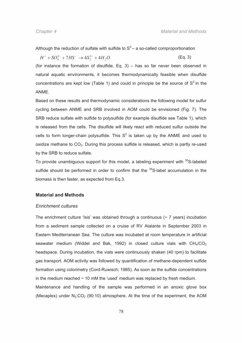
Chapter 4 Material and Methods
78
Although the reduction of sulfate with sulfide to S0 – a so-called comproportionation
(Eq. 3)
(for instance the formation of disulfide, Eq. 3) – has so far never been observed in
natural aquatic environments, it becomes thermodynamically feasible when disulfide
concentrations are kept low (Table 1) and could in principle be the source of S0 in the
ANME.
Based on these results and thermodynamic considerations the following model for sulfur
cycling between ANME and SRB involved in AOM could be envisioned (Fig. 7). The
SRB reduce sulfate with sulfide to polysulfide (for example disulfide see Table 1), which
is released from the cells. The disulfide will likely react with reduced sulfur outside the
cells to form longer-chain polysulfide. This S0 is taken up by the ANME and used to
oxidize methane to CO2. During this process sulfide is released, which is partly re-used
by the SRB to reduce sulfate.
To provide unambiguous support for this model, a labeling experiment with 35S-labeled
sulfide should be performed in order to confirm that the 35S-label accumulation in the
biomass is then faster, as expected from Eq.3.
Material and Methods
Enrichment cultures
The enrichment culture ‘Isis’ was obtained through a continuous (~ 7 years) incubation
from a sediment sample collected on a cruise of RV Atalante in September 2003 in
Eastern Mediterranean Sea. The culture was incubated at room temperature in artificial
seawater medium (Widdel and Bak, 1992) in closed culture vials with CH4/CO2
headspace. During incubation, the vials were continuously shaken (40 rpm) to facilitate
gas transport. AOM activity was followed by quantification of methane-dependent sulfide
formation using colorimetry (Cord-Ruwisch; 1985). As soon as the sulfide concentrations
in the medium reached ~ 10 mM the ‘used’ medium was replaced by fresh medium.
Maintenance and handling of the sample was performed in an anoxic glove box
(Mecaplex) under N2:CO2 (90:10) atmosphere. At the time of the experiment, the AOM
OHSHSSOH 222
24 447
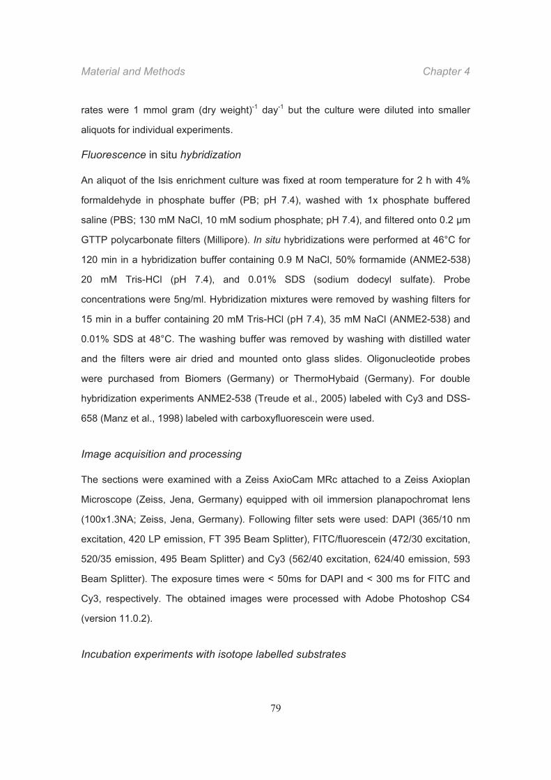
Material and Methods Chapter 4
79
rates were 1 mmol gram (dry weight)-1 day-1 but the culture were diluted into smaller
aliquots for individual experiments.
Fluorescence in situ hybridization
An aliquot of the Isis enrichment culture was fixed at room temperature for 2 h with 4%
formaldehyde in phosphate buffer (PB; pH 7.4), washed with 1x phosphate buffered
saline (PBS; 130 mM NaCl, 10 mM sodium phosphate; pH 7.4), and filtered onto 0.2 μm
GTTP polycarbonate filters (Millipore). In situ hybridizations were performed at 46°C for
120 min in a hybridization buffer containing 0.9 M NaCl, 50% formamide (ANME2-538)
20 mM Tris-HCl (pH 7.4), and 0.01% SDS (sodium dodecyl sulfate). Probe
concentrations were 5ng/ml. Hybridization mixtures were removed by washing filters for
15 min in a buffer containing 20 mM Tris-HCl (pH 7.4), 35 mM NaCl (ANME2-538) and
0.01% SDS at 48°C. The washing buffer was removed by washing with distilled water
and the filters were air dried and mounted onto glass slides. Oligonucleotide probes
were purchased from Biomers (Germany) or ThermoHybaid (Germany). For double
hybridization experiments ANME2-538 (Treude et al., 2005) labeled with Cy3 and DSS-
658 (Manz et al., 1998) labeled with carboxyfluorescein were used.
Image acquisition and processing
The sections were examined with a Zeiss AxioCam MRc attached to a Zeiss Axioplan
Microscope (Zeiss, Jena, Germany) equipped with oil immersion planapochromat lens
(100x1.3NA; Zeiss, Jena, Germany). Following filter sets were used: DAPI (365/10 nm
excitation, 420 LP emission, FT 395 Beam Splitter), FITC/fluorescein (472/30 excitation,
520/35 emission, 495 Beam Splitter) and Cy3 (562/40 excitation, 624/40 emission, 593
Beam Splitter). The exposure times were < 50ms for DAPI and < 300 ms for FITC and
Cy3, respectively. The obtained images were processed with Adobe Photoshop CS4
(version 11.0.2).
Incubation experiments with isotope labelled substrates
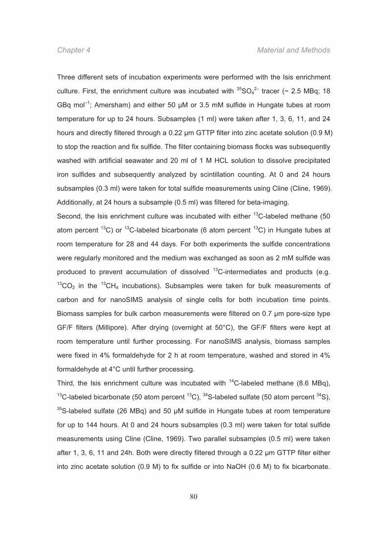
Chapter 4 Material and Methods
80
Three different sets of incubation experiments were performed with the Isis enrichment
culture. First, the enrichment culture was incubated with 35SO42– tracer (~ 2.5 MBq; 18
GBq mol–1; Amersham) and either 50 μM or 3.5 mM sulfide in Hungate tubes at room
temperature for up to 24 hours. Subsamples (1 ml) were taken after 1, 3, 6, 11, and 24
hours and directly filtered through a 0.22 μm GTTP filter into zinc acetate solution (0.9 M)
to stop the reaction and fix sulfide. The filter containing biomass flocks was subsequently
washed with artificial seawater and 20 ml of 1 M HCL solution to dissolve precipitated
iron sulfides and subsequently analyzed by scintillation counting. At 0 and 24 hours
subsamples (0.3 ml) were taken for total sulfide measurements using Cline (Cline, 1969).
Additionally, at 24 hours a subsample (0.5 ml) was filtered for beta-imaging.
Second, the Isis enrichment culture was incubated with either 13C-labeled methane (50
atom percent 13C) or 13C-labeled bicarbonate (6 atom percent 13C) in Hungate tubes at
room temperature for 28 and 44 days. For both experiments the sulfide concentrations
were regularly monitored and the medium was exchanged as soon as 2 mM sulfide was
produced to prevent accumulation of dissolved 13C-intermediates and products (e.g. 13CO2 in the 13CH4 incubations). Subsamples were taken for bulk measurements of
carbon and for nanoSIMS analysis of single cells for both incubation time points.
Biomass samples for bulk carbon measurements were filtered on 0.7 μm pore-size type
GF/F filters (Millipore). After drying (overnight at 50°C), the GF/F filters were kept at
room temperature until further processing. For nanoSIMS analysis, biomass samples
were fixed in 4% formaldehyde for 2 h at room temperature, washed and stored in 4%
formaldehyde at 4°C until further processing.
Third, the Isis enrichment culture was incubated with 14C-labeled methane (8.6 MBq), 13C-labeled bicarbonate (50 atom percent 13C), 34S-labeled sulfate (50 atom percent 34S), 35S-labeled sulfate (26 MBq) and 50 M sulfide in Hungate tubes at room temperature
for up to 144 hours. At 0 and 24 hours subsamples (0.3 ml) were taken for total sulfide
measurements using Cline (Cline, 1969). Two parallel subsamples (0.5 ml) were taken
after 1, 3, 6, 11 and 24h. Both were directly filtered through a 0.22 μm GTTP filter either
into zinc acetate solution (0.9 M) to fix sulfide or into NaOH (0.6 M) to fix bicarbonate.
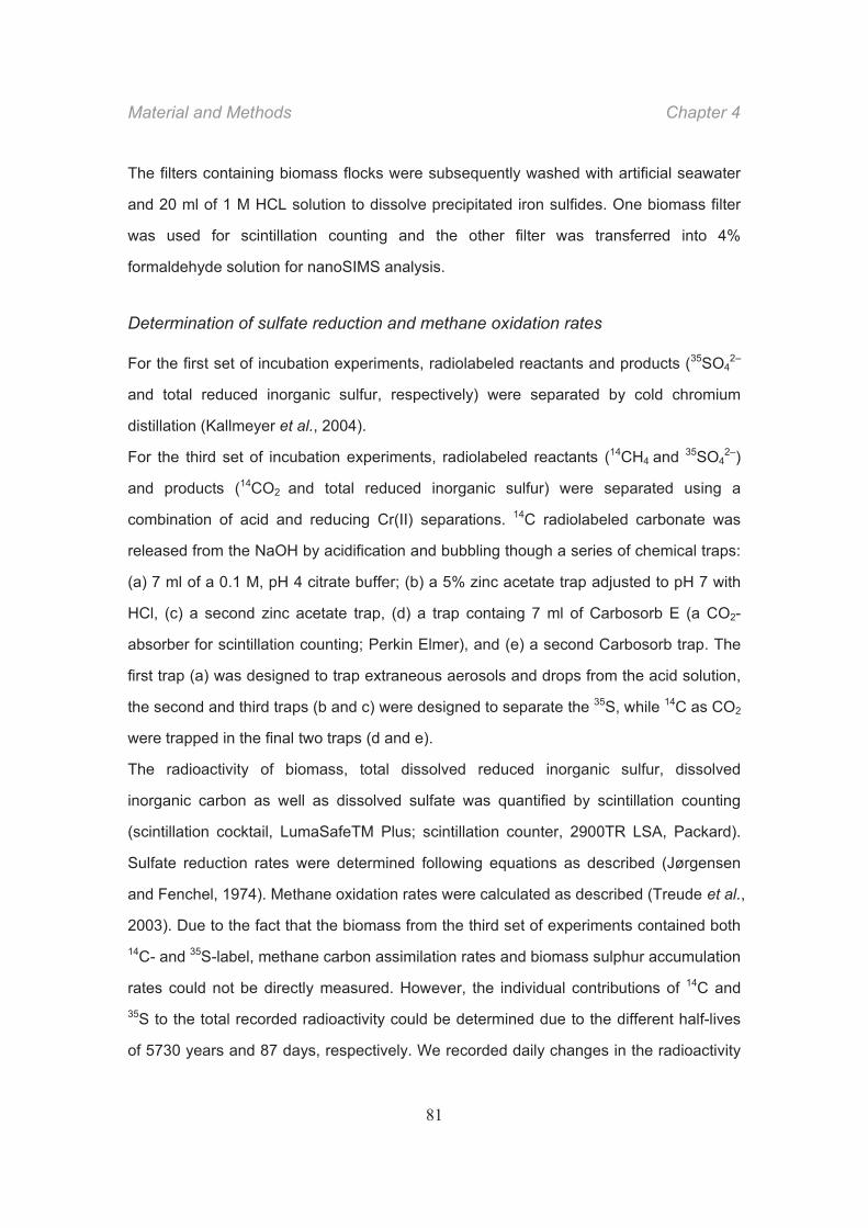
Material and Methods Chapter 4
81
The filters containing biomass flocks were subsequently washed with artificial seawater
and 20 ml of 1 M HCL solution to dissolve precipitated iron sulfides. One biomass filter
was used for scintillation counting and the other filter was transferred into 4%
formaldehyde solution for nanoSIMS analysis.
Determination of sulfate reduction and methane oxidation rates
For the first set of incubation experiments, radiolabeled reactants and products (35SO42–
and total reduced inorganic sulfur, respectively) were separated by cold chromium
distillation (Kallmeyer et al., 2004).
For the third set of incubation experiments, radiolabeled reactants (14CH4 and 35SO42–)
and products (14CO2 and total reduced inorganic sulfur) were separated using a
combination of acid and reducing Cr(II) separations. 14C radiolabeled carbonate was
released from the NaOH by acidification and bubbling though a series of chemical traps:
(a) 7 ml of a 0.1 M, pH 4 citrate buffer; (b) a 5% zinc acetate trap adjusted to pH 7 with
HCl, (c) a second zinc acetate trap, (d) a trap containg 7 ml of Carbosorb E (a CO2-
absorber for scintillation counting; Perkin Elmer), and (e) a second Carbosorb trap. The
first trap (a) was designed to trap extraneous aerosols and drops from the acid solution,
the second and third traps (b and c) were designed to separate the 35S, while 14C as CO2
were trapped in the final two traps (d and e).
The radioactivity of biomass, total dissolved reduced inorganic sulfur, dissolved
inorganic carbon as well as dissolved sulfate was quantified by scintillation counting
(scintillation cocktail, LumaSafeTM Plus; scintillation counter, 2900TR LSA, Packard).
Sulfate reduction rates were determined following equations as described (Jørgensen
and Fenchel, 1974). Methane oxidation rates were calculated as described (Treude et al.,
2003). Due to the fact that the biomass from the third set of experiments contained both 14C- and 35S-label, methane carbon assimilation rates and biomass sulphur accumulation
rates could not be directly measured. However, the individual contributions of 14C and
35S to the total recorded radioactivity could be determined due to the different half-lives
of 5730 years and 87 days, respectively. We recorded daily changes in the radioactivity
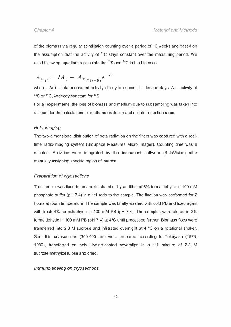
Chapter 4 Material and Methods
82
of the biomass via regular scintillation counting over a period of ~3 weeks and based on
the assumption that the activity of 14C stays constant over the measuring period. We
used following equation to calculate the 35S and 14C in the biomass.
t
tStC eATAA )0(3514
where TA(t) = total measured activity at any time point, t = time in days, A = activity of 35S or 14C, =decay constant for 35S.
For all experiments, the loss of biomass and medium due to subsampling was taken into
account for the calculations of methane oxidation and sulfate reduction rates.
Beta-imaging
The two-dimensional distribution of beta radiation on the filters was captured with a real-
time radio-imaging system (BioSpace Measures Micro Imager). Counting time was 8
minutes. Activities were integrated by the instrument software (BetaVision) after
manually assigning specific region of interest.
Preparation of cryosections
The sample was fixed in an anoxic chamber by addition of 8% formaldehyde in 100 mM
phosphate buffer (pH 7.4) in a 1:1 ratio to the sample. The fixation was performed for 2
hours at room temperature. The sample was briefly washed with cold PB and fixed again
with fresh 4% formaldehyde in 100 mM PB (pH 7.4). The samples were stored in 2%
formaldehyde in 100 mM PB (pH 7.4) at 4ºC until processed further. Biomass flocs were
transferred into 2.3 M sucrose and infiltrated overnight at 4 °C on a rotational shaker.
Semi-thin cryosections (300-400 nm) were prepared according to Tokuyasu (1973,
1980), transferred on poly-L-lysine-coated coverslips in a 1:1 mixture of 2.3 M
sucrose:methylcellulose and dried.
Immunolabeling on cryosections
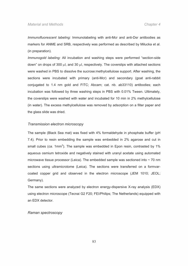
Material and Methods Chapter 4
83
Immunofluorescent labeling: Immunolabeling with anti-Mcr and anti-Dsr antibodies as
markers for ANME and SRB, respectively was performed as described by Milucka et al.
(in preparation).
Immunogold labeling: All incubation and washing steps were performed “section-side
down” on drops of 300 l, and 30 l, respectively. The coverslips with attached sections
were washed in PBS to dissolve the sucrose:methylcellulose support. After washing, the
sections were incubated with primary (anti-Mcr) and secondary (goat anti-rabbit
conjugated to 1.4 nm gold and FITC; Abcam; cat. nb. ab33110) antibodies; each
incubation was followed by three washing steps in PBS with 0.01% Tween. Ultimately,
the coverslips were washed with water and incubated for 10 min in 2% methylcellulose
(in water). The excess methylcellulose was removed by adsorption on a filter paper and
the glass slide was dried.
Transmission electron microscopy
The sample (Black Sea mat) was fixed with 4% formaldehyde in phosphate buffer (pH
7.4). Prior to resin embedding the sample was embedded in 2% agarose and cut in
small cubes (ca. 1mm3). The sample was embedded in Epon resin, contrasted by 1%
aqueous osmium tetroxide and negatively stained with uranyl acetate using automated
microwave tissue processor (Leica). The embedded sample was sectioned into ~ 70 nm
sections using ultramicrotome (Leica). The sections were transferred on a formvar-
coated copper grid and observed in the electron microscope (JEM 1010; JEOL;
Germany).
The same sections were analyzed by electron energy-dispersive X-ray analysis (EDX)
using electron microscope (Tecnai G2 F20; FEI/Philips; The Netherlands) equipped with
an EDX detector.
Raman spectroscopy
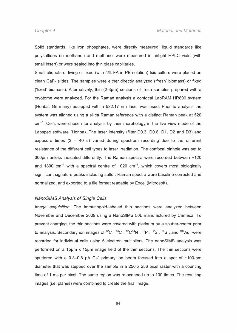
Chapter 4 Material and Methods
84
Solid standards, like iron phosphates, were directly measured; liquid standards like
polysulfides (in methanol) and methanol were measured in airtight HPLC vials (with
small insert) or were sealed into thin glass capillaries.
Small aliquots of living or fixed (with 4% FA in PB solution) Isis culture were placed on
clean CaF2 slides. The samples were either directly analyzed (‘fresh’ biomass) or fixed
(‘fixed’ biomass). Alternatively, thin (2-3μm) sections of fresh samples prepared with a
cryotome were analyzed. For the Raman analysis a confocal LabRAM HR800 system
(Horiba, Germany) equipped with a 532.17 nm laser was used. Prior to analysis the
system was aligned using a silica Raman reference with a distinct Raman peak at 520
cm–1. Cells were chosen for analysis by their morphology in the live view mode of the
Labspec software (Horiba). The laser intensity (filter D0.3, D0.6, D1, D2 and D3) and
exposure times (3 – 40 s) varied during spectrum recording due to the different
resistance of the different cell types to laser irradiation. The confocal pinhole was set to
300μm unless indicated differently. The Raman spectra were recorded between ~120
and 1800 cm–1 with a spectral centre of 1020 cm–1, which covers most biologically
significant signature peaks including sulfur. Raman spectra were baseline-corrected and
normalized, and exported to a file format readable by Excel (Microsoft).
NanoSIMS Analysis of Single Cells
Image acquisition. The immunogold-labeled thin sections were analyzed between
November and December 2009 using a NanoSIMS 50L manufactured by Cameca. To
prevent charging, the thin sections were covered with platinum by a sputter-coater prior
to analysis. Secondary ion images of 12C–, 13C–, 12C14N–, 31P–, 32S–, 34S–, and 197Au– were
recorded for individual cells using 6 electron multipliers. The nanoSIMS analysis was
performed on a 15μm x 15μm image field of the thin sections. The thin sections were
sputtered with a 0.3–0.8 pA Cs+ primary ion beam focused into a spot of ~100-nm
diameter that was stepped over the sample in a 256 x 256 pixel raster with a counting
time of 1 ms per pixel. The same region was re-scanned up to 100 times. The resulting
images (i.e. planes) were combined to create the final image.
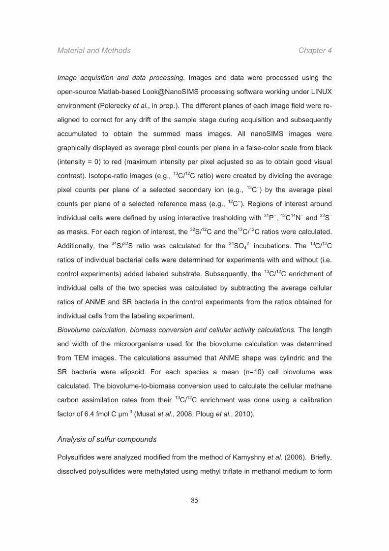
Material and Methods Chapter 4
85
Image acquisition and data processing. Images and data were processed using the
open-source Matlab-based Look@NanoSIMS processing software working under LINUX
environment (Polerecky et al., in prep.). The different planes of each image field were re-
aligned to correct for any drift of the sample stage during acquisition and subsequently
accumulated to obtain the summed mass images. All nanoSIMS images were
graphically displayed as average pixel counts per plane in a false-color scale from black
(intensity = 0) to red (maximum intensity per pixel adjusted so as to obtain good visual
contrast). Isotope-ratio images (e.g., 13C/12C ratio) were created by dividing the average
pixel counts per plane of a selected secondary ion (e.g., 13C–) by the average pixel
counts per plane of a selected reference mass (e.g., 12C–). Regions of interest around
individual cells were defined by using interactive tresholding with 31P–, 12C14N– and 32S–
as masks. For each region of interest, the 32S/12C and the13C/12C ratios were calculated.
Additionally, the 34S/32S ratio was calculated for the 34SO42– incubations. The 13C/12C
ratios of individual bacterial cells were determined for experiments with and without (i.e.
control experiments) added labeled substrate. Subsequently, the 13C/12C enrichment of
individual cells of the two species was calculated by subtracting the average cellular
ratios of ANME and SR bacteria in the control experiments from the ratios obtained for
individual cells from the labeling experiment.
Biovolume calculation, biomass conversion and cellular activity calculations. The length
and width of the microorganisms used for the biovolume calculation was determined
from TEM images. The calculations assumed that ANME shape was cylindric and the
SR bacteria were elipsoid. For each species a mean (n=10) cell biovolume was
calculated. The biovolume-to-biomass conversion used to calculate the cellular methane
carbon assimilation rates from their 13C/12C enrichment was done using a calibration
factor of 6.4 fmol C μm-3 (Musat et al., 2008; Ploug et al., 2010).
Analysis of sulfur compounds
Polysulfides were analyzed modified from the method of Kamyshny et al. (2006). Briefly,
dissolved polysulfides were methylated using methyl triflate in methanol medium to form
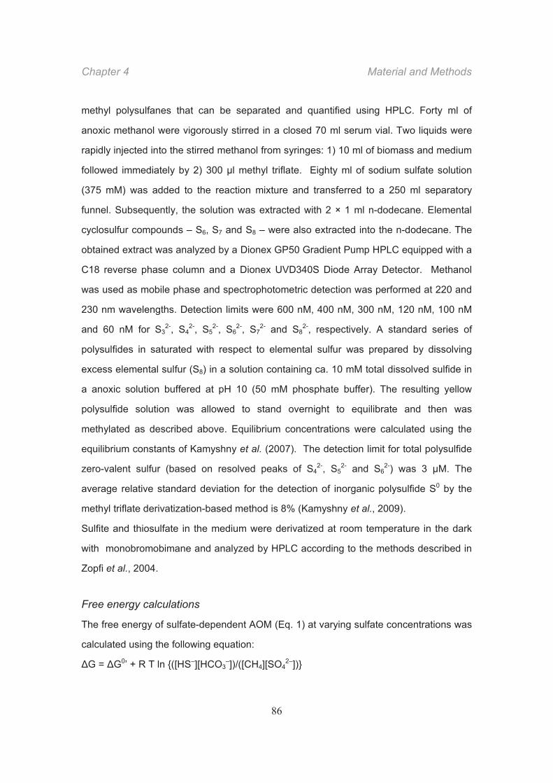
Chapter 4 Material and Methods
86
methyl polysulfanes that can be separated and quantified using HPLC. Forty ml of
anoxic methanol were vigorously stirred in a closed 70 ml serum vial. Two liquids were
rapidly injected into the stirred methanol from syringes: 1) 10 ml of biomass and medium
followed immediately by 2) 300 μl methyl triflate. Eighty ml of sodium sulfate solution
(375 mM) was added to the reaction mixture and transferred to a 250 ml separatory
funnel. Subsequently, the solution was extracted with 2 × 1 ml n-dodecane. Elemental
cyclosulfur compounds – S6, S7 and S8 – were also extracted into the n-dodecane. The
obtained extract was analyzed by a Dionex GP50 Gradient Pump HPLC equipped with a
C18 reverse phase column and a Dionex UVD340S Diode Array Detector. Methanol
was used as mobile phase and spectrophotometric detection was performed at 220 and
230 nm wavelengths. Detection limits were 600 nM, 400 nM, 300 nM, 120 nM, 100 nM
and 60 nM for S32-, S4
2-, S52-, S6
2-, S72- and S8
2-, respectively. A standard series of
polysulfides in saturated with respect to elemental sulfur was prepared by dissolving
excess elemental sulfur (S8) in a solution containing ca. 10 mM total dissolved sulfide in
a anoxic solution buffered at pH 10 (50 mM phosphate buffer). The resulting yellow
polysulfide solution was allowed to stand overnight to equilibrate and then was
methylated as described above. Equilibrium concentrations were calculated using the
equilibrium constants of Kamyshny et al. (2007). The detection limit for total polysulfide
zero-valent sulfur (based on resolved peaks of S42-, S5
2- and S62-) was 3 μM. The
average relative standard deviation for the detection of inorganic polysulfide S0 by the
methyl triflate derivatization-based method is 8% (Kamyshny et al., 2009).
Sulfite and thiosulfate in the medium were derivatized at room temperature in the dark
with monobromobimane and analyzed by HPLC according to the methods described in
Zopfi et al., 2004.
Free energy calculations
The free energy of sulfate-dependent AOM (Eq. 1) at varying sulfate concentrations was
calculated using the following equation:
G = G0’ + R T ln {([HS–][HCO3–])/([CH4][SO4
2–])}
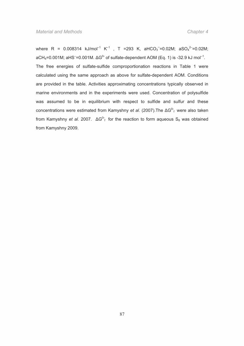
Material and Methods Chapter 4
87
where R = 0.008314 kJ/mol–1 K–1 , T =293 K, aHCO3–=0.02M; aSO4
2–=0.02M;
aCH4=0.001M; aHS–=0.001M. G0’ of sulfate-dependent AOM (Eq. 1) is -32.9 kJ mol–1.
The free energies of sulfate-sulfide comproportionation reactions in Table 1 were
calculated using the same approach as above for sulfate-dependent AOM. Conditions
are provided in the table. Activities approximating concentrations typically observed in
marine environments and in the experiments were used. Concentration of polysulfide
was assumed to be in equilibrium with respect to sulfide and sulfur and these
concentrations were estimated from Kamyshny et al. (2007).The G0’f were also taken
from Kamyshny et al. 2007. G0’f for the reaction to form aqueous S8 was obtained
from Kamyshny 2009.
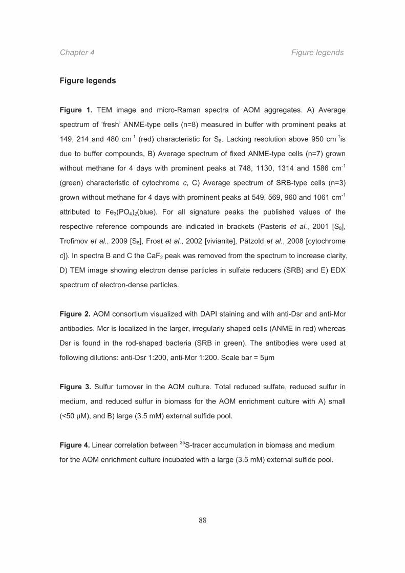
Chapter 4 Figure legends
88
Figure legends
Figure 1. TEM image and micro-Raman spectra of AOM aggregates. A) Average
spectrum of ‘fresh’ ANME-type cells (n=8) measured in buffer with prominent peaks at
149, 214 and 480 cm-1 (red) characteristic for S8. Lacking resolution above 950 cm-1is
due to buffer compounds, B) Average spectrum of fixed ANME-type cells (n=7) grown
without methane for 4 days with prominent peaks at 748, 1130, 1314 and 1586 cm-1
(green) characteristic of cytochrome c, C) Average spectrum of SRB-type cells (n=3)
grown without methane for 4 days with prominent peaks at 549, 569, 960 and 1061 cm-1
attributed to Fe3(PO4)2(blue). For all signature peaks the published values of the
respective reference compounds are indicated in brackets (Pasteris et al., 2001 [S8],
Trofimov et al., 2009 [S8], Frost et al., 2002 [vivianite], Pätzold et al., 2008 [cytochrome
c]). In spectra B and C the CaF2 peak was removed from the spectrum to increase clarity,
D) TEM image showing electron dense particles in sulfate reducers (SRB) and E) EDX
spectrum of electron-dense particles.
Figure 2. AOM consortium visualized with DAPI staining and with anti-Dsr and anti-Mcr
antibodies. Mcr is localized in the larger, irregularly shaped cells (ANME in red) whereas
Dsr is found in the rod-shaped bacteria (SRB in green). The antibodies were used at
following dilutions: anti-Dsr 1:200, anti-Mcr 1:200. Scale bar = 5 m
Figure 3. Sulfur turnover in the AOM culture. Total reduced sulfate, reduced sulfur in
medium, and reduced sulfur in biomass for the AOM enrichment culture with A) small
(<50 μM), and B) large (3.5 mM) external sulfide pool.
Figure 4. Linear correlation between 35S-tracer accumulation in biomass and medium
for the AOM enrichment culture incubated with a large (3.5 mM) external sulfide pool.
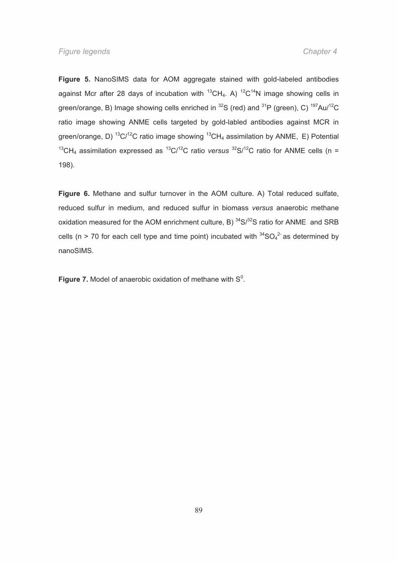
Figure legends Chapter 4
89
Figure 5. NanoSIMS data for AOM aggregate stained with gold-labeled antibodies
against Mcr after 28 days of incubation with 13CH4. A) 12C14N image showing cells in
green/orange, B) Image showing cells enriched in 32S (red) and 31P (green), C) 197Au/12C
ratio image showing ANME cells targeted by gold-labled antibodies against MCR in
green/orange, D) 13C/12C ratio image showing 13CH4 assimilation by ANME, E) Potential 13CH4 assimilation expressed as 13C/12C ratio versus 32S/12C ratio for ANME cells (n =
198).
Figure 6. Methane and sulfur turnover in the AOM culture. A) Total reduced sulfate,
reduced sulfur in medium, and reduced sulfur in biomass versus anaerobic methane
oxidation measured for the AOM enrichment culture, B) 34S/32S ratio for ANME and SRB
cells (n > 70 for each cell type and time point) incubated with 34SO42- as determined by
nanoSIMS.
Figure 7. Model of anaerobic oxidation of methane with S0.

Chapter 4 Table
90
Table 1. Gibbs free energy calculations for methane oxidation and sulfur
comproportionation. Activities of dissolved compounds used in the calculations were:
aHCO3–=0.02M; aSO4
2–=0.02M; aCH4=0.001M; aHS–=0.001M. The activity of solid
elemental sulfur was assumed to be 1. Activity of dissolved elemental sulfur (S8(aq)) was
estimated from Kamyshny (2009).
Reaction G°, kJ/rxn Grxn, J/rxn Notes
-32.9 -32.9
+207 -64.7
-240 +30.1
-438 +55.8
+21.1 -12.2
+55.3
nM S22
μM S22-
-186 +7.5
+24.3
nM S52-
μM S52-
+39.9 +57 nM S2O32-
OHHCOHSCHSO aq 23)(424
32)(40 4534 HCOHSHOHCHS aq
OHSHHSSO 202
4 4453
OHSHSSOH 222
24 447
OHSHHSSO 225
24 444
OHSHHSSOaq 2)(
24 81062 8
OHOSHHSSO 2232
24
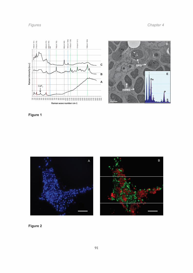
Figures Chapter 4
91
Figure 1
Figure 2
A
C
BC
O
SiSi
P
P
Fe
Fe
Fe
Cu
Cu
Cu
S S
Fe
P
S
C
O
SiSi
P
P
Fe
Fe
Fe
Cu
Cu
Cu
S S
Fe
P
S
D
E
SRB
ANMECaF2
100
150
200
250
300
350
400
450
500
550
600
650
700
750
800
850
900
950
1000
1050
1100
1150
1200
1250
1300
1350
1400
1450
1500
1550
1600
1650
1700
1750
1800
1850
1900
1950
Ramanwave number cm 1
Ram
anintensity(a.u.)
1586
(ref
. 158
3)
1314
(ref
. 131
1)
1130
(ref
. 112
8)
480(
ref.
473)
214(
ref.
216)
149(
ref.
151)
1061
(ref
. 105
0)
960(
ref.
949)
549(
ref.
545)
569
748(
ref.
750)
A B
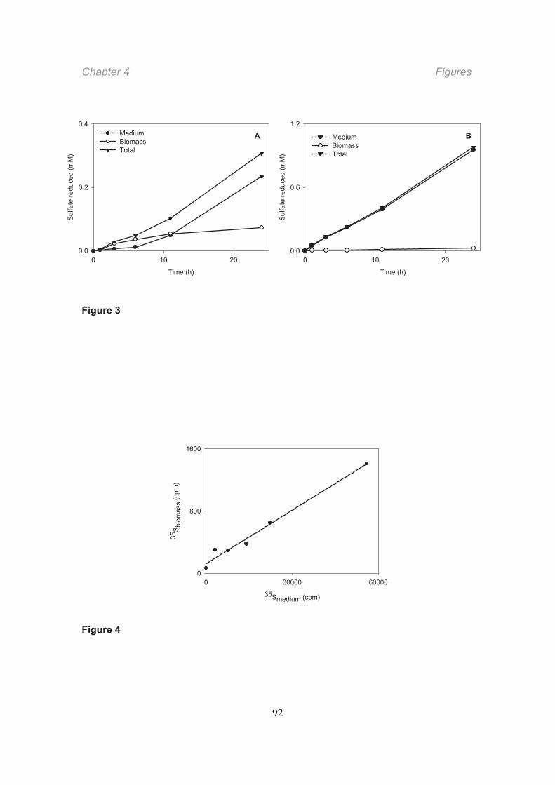
Chapter 4 Figures
92
Figure 3
Figure 4
Time (h)
0 10 20
Sulfa
te re
duce
d (m
M)
0.0
0.2
0.4MediumBiomassTotal
Time (h)
0 10 20
Sulfa
te re
duce
d (m
M)
0.0
0.6
1.2
MediumBiomassTotal
A B
35Smedium (cpm)
0 30000 60000
35S
biom
ass
(cpm
)
0
800
1600
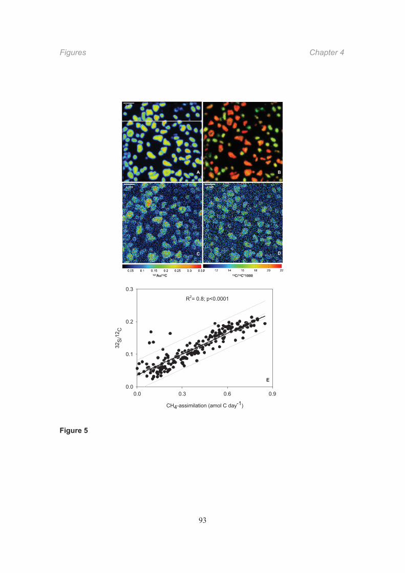
Figures Chapter 4
93
Figure 5
13C/12C*1000197Au/12C
A
C
B
D
CH4-assimilation (amol C day-1)
0.0 0.3 0.6 0.9
32S/
12C
0.0
0.1
0.2
0.3
R2= 0.8; p<0.0001
E
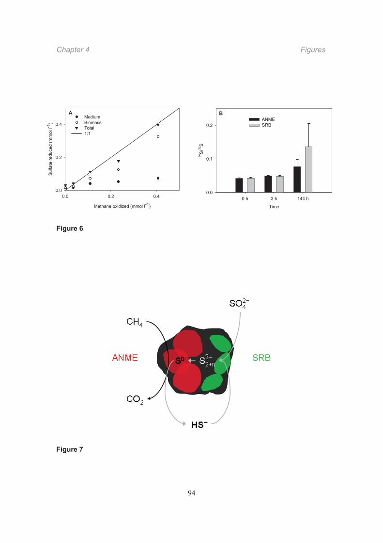
Chapter 4 Figures
94
Figure 6
Figure 7
Methane oxidized (mmol l-1)
0.0 0.2 0.4
Sulfa
te re
duce
d (m
mol
l-1)
0.0
0.2
0.4MediumBiomassTotal1:1
Time
0 h 3 h 144 h34
S/32
S
0.0
0.1
0.2ANMESRB
A B
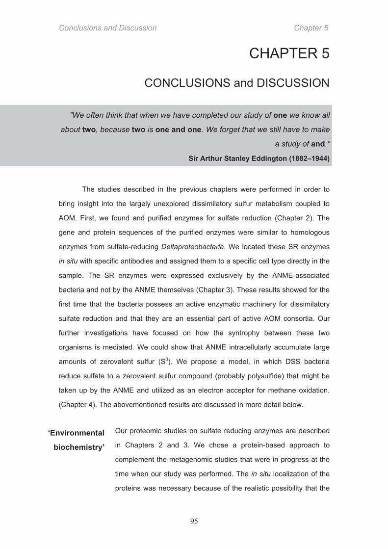
Conclusions and Discussion Chapter 5
95
CHAPTER 5
CONCLUSIONS and DISCUSSION
”We often think that when we have completed our study of one we know all
about two, because two is one and one. We forget that we still have to make
a study of and.”
Sir Arthur Stanley Eddington (1882–1944)
The studies described in the previous chapters were performed in order to
bring insight into the largely unexplored dissimilatory sulfur metabolism coupled to
AOM. First, we found and purified enzymes for sulfate reduction (Chapter 2). The
gene and protein sequences of the purified enzymes were similar to homologous
enzymes from sulfate-reducing Deltaproteobacteria. We located these SR enzymes
in situ with specific antibodies and assigned them to a specific cell type directly in the
sample. The SR enzymes were expressed exclusively by the ANME-associated
bacteria and not by the ANME themselves (Chapter 3). These results showed for the
first time that the bacteria possess an active enzymatic machinery for dissimilatory
sulfate reduction and that they are an essential part of active AOM consortia. Our
further investigations have focused on how the syntrophy between these two
organisms is mediated. We could show that ANME intracellularly accumulate large
amounts of zerovalent sulfur (S0). We propose a model, in which DSS bacteria
reduce sulfate to a zerovalent sulfur compound (probably polysulfide) that might be
taken up by the ANME and utilized as an electron acceptor for methane oxidation.
(Chapter 4). The abovementioned results are discussed in more detail below.
Our proteomic studies on sulfate reducing enzymes are described
in Chapters 2 and 3. We chose a protein-based approach to
complement the metagenomic studies that were in progress at the
time when our study was performed. The in situ localization of the
proteins was necessary because of the realistic possibility that the
‘Environmental
biochemistry’
Chapter 5 Conclusions and Discussion
96
proteins of interest were in the past subjected to horizontal gene
transfer. In that case, both gene- (and protein-) derived sequences
of these proteins would carry the signature of the donor and not the
owner and could be assigned to a wrong organism. Our study
started with the assumption that the canonical SR enzymes used
by all other known sulfate respiring prokaryotes – ATP sulfurylase,
APS reductase and dissimilatory sulfite reductase – are also
involved in the AOM-coupled SR. Indeed, we found these proteins
to be abundant and active in the crude extract from AOM samples.
We could determine their size and subunit composition by SDS-
PAGE, their N-terminal amino acid sequence by Edman
sequencing and their in vitro activity by enzyme assays. A final
assignment of the proteins to a particular organism was done with
specific antibodies. Although this approach was straightforward
some difficulties had to be overcome, the biggest of which was the
severe biomass limitation. All AOM enrichment cultures that have
been maintained and propagated in our institute in the last 10 years
could yield at maximum a few grams of wet weight, which are
minute amounts for protein purification. Therefore, we used
environmental microbial mat samples obtained from the Black Sea
instead of enriched cultures. Reports of protein purifications from
environmental samples that have not been subjected to any post-
sampling enrichment procedures are rare, which is not surprising
when one considers the complexity of the purification process. It is
undesirable to start with a sample where the target organism
comprises just a small fraction of the overall community and where,
correspondingly, the target protein concentration might be very low.
Fortunately, in our microbial mat samples the organisms of interest
constituted up to ca. 40-50% of all cells present. The mats,
however, contained high concentrations of organic extracellular
polymeric substances (EPS) that interfered with the column
Conclusions and Discussion Chapter 5
97
separation. We could remove these interfering substances by
addition of ammonium sulfate. However, the precipitate removed
apart from EPS also most of the hydrophobic proteins. The SR
proteins were soluble and thus remained in the supernatant.
Our study is one of the few reports on protein purification from
environmental samples. The described procedure might help to
solve the questions that are unanswerable by genome analysis,
such as protein abundance, sub-cellular sequestration, post-
translational modifications, oligomerization and intermolecular
interactions, in studies of complex environmental samples.
In situ protein detection in microbial cells is mostly done by
fluorescently tagging the protein of interest. This method is,
however, only established in organisms that are accessible for
genetic manipulation. The other method of protein detection –
immunolabeling – is not (yet) widespread in environmental
microbiology. Immunolabeling has occasionally been used as a
method for identification of bacterial species based on their
extracellular antigens but immunodetection of intracellular proteins
has only been reported in a few ‘pet’ microorganisms with cell walls
susceptible to routine permeabilization agents. Permeabilization is
indisputably the limiting factor for high-throughput
immunofluorescence analysis in mixed samples of microorganisms
(i.e. environmental samples). The extreme species diversity of
microorganisms leads to a large heterogeneity in their cell wall
composition and thus differences in their resistance or susceptibility
to permeabilization agents. In our case, the samples contained
mainly archaea with a thick polysaccharide S-layer and gram-
negative bacteria. In order to achieve uncompromised target
protein accessibility in both organisms we cryo-sectioned the
sample into semi-thin (200-400 nm, i.e. less than cell diameter)
Imaging
studies on thin
sections

Chapter 5 Conclusions and Discussion
98
sections. The sections were transferred to a support glass slide,
labeled with antibodies and imaged in a fluorescent microscope.
The preparation was fast (2 days) and the sample was not
subjected to drying nor incubated in any organic solvents in order
to preserve potential storage compounds. Even with conventional
epifluorescence microscopy the images of the sections were
sharper than confocal images of whole cells because the reduced
thickness of the sections decreased the depth of focus. Unlike
electron microscopy, imaging with fluorescent microscope allows
examination of large number of cells at once, which is necessary in
microbiology for statistical analyses. The sections were (with
appropriate adjustments e.g. thickness, coating, embedding) also
successfully analyzed with transmission electron microscopy,
atomic force microscopy, microRaman spectroscopy and
secondary ion mass spectrometry. This flexibility allows for
correlative imaging combining any of the above-mentioned
approaches and opens new possibilities for microbial ecologists,
especially the ones working with samples like mats, aggregates,
biofilms and sludge where even imaging with confocal microscope
often does not allow single cell discrimination.
Massive high-throughput sequencing of different microbial
communities has tremendously increased the number of newly
indentified phyla, while the number of cultivated organisms has only
moderately increased during the same time. This has resulted in a
situation where whole clusters of phylotypes do not contain any
cultured representatives and are only present as bare 16S rDNA
sequences. Hence it is difficult to deduce the metabolic properties
of microorganisms in the environment from their phylogeny and
methods for high-throughput functional identification of organisms
are desperately needed. This functional identification could come
Functional
identification of
microorganisms

Conclusions and Discussion Chapter 5
99
from metagenomic approaches (ideally automated) that would link
abundant 16S rDNA sequences with (at least some) key metabolic
genes or in situ detection of functional genes or mRNA. Although
these methods are currently still at an early stage of development,
they are the first steps in the direction of a (cheap) large scale
functional identification of microorganisms.
The immunodetection approach introduced above could, in
principle, serve the same purpose but, due to currently high
antibody prices and cell permeabilization problems, is unlikely to be
routinely used for classification of microorganisms in the near future.
Still, in a defined system like ours the antibody-based identification
of organisms as members of certain microbial ‘metabolic groups’
might greatly enhance our understanding of the nature of their
interactions. Nonetheless, the activity of microorganisms should
ideally not be deduced from the presence or abundance of
functional genes, proteins or their phylogenetic identity but should
be measured. Therefore, culturing efforts should be improved and
combined with single cell methods such as nanoSIMS, and
microRaman, or other high resolution spectroscopic methods to
unequivocally determine the metabolic activity of microorganism.
Relatively early in the history of AOM research it has been
speculated that reduced sulfur compounds might play a role in
AOM. The numerous attempts to clarify their involvement have only
yielded inconclusive results. The physiological experiments were
based on additions of different sulfur compounds (e.g. elemental
sulfur, inorganic polysulfides, methanethiol) to active AOM
enrichments. The aim was to see whether such additions stimulate
sulfide production. Generally, no increase of sulfide production was
observed (Basen, Deusner; unpublished results). Now we report
that very likely zerovalent sulfur (possibly in the form of polysulfides)
AOM-associated
sulfur chemistry
Chapter 5 Conclusions and Discussion
100
is formed and turned over during AOM (Chapter 4). What could be
the reason for these discrepancies? First of all, it is not trivial to
determine sulfur speciation in an anoxic, sulfide-rich, biological
system. Sulfur exists in 8 oxidation states (+6 to -2), including a
large palette of zerovalent sulfur compounds. The speciation and
abundance of these compounds is strongly affected by temperature,
pH and concentration of other sulfur species. Isotope labeling
experiments are complicated by the fact that added or excreted S0
and HS– rapidly equilibrate with the external polysulfide pool in
solution through abiotic isotope exchange. It is, therefore, difficult to
assign all sulfur transformations in microbial cultures to biotic
processes because abiotic reactions also cause significant
changes in sulfur speciation. The reduced sulfur compounds rapidly
oxidize when exposed to oxygen, which complicates chemical
analysis. The best methods for determining sulfur speciation in
anoxic systems would be non-invasive imaging like XANES or
Raman spectroscopy for which samples do not need to be exposed
to air.
Apart from the complex sulfur chemistry, there are several other
considerations that might explain the lack of response of the
cultures to sulfur additions. In these experiments, only small
amounts of polysulfide (μM) were added based on the assumption
that S0 is present in rate-limiting (< M) amounts. This assumption
was disproven by our measurements that show that the AOM
biomass – in particular the ANME biomass – contains several mM
of S0. The addition of small amounts of S0 (low M per day) might,
therefore, have had little or no effect on the measured sulfide
production rates. Moreover, sulfide production was measured
according to Cline, which cannot distinguish between polysulfide
(added) and sulfide (formed).
Conclusions and Discussion Chapter 5
101
The idea of adding the intermediate to stimulate or inhibit the
ANME or the SRB, respectively, might not bring the expected result
when considering the slow growth of the organisms. It might take
up to 70 years to obtain a pure culture of an organism with a
doubling time of 6 months. Moreover, if methane activation is the
rate-limiting step, the additions of an acceptor (i.e. sulfur) would not
increase the overall reaction rate.
We have designed experiments in which stable and radioactive
isotopes were added to an active culture and with extraction and
imaging techniques we have tracked the flow of the label at
different time points. The measurements were facilitated by using
‘pool trapping’ experiments. In these types of experiments, the
anticipated isotope-labeled product is ’trapped’ upon formation in a
large unlabeled pool of the same compound. The pool of unlabeled
molecules ‘buffers’ further transformations of the labeled product
(biotic or abiotic) allowing determinations of rate measurements for
intermediates with a fast turnover. Similar isotope experiments
could also be used to determine phosphorus and/or iron cycling in
the AOM cultures.
Chapter 5 Perspectives
102
PERSPECTIVES
The results obtained during this study raise some questions that I believe would be
interesting to address in the near future.
1. Based on our data we suggest that ANME reduce zerovalent sulfur during
methane oxidation. The currently known (bacterial) sulfur and polysulfide reductases
are either molybdopterin-containing proteins, sporadically associated with c-type
cytochromes, or c-type cytochromes with intrinsic sulfur reducing activity. For
archaea, however, c-type cytochromes are uncommon – only 4 out of 20 sequenced
archaeal genomes contain any. It is therefore intriguing that in the ANME genome
large amounts of genes encoding c-type cytochromes and a molybdopterin
oxidoreductase with unknown function were found. The presence of these
cytochromes was confirmed by spectrophotometry and Raman spectroscopy.
Which enzymes are responsible for sulfur reduction in ANME? Might
the peculiar multiheme cytochromes and/or the molybdopterin
oxidoreductase play a role in this process?
2. An alternative hypothesis suggests that the cytochromes in ANME might be
involved in extracellular (pure) electron transfer through nanowires. The proposed
mechanism is analogous to the one observed in iron-reducing Geobacter species. In
these organisms conclusive evidence for involvement of cytochromes was obtained
from transmission electron microscopy studies, immunolabeling and genetic knock-
outs.
Could we convincingly prove or disprove the theory of nanowires-
mediated electron transfer in AOM by cytochrome immunolocalization
with high resolution STED microscopy and/or with TEM?
3. The concentration of sulfur in ANME cells (up to 6% cell dry weight) is similar to
Beggiatoa, which are speculated to play a role in sulfur cycling in marine sediments.
?
?
Perspectives Chapter 5
103
Does the excessive sulfur storage in ANME have an effect on sulfur
biogeochemistry?
4. Transmission electron microscopy, microRaman spectroscopy and nanoSIMS
analyses have shown a high iron and phosphorus content of the ANME-associated
bacteria. These elements are stored as amorphous particles which suggests that
they might be later recruited for metabolic use. Because the respiratory metabolism
of sulfate reducing bacteria depends on phosphorylation reactions
(ATP+sulfate APS) it might require intense phosphorus cycling.
What is the role of these particles in the SRB? Do the iron/phosphorus
particles represent transient phosphorus storage for sulfate reduction?
If so, do other sulfate reducing bacteria contain such particles as well?
5. Phosphorus mobilization by the AOM organisms has not been previously
recognized and, correspondingly, the phosphorus cycling in AOM zones has not
been a subject of thorough investigations. Now that we have evidence for
phosphorus storage by the SR bacteria in the SMTZ it would be interesting to
investigate its possible implications for phosphorus and iron biogeochemistry.
What would be the effect of the release of iron phosphates from SR
bacteria (e.g. upon depletion of a methane source and a subsequent
cell death)? Could it explain the peculiar presence of phosphorites in
ancient AOM zones?
?
?
?
References
104
REFERENCES
1. Akagi, J.M. & Campbell, L.L. (1962). Studies on thermophilic sulfate-
reducing bacteria III. Adenosine triphosphate-sulfurylase of Clostridium nigrificans
and Desulfovibrio desulfuricans. Journal of Bacteriology 84, 1194-1201.
2. Aldrich, H.C., Beimborn, D.B., Bokranz, M. & Schönheit, P. (1987).
Immunocytochemical localization of methyl coenzyme-M reductase in
Methanobacterium thermoautotrophicum. Archives in Microbiology 147, 190-194.
3. Alperin, M. J. & Hoehler, T. M. (2009a). Anaerobic methane oxidation by
archaea/sulfate-reducing bacteria aggregates: 1. Thermodynamic and physical
constraints. American Journal of Science 309, 869-957.
4. Alperin, M. J. & Hoehler, T. M. (2009b). Anaerobic methane oxidation by
archaea/sulfate-reducing bacteria aggregates: 2. Isotopic constraints. American
Journal of Science 309, 958-984.
5. Alperin M. & Hoehler T. (2010). The ongoing mystery of sea-floor methane.
Science 329, 288-289.
6. Amann, R.I., Krumholz, L. & Stahl, D.A. (1990). Fluorescent-oligonucleotide
probing of whole cells for determinative, phylogenetic, and environmental studies in
microbiology. Journal of Bacteriology 172, 762 770.
7. Amann, R.I., Ludwig, W. & Schleifer, K.H. (1995). Phylogenetic
identification and in situ detection of individual microbial cells without cultivation.
Microbiological Reviews 59, 143 169.
8. Arendsen, A.F., Verhagen, M.F.J.M., Wolbert, R.B.G., Pierik, A.J., Stams,
A.J.M., Jetten, M.S.M. & Hagen, W.R. (1993). The dissimilatory sulfite reductase
References
105
from Desulfosarcina variabilis is a desulforubidin containing uncoupled metalated
sirohemes and S = 9/2 iron-sulfur clusters. Biochemistry 32, 10323 10330.
9. Barnes, R. O. & Goldberg, E. D. (1976). Methane production and
consumption in anoxic marine sediments. Geology 4, 297-300.
10. Beal, E. J., House, C. H. & Orphan, V. J. (2009). Manganese- and iron-
dependent marine methane oxidation. Science 325, 184-187.
11. Baliga, B.S., Jagannathan, V. & Vartak, H.G. (1961). Purification and
properties of sulfurylase from Desulfovibrio desulfuricans. Journal of Scientific &
Industrial Research 20C: 33-40.
12. Basen, M., Krüger, M., Milucka, J. & other authors (in revision). Bacterial
enzymes for dissimilatory sulfate reduction in a marine microbial mat (Black Sea)
mediating anaerobic oxidation of methane. Environmental Microbiology
13. Biddle, J. F., Lipp, J. S., Lever, M. A. & other authors (2006).
Heterotrophic Archaea dominate sedimentary subsurface ecosystems off Peru.
Proceedings of the National Academy of Sciences of the United States of America
103, 3846-3851.
14. Bleicher, K. & Winter, J. (1994). Formate production and utilization by
methanogens and by sewage-sludge consortia - interference with the concept of
interspecies formate transfer. Applied Microbiology and Biotechnology 40, 910-915.
15. Blumenberg, M., Seifert, R., Reitner, J., Pape, T. & Michaelis, W. (2004).
Membrane lipid patterns typify distinct anaerobic methanotrophic consortia.
Proceedings of the National Academy of Sciences of the United States of America
101, 11111 11116.
References
106
16. Bradford, M.M. (1976). Rapid and sensitive method for quantitation of
microgram quantities of protein utilizing principle of protein-dye binding. Analytical
Biochemistry 72, 248 254.
17. Boetius, A., Ravenschlag, K., Schubert, C. J. & other authors (2000). A
marine microbial consortium apparently mediating anaerobic oxidation of methane.
Nature 407, 623-626.
18. Chistoserdova, L., Vorholt, J.A. & Lidstrom, M.E. (2005). A genomic view
of methane oxidation by aerobic bacteria and anaerobic archaea. Genome Biology 6,
208.
19. Cline, J.D. (1969). Spectrophotometric determination of hydrogen sulfide in
natural waters. Limnology and Oceanography 14, 454 458.
20. Cord-Ruwisch, R. (1985). A quick method for the determination of dissolved
and precipitated sulfides in cultures of sulfate-reducing bacteria. Journal of
Microbiological Methods 4, 33 36.
21. Dahl, C., Speich, N. & Trüper, H.G. (1994a). Enzymology and molecular
biology of sulfate reduction in extremely thermophilic archaeon Archaeoglobus
fulgidus. Methods in Enzymology 243, 331 349.
22. Dahl, C. & Trüper, H.G. (1994b). Enzymes of dissimilatory sulfide oxidation
in phototrophic bacteria. Methods in Enzymology 243, 400 421.
23. Dahl, C., Rakhely, G., Pott-Sperling, A. S. & other authors (1999). Genes
involved in hydrogen and sulfur metabolism in phototrophic sulfur bacteria. FEMS
Microbiology Letters 180, 317-324.
References
107
24. Dahl, C. & Trüper, H.G. (2001). Sulfite reductase and APS reductase from
Archaeoglobus fulgidus. Methods in Enzymology. 331, 427 441.
25. Daims, H., Brühl, A., Amann, R. & Schleifer, K.H. (1999). The domain-
specific probe EUB338 is insufficient for the detection of all bacteria: development
and evaluation of a more comprehensive probe set. Systematic and Applied
Microbiology 22, 434 444.
26. Dekas, A. E., Poretsky, R. S. & Orphan, V. J. (2009). Deep-sea archaea fix
and share nitrogen in methane-consuming microbial consortia. Science 326, 422-426.
27. Devol A. H. & Ahmed S. I. (1981). Are high rates of sulphate reduction
associated with anaerobic oxidation of methane? Nature 291, 407-408.
28. Dickens, G. R. (2003). Rethinking the global carbon cycle with a large,
dynamic and microbially mediated gas hydrate capacitor. Earth and Planetary
Science Letters 213, 169-183.
29. Eisen, J. A., Nelson, K. E., Paulsen, I. T. & other authors (2002). The
complete genome sequence of Chlorobium tepidum TLS, a photosynthetic,
anaerobic, green-sulfur bacterium. Proceedings of the National Academy of Sciences
of the United States of America 99 (14), 9509-9514.
30. Ellermann, J., Rospert, S., Thauer, R.K., Bokranz, M., Klein, A., Voges, M.
& Berkessel, A. (1989). Methyl-coenzyme-M reductase from Methanobacterium
thermoautotrophicum (strain Marburg) - purity, activity and novel inhibitors. European
Journal of Biochemistry 184, 63-68.
31. Ettwig, K. F., Shima, S., van de Pas-Schoonen, K. T., Kahnt, J., Medema,
M. H., op den Camp, H. J. M., Jetten, M. S. M. & Strous, M. (2008). Denitrifying
References
108
bacteria anaerobically oxidize methane in the absence of archaea. Environmental
Microbiology 10, 3164-3173.
32. Ettwig, K. F., Butler, M. K., Le Paslier, D. & other authors (2010). Nitrite-
driven anaerobic methane oxidation by oxygenic bacteria. Nature 464, 543-548.
33. Friedrich, M.W. (2002). Phylogenetic analysis reveals multiple lateral
transfers of adenosine-5'-phosphosulfate reductase genes among sulfate-reducing
microorganisms. Journal of Bacteriology 184, 278 289.
34. Fauque G., Herve D. & Le Gall J. (1979). Structure-function relationship in
hemoproteins: the role of cytochrome c3 in the reduction of colloidal sulfur by sulfate-
reducing bacteria. Archives in Microbiology 121, 261-264.
35. Fossing H. & Jorgensen B. B. (1990). Isotope exchange reactions with
radiolabeled sulfur compounds in anoxic seawater. Biogeochemistry 9, 223-245.
36. Fritz, G., Buchert, T., Huber, H., Stetter, K.O. & Kroneck, P.M.H. (2000).
Adenylylsulfate reductases from archaea and bacteria are 1 : 1 alpha beta-
heterodimeric iron-sulfur flavoenzymes - high similarity of molecular properties
emphasizes their central role in sulfur metabolism. FEBS Letters 473, 63 66.
37. Fritz, G., Roth, A., Schiffer, A. & other authors (2002). Structure of
adenylylsulfate reductase from the hyperthermophilic Archaeoglobus fulgidus at 1.6
resolution. Proceedings of the National Academy of Sciences of the United States
of America 99 (4). 1836 1841.
38. Frost R. L., Martens W. N., Williams P., & Kloprogge J. T. (2002). Raman
and infrared spectroscopic study of the vivianite-group phosphates vivianite, baricite
and bobierrite. Mineralogical Magazine 66, 1063-1073.
References
109
39. Gorby, Y. A., Yanina, S., McLean, J. S. & other authors (2006). Electrically
conductive bacterial nanowires produced by Shewanella oneidensis strain MR-1 and
other microorganisms. Proceedings of the National Academy of Sciences of the
United States of America 103, 11358-11363.
40. Hall, T.A. (1999). BioEdit: a user-friendly biological sequence alignment
editor and analysis program for Windows 95/98/NT. Nucleic Acids Symposium Series
41, 95 98.
41. Hallam, S. J., Putnam, N., Preston, C. M., Detter, J. C., Rokhsar, D.,
Richardson, P. M. & DeLong, E. F. (2004). Reverse methanogenesis: testing the
hypothesis with environmental genomics. Science 305, 1457-1462.
42. Hallam, S.J., Girguis, P.R., Preston, C.M., Richardson, P.M. & DeLong,
E.F. (2003). Identification of methyl coenzyme M reductase A (McrA) genes
associated with methane-oxidizing archaea. Applied and Environmental Microbiology
69, 5483-5491.
43. Heller, C., Hoppert, M. & Reitner, J. (2008). Immunological localization of
coenzyme M reductase in anaerobic methane-oxidizing archaea of ANME 1 and
ANME 2 type. Geomicrobiology Journal 25, 149 156.
44. Hinrichs, K. U., Hayes, J. M., Sylva, S. P., Brewer, P. G. & DeLong, E. F.
(1999). Methane-consuming archaebacteria in marine sediments. Nature 398, 802-
805.
45. Hinrichs, K. U., Summons, R. E., Orphan, V., Sylva, S. P. & Hayes, J. M.
(2000). Molecular and isotopic analysis of anaerobic methane-oxidizing communities
in marine sediments. Organic Geochemistry 31, 1685-1701.
References
110
46. Hinrichs, K. U. & Boetius, A. (2002). The anaerobic oxidation of methane:
new insights in microbial ecology and biogeochemistry. In Ocean Margin Systems,
pp. 457–477. G. Wefer, B. D., D. Hebbeln, B. B. Jørgensen, M. Schlüter and T. C. E.
van Weering (eds.). Berlin: Springer-Verlag.
47. Hoehler, T.M., Alperin, M.J., Albert, D.B. & Martens, C.S. (1994). Field and
laboratory studies of methane oxidation in an anoxic marine sediment - evidence for
a methanogen-sulfate reducer consortium. Global Biogeochemical Cycles 8,
451 463.
48. Hoehler, T. M., Alperin, M. J., Albert, D. B. & Martens, C. S. (2001).
Apparent minimum free energy requirements for methanogenic Archaea and sulfate-
reducing bacteria in an anoxic marine sediment. FEMS Microbiology Ecology 38, 33-
41.
49. Hoppert, M. & Mayer, F. (1999). Principles of macromolecular organization
and cell function in bacteria and archaea. Cell Biochemistry and Biophysics 31, 247-
284.
50. House, C. H., Schopf, J. W., McKeegan, K. D., Coath, C. D., Harrison, T.
M. & Stetter, K. O. (2000). Carbon isotopic composition of individual Precambrian
microfossils. Geology 28, 707-710.
51. House, C. H., Orphan, V. J., Turk, K. A., Thomas, B., Pernthaler, A.,
Vrentas, J. M. & Joye, S. B. (2009). Extensive carbon isotopic heterogeneity among
methane seep microbiota. Environmental Microbiology 11, 2207-2215.
52. Jankielewicz A., Schmitz R. A., Klimmek O., & Kröger A. (1994).
Polysulfide reductase and formate dehydrogenase from Wolinella succinogenes
contain molybdopterin guanine dinucleotide. Archives in Microbiology 162, 238-242
References
111
53. Johnson, E. & Mukhopadhyay, B. (2008). A novel coenzyme F420-
dependent sulfite reductase and a small sulfite reductase in methanogenic archaea.
In Microbial Sulfur Metabolism, pp. 202-216. Edited by C. Dahl and C. G. Friedrich:
Springer
54. Johnson, E. F. & Mukhopadhyay, B. (2005). A new type of sulfite reductase,
a novel coenzyme F420-dependent enzyme, from the methanarchaeon
Methanocaldococcus jannaschii. Journal of Biological Chemistry 280, 38776-38786.
55. Jørgensen B. B. & Fenchel T. (1974). Sulfur cycle of a marine sediment
model system. Marine Biology 24, 189-201.
56. Kallmeyer J., Ferdelman T.G., Weber A., Fossing H.. & Jørgensen B.B.
(2004). Evaluation of a cold chromium distillation procedure for recovering very small
amounts of radiolabeled sulfide related to sulfate reduction measurements.
Limnology and Oceanography: Methods 2, 171-180.
57. Kamyshny A., Ekeltchik I., Gun J. & Lev O. (2006). Method for the
determination of inorganic polysulfide distribution in aquatic systems. Analytical
Chemistry 78, 2631-2639.
58. Kamyshny Jr., A., Gun, J., Rizkov, D., Voitsekovski, T. & Lev, O. (2007).
Equilibrium distribution of polysulfide ions in aqueous solutions at different
temperatures by rapid single phase derivatisation. Environmental Science and
Technology 41, 6633 - 6644.
59. Kamyshny Jr., A. (2009). Solubility of cyclooctasulfur in pure water and sea
water at different temperatures. Geochimica et Cosmochimica Acta 73, 6022-6028.
References
112
60. Kappler, O., Janssen, P. H., Kreft, J. U. & Schink, B. (1997). Effects of
alternative methyl group acceptors on the growth energetics of the O-demethylating
anaerobe Holophaga foetida. Microbiology-UK 143, 1105-1114.
61. Kiene R.P., Oremland R.S., Catena A., Miller L.G. & Capone D.G. (1986).
Metabolism of reduced methylated sulfur compounds in anaerobic sediments and by
a pure culture of an estuarine methanogen. Applied and Environmental Microbiology
52,1037–1045.
62. Klein, M., Friedrich, M., Roger, A. J. & other authors (2001). Multiple
lateral transfers of dissimilatory sulfite reductase genes between major lineages of
sulfate-reducing prokaryotes. Journal of Bacteriology 183, 6028-6035.
63. Klimes, A., Franks, A. E., Glaven, R. H., Tran, H., Barrett, C. L., Qiu, Y.,
Zengler, K. & Lovley, D. R. (2010). Production of pilus-like filaments in Geobacter
sulfurreducens in the absence of the type IV pilin protein PilA. FEMS Microbiology
Letters 310, 62-68.
64. Knittel, K., Boetius, A., Lemke, A. & other authors (2003). Activity,
distribution, and diversity of sulfate reducers and other bacteria in sediments above
gas hydrate (Cascadia margin, Oregon). Geomicrobiology Journal 20, 269 294.
65. Knittel, K., Lösekann, T., Boetius, A., Kort, R. & Amann, R. (2005).
Diversity and distribution of methanotrophic archaea at cold seeps. Applied and
Environmental Microbiology 71, 467-479.
66. Knittel, K. & Boetius, A. (2009). Anaerobic oxidation of methane: progress
with an unknown process. Annual Reviews of Microbiology 63, 311-334.
67. Kobayashi, K., Morisawa, Y., Ishituka, T. & Ishimoto, M. (1975).
Biochemical studies on sulfate-reducing bacteria. 14. Enzyme levels of
References
113
adenylylsulfate reductase, inorganic pyrophosphatase, sulfite reductase,
hydrogenase, and adenosine-triphosphatase in cells grown on sulfate, sulfite, and
thiosulfate. Journal of Biochemistry 78, 1079 1085.
68. Krämer, M. & Cypionka, H. (1989). Sulfate formation via ATP sulfurylase in
thiosulfate-disproportionating and sulfite-disproportionating bacteria. Archives in
Microbiology 151, 232 237.
69. Krüger, M., Meyerdierks, A., Glöckner, F. O. & other authors (2003). A
conspicuous nickel protein in microbial mats that oxidize methane anaerobically.
Nature 426, 878-881.
70. Krüger, M., Blumenberg, M., Kasten, S. & other authors (2008). A novel,
multi-layered methanotrophic microbial mat system growing on the sediment of the
Black Sea. Environmental Microbiology 10, 1934 1947.
71. Laemmli, U.K. (1970). Cleavage of structural proteins during assembly of
head of bacteriophage T4. Nature 227, 680-685.
72. LeGall, J. & Fauque, G. (1988). Dissimilatory reduction of sulfur compounds.
In Biology of anaerobic microorganisms, pp. 587 639. Zehnder, A.J.B. (ed.). New
York: John Wiley & Sons.
73. Lösekann, T., Knittel, K., Nadalig, T., Fuchs, B., Niemann, H., Boetius, A.
& Amann, R. (2007). Diversity and abundance of aerobic and anaerobic methane
oxidizers at the Haakon Mosby mud volcano, Barents Sea. Applied and
Environmental Microbiology 73, 3348 3362.
74. Mander, G. J., Duin, E. C., Linder, D., Stetter, K. O. & Hedderich, R.
(2002). Purification and characterization of a membrane-bound enzyme complex
from the sulfate-reducing archaeon Archaeoglobus fulgidus related to heterodisulfide
References
114
reductase from methanogenic archaea. European Journal of Biochemistry 269, 1895-
1904.
75. Mander, G. J., Pierik, A. J., Huber, H. & Hedderich, R. (2004). Two distinct
heterodisulfide reductase-like enzymes in the sulfate-reducing archaeon
Archaeoglobus profundus. European Journal of Biochemistry 271, 1106-1116.
76. Martens, C. S. & Berner, R. A. (1977). Interstitial water chemistry of anoxic
Long Island Sound sediments: 1. Dissolved gases. Limnology and Oceanography 22,
10-25.
77. Manz, W., Eisenbrecher, M., Neu, T.R. & Szewzyk, U. (1998). Abundance
and spatial organization of gram-negative sulfate-reducing bacteria in activated
sludge investigated by in situ probing with specific 16S rRNA targeted
oligonucleotides. FEMS Microbiology Ecology 25, 43 61.
78. Meulepas, R.J.W., Jagersma, C.G., Gieteling, J., Buisman, C.J.N., Stams,
A.J.M. & Lens, P.N.L. (2009). Enrichment of anaerobic methanotrophs in sulfate-
reducing membrane bioreactors. Biotechnology and Bioengineering 104, 458 470.
79. Meulepas, R. J. W., Jagersma, C. G., Khadem, A. F., Stams, A. J. M. &
Lens, P. N. L. (2010). Effect of methanogenic substrates on anaerobic oxidation of
methane and sulfate reduction by an anaerobic methanotrophic enrichment. Applied
Microbiology and Biotechnology 87, 1499-1506.
80. Meyer, B. & Kuever, J. (2007). Phylogeny of the alpha and beta subunits of
the dissimilatory adenosine-5'-phosphosulfate (APS) reductase from sulfate-reducing
prokaryotes - origin and evolution of the dissimilatory sulfate-reduction pathway.
Microbiology SGM 153, 2026 2044.
References
115
81. Meyerdierks, A., Kube, M., Lombardot, T., Knittel, K., Bauer, M.,
Glöckner, F. O., Reinhardt, R. & Amann, R. (2005). Insights into the genomes of
archaea mediating the anaerobic oxidation of methane. Environmental Microbiology
7, 1937-1951.
82. Meyerdierks, A., Kube, M., Kostadinov, I., Teeling, H., Glöckner, F. O.,
Reinhardt, R. & Amann, R. (2010). Metagenome and mRNA expression analyses of
anaerobic methanotrophic archaea of the ANME-1 group. Environmental
Microbiology 12, 422-439.
83. Michaelis, W., Seifert, R., Nauhaus, K. & other authors (2002). Microbial
reefs in the Black Sea fueled by anaerobic oxidation of methane. Science 297, 1013-
1015.
84. Moran, J. J., Beal, E. J., Vrentas, J. M., Orphan, V. J., Freeman, K. H. &
House, C. H. (2008). Methyl sulfides as intermediates in the anaerobic oxidation of
methane. Environmental Microbiology 10, 162-173.
85. Musat N., Halm H., Winterholler B. and other authors (2008). Single-cell
view on the ecophysiology of anaerobic phototrophic bacteria. Proceedings of the
National Academy of Sciences of the United States of America 105 (46), 17861-
17866.
86. Nauhaus, K., Boetius, A., Krüger, M. & Widdel, F. (2002). In vitro
demonstration of anaerobic oxidation of methane coupled to sulphate reduction in
sediment from a marine gas hydrate area. Environmental Microbiology 4, 296-305.
87. Nauhaus, K., Albrecht, M., Elvert, M., Boetius, A. & Widdel, F. (2007). In
vitro cell growth of marine archaeal-bacterial consortia during anaerobic oxidation of
methane with sulfate. Environmental Microbiology 9, 187-196.
References
116
88. Niemann, H., Lösekann, T., de Beer, D. & other authors (2006). Novel
microbial communities of the Haakon Mosby mud volcano and their role as a
methane sink. Nature 443, 854-858.
89. Odom, J.M. & Peck, H.D. (1981). Localization of dehydrogenases,
reductases, and electron-transfer components in the sulfate-reducing bacterium
Desulfovibrio gigas. Journal of Bacteriology 147, 161-169.
90. Orphan, V. J., House, C. H., Hinrichs, K. U., McKeegan, K. D. & DeLong,
E. F. (2001a). Methane-consuming archaea revealed by directly coupled isotopic and
phylogenetic analysis. Science 293, 484-487.
91. Orphan, V.J., Hinrichs, K.U., Ussler, W. & other authors (2001b).
Comparative analysis of methane-oxidizing archaea and sulfate-reducing bacteria in
anoxic marine sediments. Applied and Environmental Microbiology 67, 1922 1934.
92. Orphan, V.J., House, C.H., Hinrichs, K.U., McKeegan, K.D. & DeLong, E.F.
(2002a). Direct phylogenetic and isotopic evidence for multiple groups of archaea
involved in the anaerobic oxidation of methane. Geochimica et Cosmochimica Acta
66, A571-A571.
93. Orphan, V. J., House, C. H., Hinrichs, K. U., McKeegan, K. D. & DeLong,
E. F. (2002b). Multiple archaeal groups mediate methane oxidation in anoxic cold
seep sediments. Proceedings of the National Academy of Sciences of the United
States of America 99 (11), 7663-7668.
94. Orphan, V. J., Turk, K. A., Green, A. M. & House, C. H. (2009). Patterns of 15N assimilation and growth of methanotrophic ANME-2 archaea and sulfate-reducing
bacteria within structured syntrophic consortia revealed by FISH-SIMS.
Environmental Microbiology 11, 1777-1791.
References
117
95. Ossmer, R., Mund, T., Hartzell, P.L., Konheiser, U., Kohring, G.W., Klein,
A. & other authors (1986). Immunocytochemical localization of component C of the
methylreductase system in Methanococcus voltae and Methanobacterium
thermoautotrophicum. Proceedings of the National Academy of Sciences of the
United States of America 83, 5789-5792.
96. Pancost, R. D., Damste, J. S. S., de Lint, S., van der Maarel, M. J. E. C.,
Gottschal, J. C. & Party, M. S. S. (2000). Biomarker evidence for widespread
anaerobic methane oxidation in Mediterranean sediments by a consortium of
methanogenic archaea and bacteria. Applied and Environmental Microbiology 66,
97. Parshina S.N., Sipma J., Meint Henstra A., & Stams A.J.M. (2010). Carbon
monoxide as an electron donor for the biological reduction of sulphate. International
Journal of Microbiology, 1126-1132.
98. Pasteris J. D., Freeman J. J., Goffredi S. K. & Buck K. R. (2001). Raman
spectroscopic and laser scanning confocal microscopic analysis of sulfur in living
sulfur-precipitating marine bacteria. Chemical Geology 180, 3-18.
99. Pätzold R., Keuntje M., Theophile K., Müller J., Mielcarek E., Ngezahayo
A. & Anders-von Ahlften A. (2008). In situ mapping of nitrifiers and anammox
bacteria in microbial aggregates by means of confocal resonance Raman microscopy.
Journal of Microbiological Methods 3, 241-248.
100. Pernthaler, A., Dekas, A. E., Brown, C. T., Goffredi, S. K., Embaye, T. &
Orphan, V. J. (2008). Diverse syntrophic partnerships from deep sea methane vents
revealed by direct cell capture and metagenomics. Proceedings of the National
Academy of Sciences of the United States of America 105, 7052-7057.
References
118
101. Pimenov, N.V., Rusanov, I.I., Poglazova, M.N., Mityushina, L.L., Sorokin,
D.Y., Khmelenina, V.N. & Trotsenko, Y.A. (1997). Bacterial mats on coral-like
structures at methane seeps in the Black Sea. Microbiologiya 66, 354 360.
102. Platen, H. & Schink, B. (1987). Methanogenic degradation of acetone by an
enrichment culture. Archives of Microbiology 149, 136-141.
103. Ploug H., Musat N., Adam B. & other authors (2010). Carbon and nitrogen
fluxes associated with the cyanobacterium Aphanizomenon sp. in the Baltic Sea.
ISME journal 4, 1215-1223.
104. Polerecky L., Adam B., Musat N., Milucka J., Vagner T. & Kuypers M. M.
M. (2012). Look@NanoSIMS – a tool for the analysis of NanoSIMS data in
environmental microbiology. Environmental Microbiology DOI: 10.1111/j.1462-
2920.2011.02681.x
105. Prange A., Chauvistré R., Modrow H., Hormes J., Trüper H. G. & Dahl C.
(2002). Quantitative speciation of sulfur in bacterial sulfur globules: X-ray absorption
spectroscopy reveals at least three different species of sulfur. Microbiology 148, 267-
276.
106. Rabus, R., Hansen, T.A. & Widdel, F. (2006). Dissimilatory sulfate- and
sulfur-reducing prokaryotes. In The Prokaryotes A handbook on the biology of
bacteria, pp. 659 768. Dworkin, M., Falkow, S., Rosenberg, E., Schleifer, K.H., and
Stackebrandt, E. (eds). New York: Springer.
107. Raghoebarsing, A. A., Pol, A., van de Pas-Schoonen, K. T. & other
authors (2006). A microbial consortium couples anaerobic methane oxidation to
denitrification. Nature 440, 918-921.
References
119
108. Reeburgh, W. S. (1976). Methane consumption in Cariaco Trench waters
and sediments. Earth and Planetary Science Letters 28, 337-344.
109. Reeburgh, W. S. (2007). Oceanic methane biogeochemistry. Chemical
Reviews 107, 486-513.
110. Reguera, G., McCarthy, K. D., Mehta, T., Nicoll, J. S., Tuominen, M. T. &
Lovley, D. R. (2005). Extracellular electron transfer via microbial nanowires. Nature
435, 1098-1101.
111. Reitner, J., Peckmann, J., Blumenberg, M., Michaelis, W., Reimer, A. &
Thiel, V. (2005a). Concretionary methane-seep carbonates and associated microbial
communities in Black Sea sediments. Palaeogeography Palaeoclimatology
Palaeoecology 227, 18-30.
112. Reitner, J., Peckmann, J., Reimer, A., Schumann, G. & Thiel, V. (2005b).
Methane-derived carbonate build-ups and associated microbial communities at cold
seeps on the lower Crimean shelf (Black Sea). Facies 51, 71-84.
113. Renosto, F., Martin, R.L., Borrell, J.L., Nelson, D.C. & Segel, I.H. (1991).
ATP sulfurylase from trophosome tissue of Riftia pachyptila (hydrothermal vent tube
worm). Archives of Biochemistry and Biophysics 290, 66 78.
114. Rossi, M., Pollock, W. B. R., Reij, M. W., Keon, R. G., Fu, R. D. &
Voordouw, G. (1993). The hmc operon of Desulfovibrio vulgaris subsp vulgaris
Hildenborough encodes a potential transmembrane redox protein complex. Journal
of Bacteriology 175, 4699-4711.
115. Scheller, S., Goenrich, M., Boecher, R., Thauer, R. K. & Jaun, B. (2010).
The key nickel enzyme of methanogenesis catalyses the anaerobic oxidation of
methane. Nature 465, 606-608.
References
120
116. Schreiber, L., Holler, T., Knittel, K., Meyerdierks, A. & Amann, R. (2010).
Identification of the dominant sulfate-reducing bacterial partner of anaerobic
methanotrophs of the ANME-2 clade. Environmental Microbiology 12, 2327 2340.
117. Shima S. & Thauer R. K. (2005). Methyl-coenzyme M reductase and the
anaerobic oxidation of methane in methanotrophic Archaea. Current Opinion in
Microbiology 6, 643-648.
118. Sirevag, R., Buchanan, B. B., Berry, J. A. & Troughton, J. H. (1977).
Mechanisms of CO2 fixation in bacterial photosynthesis studied by carbon isotope
fractionation technique. Archives of Microbiology 112, 35-38.
119. Skyring, G.W., Trudinger P. A. & Shaw, W.H. (1972). Electophoretic
characterization of ATP-sulfate adenylyltransferase (ATP-sulfurylase) using
acrylamide gels. Analytical Biochemistry 48, 259-265.
120. Sørensen, K.B., Finster, K. & Ramsing, N.B. (2001). Thermodynamic and
kinetic requirements in anaerobic methane oxidizing consortia exclude hydrogen,
acetate, and methanol as possible electron shuttles. Microbial Ecology 42, 1-10.
121. Sperling, D., Kappler, U., Wynen, A., Dahl, C. & Trüper, H.G. (1998).
Dissimilatory ATP sulfurylase from the hyperthermophilic sulfate reducer
Archaeoglobus fulgidus belongs to the group of homo-oligomeric ATP sulfurylases.
FEMS Microbiology Letters 162, 257 264.
122. Sprott, G. D. & Beveridge, T. J. (1993). Microscopy. In Methanogenesis:
ecology, physiology, biochemistry & genetics, pp. 81-128. Edited by J. G. Ferry:
Chapman & Hall.
References
121
123. Stetter, K.O. (1988). Archaeoglobus fulgidus gen. nov., sp. nov.: a new taxon
of extremely thermophilic archaebacteria. Systematic and Applied Microbiology 10,
172 173.
124. Steudel R. (2002). Organic polysulfanes R2Sn (n > 2). Chemical Reviews 102,
3905-3945.
125. Thauer, R. K., Jungermann, K. & Decker, K. (1977). Energy conservation in
chemotrophic anaerobic bacteria. Bacteriological Reviews 41, 100-180.
126. Thauer, R. K. & Shima, S. (2008). Methane as fuel for anaerobic
microorganisms. Incredible Anaerobes: From Physiology to Genomics to Fuels 1125,
158-170.
127. Thauer, R.K. (1998). Biochemistry of methanogenesis: a tribute to Marjory
Stephenson. Microbiology-SGM 144: 2377 2406.
128. Treude T., Boetius A., Knittel K., Wallmann K. & Jørgensen B.B. (2003).
Anaerobic oxidation of methane above gas hydrates at Hydrate Ridge, NE Pacific.
Ocean Marine Ecology – Progress Series 264, 1-14.
129. Treude, T., Knittel, K., Blumenberg, M., Seifert, R. & Boetius, A. (2005).
Subsurface microbial methanotrophic mats in the Black Sea. Applied and
Environmental Microbiology 71, 6375 6378.
130. Treude, T., Orphan, V., Knittel, K., Gieseke, A., House, C.H. & Boetius, A.
(2007). Consumption of methane and CO2 by methanotrophic microbial mats from
gas seeps of the anoxic Black Sea. Applied and Environmental Microbiology 73,
3770 3770.
References
122
131. Trofimov B. A., Sinegovskaya L. M. & Gusarova N. K. (2009). Vibrations of
the S S bond in elemental sulfur and organic polysulfides: a structural guide. Journal
of Sulfur Chemistry 5, 518-554.
132. Valente, F. M. A., Saraiva, L. M., LeGall, J., Xavier, A. V., Teixeira, M. &
Pereira, I. A. C. (2001). A membrane-bound cytochrome c(3): A type II cytochrome
c(3) from Desulfovibrio vulgaris Hildenborough. Chembiochem 2, 895-905.
133. Valentine, D.L., Blanton, D.C. & Reeburgh, W.S. (2000). Hydrogen
production by methanogens under low-hydrogen conditions. Archives in Microbiology
174, 415 421.
134. Valentine D. L. & Reeburgh W. S. (2000). New perspectives on anaerobic
methane oxidation. Environmental Microbiology 2,477–84.
135. Wagner, M., Roger, A.J., Flax, J.L., Brusseau, G.A. & Stahl, D.A. (1998).
Phylogeny of dissimilatory sulfite reductases supports an early origin of sulfate
respiration. Journal of Bacteriology 180, 2975 2982.
136. Wallner, G., Amann, R. & Beisker, W. (1993). Optimizing fluorescent in situ
hybridization with rRNA-targeted oligonucleotide probes for flow cytometric
identification of microorganisms. Cytometry 14, 136 143.
137. Warikoo, V., McInerney, M. J., Robinson, J. A. & Suflita, J. M. (1996).
Interspecies acetate transfer influences the extent of anaerobic benzoate
degradation by syntrophic consortia. Applied and Environmental Microbiology 62, 26-
32.
138. Wegener, G., Niemann, H., Elvert, M., Hinrichs, K. U. & Boetius, A. (2008).
Assimilation of methane and inorganic carbon by microbial communities mediating
the anaerobic oxidation of methane. Environmental Microbiology 10, 2287-2298.
References
123
139. Widdel, F., Musat, F., Knittel, K. & Galushko, A. (2007). Anaerobic
degradation of hydrocarbons with sulphate as electron acceptor. In Sulphate-
Reducing Bacteria: Environmental and Engineered Systems, pp. 265-303. L. L.
Barton & W. A. Hamilton (eds.). Cambridge University Press.
140. Widdel, F. & Bak, F. (1992). Gram-negative mesophilic sulfate-reducing
bacteria. In The Prokaryotes, pp. 3352 3378. Balows, A.T., H.G.; Dworkin, M.,
Harder, W.; and Schleifer, K.-H. (eds.). New York: Springer.
141. Zehnder, A.J.B., & Brock, T.D. (1979). Methane formation and methane
oxidation by methanogenic bacteria. Journal of Bacteriology 137, 420 432.
142. Zopfi J., Ferdelman T.G. & Fossing H. (2004). Distribution and fate of sulfur
intermediates – sulfite, thiosulfate, and elemental sulfur – in marine sediments. In
Sulfur Biogeochemistry – Past and Present, pp. 97-116. Amend J., Edwards K.J.,
and Lyons T.J. (eds.). Geological Society of America Special Paper 379.
List of Publications
124
LIST of PUBLICATIONS
Manuscript 1.
Bacterial enzymes for dissimilatory sulfate reduction in a marine microbial mat (Black
Sea) mediating anaerobic oxidation of methane (in revision)
Mirko Basen, Martin Krüger, Jana Milucka, Jan Kuever, Olav Grundmann, Anke
Meyerdierks, Friedrich Widdel, and Seigo Shima
Manuscript 2.
Immunological detection of enzymes for sulfate reduction in bacterial cells of
anaerobic methane-oxidizing microbial consortia (in preparation)
Jana Milucka, Friedrich Widdel, Seigo Shima
Manuscript 3.
Sulfur cycling between the archaea and bacteria is involved in anaerobic oxidation of
methane (in preparation)
Jana Milucka, Tim Ferdelman, Gabriele Klockgether, Lubos Polerecky, Tomas
Vagner, Michael Formolo, Gunter Wegener, Markus Schmid, Michael Wagner,
Friedrich Widdel, Marcel Kuypers
Manuscript 4.
Look@NanoSIMS – a tool for the analysis of NanoSIMS data in environmental
microbiology (in preparation)
Lubos Polerecky, Birgit Adam, Niculina Musat, Jana Milucka, Tomas Vagner, Marcel
M.M. Kuypers
Manuscript 5.
Vacuolar respiration of nitrate coupled to energy conservation in Beggiatoa sp. (in
revision)
Martin Beutler, Susanne Borgwardt, Frank Schreiber, Jana Milucka, Jörg Brock, Marc
Mussman, Heide Schulz-Vogt, Dirk DeBeer
Acknowledgments
125
ACKNOWLEDGMENTS
Let me use this last page of my thesis to express my thanks to the many people
whose support and help has brought me through the last years.
… Mr. Widdel, it was very nice of you to entrust me my AOM project. I appreciate
your belief in me and my decisions. I very much enjoyed our inventive sessions with
AOM schemes and planning experiments.
… Rudi, thank you for your guidance and your assistance with the last steps of my
PhD as my ‘substitute’ supervisor.
… Marc, thank you for your time and effort spent by reviewing my thesis and for the
nice discussions.
… Professor Stick and Tim, thank you for being members of my defence committee.
… Seigo and professor Thauer, thank you and your ‘Marburg’ group for the pleasant
and prolific days that I spent working with you.
… Anke and Jens, thank you for your time and for your valued contributions during
our thesis committee meetings.
… Tim and Marcel – your enthusiasm came at the right time during my work. I
consider the time we spent together in discussions about AOM as most precious.
Thank you for your encouragement and the support during late night-early morning
sampling hours. Thank you for your introduction to the world of chemistry and its bio-
and geo- prefixes. Most of all, thank you for letting me feel the satisfaction when data
start making sense.
… Thomas, Mirko and Christian, I profited a lot from your experiences and advice
during the past few years.
… thanks to the whole microbio department for the pleasant times.
… I am very indebted to Marga and Thomas from the MPI in Munich for the pleasant
atmosphere, friendliness and great deal of help that made me enjoy my every stay
despite being most of the time closed in the dry, dark and silent microtome chamber.
… Karsten from the Fraunhofer institute, thank you for the great help with electron
microscopy and for the cheerful discussions about bacteria.
Acknowledgments
126
… Verena, Julia and Anne, you have been such fantastic officemates! Whether it was
discussion about introns or outlet stores, I loved them all.
… my two doctorate Musketeers – Laura and Caro, thanks for your friendship and
care, for filling my days and nights (and hungry stomach when necessary). There
should have been more ‘Sardinias’ and more ‘Corsicas’… Sylvain and Roy, I have
nearly forgotten you two…! (Bazinga!)
… to my friends and flatmates, and their smaller or larger families - Manuel and Sibyl,
Sergio and Agnieszka and Clelia
… my ‘Slovakian horde’ who made me feel home upon my arrival to Bremen and
during the whole time since then, thank you for our jazz Tuesdays and our Sunday
Sportwelt visits. Thank you, Lubos, for the friendship, and the motivating and
encouraging discussions.
… thank you, ‘MPI Munte badminton squad’
… and ultimately, big big thanks to my incredibly tolerant and undeservedly
neglected friends from the old days: Mikey, Matka, Lenuska, Anjelik, Betus, Zorka,
Janka (a Robo a Eliska), Lenka, Vendy, Jarmilka, Rastik, Michal a Karel.
Ale hlavne:
… mojej najsamlepsej rodine. Za tu vzdy spravnu davku povzbudenia a strachovania
a za tu trochu cynicizmu, co vratila moj zadok zase pekne spat na zem. Ako tazko by
sa mi tu darilo bez vas!
and most of all
to Marcel - for being a fantastic Y.


