Struktur und molekulare Interaktionsanalyse von monoklonalen Antikörpern in Komplex mit Rezeptor-Tyrosinkinasen Dissertation zur Erlangung des Doktorgrades der Naturwissenschaften vorgelegt beim Fachbereich 14 Biochemie, Chemie und Pharmazie der Johann Wolfgang Goethe-Universität in Frankfurt am Main von Judith Schmiedel aus Berlin Frankfurt 2009 D30
Transcript
Struktur und molekulare Interaktionsanalyse von monoklonalen Antikörpern in Komplex mit Rezeptor-Tyrosinkinasen
Dissertation zur Erlangung des Doktorgrades
der Naturwissenschaften
vorgelegt beim Fachbereich 14 Biochemie, Chemie und Pharmazie
der Johann Wolfgang Goethe-Universität in Frankfurt am Main
von Judith Schmiedel
aus Berlin
Frankfurt 2009 D30
vom Fachbereich 14 „Biochemie, Chemie und Pharmazie“ der Johann Wolfgang Goethe-Universität Frankfurt am Main als Dissertation angenommen. Dekan: Prof. Dr. Dieter Steinhilber 1. Gutachter: Prof. Dr. Volker Dötsch 2. Gutachter: Prof. Dr. Martin Pos Datum der Disputation:
Table of contents
1
Table of contents
1. ZUSAMMENFASSUNG................................................................................................... 5 2. AIM OF THE THESIS..................................................................................................... 11 3. RECEPTOR TYROSINE KINASES............................................................................... 13
3.1. Introduction .......................................................................................................... 13 3.2. Structures of RTKs............................................................................................... 14 3.3. RTK activation ..................................................................................................... 16 3.4. Signaling mechanisms downstream of activated RTKs....................................... 17 3.5. RTKs and cancer .................................................................................................. 19
5. Matuzumab binding to EGFR prevents the conformational rearrangement required for dimerization..................................................................................................................... 37
5.1. Introduction .......................................................................................................... 37 5.1.1. Ligand-induced EGFR activation..................................................................... 39 5.1.2. Structures of ErbB receptor family extracellular domains............................... 40 5.1.3. ErbB receptor dimerization at the cell surface ................................................. 41 5.1.4. EGFR and cancer ............................................................................................. 42 5.1.5. Anti-EGFR antibodies...................................................................................... 43
5.2. Results .................................................................................................................. 46 5.2.1. Matuzumab binding to sEGFR......................................................................... 46 5.2.2. Ligand competition analysis of matuzumab..................................................... 47 5.2.3. Matuzumab binding prevents receptor dimerization........................................ 48 5.2.4. The matuzumab epitope ................................................................................... 49
Table of contents
2
5.2.5. The matuzumab epitope is distinct from the ligand binding site on domain III of sEGFR.......................................................................................................... 52
5.3. Discussion ............................................................................................................ 54 5.3.1. Matuzumab binding characteristics to soluble and cell surface EGFR............ 54 5.3.2. The matuzumab epitope on sEGFR domain III ............................................... 55 5.3.3. Matuzumab and ligand epitopes do not overlap on sEGFR domain III........... 56 5.3.4. The matuzumab inhibition mechanism ............................................................ 57 5.3.5. Matuzumab binding properties interpreted with structural information .......... 59 5.3.6. Implications for the therapeutic application of matuzumab............................. 60
5.4. Conclusion............................................................................................................ 63 6. Antibody binding and dimerization properties of the mutant EGFR variant III
6.2. Results .................................................................................................................. 70 6.2.1. Expression and purification sEGFRvIII ........................................................... 70 6.2.2. sEGFRvIII dimerization properties.................................................................. 71 6.2.3. Antibody and ligand binding properties of sEGFRvIII.................................... 73 6.2.4. The sEGFRvIII structure.................................................................................. 74 6.2.5. The sEGFRvIII solution structure .................................................................... 76
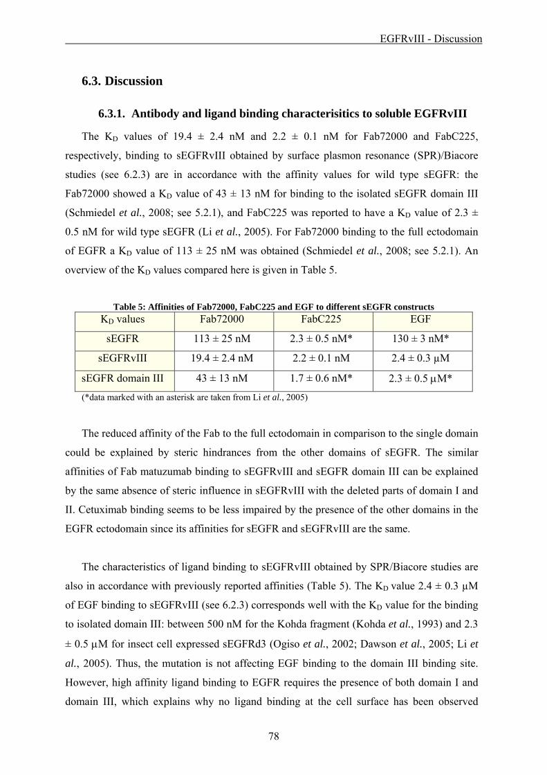
6.3. Discussion ............................................................................................................ 78 6.3.1. Antibody and ligand binding characterisitics to soluble EGFRvIII ................. 78 6.3.2. The structure of EGFRvIII domain III and IV is unaffected by the mutation . 79 6.3.3. sEGFRvIII in solution ...................................................................................... 80 6.3.4. sEGFRvIII dimerization and activation ........................................................... 80 6.3.5. Implications for a therapeutic approach against EGFRvIII driven cancers ..... 81
6.4. Conclusion............................................................................................................ 83 7. Characterization of the antibody EMD1159476 binding to the insulin-like growth factor-
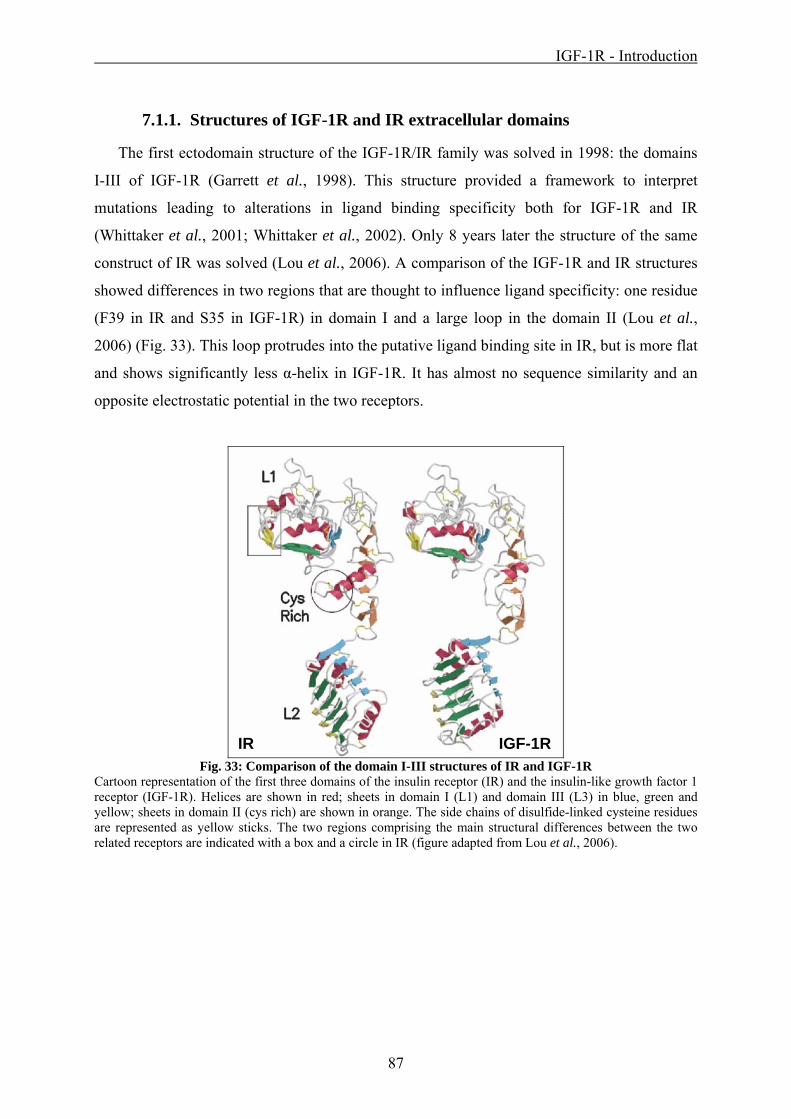
7.1.1. Structures of IGF-1R and IR extracellular domains......................................... 87 7.1.2. Ligand-induced IR/IGF-1R activation ............................................................. 89 7.1.3. IGF-1R and cancer ........................................................................................... 90 7.1.4. Anti-IGF-1R antibodies.................................................................................... 92
7.2. Results .................................................................................................................. 94 7.2.1. Expression and purification sIGF-1R............................................................... 94 7.2.2. Fab1159476 structure....................................................................................... 95 7.2.3. Antibody binding to sIGF-1R domain I-III and domain II .............................. 97
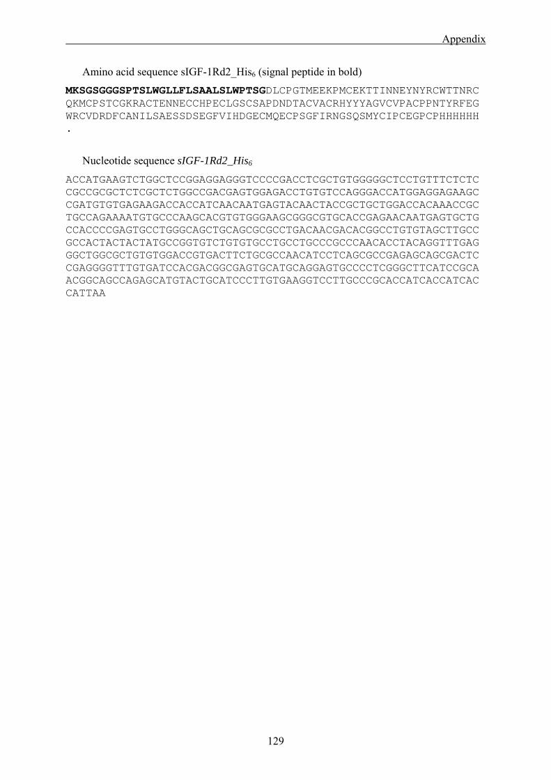
11.1. Primer sequences................................................................................................ 125 11.2. Protein constructs ............................................................................................... 126 11.3. Supplementary data ............................................................................................ 130
Figures
3
List of figures
Fig. 1: Human receptor tyrosine kinases .................................................................................. 14 Fig. 2: Simplified RTK intracellular signaling pathway overview .......................................... 18 Fig. 3: Cloning scheme of sEGFRvIII...................................................................................... 22 Fig. 4: Crystals of the complex sEGFRd3:Fab72000 .............................................................. 33 Fig. 5: Domain organization of ErbB receptors ....................................................................... 38 Fig. 6: Ligand induced EGF receptor dimerization.................................................................. 39 Fig. 7: ErbB family extracellular domain structures without ligand........................................ 41 Fig. 8: Antibody receptor co-structures.................................................................................... 45 Fig. 9: Characterization of matuzumab binding to sEGFR...................................................... 46 Fig. 10: ITC sEGFR and Fab72000 ......................................................................................... 47 Fig. 11: Ligand competition properties of matuzumab ............................................................ 48 Fig. 12: Does the sEGFR:Fab72000 complex dimerize? Analysis by AUC ........................... 49 Fig. 13: Structure of the complex between the matuzumab Fab fragment and domain III of
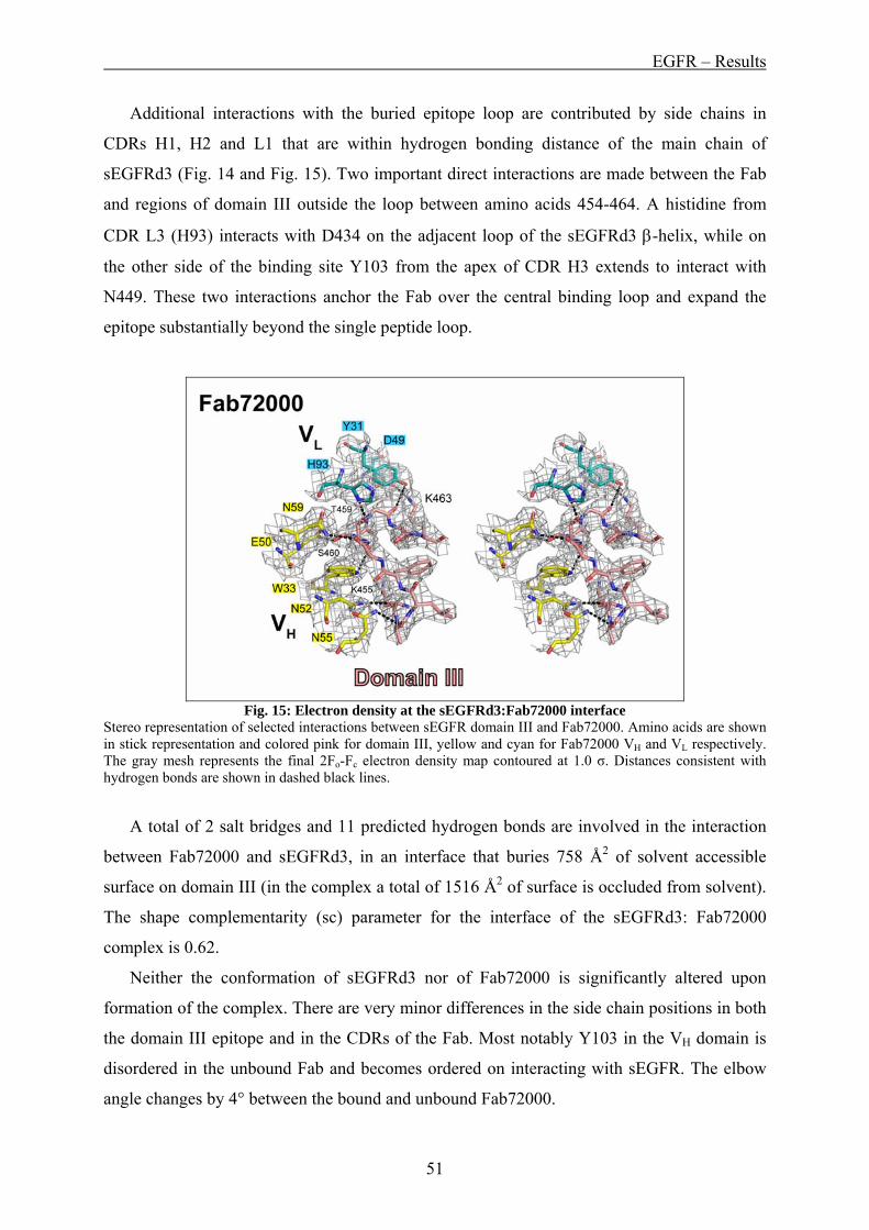
sEGFR .......................................................................................................................... 50 Fig. 14: The epitope of matuzumab in detail ........................................................................... 50 Fig. 15: Electron density at the sEGFRd3:Fab72000 interface................................................ 51 Fig. 16: The matuzumab epitope is distinct from the ligand binding site on domain III of
sEGFR .......................................................................................................................... 52 Fig. 17: Effects of sEGFR mutant binding to matuzumab or EGF .......................................... 53 Fig. 18: Implications for the mechanism of inhibition of EGFR by matuzumab..................... 58 Fig. 19: The matuzumab and cetuximab epitopes do not overlap............................................ 61 Fig. 20: Matuzumab and cetuximab use different mechanisms to block ligand induced EGFR
dimerization.................................................................................................................. 63 Fig. 21: Domain organization of EGFR and EGFRvIII in comparison ................................... 66 Fig. 22: SDS-PAGE sEGFRvIII purification........................................................................... 70 Fig. 23: sEGFR and sEGFRvIII dimerization properties analysed by static light scattering... 72 Fig. 24: Characterization of cetuximab and matuzumab binding to sEGFRvIII ..................... 73 Fig. 25: Characterization of EGF binding to sEGFRvIII ......................................................... 74 Fig. 26: Structure of sEGFRvIII............................................................................................... 75 Fig. 27: Electron density of sEGFRvIII domain III ................................................................. 75 Fig. 28: Experimental and calculated SAXS scattering curves sEGFRvIII ............................. 76 Fig. 29: Model of the disordered sEGFRvIII regions calculated by BUNCH ......................... 77 Fig. 30: Ab initio solution structure of sEGFRvIII calculated by DAMMIN .......................... 77 Fig. 31: sEGFRvIII and sEGFR wild type in comparison ....................................................... 79 Fig. 32: Domain organization of IGF-1R................................................................................. 86 Fig. 33: Comparison of the domain I-III structures of IR and IGF-1R.................................... 87 Fig. 34: Structure of the insulin receptor ectodomain monomer.............................................. 88 Fig. 35: Insulin receptor ligand binding model ........................................................................ 89 Fig. 36: SDS-PAGE sIGF-1R domain I-III and domain II purification................................... 95 Fig. 37: Fab1159476 structure ................................................................................................. 95 Fig. 38: Fab1159476 electron density ...................................................................................... 96 Fig. 39: Characterization of EMD1159476 binding to sIGF-1R ............................................. 97 Fig. 40: ITC sIGF-1R domain I-III and Fab1159476............................................................... 98 Fig. 41: ITC sIGF-1R domain II and Fab1159476................................................................... 99 Fig. 42: Preliminary ligand competition properties of EMD1159476 ................................... 100 Fig. 43: Thermodynamic characteristics of Fab binding to IGF-1R ...................................... 102 Fig. 44: sEGFR in complex with matuzumab binding to EGF .............................................. 130 Fig. 45: sEGFR binding to mAb72000 immobilized by protein A........................................ 131
Tables
4
List of tables
Table 1: Overview structural information of RTK extracellular domains ............................... 15 Table 2: Data collection and refinement statistics Fab72000 and sEGFRd3:Fab72000.......... 34 Table 3: Data collection and refinement statistics sEGFRvIII................................................. 35 Table 4: Data collection and refinement statistics Fab1159476 .............................................. 36 Table 5: Affinities of Fab72000, FabC225 and EGF to different sEGFR constructs .............. 78 Table 6: ITC-derived characteristics of antibody binding to IGF-1R at 25°C....................... 130
Zusammenfassung
5
1. ZUSAMMENFASSUNG Rezeptor-Tyrosinkinasen (RTKs) sind essentielle Bestandteile der inter- und
intrazellulären Kommunikation und der Signaltransduktion in Metazoen. Sie sind involviert in
die Steuerung wichtiger zellulärer Prozesse wie Zellteilung, Zellwachstum, Zelldifferen-
zierung und Zelltod (Hubbard and Miller, 2007). RTKs gehören zu der Enzymfamilie der
Protein-Tyrosinkinasen, die den Transfer einer Phosphatgruppe von ATP auf Tyrosinreste des
Substrates katalysieren. Im menschlichen Genom sind 58 RTKs und 32 nicht-Rezeptor
Protein-Tyrosinkinasen kodiert. Die Rezeptoren sind Typ I Transmembranproteine mit einer
extrazellulären Liganden-Bindungsdomäne und einer intrazellulären Tyrosinkinasedomäne.
Der extrazelluläre Bereich ist mit der intrazellulären Kinasedomäne durch eine einfache
Transmembranhelix verbunden (Schlessinger, 2000).
Generell werden RTKs durch Liganden-induzierte Dimerisierung aktiviert, die die
intrazellulären Kinasendomänen nahe genug zueinander bringt um eine Autophospho-
rylierung in trans zu ermöglichen. Die phosphorylierten Proteinsequenzen rekrutieren
Proteinsubstrate, die eine Signalkaskade in das Zellinnere und in den Zellkern initiieren.
Letztendlich werden so Transkriptionsfaktoren reguliert, die in Prozesse involviert sind wie
zum Beispiel die Zelldifferenzierung oder das Zellüberleben (Hunter, 2000).
Ausgehend von ersten Untersuchungen in den 1980er Jahren zeigte sich, dass viele RTKs
an der Entstehung verschiedener Neoplasien beteiligt sind und sogar Malignome hervorrufen
können, wenn Störungen in der normalen Regulation der Rezeptoren vorliegen.
Missregulierungen dieser Art können u.a. durch Genamplifikationen oder durch Mutationen
verursacht werden, die eine konstitutive Aktivierung der Rezeptoren zur Folge haben
(Weinberg, 2007).
In der Klinik werden verschiedene Therapieansätze gegen Neoplasien, die durch RTKs
hervorgerufen werden, genutzt (Mendelsohn and Baselga, 2006). Unter anderem können
einerseits Tyrosinkinase-Inhibitoren intrazellulär die Signaltransduktionskaskaden blockieren,
die zu einer weiteren Zellteilung und –amplifikation führen würden. Andererseits werden
monoklonale Antikörper eingesetzt, die die Rezeptoren extrazellulär binden. Hierdurch wird
das Immunsystem des Körpers gegen Zellen aktiviert, die eine große Anzahl der Rezeptoren
an der Oberfläche tragen. Zusätzlich können Antikörper die Aktivierung der RKTs
verhindern, indem sie das Binden von Liganden oder die Rezeptordimerisierung blockieren.
Zusammenfassung
6
Verschiedene Studien über die Anwendung von monoklonalen Antikörpern in der
Krebstherapie haben gezeigt, dass aktivierende Mutationen in Mediatoren der Signalkaskaden
(zum Beispiel K-ras), Kompensationsmechanismen bzw. Resistenzen der Zelle und sich
gegenseitig beeinflussende Signaltransduktionswege von verschiedenen RTKs Einfluss auf
die Wirksamkeit der Therapie haben (Dempke and Heinemann, 2009). Eine für jeden
Patienten individuell angepasste Kombination von Chemotherapie, Strahlentherapie und
Antikörpern bzw. Inhibitoren könnte ein Weg sein um die Effektivität der Behandlung zu
steigern und Nebenwirkungen zu minimieren (Friedman et al., 2005).
In dieser Arbeit wurde mit zwei verschiedenen RTKs gearbeitet: der Epidermale
Wachstumsfaktorrezeptor EGFR und der Insulin-ähnliche Wachstumsfaktorrezeptor 1 IGF-
1R. Beide Rezeptoren können bei Missregulation Tumoren hervorrufen, u.a. epitheliale
Neoplasien wie Bronchialkarzinome oder Kolonkarzinome. Eine ansteigende Anzahl von
Antikörpern gegen EGFR and IGF-1R ist in der klinischen Untersuchungsphase oder schon in
der Klinik in Anwendung. Gegen EGFR sind die Antikörper Cetuximab/Erbitux® und
Panitumumab/Vectibix® seit 2004 beziehungsweise 2006 zugelassen. Des Weiteren ist der
monoklonalen Antikörper Trastuzumab/Herceptin® seit 1998 in der klinischen Anwendung
gegen Mammkarzinome, die das zweite Familienmitglied der EGFR Familie ErbB2
überexprimieren.
Das Ziel dieser Arbeit war die Charakterisierung der Interaktionen von löslichen RTK
extrazellulären Domänen mit Antikörper Fab-Fragmenten sowie der Inhibitionsmechanismen
von verschiedenen Antikörpern. Ein besseres Verständnis der Epitope der Antikörper, ihrer
Affinitäten und Liganden-Kompetitionscharakteristiken könnte dazu beitragen die klinische
Anwendung der Antikörper in der Krebstherapie zu verbessern. Es wurden die folgenden
Fragestellungen untersucht:
1. an welcher Stelle der extrazellulären Domäne bindet der Antikörper?
2. welche Affinität hat der Antikörper zum löslichen Rezeptor?
3. wie beeinflusst die Bindung des Antikörpers die Aktivierung des Rezeptors?
4. ist es den natürlichen Liganden des Rezeptors noch möglich zu binden, wenn der
Antikörper vorhanden ist?
5. welchen Effekt hat der gebundene Antikörper auf die Rezeptordimerisierung?
6. ist die strukturelle Reorganisation, die Voraussetzung für die Rezeptoraktivierung ist,
noch möglich mit gebundenem Antikörper?
Zusammenfassung
7
Die Arbeit wurde in drei Abschnitte gegliedert. Im ersten Abschnitt (Kapitel 5) werden
die Interaktionen von EGFR mit dem monoklonalen Antikörper Matuzumab (EMD72000)
beschrieben. Der zweite Abschnitt (Kapitel 6) zeigt Untersuchungen zu einer EGFR Mutante
(EGFR Variante III oder EGFRvIII), die bisher ausschließlich auf neoplastischen Zellen
nachgewiesen werden konnte. Im dritten Abschnitt wird die Bindung des monoklonalen
Antikörpers EMD1159476 an den Insulin-ähnlichen Wachstumsfaktor-rezeptor 1 IGF-1R
beschrieben (Kapitel 7).
(1) EGFR – Antikörper Interaktionen (Kapitel 5)
In diesem Teil der Arbeit wurden die Eigenschaften des gegen EGFR gerichteten
monoklonalen Antikörpers Matuzumab (EMD72000) untersucht. Matuzumab ist die
humanisiert Form des murinen anti-EGFR Antikörpers 425 und hat die Phase II der klinischen
Studien erreicht. Es konnte die Komplexkristallstruktur des Matuzumab Fab-Fragments mit
der Domäne III des Rezeptors gelöst und so erstmals das Epitop des Antikörpers identifiziert
werden. Das Epitop wurde durch Rezeptor-Mutationsstudien in Lösung bestätigt.
Interessanterweise überlappt die Matuzumab Bindestelle nicht mit dem Epitop des natürlichen
Liganden EGF. Das Gegenteil wurde zuvor für den bereits in der Klinik eingesetzten
Antikörper Cetuximab beobachtet, dessen Bindungsstelle sich mit dem Epitop von EGF
überschneidet. Zudem sind die Epitope der beiden Antikörper Matuzumab und Cetuximab
unterschiedlich und nicht überlappend. Während Cetuximab direkt das Binden des
aktivierenden Liganden an EGFR verhindert, konnte für Matuzumab in dieser Arbeit ein
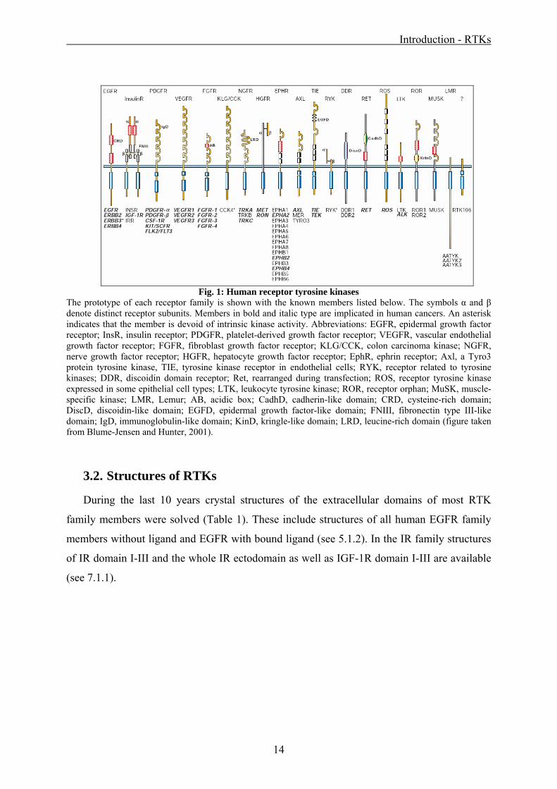
The prototype of each receptor family is shown with the known members listed below. The symbols α and β denote distinct receptor subunits. Members in bold and italic type are implicated in human cancers. An asterisk indicates that the member is devoid of intrinsic kinase activity. Abbreviations: EGFR, epidermal growth factor receptor; InsR, insulin receptor; PDGFR, platelet-derived growth factor receptor; VEGFR, vascular endothelial growth factor receptor; FGFR, fibroblast growth factor receptor; KLG/CCK, colon carcinoma kinase; NGFR, nerve growth factor receptor; HGFR, hepatocyte growth factor receptor; EphR, ephrin receptor; Axl, a Tyro3 protein tyrosine kinase, TIE, tyrosine kinase receptor in endothelial cells; RYK, receptor related to tyrosine kinases; DDR, discoidin domain receptor; Ret, rearranged during transfection; ROS, receptor tyrosine kinase expressed in some epithelial cell types; LTK, leukocyte tyrosine kinase; ROR, receptor orphan; MuSK, muscle-specific kinase; LMR, Lemur; AB, acidic box; CadhD, cadherin-like domain; CRD, cysteine-rich domain; DiscD, discoidin-like domain; EGFD, epidermal growth factor-like domain; FNIII, fibronectin type III-like domain; IgD, immunoglobulin-like domain; KinD, kringle-like domain; LRD, leucine-rich domain (figure taken from Blume-Jensen and Hunter, 2001).
3.2. Structures of RTKs
During the last 10 years crystal structures of the extracellular domains of most RTK
family members were solved (Table 1). These include structures of all human EGFR family
members without ligand and EGFR with bound ligand (see 5.1.2). In the IR family structures
of IR domain I-III and the whole IR ectodomain as well as IGF-1R domain I-III are available
(see 7.1.1).
Introduction - RTKs
15
Table 1: Overview structural information of RTK extracellular domains Structural information available
Receptor Structures of parts or the full extracellular domain solved
EGFR
all family members, EGFR with ligands EGF and TGF-α (Cho and Leahy,
2002; Ogiso et al., 2002; Garrett et al., 2002; Ferguson et al., 2003; Cho et
al., 2003; Garrett et al., 2003; Franklin et al., 2004; Bouyain et al., 2005)
IR family IR domain I-III and IR ectodomain, IGF-1R domain I-III (Garrett et al.,
1998; McKern et al., 2006; Lou et al., 2006)
PDGFR KIT with and without ligand (Yuzawa et al., 2007)
VEGFR1 domain II (Christinger et al., 2004)
FGFR
parts of the extracellular domain in complex with different ligands
(Plotnikov et al., 1999; Stauber et al., 2000; Plotnikov et al., 2000; Yeh et
al., 2003; Olsen et al., 2004; Olsen et al., 2006)
NGFR
full and fragmented ectodomains with and without ligand (Wiesmann et al.,
1999; Ultsch et al., 1999; Robertson et al., 2001; Banfield et al., 2001;
Wehrman et al., 2007)
HGFR partial ectodomain with ligand bound (Stamos et al., 2004)
EPHR
several extracellular domains with and without ligand (Himanen et al.,
2001; Himanen et al., 2004; Chrencik et al., 2006; Qin et al., 2008;
Goldgur et al., 2009)
AXL two family members alone and in complex with ligand (Heiring et al.,
2004; Sasaki et al., 2006)
TIE partial ectodomain alone and in complex with ligand (Barton et al., 2006)
DDR discoidin domain of DDR2 (Ichikawa et al., 2007)
MuSK first and second immunoglobulin-like domain (Stiegler et al., 2006)
No structural information available
KLG/CCK, RYK, RET, ROS, LTK, ROR
Introduction - RTKs
16
3.3. RTK activation
Generally, RTKs are activated through ligand induced receptor dimerization, which brings
the tyrosine kinase domains into close proximity promoting the allosteric activation of the
kinase domains (Zhang et al., 2006a; Hubbard and Miller, 2007). The phosphorylated tyrosine
residues are located in the kinase activation loop or juxtamembrane region, inducing
conformational changes that stabilize the active state of the kinase (Hubbard, 2004). Induced
by the phosphorylation event, the activated kinase domains recruit downstream substrate
molecules which initiate an intracellular signal cascade (see 3.4). The signaling pathways
regulate transcription factors involved in cell survival or cell differentiation (Blume-Jensen
and Hunter, 2001; Murphy and Blenis, 2006).
Within the RTK family different ligands employ varying modes for inducing the active
dimeric state of the receptors. The following mechanisms have been described:
1. The simplest mechanism is represented by bivalent ligands, binding simultaneously to
two receptor molecules (1:2 ligand:receptor complex). This binding mode has been
observed e.g. in structural studies investigating the growth hormone receptor (GHR,
not included in Fig. 1) in complex with growth hormone (GH) (Kossiakoff and de
Vos, 1998).
2. A 2:2 ligand:receptor complex was described for homodimeric growth factors, e.g.
VEGF, FGF or PDGF (Wiesmann et al., 1997; Plotnikov et al., 1999). Using electron
microscopy and small-angle x-ray scattering also the RTK Met was described to be
activated in a similar 2:2 ligand:receptor mode with no direct receptor contact in the
complex (Gherardi et al., 2006). In case of FGF receptor activation it was shown in
crystallographic studies that the receptor requires heparin sulfate proteoglycans in
addition to the ligands to stabilize the dimeric complex (Mohammadi et al., 2005).
3. The structures of complexes of EGFR and its ligands EGF and TGF-α (Ogiso et al.,
2002; Garrett et al., 2002) also showed a 2:2 ligand:receptor complex. But in contrast
to the complexes mentioned above the dimer interface is entirely receptor mediated
and the ligands do not touch each other (see 5.1 and Fig. 6).
4. Unlike the majority of RTKs the insulin receptor family is not a single-chain receptor,
but a α2β2 homodimer (see 7.1). Recently, the structure of the entire disulfide-linked
ectodomain of the insulin receptor has been solved (McKern et al., 2006) (Fig. 34).
The current activation model suggests a 2:1 ligand:receptor dimer complex with the
ligands mediating the contact between the two halfes of the homodimer.
Introduction - RTKs
17
5. A subset of RTKs, including Ret (rearranged during transfection) and MuSK (muscle-
specific kinase), do not bind their ligands directly, but require co-receptors for ligand-
induced activation. Ret dimerizes as 1:2:2 ligand:receptor:co-receptor complex
(Schlee et al., 2006). Ligand and co-receptor of MuSK were recently identified as the
heparan sulfate proteoglycan agrin and the low density lipoprotein receptor (LDLR)
family member Lrp4 (Stiegler et al., 2006; Kim et al., 2008).
3.4. Signaling mechanisms downstream of activated RTKs
The phosphotyrosine residues in RTKs are bound by cytoplasmic enzymes and
adapter/scaffolding proteins containing SRC homology-2 (SH2) or phosphotyrosine-binding
(PTB) domains (Hubbard and Miller, 2007). SH2 domain-containing enzymes (SHC) are e.g.
protein tyrosine kinases (SRC kinases), protein tyrosine phosphatases (SHP2), phospholipase
C (PLCγ) or guanine exchange factors (Ras-GAP). With their SH2 and SH3 domains adapter
proteins (e.g. GRB2, NCK, CRK, SHC) form scaffolds that link different proteins involved in
signal transduction.
Simplified, there are three main intracellular signal transduction pathways that are
activated through RTK phosphorylation (Fig. 2).
1. The Ras/MAP kinase (mitogen-activated protein kinase) signaling cascade
(Schlessinger, 2000). The adapter protein GRB2 forms a complex with the guanine
nucleotide exchange factor mSOS (mammalian son of sevenless). The GRB2:SOS
complex binds to RTK phosphotyrosine residues thus translocating SOS to the plasma
membrane and close to Ras. Here it stimulates the exchange of GTP for GDP
(Gureasko et al., 2008). Once in the active GTP-bound state, Ras interacts with several
effector proteins such as Raf and phosphatidylinositol 3-kinase (PI-3K) to trigger
Upon extracellular ligand binding and receptor dimerization, tyrosine trans-autophosphorylation occurs. This triggers the binding of downstream effectors, such as Grb2. Subsequently the recruitment of son-of-sevenless (SOS) and Ras, Raf, MEK leads to the activation of the entire mitogen-activated protein kinase (MAPK) cascade (MEK stands for ‘MAPK and extracellular signal-regulated kinase (ERK) kinase’). Other signaling pathways include the activation of phosphatidylinositol 3-kinase (PI-3K) and Akt or phospholipase C (PLCγ). RTK signaling leads to enhanced cell survival, growth and differentiation through the activation of transcription factors (e.g. ELK, FOS, STAT, not shown here).
The phosphorylation events downstream of RTK activation involve many proteins and
expand quickly in the cell. Phosphotyrosine studies in the EGFR signaling network showed
significant changes in the phosphorylation state of 81 proteins within 20 min after EGF
stimulation (Blagoev et al., 2004; Zhang et al., 2005).
The signaling pathways are subjected to multiple negative feedback mechanisms at the
level of the receptor itself by inhibitory protein tyrosine phosphatases and by receptor
endocytosis and degradation (Schlessinger, 2000; Le Roy and Wrana, 2005). In addition, the
specific activity of downstream effector proteins can be negatively regulated by inhibitory
signals, e.g. through MAPK specific phosphatases. The strength and duration of the signals
that are transmitted through the networks of signaling cascades are modulated through factors
such as cell-surface receptor density, expression levels of scaffolding proteins, the
Introduction - RTKs
19
surrounding extracellular matrix and the balance between kinases and phosphatases (Murphy
and Blenis, 2006).
Taken together, the downstream signaling pathways are not linear but consist of
multilayered and cross-connected networks. This allows for horizontal interactions and
permits multiple combinatorial and integrated responses (Mendelsohn and Baselga, 2006).
The complexity of this network makes it especially difficult to treat RTK misregulation in
cancer (see next section).
3.5. RTKs and cancer
When mutated or altered structurally, RKTs can become potent oncoproteins. More than
half of the known receptors tyrosine kinases (marked in bold in Fig. 1) have been repeatedly
found to be either mutated or overexpressed in human malignancies (Blume-Jensen and
Hunter, 2001). Once their normal tight regulation is impaired, RTKs can cause deregulated
autonomous cell growth and support the capacity to invade other tissues.
This oncogenic transformation can be induced by four main principles: retroviral
transduction of a proto-oncogene corresponding to a RTK with deregulating structural
changes (commonly found in rodents and chicken); genomic re-arrangement, i.e.
chromosomal translocations, resulting in oncogenic fusion proteins; gain-of-function
mutations or small deletions; or receptor/ligand overexpression resulting from gene
amplification. In general, the transforming effects are based upon enhanced or constitutive
kinase activity with quantitatively or qualitatively altered downstream signaling (Murphy and
Blenis, 2006; Weinberg, 2007).
In consequence much effort has gone into designing and identifying potent and specific
RTK inhibitors. Targeted therapeutics were developed both to the extracellular regions of
RTKs using e.g. monoclonal antibodies, and to the cytoplasmic (kinase) domains using small-
molecule inhibitors (Mendelsohn and Baselga, 2006).
20
Materials & Methods
21
4. MATERIALS & METHODS
4.1. Molecular Biology
4.1.1. EGFR
The vector constructs of the full length extracellular domain of the epidermal growth
factor receptor sEGFR (pFastBac_sEGFR_His6) and the isolated domain III with the amino
acids 310-500 of mature sEGFR (sEGFRd3, pFastBac_sEGFRd3_His6) were provided by K.
M. Ferguson, University of Pennsylvania. These constructs were used for all experiments
presented in section 5 beside the mutational studies. The same construct sEGFR
(pFastBac_sEGFR_His6) was cloned by standard PCR and molecular biology procedures for
the experiments described in section 6 (primer, DNA and protein sequences in the Appendix
in 11.1 and 11.2). Human EGFR cDNA was provided by Merck KGaA, Germany.
Site-directed mutagenesis to introduce alanine mutation into sEGFR was carried out using
the QuikChange Kit (Stratagene) following a two-stage PCR protocol (Wang and Malcolm,
2002). To generate the mutant sEGFR K454A the primers K454 up and K454 rev were used,
for the mutant sEGFR K463A the primers K463 up and K463 rev were used and for the
double mutant sEGFR T459A/S460A the primers T459A/S460A up and T459A/S460A rev
were used (sequences in 11.1).
The residues K454 or K463 for the triple mutants are sequentially close to the double
mutant residues T459A and S460A. To prevent back-mutation of already introduced
alterations the mutagenesis was carried out in two PCR stages: a first round with the primers
of the T459A/S460A mutation (see above) and a second stage performed with the primers
tripleK454A up and tripleK454A rev to generate the mutant sEGFR T459A/S460A/K454A or
the primers tripleK463A up and tripleK463A rev for the mutant sEGFR
T459A/S460A/K463A (sequences in 11.1). The successful introduction of the mutations was
verified by DNA sequencing of the respective pFastBac constructs.
Protein of the mutant sEGFR D355T/F357A was provided by K. M. Ferguson, University
of Pennsylvania. Protein of the mutant sEGFR Y251A/R285S was a donation of J. Dawson,
University of Pennsylvania.
Materials & Methods
22
4.1.2. sEGFRvIII
The deletion mutant sEGFR variant III (sEGFRvIII) was amplified by PCR in two
fragments from EGFR cDNA (provided by Merck KGaA, Germany). Both fragments were
generated with a complementary base pair overlap resulting in a novel glycine residue at the
fusion junction (Fig. 3).
Fig. 3: Cloning scheme of sEGFRvIII
sEGFR variant III consists of two fragments of the wild type EGFR gene, which are fused by a complementary overlap at the fusion junction introduced by the primers. Thus residue 5 of domain I is directly connected to residue 274 of domain II via a novel glycine residue.
The DNA of sEGFRvIII was amplified and purified using standard PCR and molecular
biology procedures. The construct was cloned with the N-terminal native secretion signal
peptide and a C-terminal hexa-histidine tag. In addition attB-sequences were introduced at the
start and the end of the PCR product to enable the fusion of the gene into a Gateway® entry
vector (Invitrogen, 2003). The primers sEGFRvIII f1 up and sEGFRvIII f1 rev were used to
generate the sEGFRvIII_His6 N-terminal fragment and the primers sEGFRvIII f2 up and
sEGFRvIII f2 rev for the C-terminal fragment (sequences in 11.1). The sequence of the
construct sEGFRvIII_His6 was confirmed by DNA sequencing (DNA and protein sequences
in 11.2).
Materials & Methods
23
4.1.3. sIGF-1R
Based on IGF-1R cDNA provided by Merck KGaA, Germany the domains I-III of the
extracellular domain (amino acids 31-492 of mature IGF-1R) as well as the isolated domain II
(amino acids 180-329 of mature IGF-1R) were amplified by standard PCR techniques. Both
constructs were cloned with the N-terminal native secretion signal peptide and a C-terminal
hexa-histidine tag. The constructs were transferred into the expression vectors of the
respective insect cell or mammalian expression system using the Gateway® technology
(Invitrogen, 2003). The native secretion signal peptide was directly fused to the domain II by
blunt end ligation. The primers sIGF-1Rd1-3 up and sIGF-1Rd1-3 rev were used for the
generation of the sIGF-1Rd1-3 entry vector, the primers sIGF-1Rd2 blunt up and sIGF-1Rd2
blunt rev for the sIGF-1Rd2 blunt end ligation construct and the primers sIGF-1Rd2 up and
sIGF-1Rd2 rev for the sIGF-1Rd2 amplification (sequences in 11.1). The sequences of the
constructs were confirmed by DNA sequencing (DNA and protein sequences in 11.2).
4.1.4. Generation of recombinant baculovirus
Recombinant baculoviruses for the expression of sEGFR, sEGFR domain III, sEGFRvIII,
the sEGFR mutants, sIGF-1R domain I-III and sIGF-1R domain II were produced as
described (Invitrogen, 2009).
4.2. Protein expression
4.2.1. sEGFR and sEGFRvIII
The soluble extracellular part of the EGFR wild type (sEGFR), the isolated domain III of
the receptor (sEGFRd3) and the sEGFR mutants (see 4.1.1) were expressed in Sf9 insect cells
infected by recombinant baculovirus exactly as described (Ferguson et al., 2000) (see 4.1.4).
Briefly, 5-10 L insect cell culture was infected with freshly amplified baculovirus at a density
of 2.0 x 10⁶ c/ml (viability > 98%) and incubated for 96 h at 27°C in multiple 1 L spinner
flasks that each contained <500 ml (to ensure adequate aeration) The cells were separated
from the protein containing medium by centrifugation.
Materials & Methods
24
4.2.2. sIGF-1R
Insect cell expression. The isolated domains 1-3 and domain 2 of the IGF-1R
extracellular part (sIGF-1Rd1-3 and sIGF-1Rd2, respectively) were expressed both in Sf9 and
Hi5 insect cells infected by recombinant baculovirus (see 4.1.4). The cells grew at 27°C in
500 ml shaking flasks in Sf-900 II serum free medium (Invitrogen) or Express Five serum free
medium (Invitrogen), respectively,. They were infected with recombinant baculovirus at a
density of 2x10⁶ cells/ml and incubated for 24-96h at 27°C. The highest yield was obtained
with a multiplicity of infection (MOI) 4 and an expression for 48 h, after which protein
degradation started to occur. The cells were separated from the protein containing medium by
centrifugation.
Mammalian cell expression. Both sIGF-1R constructs sIGF-1Rd1-3 and sIGF-1Rd2
were transiently expressed in human kidney HEK293 Ebna cells. The cells were cultured in
suspension in Ex-Cell VPRO Serum Free Medium (SAFC, Sigma Aldrich) with 4 mM
glutamine (Invitrogen) and 0.1% Pluronic (Invitrogen) at 37°C, 25% O2, 75 rpm in a 8 L
fermenter. For transfection, cells harvested after 24h cells at 2.5 x10⁶ cells/ml were
resuspended in transfection medium consisting of DMEM F-12 1:1 (Invitrogen) with 8 mM
was immobilized at a flow rate of 5 µl/min for 10 min on an activated CM5 chip surface
(Ferguson et al., 2000; Li et al., 2005). The final immobilization level was 250 RU.
Regeneration of the EGF surface was carried out with 1 M NaCl in 10 mM sodium acetate
(pH 5.0).
Titration and competition experiments. sEGFR, sEGFR domain III and sEGFRvIII
were flown as twofold serial dilutions covering a concentration range of 0-1000 nM over the
Fab72000 or FabC225 surface. sEGFR wild type, sEGFR mutants and EGFRvIII binding to
the immobilized ligand EGF was observed with twofold serial dilutions covering a
concentration range of 0-20 µM. sIGF-1R domain I-III and domain II binding to immobilized
Fab1159476 was observed with twofold serial dilutions in the range of 0-1000 nM.
Competition experiments were carried out with a constant concentration of the receptor
protein (600 nM). The binding to a ligand surface was monitored while increasing amounts of
Fab fragments ranging from 0-30 µM were added to the receptor sample.
Materials & Methods
29
4.4.4. Analytical ultracentrifugation
Analytical ultracentrifugation sedimentation equilibrium (AUC SE) experiments were
performed to investigate the dimerization state of sEGFR in the presence of ligand and
Fab72000 using an XL-A analytical ultracentrifuge (Beckman, USA). Samples (4 µM) of
wild type or mutated sEGFR protein were analyzed both in the presence and in the absence of
a 1.5-fold molar excess of EGF. As control the molecular weight of a dimerization
incompetent sEGFR in complex with Fab was obtained with and without EGF. The
dimerization incompetent receptor was provided by Jessica Dawson, University of
Pennsylvania. Each sample contained 4µM of the relevant protein or sEGFR:Fab72000
complex in 20 mM HEPES, 100 mM NaCl (pH 7.5). Samples were loaded in six-channel
charcoal-Epon cells with quartz windows at both ends. Radial scans were performed at 20°C
at 6,000, 9,000, and 12,000 rpm in an An Ti 60 rotor, with detection over a wavelength range
of 236 to 285 nm. Equilibrium was reached in each speed step within 18h. The partial specific
volume of sEGFR proteins was estimated as 0.71 ml/g as described before (Ferguson et al.,
2000), and solvent density was taken as 1.003 g/ml. Molecular masses were determined by
fitting multiple data sets to a simple model for a single species in Sedfit version 9.4c and
Sedphat version 4.4b.
4.4.5. Isothermal titration calorimetry
Isothermal titration calorimetry (ITC) was carried out to investigate the binding affinity of
the receptor ectodomains to the antibody Fab fragments and the thermodynamics of the
interaction. The experiments were carried out using a VP-ITC microcalorimeter (Microcal
LLC) and evaluated with the Origin 7 calorimetry sofware (MicroCal LLC) to calculate the
binding constant (KA) and the binding affinity (KD=1/KA), the observed binding enthalpy
(ΔHobs) as well as the stoichiometry (N) of the formed complex. For all receptor Fab binding
experiments a model of one binding site was assumed. ΔHobs values were calculated based on
the difference between the heat liberated during the binding phase of the injections and the
average heat of dilution found once the receptor was saturated with antibody.
10 µl Fab solution (16.7-50 µM) (see 4.3.3) was titrated to 2 ml receptor in the cell (1.7-5
µM) (see 4.3). More precisely, Fab72000 (20 µM) in 10 mM HEPES, 50 mM NaCl (pH 7.5)
was injected in 10 µl steps into a cell containing 2 µM sEGFR. Fab1159476 (16.7 µM) in
PBS was injected in 11 µl steps into a cell containing 1.7 µM sIGF-1R domain I-III (sIGF-
1Rd1-3). In addition Fab1159476 was investigated for sIGF-1R domain II (sIGF-1Rd2)
Materials & Methods
30
binding and was injected at 50 µM in PBS in 11 µl steps into a cell containing 5 µM sIGF-
1Rd2. All binding experiments were carried out at 25°C with a spacing time between the
injections of 320 sec.
4.4.6. Small angle X-ray scattering
Small angle X-ray scattering (SAXS) experiments were carried out to determine a low
resolution shape (Koch et al., 2003) of sEGFRvIII in solution. The scattering data from
sEGFRvIII samples (see 4.3.2) were collected at the SAXS beamline EMBL, DESY,
Germany. Using a MAR345 image plate detector at a sample-detector distance of 2.7 m and a
wavelength of λ = 1.5 Å a range of 0.01<s<0.5 Å-1 was covered (s = 4π sinθ/λ, where 2θ is the
scattering angle and λ the X-ray wavelength). 100 µl samples of three different concentrations
(1 mg/ml, 5 mg/ml and 10 mg/ml in 20 mM HEPES, 100 mM NaCl, pH 7.5) were measured
at 10°C for 120 sec. To monitor for radiation damage two successive measurements of protein
solutions were compared and no significant changes were observed. The scattering intensities
of buffer backgrounds were measured both before and after the sample and the averaged
background scattering was subtracted from the scattering of the sample.
The low angle data measured at lower protein concentrations were extrapolated to infinite
dilution and merged with the higher concentration data to yield the final composite scattering
curve. Data processing was performed using the program PRIMUS (Konarev et al., 2003).
The radius of gyration Rg was calculated using the Guinier approximation (Guinier, 1939)
and the program GNOM (Svergun, 1992), which also provided the distance distribution
function of the particle p(r) and the maximum particle size Dmax. The molecular mass of the
solute was estimated based on the excluded (Porod) volume (Porod, 1982). For globular
proteins, the Porod volume in nm3 is about twice the molecular mass in kDa.
Molecular modeling. The theoretical scattering from the low resolution crystal structure
of sEGFRvIII (see 4.5.4) was calculated using the program CRYSOL (Svergun et al., 1995).
Given the atomic coordinates, the program uses the scattering amplitudes to calculate the
spherically averaged scattering pattern and takes into account the hydration shell of the
protein.
Domain I and II of sEGFRvIII, which are disordered in the crystal structure (see 6.2.4),
were modeled using the program BUNCH (Petoukhov and Svergun, 2005). The program
combines rigid body and ab initio modeling of proteins consisting of domains linked by
Materials & Methods
31
flexible loops of unknown structure. A simulated annealing protocol is employed to model the
probable conformation of the flexible linkers with the structurally known domains kept as
rigid bodies. The ab initio modeled loops are represented as interconnected chains of dummy
residues (Petoukhov et al., 2002). Domain/loop arrangements with steric clashes, dummy
residue loops with improper distribution of bond or dihedral angles as well as too extended
loops are penalized.
Ab initio shape determination. The scattering curve of sEGFRvIII was further used to
model the low resolution ab initio shape of solution sEGFRvIII by the program DAMMIN
(Svergun, 1999). This program represents the particle shape by a densely packed bead model,
which is fitted through simulated annealing procudures to the experimental data Iexp(s). The
models of 10 DAMMIN runs were averaged to determine common structural features using
the programs DAMAVER (Volkov and Svergun, 2003) and SUPCOMB (Kozin and Svergun,
2001).
Materials & Methods
32
4.5. Protein Crystallography
4.5.1. sEGFR:Fab72000
sEGFR in complex with Fab72000 (see 4.3.4) was concentrated and buffer exchanged by
gel filtration into 10 mM HEPES, 50 mM NaCl (pH 7.5) and crystallized using the hanging
drop vapor diffusion method. The polydispersity of sEGFR:Fab72000 samples as determined
by dynamic light scattering was 15.7%. The complex crystallized in several conditions with a
low pH value [0.1 M sodium acetate, 1.7 M ammonium sulfate (pH 4.5) at 4°C; 50 mM
citrate, 17% PEG-3350, 1.6 M NaCl, 3% ethylene glycol (pH 5.0) at 20°C; 0.1 M phosphate-
citrate, 20% PEG-1000, 0.25 M lithium sulfate (pH 4.2) at 20°C], but the crystals proved to
be unstable and/or with low diffraction quality.
4.5.2. Fab72000
Freshly purified protein (see 4.3.3) was concentrated and buffer exchanged by gel
filtration into 10 mM HEPES, 50 mM NaCl (pH 7.5) and crystallized using the hanging drop
vapor diffusion method. Single crystals of Fab72000 (0.1x0.5x0.1 mm) were obtained by
mixing equal volumes (1:1) of the Fab (13 mg/ml) with a solution containing 1.8 M
ammonium sulfate, 0.1 M MES (pH 6.5) and equilibrating over a reservoir of this buffer at
20°C. Crystals were flash frozen in reservoir solution that was supplemented with 9%
sucrose, 2% glucose, 8% glycerol, 8% ethylene glycol. X-ray diffraction data were collected
at the Cornell High Energy Synchrotron Source (CHESS) beamline F1, using an ADSC
Quantum-210 CCD detector. The data were processed with HKL2000 (Otwinowski and
Minor, 1997). Data collection statistics are summarized in Table 2 (see 4.5.3).
The structure of Fab72000 was solved by the method of molecular replacement using the
program PHASER (CCP4, 1994). The coordinates for Fab2C4 (PDB ID 1L7I) (Vajdos et al.,
2002) were selected as the initial search model based on the sequence identity between
Fab2C4 and Fab72000. Coordinates were manually rebuilt in COOT (Emsley and Cowtan,
2004) and refined using CNS (Brünger et al., 1998) and Refmac (CCP4, 1994). New maps
were calculated following each iteration of refinement, including solvent flattened maps with
minimized model bias calculated using the program DM (CCP4, 1994). Refinement statistics
are summarized in Table 2 (see 4.5.3).
Coordinates of the Fab72000 structures have been deposited with the PDB ID code 3C08.
Materials & Methods
33
4.5.3. sEGFRd3:Fab72000
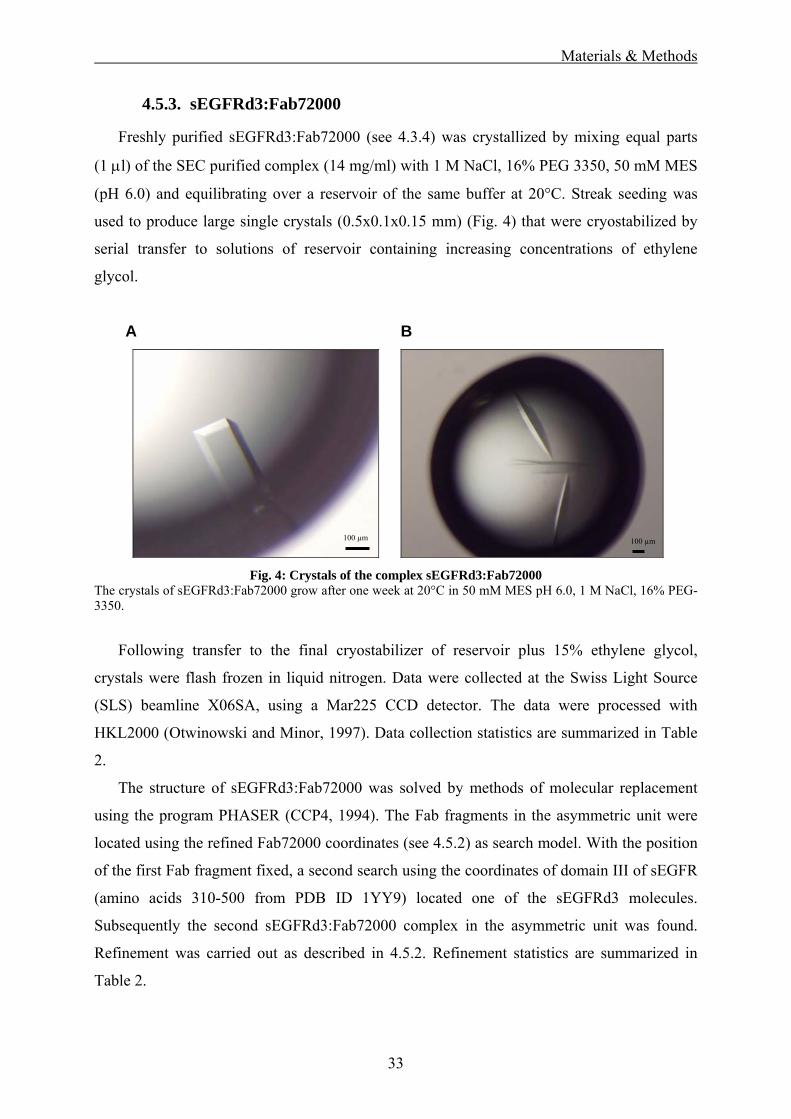
Freshly purified sEGFRd3:Fab72000 (see 4.3.4) was crystallized by mixing equal parts
(1 μl) of the SEC purified complex (14 mg/ml) with 1 M NaCl, 16% PEG 3350, 50 mM MES
(pH 6.0) and equilibrating over a reservoir of the same buffer at 20°C. Streak seeding was
used to produce large single crystals (0.5x0.1x0.15 mm) (Fig. 4) that were cryostabilized by
serial transfer to solutions of reservoir containing increasing concentrations of ethylene
glycol.
A
B
Fig. 4: Crystals of the complex sEGFRd3:Fab72000 The crystals of sEGFRd3:Fab72000 grow after one week at 20°C in 50 mM MES pH 6.0, 1 M NaCl, 16% PEG-3350.
Following transfer to the final cryostabilizer of reservoir plus 15% ethylene glycol,
crystals were flash frozen in liquid nitrogen. Data were collected at the Swiss Light Source
(SLS) beamline X06SA, using a Mar225 CCD detector. The data were processed with
HKL2000 (Otwinowski and Minor, 1997). Data collection statistics are summarized in Table
2.
The structure of sEGFRd3:Fab72000 was solved by methods of molecular replacement
using the program PHASER (CCP4, 1994). The Fab fragments in the asymmetric unit were
located using the refined Fab72000 coordinates (see 4.5.2) as search model. With the position
of the first Fab fragment fixed, a second search using the coordinates of domain III of sEGFR
(amino acids 310-500 from PDB ID 1YY9) located one of the sEGFRd3 molecules.
Subsequently the second sEGFRd3:Fab72000 complex in the asymmetric unit was found.
Refinement was carried out as described in 4.5.2. Refinement statistics are summarized in
Table 2.
100 µm 100 µm
Materials & Methods
34
Coordinates of the sEGFRd3:Fab72000 structures have been deposited with the PDB ID
code 3C09.
Table 2: Data collection and refinement statistics Fab72000 and sEGFRd3:Fab72000
Data collection statisticsa Fab72000 sEGFRd3:Fab72000 Space group P212121 C2
Unique cell dimensions
a = 56.8 Å, b = 61.4 Å, c = 102.7Å
a = 141.1 Å, b = 205.0 Å, c = 81.6Åβ = 117.5°
X-ray source CHESS F1 SLS X06SA Resolution limit 2.15 Å 3.2 Å Observed/unique 107,297 / 20,191 120,206 / 33,886 Completeness (%) 99.9 (99.9) 99.7 (98.7) Rsym
b 0.10 (0.42) 0.12 (0.35) <I/σ> 20.7 (3.6) 11.4 (3.4) Refinement statistics Resolution limits 50 – 2.15 Å 50 – 3.2 Å No. of reflections/no. test set
19,098 / 1,029 32,028 / 1,709
R factor (Rfree)c 0.22 (0.26) 0.24 (0.29) Asymmetric unit One Fab72000 molecule Two sEGFRd3:Fab72000 complexesProtein aa 4-211 of light chain;
aa 1-224 of heavy chain aa 310-500 of mature sEGFR with 13 saccharide units; aa 1-211 of Fab light chain; aa 1-135, 142-222 of Fab heavy chaind
Water/ions 99 water molecules; 2 sulfates
-
Total number of atoms 3,209 8,517 RMSD bond length (Å) 0.012 0.015 RMSD bond angles (°) 1.35 1.6 aNumbers in parentheses refer to highest resolution shell. bRsym=Σ|Ih-<Ih>|/ΣIh, where <Ih>=average intensity over symmetry equivalent measurements. cR factor=Σ|Fo-Fc|/ΣFo, where summation is over data used in the refinement; Rfree includes 5% of the data excluded from the refinement. dThe number of missing amino acids in the heavy and light chains differs in the two complexes
Materials & Methods
35
4.5.4. sEGFRvIII
Freshly purified sEGFRvIII (see 4.3.2) was crystallized using the hanging drop vapor
diffusion method. Initial crystals were obtained by mixing equal volumes (1:1) of sEGFRvIII
concentrated to 4.5 mg/ml with a solution containing 50 mM acetate (pH 4.8), 22% PEG3350,
10 mM EDTA and equilibrating over a reservoir of this buffer at 20°C. Streak seeding
techniques were used to obtain large single crystals that were cryostabilized in reservoir
solution supplemented with 25% glycerol. X-ray diffraction data were collected at the Swiss
Light Source (SLS) beamline X06SA using a PILATUS 6M detector. The data were
processed with XDS (Kabsch, 1993). Data collection statistics are summarized in Table 3.
The structure of sEGFRvIII was solved by molecular replacment using the program
PHASER (CCP4, 1994). As search models the domain III and domain IV of sEGFR (amino
acids 310-500 and 501-614 from PDB ID 1YY9) were used. Coordinates were manually
rebuilt in COOT (Emsley and Cowtan, 2004) and refined with Refmac (CCP4, 1994). Current
refinement statistics are summarized in Table 3.
Table 3: Data collection and refinement statistics sEGFRvIII
Data collection statisticsa Space group P65
Unique cell dimensions
a = 150 Å, b = 150 Å, c = 44 Å α = 90°, β = 90°, γ = 120°
b 0.096 (0.701) <I/σ> 20.2 (3.4) Refinement statistics Resolution limits 50 – 3.9 Å R factor (Rfree)c 28.4 (37.6) Asymmetric unit One sEGFRvIII molecule Protein aa 300 - 501 of sEGFR wild type with
three saccharide units Water/ions - Total number of atoms 2,382 RMSD bond length (Å) 0.032 RMSD bond angles (°) 3.2 aNumbers in parentheses refer to highest resolution shell. bRsym=Σ|Ih-<Ih>|/ΣIh, where <Ih>=average intensity over symmetry equivalent measurements. cR factor=Σ|Fo-Fc|/ΣFo, where summation is over data used in the refinement; Rfree includes 5% of the data excluded from the refinement.
Materials & Methods
36
4.5.5. Fab1159476
Crystals of Fab EMD1159476 (Fab1159476) (see 4.3.3) were obtained by mixing equal
volumes (1 µl) of the Fab (19 mg/ml) with a solution containing 0.1 M Tris, 25% PEG-3350
(pH 8.8) and equilibrating over a reservoir of this buffer at 20°C. Streak seeding was used to
produce single crystals. The crystals were flash frozen in reservoir solution that was
supplemented with 25% glycerol. X-ray diffraction data were collected at the Swiss Light
Source (SLS) beamline X06SA, using a PILATUS 6M detector. The data were processed with
XDS (Kabsch, 1993). Data collection statistics are summarized in Table 4.
The structure of the Fab1159476 was solved by molecular replacement using the program
PHASER (CCP4, 1994). As initial search model the coordinates of an anti- steroid Fab (PDB
ID 1DBA) (Arevalo et al., 1993) was chosen based on similarity of the elbow angle (Stanfield
et al., 2006). Refinement was carried out exactly as described in 4.5.2. Data collection and
refinement statistics of the EMD1159476 Fab fragment structure are given in Table 4.
Table 4: Data collection and refinement statistics Fab1159476
Data collection statisticsa Space group P1211 Unique cell dimensions
b 0.07 (0.41) <I/σ> 12.8 (2.9) Refinement statistics Resolution limits 50 – 1.7 Å R factor (Rfree)c 0.19 (0.23) Asymmetric unit One Fab1159476 molecule Protein aa 1-212 of light chain; aa 1-219 of heavy
chain Water/ions 655 water molecules Total number of atoms 7,045 RMSD bond length (Å) 0.014 RMSD bond angles (°) 1.42 aNumbers in parentheses refer to highest resolution shell. bRsym=Σ|Ih-<Ih>|/ΣIh, where <Ih>=average intensity over symmetry equivalent measurements. cR factor=Σ|Fo-Fc|/ΣFo, where summation is over data used in the refinement; Rfree includes 5% of the data excluded from the refinement.
EGFR - Introduction
37
5. Matuzumab binding to EGFR prevents the
conformational rearrangement required for
dimerization*
5.1. Introduction
The epidermal growth factor receptor (EGFR) belongs to the best studied receptor
tyrosine kinases (RTKs). In mammals, EGFR is one of a family of four RTKs collectively
known as the ErbB or HER receptors (Holbro and Hynes, 2004) that is involved in critical
cellular processes such as proliferation, differentiation and apoptosis (Schlessinger, 2000;
Hubbard and Miller, 2007). Beside EGFR (ErbB1), the family includes ErbB2/HER2/Neu
(Citri et al., 2003) as well as the neuregulin receptors ErbB3/HER3 (Citri et al., 2003) and
ErbB4/HER4 (Carpenter, 2003). Each has a large extracellular ligand-binding domain (~620
amino acids), a single transmembrane α-helix, and an intracellular region that contains a
juxtamembrane region (~45 amino acids), a tyrosine kinase domain (~270 amino acids) and a
* The work described in this part of the thesis has been published in Schmiedel et al. (2008)
Cancer Cell 13, 365-373 and commented in Leahy (2008) Cancer Cell 13, 291-293 (see
Appendix 11.3).
EGFR – Introduction
38
Fig. 5: Domain organization of ErbB receptors
ErbB receptors comprise an extracellular region consisting of domains I-IV, a transmembrane helix and an intracellular region with a juxtamembrane domain, a tyrosine kinase and a regulatory region. Residue numbers of domain boundaries refer to EGFR. L domain, large domain; CR domain, cysteine-rich domain (figure taken from Burgess et al., 2003).
The extracellular region of the ErbB receptors comprises four distinct domains of two
different types. There are two homologous large (L) domains (red in Fig. 5), and two
cysteine-rich (CR) domains (green in Fig. 5), which occur in the order L1 (I) -CR1 (II) -L2
(III) -CR2 (IV) (Ward et al., 1995). Domains I and III share 37% sequence identity in EGFR
(Burgess et al., 2003).
EGFR was one of the first RTKs for which ligand-induced dimerization was described as
initial event in transmembrane signaling (Yarden and Schlessinger, 1987a; Yarden and
Schlessinger, 1987b; Jorissen et al., 2003). Binding of ligand shifts a monomer-dimer
equilibrium to favor the dimeric state (Schlessinger, 2000; Carpenter, 2003). EGFR is
regulated by a family of at least seven distinct peptide ligands (Harris et al., 2003), including
and heparin binding EGF-like growth factor (HB-EGF). ErbB2 has no known direct activating
ligand (Citri et al., 2003), while ErbB3 and ErbB4 are bound by the four known neuregulins
(NRGs) (Falls, 2003).
EGFR – Introduction
39
Upon ligand binding and receptor dimerization the intracellular tyrosine kinase activity is
stimulated. In EGFR and ErbB4 homodimers, this occurs through an allosteric mechanism
(Zhang et al., 2006b). Kinase autophosphorylation leads to the stimulation of a complex
intracellular signaling network (Oda et al., 2005) (see 3.4 and Fig. 2).
5.1.1. Ligand-induced EGFR activation
From 2002 onwards, x-ray crystal structures of the extracellular regions of all human
EGFR family members in the absence of ligand were solved (Cho and Leahy, 2002; Ferguson
et al., 2003; Cho et al., 2003; Garrett et al., 2003; Franklin et al., 2004; Bouyain et al., 2005).
In addition, structures of a large part of the EGFR extracellular region in ligand-induced
dimers were published (Ogiso et al., 2002; Garrett et al., 2002). Based on these structures, a
model for ligand dependent dimerization and activation of the ErbB receptors has been
proposed (Burgess et al., 2003) (Fig. 6).
Fig. 6: Ligand induced EGF receptor dimerization
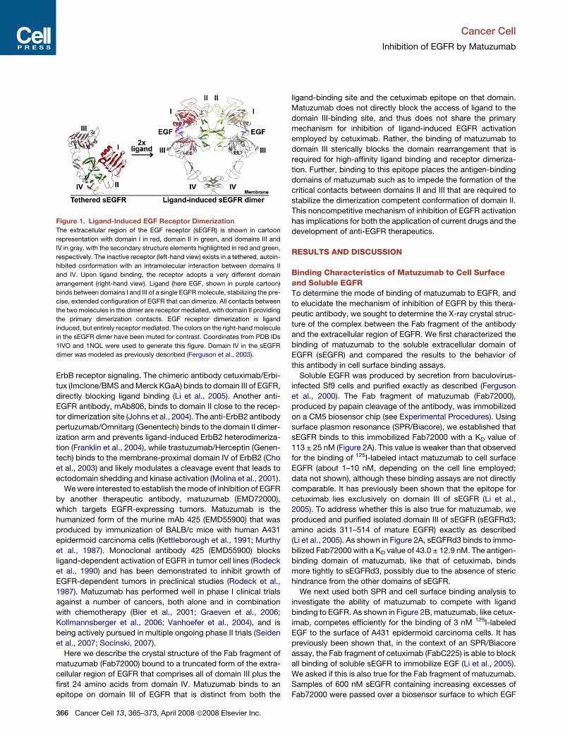
The extracellular region of the EGF receptor (sEGFR) is shown in cartoon representation with domain I in red, domain II in green and domains III and IV in gray with the secondary structure elements highlighted in red and green, respectively. The inactive receptor (left hand view) exists in a tethered, autoinhibited conformation with an intramolecular interaction between the domains II and IV. Upon ligand binding the receptor adopts a very different domain arrangement (right hand view). Ligand (here EGF, shown in purple cartoon) binds between domains I and III of a single EGFR molecule, stabilizing the precise, extended configuration of EGFR that can dimerize. All contacts between the two molecules in the dimer are receptor mediated with domain II providing the primary dimerization contacts. EGF receptor dimerization is ligand induced, but entirely receptor mediated. The colors on the right hand molecule in the sEGFR dimer have been muted for contrast. Coordinates from PDB IDs 1IVO and 1NQL were used to generate this figure. Domain IV in the sEGFR dimer was modeled as previously described (Ferguson et al., 2003).
EGFR – Introduction
40
In a dimer of the EGFR extracellular domains, all intermolecular interactions are
contributed by the receptor (Ogiso et al., 2002; Garrett et al., 2002). This entirely receptor
mediated dimerization is unique among the RTKs with known ligand-bound structures. In all
other ligand RTK complex structures the ligand is located in between the two monomers
mediating the dimerization (see 0).
The majority of interactions in the dimer of the EGFR extracellular domains (sEGFR) is
contributed by domain II. A ‘dimerization arm’ (Ogiso et al., 2002) protrudes into the dimer
interface directly contacting the other receptor monomer. However, it was shown through
mutation and deletion studies that simply exposing the dimerization arm is not sufficient to
promote sEGFR dimerization in the absence of ligand (Elleman et al., 2001; Ferguson et al.,
2003). Additional conformational changes induced by the ligand are required to stabilize the
precise conformation of domain II (Dawson et al., 2005; Lemmon, 2009). Further interactions
in the sEGFR dimer are contributed by parts of domain IV that are close to or contacting each
other as suggested by modeled structures (Ferguson et al., 2003) and biochemical and
biophysical data (Berezov et al., 2002; Dawson et al., 2007; Mi et al., 2008).
In the unliganded state the receptor adopts a very different conformation that occludes
much of the domain II dimerization interface in an intramolecular interaction or tether with
domain IV (Cho and Leahy, 2002; Ferguson et al., 2003) (left hand in Fig. 6). This
conformation is thought to be autoinhibited (Burgess et al., 2003). Upon ligand binding both
the domain I and domain III are contacting the ligand, which exposes the domain II and
domain IV dimerization interface. Thus, promoted by ligand binding the extracellular region
of EGFR must undergo a dramatic domain rearrangement to be able to dimerize.
5.1.2. Structures of ErbB receptor family extracellular domains
Interestingly, the EGFR family includes an orphan receptor that nonetheless shows
tyrosine kinase activity (ErbB2) and an NRG binding receptor (ErbB3) that lacks tyrosine
kinase activity (Burgess et al., 2003). As shown in Fig. 7, the unliganded extracellular regions
of EGFR (Ferguson et al., 2003), ErbB3 (Cho and Leahy, 2002) and ErbB4 (Bouyain et al.,
2005) adopt the tethered, autoinhibited conformation. Based on solution scattering studies,
binding of neuregulins is thought to promote a similar structural reorganization of the receptor
as seen for EGF (Dawson et al., 2007). In contrast, structures of the ErbB2 extracellular
domain (Cho et al., 2003; Garrett et al., 2003; Franklin et al., 2004) revealed a conformation
that is similar to the extended, dimerization competent receptor form. ErbB2 has no known
ligand (Citri et al., 2003). Nevertheless this receptor is able to transform cells just by
EGFR – Introduction
41
overexpression (Di Fiore et al., 1987). It was shown that these impaired receptors are
signaling at the cell surface through heterodimerization (Wada et al., 1990).
Fig. 7: ErbB family extracellular domain structures without ligand
The structures of the extracellular domains of each ErbB receptor family member in the absence of ligand are shown in cartoon presentation. The coloring is the same as in Fig. 6. EGFR, ErbB3 and ErbB4 adopt the autoinhibited conformation with an intramolecular tether between domain II and IV. ErbB2 in contrast adopts an extended conformation that resembles the ligand-induced dimerization-competent form described in Fig. 6 (structures from Lemmon, 2009).
5.1.3. ErbB receptor dimerization at the cell surface
The model of ligand-induced dimerization (Fig. 6) is in accordance with results for EGFR
and ErbB4 receptor homodimerization both at the cell surface and in solution (Lemmon,
2009). However, it fails to answer all questions about ErbB receptor heterodimerization at the
cell surface. It is e.g. not clear, why ErbB2 forms heterodimers with all other EGF receptor
family members at the cell surface (Graus-Porta et al., 1997; Berger et al., 2004; Wehrman et
al., 2006), while it remains monomeric in solution (Horan et al., 1995; Ferguson et al., 2000).
Furthermore, the dimerization model can not explain results from EGF binding studies at
the cell surface. Scatchard plots showing a characteristic curvilinear (concave-up) form and
ligand competition assays at the cell surface indicate heterogenic ligand binding sites and a
negative cooperativity of EGF receptor binding (Shoyab et al., 1979; Magun et al., 1980;
Macdonald and Pike, 2008). These findings resulted in the proposal of two different receptor
affinity classes at the cell surface, with 2%–5% of receptors binding EGF with high affinity
(KD < 0.1 nM) and 92%–95% binding with lower affinity (KD 6–12 nM) (Hunter et al., 1984;
Livneh et al., 1986; Defize et al., 1989; Ullrich and Schlessinger, 1990; Bellot et al., 1990;
Burgess et al., 2003). However, the two states can not just be equalized with the tethered and
extended conformations of the extracellular domains (Fig. 6). Such a model would lead to
positive cooperativity and concave-down Scatchard plots (Wofsy et al., 1992; Lemmon et al.,
1997; Özcan et al., 2006). Negative cooperativity requires that the binding of a second EGF to
a dimer plus one EGF would need to have a substantially lower affinity than the first EGF
EGFR – Introduction
42
binding event. At the cell surface the receptor dimer with a single EGF bound would be the
major species at subsaturating ligand concentrations. This was indeed seen in studies with
excess ligand binding to high affinity EGFR and interestingly also for the insulin receptor
(Wofsy et al., 1992; Lemmon, 2009). To further complicate interpretation, there is evidence
for EGF binding to higher oligomeric EGFR states beside the dimer (Pråhl et al., 1991; De
Meyts, 1994; Macdonald and Pike, 2008; De Meyts, 2008).
These results imply that additional factors beside the extracellular receptor domains that
are responsible for negative cooperativity and heterodimerization at the cell surface (Clayton
et al., 2005; Saffarian et al., 2007). It was shown that the transmembrane domains (Holbrook
et al., 2000; Domagala et al., 2000; Klein et al., 2004; Mayawala et al., 2005; Lemmon, 2009)
as well as the intracellular domains (Mendrola et al., 2002; Duneau et al., 2007) are triggering
dimerization and could be crucial for regulating the association of two ErbB receptors. Thus,
for a complete picture of ErbB receptor regulation it seems to be necessary to consider the
intact EGFR structure and to combine cellular and structural data. This is especially important
for the development of anti-cancer drugs that inhibit misregulated ErbB receptors.
5.1.4. EGFR and cancer
In the 1980s EGFR was the first cell-surface receptor to be linked directly to cancer as
described in fibroblasts infected with oncogenic viruses (De Larco and Todaro, 1987). It was
found that the neu oncogene encodes a protein related to EGFR (Schechter et al., 1984;
Coussens et al., 1985) and that the product of the v-erbB oncogene from avian
erythroblastoma virus is a truncated form of EGFR (Downward et al., 1984). These findings
revolutionized both the field of growth factors and of cancer biology. It is now known that
EGFR is aberrantly activated in a variety of epithelial tumors e.g. metastatic non-small-cell
lung cancer, colorectal cancer, squamous-cell carcinoma of head and neck and pancreatic
cancer (Mendelsohn and Baselga, 2006). ErbB2/HER2 overexpression is connected to breast
cancer (Park et al., 2008). Malignant transformation of the cell in these cancers can be caused
through EGFR overexpression or mutation, which leads to constitutive activity or impaired
receptor down-regulation (Mendelsohn and Baselga, 2006). Anti-EGFR agents are now
approved since the late 1990s in the therapy of non-small cell lung cancer (NSCLC), colon
cancer and head and neck cancer, pancreatic cancer and breast cancer, in which they provide
significant clinical benefit (Baselga, 2008). The next step in targeted therapy will be the
development of predictive markers of response to anti-EGFR agents to identify suitable
EGFR – Introduction
43
patients that will benefit from the treatment. Such markers are downstream effector proteins
of the signaling cascade, e.g. Ras and PTEN (phosphatase and tensin homologue) (Nagata et
al., 2004; Khambata-Ford et al., 2007; Benvenuti et al., 2007). Increased response rates might
be achieved by combination of inhibitors against several members of the same signaling
pathway (Zhang et al., 2007; Baselga, 2008) (see also 3.4).
The ErbB regulation mechanism (see 5.1.1 and 5.1.3) suggests a number of ways to
inhibit EGFR activation (Baselga, 2002). Intracellularly the kinase domain can be blocked
with low molecular weight ATP-competitive tyrosine kinase inhibitors (TKIs), e.g. gefitinib
(Iressa®), erlotinib (Tarceva®) or lapatinib (Tykerp®) (Zhang et al., 2007). Gefitinib
(AstraZeneca) was approved in 2003 by the US American Food and Drug administration
(FDA) for NSCLC; erlotinib (OSI Pharmaceuticals) was approved in 2004 for NSCLC and
pancreatic cancer. In 2005 gefitinib failed to show an advantage for patients with NSCLC and
was withdrawn from the market (Singer, 2005). After retrospectively studying lung cancer
samples from patients enrolled in that studies (Shepherd et al., 2005; Thatcher et al., 2005) it
became clear that a part of these patients did not express EGFR at high levels and thus was
less gefitinib sensitive from the beginning (Hirsch et al., 2007). This highlights the necessity
to develop bio-markers in order to identify patients that will benefit from a targeted therapy.
Lapatinib (GlaxoSmithKline), a dual EGFR and ErbB2 inhibitor, was approved in 2007 for
HER2 overexpressing breast cancer.
From the extracellular side ErbB family members can be targeted in cancer therapy by
monoclonal antibodies as described in the next section.
5.1.5. Anti-EGFR antibodies
The first study with monoclonal antibodies (mAbs) directed against the rat ErbB2
extracellular region were carried out in the early 1980s and found that some mAbs are able to
reverse the transformed phenotype of HER2 overexpressing cells (Drebin et al., 1985). Based
on this defining study several mAbs to the human extracellular domains of EGFR and ErbB2
were generated with varying effects on the receptor regulation (Hudziak et al., 1989; Lewis et
al., 1993). Some induced receptor aggregation thus mimicking ligand activation (Schreiber et
al., 1981; Schreiber et al., 1983), while others blocked receptor activation and showed the
desired antiproliferative effects (Kawamoto et al., 1983; Sato et al., 1983; Masui et al., 1984;
Gill et al., 1984). X-ray crystallographic and biochemical analysis of receptor-antibody
complexes have indicated several modes of binding that lead to effective inhibition of ErbB
receptor signaling: direct steric blockage of ligand binding or receptor dimerization,
EGFR – Introduction
44
stabilization of the tethered conformation, block of the domain rearrangement required for
receptor dimerization, antibody-dependent cellular cytotoxicity (ADCC) and complement-
dependent cytotoxicity (CDC), antibody-mediated receptor down-regulation and
augmentation of the antitumor effects of chemo- and radiotherapy (Mendelsohn and Baselga,
2006; Leahy, 2008; Schmitz and Ferguson, 2009). Improved efficacy of mAbs in cancer
therapy might be achieved by arming the antibodies with radionuclides or toxins (Carter,
2001).
Examples of anti-ErbB receptor antibodies already approved or in clinical trials are listed
below.
Cetuximab/Erbitux®. The chimeric antibody cetuximab/Erbitux® (ImClone/BMS and
Merck KGaA) binds to domain III of EGFR, directly blocking ligand binding (Li et al., 2005).
Cetuximab was approved by the FDA in 2004 for the treatment of patients with colorectal and
head and neck cancer. Clinical trials for cetuximab as a first line treatment are in progress
(Bokemeyer et al., 2009; Han et al., 2009).
Panitumumab/Vectibix®. The antibody panitumumab/Vectibix® (Amgen) was developed
from transgenic mice that express fully human antibodies and also binds to EGFR domain III
(Yang et al., 2001). Probably it employs a similar ligand binding competition mechanism as
cetuximab. As an antibody of the subtype IgG2 it does not stimulate robust antibody
dependent ADCC (Schmitz and Ferguson, 2009). In 2006 it was FDA approved for colorectal
cancer in combination with chemotherapy and is currently under investigation for first line
treatment in colorecetal cancer (Stephenson et al., 2008). Recently the addition of
panitumumab to the anti-angiogenesis mAb bevacizumab and chemotherapy for the first-line
treatment of metastatic colorectal cancer was found to be harmful when compared with
bevacizumab and chemotherapy alone (Giusti et al., 2009). Evaluation of this result is still
ongoing.
IMC-11F8. The fully human anti-EGFR antibody 11F8 (ImClone) binds to the same
epitope on EGFR domain III as cetuximab competing with ligand binding (Li et al., 2008). It
has performed well in phase I and is currently investigated in phase II clinical trials.
MAb806, another anti-EGFR antibody, binds to domain II close to the receptor
dimerization site (Johns et al., 2004). It was generated using cells expressing EGFR variant III
(EGFRvIII, see section 1) as antigen, but also binds to overexpressed wild-type EGFR
(Jungbluth et al., 2003). The antibody has performed well in a phase I study (Scott et al.,
2007).
EGFR – Introduction
45
Pertuzumab/ Omnitarg® (Genentech) binds to the domain II dimerization arm of ErbB2
and directly blocks ligand induced ErbB2 heterodimerization (Franklin et al., 2004). It is a
recombinant, humanized mAb and after a phase II study treating breast cancer patients in
combination with trastuzumab (Portera et al., 2008) it is currently investigated in a phase III
clinical trial (Baselga, 2008).
Trastuzumab/Herceptin® (Genentech) binds to the membrane proximal domain IV of
ErbB2 (Cho et al., 2003) and likely modulates a cleavage event that leads to ectodomain
shedding and kinase activation (Molina et al., 2001). Trastuzumab was FDA approved in
1998 for use in combination with first line chemotherapeutic agents in patients with metastatic
breast cancer expressing high levels of ErbB2 (Slamon et al., 2001).
The antibodies with structurally known epitopes are summarized in Fig. 8.
Fig. 8: Antibody receptor co-structures
Cartoon presentation of receptor antibody Fab fragment co-structures. The coloring is the same as in Fig. 6 with the receptor ectodomain in red (domain I) and grey/red (domain III) and green (domain II) and grey/green (domain IV). The antibody Fab fragments of cetuximab/Erbitux® is in ocher/orange (Li et al., 2005), of 11F8 in turquoise/violet (Li et al., 2008), of trastuzumab/Herceptin® in light violet/yellow (Cho et al., 2003) and of pertuzumab/Omnitarg® in red/blue (Franklin et al., 2004). The first two antibodies are directed against EGFR, while the latter two are targeted against ErbB2.
The mode of action of another therapeutic antibody, matuzumab (EMD72000), which
targets EGFR expressing tumors, is investigated in this thesis. Matuzumab is the humanized
form of the murine mAb 425 (EMD55900) that was produced by immunization of BALB/c
mice with human A431 epidermoid carcinoma cells (Murthy et al., 1987; Kettleborough et
al., 1991). Matuzumab has performed well in phase I clinical trials against a number of
cancers, both alone and in combination with chemotherapy (Bier et al., 2001; Vanhoefer et
al., 2004; Graeven et al., 2006; Kollmannsberger et al., 2006; Rao et al., 2008), and has
reached phase II trials (Seiden et al., 2007; Socinski, 2007).
EGFR - Results
46
5.2. Results
5.2.1. Matuzumab binding to sEGFR
Surface plasmon resonance (SPR)/Biacore experiments were carried out to characterize
the binding of Fab72000 (see 4.3.3) to the soluble extracellular domain of the EGF receptor
(sEGFR) and the isolated receptor domain III (sEGFRd3) (see 4.3.1). The apparent KD values
obtained were 113 ± 25 nM for sEGFR and 43 ± 13 nM for sEGFRd3 (Fig. 9).
Fig. 9: Characterization of matuzumab binding to sEGFR
Surface plasmon resonance (SPR)/Biacore analysis of the binding of sEGFR and sEGFRd3 to immobilized Fab72000. A series of samples of sEGFR or sEGFRd3, at the indicated concentrations, was passed over a biosensor surface to which Fab72000 had been amine coupled. Data points show the equilibrium SPR response value for a representative set of samples of sEGFR (black squares) and of sEGFRd3 (open triangles), expressed as a percentage of the maximal SPR binding response. The curves represent the fit of these data to a simple one-site Langmuir binding equation. KD values, based on at least three independent binding experiments, are 113 ± 25 nM for sEGFR and 43 ± 13 nM for sEGFRd3.
The affinity measurement was verified with an ITC experiment, which gave a KD value of
2.1 ± 0.5 nM for the interaction of Fab72000 with sEGFR (Fig. 10).
EGFR – Results
47
Fig. 10: ITC sEGFR and Fab72000
Fab72000 (20 µM) was injected in 10 µl steps into a cell containing 2 µM sEGFR at 25°C. Each peak represents the heat of binding following one injection (upper plot). The lower plot shows the integrated results, where each point represents the normalized heat change for each injection. The calculated KD for this interaction is 2.1 ± 0.5 nM.
5.2.2. Ligand competition analysis of matuzumab
Competition assays were carried out to investigate the ability of matuzumab to compete
with ligand binding to EGFR. As shown in Fig. 11, there is an initial decrease in the
equilibrium SPR response as increasing Fab72000 is added. At a 1:1 molar ratio of
Fab72000:sEGFR the SPR response is about 45 % of that obtained with no added Fab.
Addition of increasing excesses of Fab72000 does not further reduce this binding level. Even
at higher concentration of sEGFR and with up to a 50 fold excess of Fab72000 the
equilibrium SPR response does not fall below 40 % of the value in the absence of added Fab.
EGFR – Results
48
Equilibrium binding analysis to immobilized EGF for samples of sEGFR containing a 10
fold molar excess of Fab72000 indicates an apparent KD value that is approximately five fold
weaker than for sEGFR alone (see Appendix 11.3 Fig. 44).
Fig. 11: Ligand competition properties of matuzumab
A: A competition experiment showing the effect of addition of Fab72000 upon the binding of 600 nM sEGFR to immobilized EGF. Mixtures of 600 nM sEGFR plus the indicated concentrations of Fab72000 were passed over a biosensor surface to which EGF had been amine coupled. The equilibrium SPR responses for each mixture is shown, normalized to the response obtained with no added Fab. Error bars indicate the standard deviation on at least three independent measurements. The line simply connects the data points. B: The ability of FabC225 (the antigen binding domain of cetuximab; gray shades) and Fab72000 (red shades) to compete for the binding of 600 nM sEGFR to immobilized EGF, determined exactly as described in A. Samples of each Fab alone show no binding to the immobilized EGF (data not shown). Data for FabC225 are taken from Li et al., 2005. Error bars indicate the standard deviation on at least three independent measurements.
Analytical ultracentrifugation sedimentation equilibrium experiments (SE) were carried
out to determine the oligomeric state of sEGFR:Fab72000 (see 4.3.4) in the presence of
excess ligand. The control samples show a doubling of the molecular weight for sEGFR in
presence of EGF. However, only a slight increase in molecular weight is observed for the
complex sEGFR:Fab72000 with added ligand (Fig. 12).
EGFR – Results
49
Fig. 12: Does the sEGFR:Fab72000 complex dimerize? Analysis by AUC
Analytical ultracentrifugation sedimentation equilibrium data of the oligomeric state of sEGFR:Fab72000 in the presence of EGF. Depicted are the theoretical molecular weights by sequence of the EGF receptor, the complex sEGFR:Fab72000 and the mutated receptor sEGFR Y251A/R285S:Fab72000 (sEGFRmut:Fab72000) (yellow bars), each alone and in presence of excess EGF. The experimentally determined molecular weights of the respective molecules are shown as green bars. The data represent a fit of 4 µM samples measured in 20 mM HEPES, 100 mM NaCl (pH 7.5). Equilibrium data after 18 h were obtained at three different speeds 6,000 rpm, 9,000 rpm and 12,000 rpm at 20°C.
The same slight increase in the molecular weight is seen for the dimerization incompetent
receptor:antibody complex sEGFR Y251A/R285S:Fab72000 in presence of EGF. This protein
has two mutations in the dimerization arm of domain II (Y251A/R285S) that lead to the loss
of an inter-receptor hydrogen bond. The mutated receptor binds ligand, albeit with lower
affinity, but does not dimerize (Dawson et al., 2005). It is not known why the mutated
receptor shows a higher molecular weight than the wild type receptor, but the same was
observed in former experiments (personal communication Dr. Dawson).
5.2.4. The matuzumab epitope
The crystal structure of EGFRd3 in complex with Fab72000 (see 4.5.3) reveals that
Fab72000 binds primarily to the loop that precedes the most C-terminal strand of the domain
III β-helix (amino acids 454-464; highlighted in red in Fig. 13). This loop penetrates into a
cleft between the VL and VH domains of the Fab. The tip of this loop forms a type I beta turn,
with T459 and S460 in this turn protruding the farthest into the cleft. All of the key
interactions made by the Fab are from the complementarity determining regions (CDRs), with
the major specificity determining contacts coming from CDRs H3 and L3. All CDRs
contribute to binding to domain III.
EGFR – Results
50
Fig. 13: Structure of the complex between the matuzumab Fab fragment and domain III of sEGFR
Cartoon of the sEGFRd3:Fab72000 complex. Domain III is colored in gray with the epitope highlighted in red. The orientation of domain III is the same as for the tethered sEGFR (left hand view) in Fig. 6. Fab72000 is colored cyan for the light chain and yellow for the heavy chain.
The tip of the buried loop from sEGFR makes interactions with both the heavy and light
chain CDRs (Fig. 14); the side chain of T459 interacts with that of H93 from the Fab light
chain, while the side chain of S460 contacts the CDR H2 side chain E50. Two lysines, one on
either end of the sEGFRd3 epitope loop, form salt bridge interactions with aspartic acids on
the Fab (K454 with D100 from CDR H3 and K463 with CDR L2 D49).
Fig. 14: The epitope of matuzumab in detail
A closeup view of the interactions between Fab72000 and domain III of sEGFR. Domain III is in gray with the secondary structure elements highlighted in red. The VL and VH domains of Fab72000 are in gray with cyan and yellow highlights, respectively. The CDRs of Fab72000 are shown in cyan for L1, L2 and L3 of the VL domain, and in yellow for H1, H2 and H3 of the VH domain. The side chains of the amino acids participating in key interactions are shown, colored as for the CDRs for the Fab and in pink for domain III. The amino acids are labeled on a cyan background for those from VL, on a yellow background for VH and in black for sEGFRd3. Distances corresponding to hydrogen bonds are shown with dashed black lines.
EGFR – Results
51
Additional interactions with the buried epitope loop are contributed by side chains in
CDRs H1, H2 and L1 that are within hydrogen bonding distance of the main chain of
sEGFRd3 (Fig. 14 and Fig. 15). Two important direct interactions are made between the Fab
and regions of domain III outside the loop between amino acids 454-464. A histidine from
CDR L3 (H93) interacts with D434 on the adjacent loop of the sEGFRd3 β-helix, while on
the other side of the binding site Y103 from the apex of CDR H3 extends to interact with
N449. These two interactions anchor the Fab over the central binding loop and expand the
epitope substantially beyond the single peptide loop.
Fig. 15: Electron density at the sEGFRd3:Fab72000 interface
Stereo representation of selected interactions between sEGFR domain III and Fab72000. Amino acids are shown in stick representation and colored pink for domain III, yellow and cyan for Fab72000 VH and VL respectively. The gray mesh represents the final 2Fo-Fc electron density map contoured at 1.0 σ. Distances consistent with hydrogen bonds are shown in dashed black lines.
A total of 2 salt bridges and 11 predicted hydrogen bonds are involved in the interaction
between Fab72000 and sEGFRd3, in an interface that buries 758 Å2 of solvent accessible
surface on domain III (in the complex a total of 1516 Å2 of surface is occluded from solvent).
The shape complementarity (sc) parameter for the interface of the sEGFRd3: Fab72000
complex is 0.62.
Neither the conformation of sEGFRd3 nor of Fab72000 is significantly altered upon
formation of the complex. There are very minor differences in the side chain positions in both
the domain III epitope and in the CDRs of the Fab. Most notably Y103 in the VH domain is
disordered in the unbound Fab and becomes ordered on interacting with sEGFR. The elbow
angle changes by 4° between the bound and unbound Fab72000.
EGFR – Results
52
5.2.5. The matuzumab epitope is distinct from the ligand binding site on
domain III of sEGFR
Based on the crystal structures sEGFRd3:Fab72000 (see 4.5.3), sEGFR:FabC225 (PDB
ID 1YY9) and sEGFR:EGF (PDB ID 1NQL) the epitopes of matuzumab and EGF were
mapped onto sEGFR domain III to investigate the spatial arrangement of their binding sites
(Fig. 16A). The same was done with the epitopes of cetuximab and EGF (Fig. 16B). While
the binding sites of cetuximab and the ligand clearly overlap, the epitopes of matuzumab and
EGF show no overlap.
Fig. 16: The matuzumab epitope is distinct from the ligand binding site on domain III of sEGFR
A: surface representation of domain III is shown in gray viewed in approximately the same orientation as in Fig. 13. On the left hand side the amino acids on domain III that are within 4 Å of Fab72000 (red) or of EGF (green) are indicated on this surface. The amino acids that were mutated (Fig. 17) are labeled in white. B: the same surface representation of domain III is shown with the contacting amino acids for FabC225 in yellow, for EGF in green and for the region of overlap between FabC225 and EGF in blue.
To confirm that the crystallographically defined epitope for matuzumab precisely
represents what is seen in solution, site specific alterations in sEGFR at key amino acids in the
domain III matuzumab epitope were generated (Fig. 14) (see 4.1.1). Each purified, altered
sEGFR (see 4.3.1) was analyzed for binding to immobilized Fab72000 and to immobilized
EGF. Alteration to alanine of either of the two lysines on the epitope loop (K454A or K463A)
leads to an approximate 100-fold reduction in the affinity of sEGFR for Fab72000 (Fig. 17).
Substitution of alanines at T459 and S460 (T459A/T460A) also dramatically reduces the
binding affinity. The combination of either lysine to alanine substitution with T459A/T460A
abolishes all detectable interaction between sEGFR and the immobilized Fab72000.
EGFR – Results
53
Fig. 17: Effects of sEGFR mutant binding to matuzumab or EGF
Surface plasmon resonance (SPR)/Biacore analysis of the binding of altered sEGFR proteins to immobilized Fab72000 or EGF. The equilibrium binding KD values for each protein were determined exactly as described in Fig. 9. The fold change in this KD value for each altered protein relative to that for the binding of wild type sEGFR to each immobilized ligand is plotted. Error bars indicate the standard deviation on at least three independent sets of measurements.
sEGFR proteins with alterations in the Fab72000 epitope bind to immobilized EGF with
near wild type affinity (Fig. 17). However, substitution of two amino acids that are known to
be critical for EGF binding (D355T/F357A) have negligible effect on binding of sEGFR to
Fab72000.
EGFR - Discussion
54
5.3. Discussion
5.3.1. Matuzumab binding characteristics to soluble and cell surface
EGFR
The KD value of 113 ± 25 nM for Fab matuzumab binding to sEGFR obtained by surface
plasmon resonance (SPR)/Biacore studies (see 5.2.1) is weaker than observed for the binding
of 125I-labeled intact matuzumab to cell surface EGFR with about 1-10 nM, depending on the
cell line employed (Schmiedel et al., 2008). The value obtained by ITC with 2.1 ± 0.5 nM
corresponds better to the cellular data. However, binding assays with immobilized soluble
receptor and with cell surface receptor are not directly comparable. The isolated domain III
of sEGFR (sEGFRd3) binds to immobilized Fab72000 with a KD value of 43.0 ± 12.9 nM.
The antigen binding domain of matuzumab, like the ones of the antibodies cetuximab and
11F8 (Li et al., 2005; Li et al., 2008), binds more tightly to sEGFRd3, possibly due to the
absence of steric hindrance from the other domains of sEGFR.
It is possible that the immobilization of the Fab72000 could affect the measured affinity in
the Biacore experiments. However, essentially the same affinity for sEGFR binding to
immobilized (amine coupled or bound via immobilized protein A) mAb matuzumab was
obtained (see Appendix 11.3 Fig. 45). The absolute value of the affinity of sEGFR for
immobilized Fab72000 obtained from the Biacore experiment is of less significance. This
assay is however relevant in comparing the binding of sEGFRd3 and of the sEGFR proteins
with alteration in the Fab72000 epitope to this same Biacore surface (see 5.3.3).
It has previously been shown that, in the context of an SPR/Biacore assay, the Fab
fragment of cetuximab (FabC225) is able to block all binding of soluble sEGFR to
immobilized EGF (Li et al., 2005). Therefore the ability of matuzumab to compete with
ligand binding to sEGFR was investigated by SPR. As shown in Fig. 11, matuzumab, in
contrast to cetuximab, is not able to completely block the binding of the soluble receptor to
immobilized EGF. Instead the equilibrium SPR response plateaus at 40% of the value in the
absence of added Fab. One possible explanation for the observed SPR responses in Fig. 11 is
that both unbound sEGFR and the sEGFR:Fab72000 complex can interact with the
immobilized EGF, but that the complex binds with substantially weaker affinity. Equilibrium
binding analysis to immobilized EGF for samples of sEGFR containing a 10 fold molar
excess of Fab72000 indicates an apparent KD value that is approximately five fold weaker
than for sEGFR alone.
EGFR - Discussion
55
Still, matuzumab, like cetuximab, competes efficiently for the binding of 3 nM 125I-
labeled EGF to the surface of A431 epidermoid carcinoma cells (Schmiedel et al., 2008).
Taken together, these data suggest that there must be something quite different about the
mode of binding to sEGFR of the Fab fragment of matuzumab compared to that of cetuximab.
Both antibodies are able to compete for binding of low concentrations of EGF to cell surface
EGFR, yet the Fab fragments from the two antibodies have very different effects on the
ability of soluble EGFR to bind to immobilized EGF in the Biacore assay. This apparent
discrepance can be explained after examining the complex structure of the matuzumab Fab
fragment and sEGFRd3 (see 5.3.5).
5.3.2. The matuzumab epitope on sEGFR domain III
The epitope of matuzumab on sEGFR domain III consists of a single loop of 10 amino
acids with two additional interactions probably stabilizing the antibody receptor interaction
(Fig. 13). Kamat et al. identified through mutagenesis experiments the residues S460/G461 to
be essential for binding of the murine progenitor of matuzumab 425 (Kamat et al., 2008).
These residues comprise exactly the tip of the epitope loop as seen in the crystal structure.
This mode of binding of an antibody to a large protein antigen is unusual. It is more
common for the epitope on a large protein antigen to comprise a large flat surface (Sundberg
and Mariuzza, 2002), as was observed for the binding of cetuximab and 11F8 to EGFR (Li et
al., 2005; Li et al., 2008). The fact that all complementarity determining regions (CDRs) of
matuzumab contribute to the binding to sEGFRd3 is also an unusual feature compared to most
antigen-antibody complexes (Sundberg and Mariuzza, 2002).
The shape complementarity parameter for the interface of the sEGFRd3:Fab72000
complex of 0.62 is slightly lower than is typically observed for antigen-antibody interfaces
(0.64 to 0.68) (Lawrence and Colman, 1993). The sc values reported for cetuximab and 11F8
bound to EGFR (Li et al., 2005; Li et al., 2008) and for the pertuzumab and trastuzumab
complexes with the extracellular region ErbB2 (Cho et al., 2003; Franklin et al., 2004) are all
somewhat higher, in the range from 0.69 to 0.75. This perhaps reflects the more convex shape
of the matuzumab epitope compared to those of these other antibody drugs.
The elbow angle change of only 4° between the bound and unbound Fab72000 is in within
the range expected due to dynamic elbow flexibility (Stanfield et al., 2006) and thus probably
not induced by the binding event.
EGFR - Discussion
56
Neither the conformation of the domain III and nor that of Fab72000 is significantly
altered upon complex formation. Additionally, the binding of Fab72000 would not be
expected to disrupt the tethered configuration of sEGFR (Fig. 6, left panel), the preferred
solution conformation of the receptor (Dawson et al., 2007) and the likely conformation of the
unliganded receptor at the cell surface. Fab72000 can readily be docked onto its epitope on
either of the two known structures of tethered sEGFR (PDB IDs 1NQL and 1YY9) without
hindrance from any of the other domains of sEGFR.
5.3.3. Matuzumab and ligand epitopes do not overlap on sEGFR domain
III
SPR/Biacore experiments showed a dramatically reduced affinity of Fab72000 to sEGFR
carrying mutations in the Fab72000 epitope in comparison to wild type receptor (Fig. 17).
However, the affinity of EGF to the mutated receptor is only slightly reduced in comparison
to the wild type. This confirms that the striking reduction in binding affinity for Fab72000 is
not due to a global disruption of the structure of domain III of sEGFR. The reverse effect is
seen for receptor protein carrying mutations in the ligand binding site: the affinity of EGF is
reduced while the binding to Fab72000 remains unaffected. These results confirm that the
epitopes of the ligand and matuzumab are distinct as seen in 5.2.5. Indeed, not only is there no
overlap of the epitope for matuzumab and the ligand binding region on domain III (Fig. 16A),
but a bound Fab72000 would impose no steric hindrance to the binding of EGF or of TGF-α
to domain III. With domain III from the sEGFRd3:Fab72000 complex overlaid on domain III
from the sEGFR:EGF complex (PDB ID 1IVO) the closest approach of the Fab and EGF is 9
Å. This is in stark contrast to the situation for cetuximab binding. There is a high degree of
overlap between the cetuximab and EGF binding sites on domain III (Fig. 16B). The steric
block of this ligand binding site is the primary mechanism of cetuximab mediated inhibition
of ligand induced dimerization and activation of EGFR (Li et al., 2005). Clearly the
mechanism of inhibition of EGFR activation by matuzumab must be different.
EGFR - Discussion
57
5.3.4. The matuzumab inhibition mechanism
If matuzumab does not directly block access of the ligand to the domain III ligand binding
site, how does it prevent high affinity ligand binding, receptor dimerization and activation as
seen in ultracentrifugation studies (Fig. 12)? To understand this, the effect of the binding of
Fab72000 upon the formation of the ligand induced dimeric form of the receptor is
considered.
As shown in Fig. 6, sEGFR undergoes a dramatic domain rearrangement in going from
the tethered inactive state to the ligand bound dimeric state (Burgess et al., 2003). Additional
local structural changes in domain II are known to be key for high affinity ligand binding,
receptor dimerization and activation (Ogiso et al., 2002; Dawson et al., 2005). As shown in
Fig. 18 and discussed in detail below, when domain III from the sEGFRd3:Fab72000
complex is overlaid on domain III from the receptor in its extended, dimerization competent
conformation (PDB ID 1MOX), there are direct clashes between the bound Fab72000 and
both domains I and II of the extended receptor. Thus, with matuzumab bound to domain III of
EGFR, the receptor cannot undergo the large scale domain rearrangement that is required for
dimerization. Further, the binding of Fab72000 blocks the critical local conformational
changes in domain II.
EGFR - Discussion
58
Fig. 18: Implications for the mechanism of inhibition of EGFR by matuzumab
A. Cartoon of the extended sEGFR with Fab72000, in surface representation, docked onto its domain III epitope. The orientation of the receptor is the same as for the right hand protomer in the sEGFR dimer shown in Fig. 6 (with domains colored as for the left hand protomer; EGF is omitted for clarity). Fab72000 is colored as in Fig. 13. The N-terminal region of domain I clashes with the VL domain (indicated with an arrow). Additional clashes occur along the C-terminal half of domain II (see part B). The C-terminal loop on domain II (D278, H280) that makes critical contacts across the dimer interface is marked with an asterisk. B. In this view, an approximate 50° rotation about the vertical axis relative to part A, domain II is shown in sphere representation in dark green. A cartoon of domain II of the other molecule in the dimer is shown (light green) for reference. Domain I has been omitted for clarity. The VL domain of the Fab clashes with domain II in the critical C-terminal region that forms the binding pocket for the dimerization arm and makes important contacts with domain III (from N274 and E293 in domain II, colored orange). These interactions are known to be crucial for stabilizing the dimerization competent conformation of domain II. The Fab72000 epitope loop on domain III is colored in red.
With the receptor in the extended conformation, the N-terminal region of the domain I
clashes with the light chain of Fab72000 preventing domain I from reaching the position that
is required for high affinity ligand binding (indicated with an arrow in Fig. 18). This is
reminiscent in nature and extent to clashes between the antigen-binding fragment of
cetuximab (FabC225) and domain I that were previously implicated as part of the mechanism
of inhibition of EGFR dimerization by that antibody (Li et al., 2005). In that case, the
different orientation of FabC225 on domain III positions th VH domain such as to occlude the
N-terminal portion of domain I from its required position in the receptor dimer.
Clashes between domain II of the extended receptor and the Fab were not seen in the
cetuximab complex, and are significant. With Fab72000 bound to domain III of EGFR it
would not be possible for the C-terminal portion of domain II to adopt the conformation
observed in the ligand bound dimeric form of the receptor. As shown in Fig. 18B, if Fab72000
is docked onto its epitope on domain III of an sEGFR molecule in the extended conformation,
there are clashes along the C-terminal half of domain II, predominantly with the VL domain of
EGFR - Discussion
59
the Fab. This C-terminal half of domain II forms the binding pocket for the dimerization arm
from the other molecule in the receptor dimer. Additional interactions across the dimer
interface from a C-terminal loop on domain II (D279 and H280, marked with an asterisk in
Fig. 18A) contribute substantially to the stability of the EGFR dimer. The conformation of
domain II in this region is stabilized by interactions with domain III that have been
demonstrated to be critical for EGFR dimerization and activation (Ogiso et al., 2002; Dawson
et al., 2005). The binding of Fab72000 to domain III would disrupt all of these interactions.
Thus, Fab72000 binding to domain III of EGFR blocks the global domain rearrangement
of EGFR and the local conformational changes in domain II. The blocking both of these key
elements in formation of the productive EGFR dimer is critical for the effective inhibition of
EGFR activation by matuzumab.
5.3.5. Matuzumab binding properties interpreted with structural
information
The steric restriction on EGFR conformation imposed by the binding of matuzumab
offers a structural framework to explain the competition data presented in Fig. 11. In binding
studies at the cell surface matuzumab and cetuximab are both efficiently competing with
EGF to receptor binding, while in SPR/Biacore assays the two antibodies show differing
competition characteristics. A major difference between the two competitions assays is the
concentrations of the soluble ligands that are used. In the cell based competition assay EGF
is present at 3 nM. This concentration is well below the KD value for the binding of EGF to
isolated domain III: between 500 nM (for the Kohda fragment) and 2 μM (for insect cell
expressed sEGFRd3) (Ogiso et al., 2002; Dawson et al., 2005). To observe binding of EGF
to cell surface EGFR under these conditions both domains I and III must be engaged to form
a high affinity EGF binding site. However, binding of matuzumab to EGFR prevents the
receptor from adopting the conformation required to form the high affinity ligand binding
site. Thus at this relatively low EGF concentration matuzumab blocks detectable binding of
EGF to the cell surface and thus competes as effectively for the binding of EGF as does
cetuximab. Any EGF that binds to the exposed ligand binding site on domain III of a
matuzumab occupied cell surface EGFR would be so weakly bound that it would be washed
out in this assay. By preventing the receptor from adopting the conformation required for the
bipartite binding of EGF between domains I and III, matuzumab blocks all detectable
binding of EGF to cell surface EGFR in this assay.
EGFR - Discussion
60
By contrast the Biacore assay is performed at a much higher concentration of soluble
ligand (in this case sEGFR, which binds to immobilized EGF). The soluble EGFR is passed
over this surface at 600 nM, a concentration that is close to the KD for the binding of isolated
domain III of EGFR to immobilized EGF. Under these conditions the monovalent binding of
domain III alone to EGF can be detected. There are probably two populations of sEGFR with
two different affinities. Unbound sEGFR has a higher affinity for EGF, while the Fab72000
bound sEGFR has a lower affinity. In the Biacore assay, the residual binding to immobilized
EGF observed for sEGFR in the presence of excess Fab72000 (Fig. 44) is due, at least in
part, to binding to EGF of the exposed domain III in an sEGFR:Fab72000 complex.
5.3.6. Implications for the therapeutic application of matuzumab
As discussed above, the mechanism of inhibition of matuzumab is different from that
previously described for cetuximab. Both antibodies effectively block productive binding of
EGF to cell surface EGFR, but do so by interacting with distinct epitopes on domain III. Not
only are the epitopes of matuzumab and cetuximab non-overlapping, but the structures
suggest that both matuzumab and cetuximab could simultaneously bind to EGFR. As shown
in Fig. 19, when FabC225 and Fab72000 are simultaneously docked onto their respective
epitopes on domain III the two Fab fragments occupy different positions and do not overlap.
EGFR - Discussion
61
Fig. 19: The matuzumab and cetuximab epitopes do not overlap
A surface representation of the domain III as in Fig. 16 is shown. Cartoons of Fab72000, FabC225 (PDB ID 1YY9) and EGF (PDB ID 1IVO) are shown docked onto their respective binding sites on domain III. Fab72000 is colored as in Fig. 13, FabC225 is shown with the heavy chain in orange and the light chain in light green, and EGF is in purple.
This observation was experimentally confirmed by surface plasmon resonance/Biacore,
size exclusion chromatography and analytical ultracentrifugation analysis investigating the
simultaneous binding properties of cetuximab and the murine progenitor of matuzumab 425 to
soluble and cell surface EGFR (Kamat et al., 2008). Cellular competition assays showed that
neither antibody competes with the binding of the other (Schmiedel et al., 2008; Kamat et al.,
2008). Further it has been reported that there is an increased number of cell surface antibody
binding sites for a mixture of matuzumab and cetuximab compared to either antibody alone
(Kreysch and Schmidt, 2004). This suggests that both matuzumab and cetuximab can bind to
a single receptor molecule at the cell surface.
Treatment of cells with combinations of antibodies against distinct epitopes on the
extracellular domain of EGFR, and on the related family member ErbB2, lead to enhanced
receptor internalization and degradation (Ye et al., 1999; Spiridon et al., 2002; Friedman et
al., 2005), a factor that contributes to the antitumor activity of many therapeutic antibodies
(Logtenberg, 2007). Matuzumab and cetuximab can both bind simultaneously to EGFR and
this has the potential to lead to synergistic antitumor effects.
EGFR - Discussion
62
Indeed, a combination of cetuximab and the murine progenitor of matuzumab 425 reduced
growth and survival of EGFR overexpressing breast cancer cells more effectively than either
antibody alone (Kamat et al., 2008). Furthermore, it was shown that combinations of
antibodies binding to different epitopes on EGFR trigger potent complement-dependent
cytotoxicity (CDC) (Dechant et al., 2008). The authors particularly emphasized the
combination of cetuximab and matuzumab for effectivity. Combination of matuzumab and
cetuximab could, thus, be beneficial in cancer therapy.
EGFR- Conclusion
63
5.4. Conclusion
EGFR dimerization requires a conformational reorganization of the receptor extracellular
region that is promoted by ligand binding to domain I and III (Fig. 6). As shown
schematically in Fig. 20, cetuximab acts as a competitive inhibitor, preventing ligand induced
dimerization by directly blocking access of ligand to the domain III ligand binding site.
Fig. 20: Matuzumab and cetuximab use different mechanisms to block ligand induced EGFR dimerization In the center of the scheme the ligand induced sEGFR dimer is represented, with domain I in red, domain II in green, domain III in gray with red border, domain IV in gray with green border and the ligand (E) in violet. The colors for one protomer are lightened for contrast. On the left hand side a scheme is shown to illustrate the mechanism of inhibition of ligand induced dimerization by matuzumab. Fab72000 binds to domain III of sEGFR and sterically prevents the receptor from adopting the conformation required for dimerization. Importantly, Fab72000 blocks the local conformational changes in domain II that are critical for both high affinity ligand binding and dimerization. The inhibition is non-competitive; the ligand binding site on domain III is not blocked. This contrasts with the mechanism of inhibition previously reported for cetuximab (Li et al., 2005). FabC225 (right hand side) is a competitive inhibitor that blocks the ligand binding site on domain III. This is the primary mechanism of inhibition of ligand mediated dimerization by cetuximab.
By contrast matuzumab does not occlude the ligand binding site on domain III. Rather
matuzumab exploits a non-competitive mechanism to inhibit sEGFR dimerization and
activation. Inhibition of ligand induced EGFR activation by matuzumab is entirely dependent
on sterically blocking the receptor from adopting the conformation that is required for high
affinity ligand binding and dimerization. These different mechanisms of inhibition suggest
opportunities to exploit multiple EGFR targeting drugs to act synergistically for optimal
therapeutic gain.
64
EGFRvIII - Introduction
65
6. Antibody binding and dimerization properties of the
mutant EGFR variant III ectodomain
6.1. Introduction
Beside cancer related mutations in the tyrosine kinase domain of the epidermal growth
factor receptor (EGFR), several mutations in the extracellular part of EGFR were described to
promote tumorigenesis (Lee et al., 2006). The type III EGFR mutation (EGFRvIII, de2-7
EGFR or ΔEGFR) is the most common one and clinically connected with enhanced tumor
aggresivity and chemoresistance (Nishikawa et al., 1994; Huang et al., 1997; Heimberger et
al., 2005; Weppler et al., 2007; Wang et al., 2009). The mutated receptor EGFRvIII is a
truncated version of the wild type EGFR showing constitutive signaling activity and impaired
down-regulation (Pedersen et al., 2001).
The presence of EGFRvIII was described for many different cancer types, among them
lung and prostate cancer as well as gliomas, where the mutation is found in up to 50% of
glioblastomas (Moscatello et al., 1995; Frederick et al., 2000; Cavenee, 2002). Expression of
EGFRvIII is a negative prognostic indicator in glioblastoma and mediates resistance to TKIs
targeted against EGFR (Learn et al., 2004).
6.1.1. EGFRvIII in-frame deletion
EGFRvIII contains a deletion of exons 2-7 of the wild tpye gene, resulting in the in-frame
loss of most of domain I and II including the dimerization arm (Pedersen et al., 2001). At the
fusion junction a novel glycine residue is generated. Thus, the protein consists of the residues
1-5 of domain I, the glycine residue and continues with residue 274 of domain II (Fig. 21).
EGFRvIII - Introduction
66
Fig. 21: Domain organization of EGFR and EGFRvIII in comparison
Human EGFR and EGFRvIII comprise an extracellular region consisting of domains I-IV, a transmembrane region and an intracellular tyrosine kinase domain. The truncated EGFRvIII consists of residues 1-5 of domain I of the wild type EGFR, a glycine residue at the fusion junction and continues with residue 274 of domain II. The sequence of domain III and IV are unaltered in mutant EGFRvIII in comparison to full length EGFR (figure adapted from Pedersen et al., 2001).
Based on the sequence the ligand binding site on domain III is unaltered as well as the
epitope of the monoclonal antibodies cetuximab (Li et al., 2005) and matuzumab (Schmiedel
et al., 2008). However, no ligand binding is observed through the mutated receptor on the cell
surface (Wikstrand et al., 1997). It is unclear if the mutant receptor is able to form
homodimers at the cell surface upon ligand stimulation since conflicting data were published
(Chu et al., 1997; Fernandes et al., 2001). Heterodimerization of the EGFRvIII with the wild
type EGFR was shown in murine BaF/3 cells and in human glioblastoma cells (O'Rourke et
al., 1998; Luwor et al., 2004). Structural or biophysical investigations of the isolated
ectodomain of EGFRvIII are not available so far.
EGFRvIII was so far only shown to be expressed on cancer cells and is therefore an ideal
target for anticancer therapy (Wikstrand et al., 1997; Kuan et al., 2000; Li and Wong, 2008).
It is unknown if untransfected cells in tumors are able to co-express both the receptor wild
type and EGFRvIII within the same cells or if a mixture of cells is present expressing either
one or the other receptor controversially discussed in literature (Nishikawa et al., 1994;
Aldape et al., 2004; Yang et al., 2008; Zhu et al., 2009).
EGFRvIII - Introduction
67
6.1.2. EGFRvIII signaling activity
In comparison to the wild type receptor, which signals via the phosphatidylinositol 3-
kinase (PI3K)/Akt, ras/raf/MEK/ERK, phospholipase C gamma (PLCγ), and signal transducer
and activator of transcription (STAT3) signaling pathways (Sebastian et al., 2006) (see 3.4),
EGFRvIII seems to activate different signaling pathways (Zhu et al., 2009).
The phosphorylation of the mutant was reported to be lower in comparison to the wild
type, but to be constitutive. Recently it was found that tyrosine residue 992 in human
EGFRvIII expressed by mouse cells is constitutively phosphorylated (Zhu et al., 2009).
Attempts to identify intracellular molecules mediating the mutant signaling have been
inconclusive so far. PLCγ was reported not to be phosphorylated in NR6M cells expressing
only EGFRvIII (Chu et al., 1997), while it was recently described as persistently activated in
mouse glioblastoma cells expressing human EGFRvIII (Zhu et al., 2009). The last group
implies a novel MAPK independent signaling pathway for PLCγ in glioblastomas.
Several authors describe elevated PI3K activity in murine fibroblasts and human glioma
cells expressing EGFRvIII (Moscatello et al., 1998; Narita et al., 2002; Klingler-Hoffmann et
al., 2003). However, both cell lines express endogenous wild type EGFR, which might have
influenced the results obtained. In contrast, mouse glioma cells expressing human EGFRvIII
were reported to show phosphorylated Akt only on Ser-473, which is not mediated through
PI3K but through mTORC2 kinase activity (Zhu et al., 2009).
The activation of the mitogen-activated protein kinase (MAPK) by EGFRvIII also remains
controversial. Two groups reported activation of the MAPK pathway in NR6 cells and in
U87MG glioma cells (Wu et al., 1999; Lorimer and Lavictoire, 2001). Other groups
suggested that there was no MAPK activation in NIH3T3 cells, NR6 cells or in mouse glioma
cells (Moscatello et al., 1996; Chu et al., 1997; Zhu et al., 2009). Therefore, it remains
unclear so far which pathway contributes significantly to the enhanced tumorigenicity of
EGFRvIII in vivo.
6.1.3. EGFRvIII down-regulation
There are two main mechanisms described for negative EGF receptor regulation:
(1) intracellular binding of the ubiqutin ligase Cbl at phosphorylated tyrosine residues leading
to internalization plus lysosomal receptor degradation and (2) extracellular binding of the
leucine rich repeat and immunoglobulin-like domain-1 protein (LRIG1), which enhances
receptor degradation by a so far unknown mechanism (Davies et al., 2006; Stutz et al., 2008).
EGFRvIII - Introduction
68
The exact mechanism by which EGFRvIII evades down-regulation is not fully understood
yet, but it seems that only the first of the two described mechanisms is impaired for
EGFRvIII. Studies suggest that the interaction of the mutant receptor with Cbl may be
compromised (Davies et al., 2006). Recently it was reported that in spite of the mutation in
the extracellular domain of the EGFRvIII the mutual interaction with LRIG1 is not disrupted
(Stutz et al., 2008). It was suggested that the loss of LRIG1 might promote EGFRvIII driven
oncogenesis. Similar promotion of gliomagenesis especially for EGFRvIII expressing tumors
was observed through the loss of the tumor suppressors Ink4a/Arf and PTEN (Zhu et al.,
2009).
In addition to inefficient internalization, the EGFRvIII was reported to be efficiently
recycled to the plasma membrane resulting in a long half-life of the mutant receptor (Grandal
et al., 2007).
6.1.4. Therapeutic strategies against EGFRvIII
Several strategies are under investigation to treat cancer patients with EGFRvIII
expressing tumors including active vaccination and monoclonal antibodies.
Vaccination with a peptide containing the EGFRvIII specific mutated junction sequence
has now progressed to clinical trial. The efficiency of anti-cancer vaccination was first shown
in mice injected with a peptide derived from EGFRvIII. The immunized animals showed a
significantly decreased tumor incidence in comparison to control mice (Moscatello et al.,
1997). Currently there are five clinical trials evaluating active immunization with exactly the
EGFRvIII peptide (now produced under the name CDX-110) described by Moscatello et al.
(1997) reaching from Phase I to Phase II/III (Sonabend et al., 2007; Li and Wong, 2008;
Sampson et al., 2008)
Since EGFRvIII seems to be a tumor-specific marker (Wikstrand et al., 1995), the mutant
is currently investigated as therapeutic target in anti-cancer immunotherapy through
monoclonal antibodies, i.e. mAb806 or cetuximab, and single chain antibody variable
domains (scFv) (Kuan et al., 2000; Shankar et al., 2006; Aerts et al., 2007; Yoshimoto et al.,
2008; Yang et al., 2008). In summary, several monoclonal antibodies including the antibody
cetuximab were described to recognize the mutated receptor in vitro (Wikstrand et al., 1995;
Modjtahedi et al., 2003). However, in vivo effectivity of cetuximab was reported to be
reduced in comparison with mAb806 (Li et al., 2007). Cetuximab was described to bind to
EGFRvIII expressed by human glioma cell lines and to trigger ADCC in a dose-dependent
EGFRvIII - Introduction
69
manner, but not to exhibit a growth-inhibitory effect (Fukai et al., 2008). Furthermore,
boronated cetuximab was used for boron neutron capture therapy of rat gliomas, but showed
in vivo reduced binding to EGFRvIII in comparison to the wild type EGFR (Yang et al.,
2008). A chimeric version of the mouse mAb 806 (ch806) has been engineered and has
performed well in phase I trials (Scott et al., 2007). The antibody was reported to accumulate
in patient tumor tissue in comparison with normal tissue. This is in accordance with the
observation that mAb806 binds to EGFRvIII and to wild type EGFR in cells expressing
elevated levels of the receptor, but not to wild type EGFR in tissue expressing normal levels
of EGFR (Jungbluth et al., 2003).
In addition, combination therapy of different tyrosine kinase inhibitors with chemotherapy
was described to be potentially beneficial for glioblastoma patients with high EGFRvIII
expression levels (Huang et al., 2007). Methods to detect EGFRvIII in tissue samples of
patients are developed to enable EGFRvIII-directed therapies (Yoshimoto et al., 2008).
EGFRvIII - Results
70
6.2. Results
In this chapter the structure of the isolated extracellular domain of sEGFRvIII is
investigated as well as its dimerization properties. In addition the binding characteristics of
EGF and the monoclonal antibodies matuzumab (see 5) and cetuximab to the EGFRvIII
ectodomain are analyzed.
6.2.1. Expression and purification sEGFRvIII
This section describes for the first time the expression and purification of the mutant
EGFR variant III ectodomain for crystallization experiments and antibody and ligand binding
studies. The soluble extracellular domain of EGFR variant III (sEGFRvIII) was expressed in a
6 L scale Sf9 cell culture (see 4.2.1 and 4.3.2). The yield was about 0.2 mg/L purified protein
depending on the age and condition of the Sf9 cells. The sEGFRvIII C-terminal end was
confirmed by Western blot with an anti-His6-antibody. The N-terminal sequence and the
mutation fusion junction (Fig. 3) were confirmed by Edman degradation. The purity of
sEGFRvIII was confirmed both by reducing and non-reducing SDS-PAGE (Fig. 22), by
dynamic light scattering (DLS) and by analytical size exclusion chromatography (SEC)/static
light scattering (SLS) methods. sEGFRvIII samples showed a polydispersity of 17.2 % at
10 mg/ml measured by DLS and a defined SEC peak with a corresponding molar mass of
47 kDa. This is in accordance with a molecular weight of sEGFRvIII by sequence of 39.2 kDa
plus glycosylation.
Fig. 22: SDS-PAGE sEGFRvIII purification
The non-reducing SDS-PAGE of sEGFRvIII gel filtration fractions shows a single band in lane 5 corresponding to a protein of the expected size (39.2 kDa by sequence plus glycosylation, marked with an arrow). Aggregated or misfolded protein (lanes 2-4) was separated through the gel filtration run using a HiLoadTM Superdex200 16/60 preparation grade column (GE Healthcare) pre-equilibrated with 20 mM HEPES, 100 mM NaCl (pH 7.5). The protein marker is shown in lane 1 with the sizes indicated in kDa.
EGFRvIII - Results
71
6.2.2. sEGFRvIII dimerization properties
Soluble EGFR wild type (sEGFR) and mutant EGFR variant III (sEGFRvIII) ectodomains
(see 4.2.1and 4.3) were analyzed for their ligand dependent dimerization properties by
analytical SEC/SLS (Fig. 23).
Addition of a 1.2 molar excess of the ligand EGF to sEGFR leads to a doubling of
molecular weight of the species in the sample from 77 kDa to 133 kDa. This was not seen for
sEGFRvIII samples, which showed a shift in molar mass from 45 Da to 48 kDa in the
presence of excess ligand EGF.
In addition the heterodimerization properties of sEGFR and sEGFRvIII were investigated
in absence and presence of excess ligand. Beside clear homodimeric sEGFR and monomeric
sEGFRvIII peaks as seen in Fig. 23 no defined heterodimer peaks could be oberserved.
However, this could be due to the resolution limit of the column.
Fig. 23: sEGFR and sEGFRvIII dimerization properties analysed by static light scattering
sEGFR (A) and sEGFRvIII (B) ligand dependend dimerization was analysed by analytical SEC/static light scattering (SLS). The red line indicates a sample with a 1.2 molar excess of EGF, the green line a 0.5 molar excess of EGF and the blue line the respective receptor without added ligand. The straight lines indicate the molecular mass distribution in the sample, wheres the curves show the elution profile as determined by the refractive index. 30-40 µl (2 mg/ml) protein solution was injected onto a Superdex75 HR analytical SEC column (GE Healthcare) and the molecular weight determined from light scattering data. The column was equilibrated with 20 mM HEPES, 100 mM NaCl (pH 7.5) using an Agilent 1200 HPLC system. SLS data for protein eluting from the SEC column were collected using a DAWN-HELEOS-II static light scattering detector coupled to an in-line refractive index meter (Wyatt Technologies). The data were analyzed using the Astra V software (Wyatt Technologies). The molar weight of sEGFR (77 kDa) doubled in the presence of EGF (133 kDa), indicating a dimerization event. This was not seen for sEGFRvIII (47 kDa) in the presence of excess ligand (48 kDa).
EGFRvIII - Results
73
6.2.3. Antibody and ligand binding properties of sEGFRvIII
Surface plasmon resonance (SPR)/Biacore experiments were carried out to characterize
the binding of the matuzumab Fab fragment (Fab72000) and the cetuximab Fab fragment
(FabC225) to sEGFRvIII. The apparent KD values obtained were 19.4 ± 2.4 nM and 2.2 ± 0.1
nM for matuzumab and cetuximab, respectively (Fig. 24).
Fig. 24: Characterization of cetuximab and matuzumab binding to sEGFRvIII
Surface plasmon resonance (SPR)/Biacore analysis of the binding of sEGFRvIII to immobilized FabC225 or Fab72000. A series of sEGFRvIII samples of the indicated concentrations was passed over a biosensor surface to which FabC225 or Fab72000 had been amine coupled. Data points show the equilibrium SPR response value for a representative set of samples for FabC225 (black squares) and Fab72000 (black triangles), expressed as a percentage of the maximal SPR binding response. The curves represent the fit of these data to a simple one-site Langmuir binding equation. The inset shows that there is no additional binding at higher concentrations. KD values, based on at least three independent binding experiments, are 19.4 ± 2.4 nM and 2.2 ± 0.1 nM for sEGFRvIII binding to Fab matuzumab and Fab cetuximab, respectively.
EGFRvIII - Results
74
SPR/ Biacore experiments were carried out to characterize the binding of the ligand to
sEGFRvIII. The apparent KD value obtained was 2.4 ± 0.3 µM (Fig. 24).
Fig. 25: Characterization of EGF binding to sEGFRvIII
Surface plasmon resonance (SPR)/Biacore analysis of the binding of sEGFRvIII to immobilized EGF. A series of sEGFRvIII samples of the indicated concentrations was passed over a biosensor surface to which EGF had been covalently coupled. Data points show the equilibrium SPR response value for a representative set of samples expressed as a percentage of the maximal SPR binding response. The curve represents the fit of these data to a simple one-site Langmuir binding equation. The KD value, based on at least three independent binding experiments, is 2.4 ± 0.3 µM.
6.2.4. The sEGFRvIII structure
The crystal structure of sEGFRvIII was determined at 3.9 Å resolution. It reveals the
intact sEGFR wild type domain III and IV. However, for domain I and II including the
deletion junction (residues 1-5 and 274-309 of wild type sEGFR) almost no electron density
was visible. These domains are probably disordered in the crystal structure and are therefore
not included in the model of sEGFRvIII (Fig. 26 and Fig. 27).
EGFRvIII - Results
75
Fig. 26: Structure of sEGFRvIII
Cartoon representation of the extracellular domain of the mutant EGFR variant III (sEGFRvIII). Domain III is colored in gold and domain IV in blue. No electron density was observed for domain II and domain I including the deletion junction indicating a disordered region in the crystal (marked by an asterisk) (see Fig. 27).
Fig. 27: Electron density of sEGFRvIII domain III
Stereo representation of a slab of the sEGFRvIII domain III electron density. Amino acids are shown in stick representation and colored in gold. The grey mesh represents the 2Fo-Fc electron density map contoured at 1.0 σ. Oxygen atoms are colored in red, nitrogen in blue and disulfide bridges in yellow. The askterisks mark the last amino acid that is visible at the N-terminus of domain III.
EGFRvIII - Results
76
6.2.5. The sEGFRvIII solution structure
Small-angle X-ray solution scattering studies were carried out to calculate a low
resolution shape of sEGFRvIII in solution and to model the disordered regions in the crystal
structure. An estimate of the molecular mass was assessed from the Porod volume of the
particle in solution, which is equal to 1230 Å3. The experimental radius of gyration Rg and
maximum size Dmax of sEGFRvIII were estimated as 40±5 Å and 160±10 Å, respectively.
The processed and merged SAXS scattering curve in the range 0.05<s<0.30 Å-1 from
sEGFRvIII is displayed in Fig. 28. Overlayed are the by CRYSOL calculated scattering
curves of sEGFRvIII as seen in the crystal structure and sEGFRvIII (χ = 9.39) modeled by
BUNCH (χ = 3.21, see below) (Svergun et al., 1995; Petoukhov and Svergun, 2005).
Fig. 28: Experimental and calculated SAXS scattering curves sEGFRvIII
Experimental and theoretical scattering intensities calculated by CRYSOL (relative scale) as a function of the momentum transfer 0.05<s<0.30 Å-1 (s = 4π sinθ/λ) for sEGFRvIII. Displayed are the experimental scattering data (green), the theoretical scattering curve of the sEGFRvIII fragment seen in the crystal structure (sEGFRvIII x-ray, black) and the theoretical scattering of sEGFRvIII modeled by BUNCH (sEGFRvIII BUNCH, pink). The scattering of sEGFRvIII x-ray and sEGFRvIII BUNCH fit the experimental data with discrepancies of χ = 9.39 and χ = 3.21, respectively.
The model of sEGFRvIII calculated by BUNCH (Petoukhov et al., 2002; Petoukhov and
Svergun, 2005) shows domain I and II as well as the receptor ectodomain C-terminus as
dummy residues (DR) (Fig. 29). The model fits the experimental scattering with a discrepancy
of χ = 3.21 (Fig. 28).
EGFRvIII - Results
77
Fig. 29: Model of the disordered sEGFRvIII regions calculated by BUNCH
Cartoon representation of sEGFRvIII modeled by BUNCH (Petoukhov and Svergun, 2005) with domain III and IV colored according to Fig. 26. The model of the C-terminus (blue) as well as domain I (orange) and II (purple) are displayed as dummy residue chain. The optimal position of domain III and IV as rigid body domain and the probable conformations of the C-terminus and domain I/II were found by a simulated annealing protocol using the program BUNCH. The program combines rigid body with ab initio modeling.
The low resolution shape of sEGFRvIII was reconstructed ab initio using the bead
modelling program DAMMIN (Svergun, 1999), which employs the range of scattering
vectors up to s = 0.3 Å -1 (resolution about 20 Å). The most probable model averaged out of a
10 reconstructions (Fig. 30) displays a very extended structure and fits the experimental data
with discrepancy χ = 1.69.
Fig. 30: Ab initio solution structure of sEGFRvIII calculated by DAMMIN
Cartoon representation of sEGFRvIII as seen in the crystal structure (Fig. 26) fitted into the ab initio shape (displayed as a grid) of solution sEGFRvIII calculated by the program DAMMIN (Svergun, 1999). 10 densely packed bead models were calculated based on simulated annealing procedures and averaged to determine common structural features using the programs DAMAVER (Volkov and Svergun, 2003) and SUPCOMB (Kozin and Svergun, 2001).
EGFRvIII - Discussion
78
6.3. Discussion
6.3.1. Antibody and ligand binding characterisitics to soluble EGFRvIII
The KD values of 19.4 ± 2.4 nM and 2.2 ± 0.1 nM for Fab72000 and FabC225,
respectively, binding to sEGFRvIII obtained by surface plasmon resonance (SPR)/Biacore
studies (see 6.2.3) are in accordance with the affinity values for wild type sEGFR: the
Fab72000 showed a KD value of 43 ± 13 nM for binding to the isolated sEGFR domain III
(Schmiedel et al., 2008; see 5.2.1), and FabC225 was reported to have a KD value of 2.3 ±
0.5 nM for wild type sEGFR (Li et al., 2005). For Fab72000 binding to the full ectodomain
of EGFR a KD value of 113 ± 25 nM was obtained (Schmiedel et al., 2008; see 5.2.1). An
overview of the KD values compared here is given in Table 5.
Table 5: Affinities of Fab72000, FabC225 and EGF to different sEGFR constructs
(*data marked with an asterisk are taken from Li et al., 2005)
The reduced affinity of the Fab to the full ectodomain in comparison to the single domain
could be explained by steric hindrances from the other domains of sEGFR. The similar
affinities of Fab matuzumab binding to sEGFRvIII and sEGFR domain III can be explained
by the same absence of steric influence in sEGFRvIII with the deleted parts of domain I and
II. Cetuximab binding seems to be less impaired by the presence of the other domains in the
EGFR ectodomain since its affinities for sEGFR and sEGFRvIII are the same.
The characteristics of ligand binding to sEGFRvIII obtained by SPR/Biacore studies are
also in accordance with previously reported affinities (Table 5). The KD value 2.4 ± 0.3 µM
of EGF binding to sEGFRvIII (see 6.2.3) corresponds well with the KD value for the binding
to isolated domain III: between 500 nM for the Kohda fragment (Kohda et al., 1993) and 2.3
± 0.5 μM for insect cell expressed sEGFRd3 (Ogiso et al., 2002; Dawson et al., 2005; Li et
al., 2005). Thus, the mutation is not affecting EGF binding to the domain III binding site.
However, high affinity ligand binding to EGFR requires the presence of both domain I and
domain III, which explains why no ligand binding at the cell surface has been observed
EGFRvIII - Discussion
79
(Wikstrand et al., 1997). At the cell surface the concentration of EGF is well below the KD
value for the binding of EGF to domain III alone. By contrast the Biacore assay is performed
at a much higher concentration of soluble ligand (in this case sEGFRvIII, which binds to
immobilized EGF). Under these conditions the monovalent binding of domain III alone to
EGF can be detected, which is not seen at the cell surface.
Based on the sequence of sEGFRvIII the receptor domain III is predicted to be unaffected
by the deletion mutation. The unchanged affinities of ligand and of antibody binding to
different epitopes on this domain indeed indicate the same overall fold of domain III in wild
type sEGFR and mutant sEGFRvIII. To further investigate the structure of the EGFRvIII
ectodomain and to gain insight into the activation of the mutant, its crystal and solution
structure were determined by x-ray crystallography and SAXS, respectively.
6.3.2. The structure of EGFRvIII domain III and IV is unaffected by the
mutation
The 3.9 Å low resolution crystal structure of sEGFRvIII (Fig. 26) shows the intact sEGFR
wild type domain III and IV. The flexible N-terminus of sEGFRvIII that is not seen in the
crystal structure might be one reason for the low diffraction quality of the crystals.
Superposition of the domains III of sEGFRvIII and sEGFR shows a shift of domain IV of
about 20° relative to domain III (Fig. 31).
Fig. 31: sEGFRvIII and sEGFR wild type in comparison
Cartoon presentation of sEGFR wild type and sEGFRvIII aligned with their domains III. sEGFR (PDB ID 1YY9, Li et al., 2005) is colored in green and red analogue to Fig. 6, sEGFRvIII is shown in yellow and blue analogue to the coloring in Fig. 26. The receptor domain IV shows a shift of about 20° in respect to domain III.
EGFRvIII - Discussion
80
The shift of domain IV in respect to domain III in comparison to sEGFR in PDB ID 1YY9
could be induced by crystal packing or could signify a biological relevance. Crystal packing
as a reason for the shift seems to be more reasonable, since the position of domain IV in
1YY9 would be impaired in the crystal of sEGFRvIII through symmetry mates. Furthermore
other ErbB structures indicate a flexibility in the linker region between domain III and IV.
Superposition of the domains III of other EGFR structures (PDB IDs 1NQL, 3B2V) or other
ErbB family members (PDB IDs 1N8Z, 1M6B, 1S78, 2AHX) also shows slight shifts (~ 10°)
of domain IV relative to domain III.
6.3.3. sEGFRvIII in solution
An estimate of the molecular mass of sEGFRvIII in solution was assessed from the Porod
volume of the particle, which was calculated as 1230 Å3. Noting that, for globular proteins,
the hydrated volume in Å3 should be about twice the molecular mass in Da (Porod, 1982), a
molecular weight estimate of 61.5 kDa is obtained. This is in line with monomeric sEGFRvIII
(47 kDa by SLS/SEC). The experimental radius of gyration Rg and maximum size Dmax (40±1
Å and 160±5 Å, respectively) are larger than expected from the crystal structure of
sEGFRvIII, where the maximum distance is 100 Å (Fig. 26). The additional extension of the
particle in solution could be due to partial receptor aggregation. This leads to the generation
of a very extended molecule in the ab initio shape reconstitutions (Fig. 29 and Fig. 30) and to
a high discrepancy (χ = 3.21) of the experimental scattering and the theoretical scattering of
the model calculated by BUNCH (Fig. 28 and Fig. 29). The ab initio models calculated by
DAMMIN (Fig. 30) fit the experimental data with a lower discrepancy (χ = 1.69), but is still
not close to the ideal fit with a χ value 1.
The SAXS measurements need to be repeated with samples closer to monodispersity.
6.3.4. sEGFRvIII dimerization and activation
Analytical size exclusion chromatography (SEC) and static light scattering (SLS)
experiments (Fig. 23) showed that the isolated extracellular domain of sEGFRvIII is unable to
dimerize in the presence of ligand. This is maybe not surprising in the absence of the
dimerization arm. However, this result shows that the amino acids N274 and E293 in domain
II, which are unaffected by the deletion mutation, are not sufficient for soluble receptor
EGFRvIII - Discussion
81
dimerization. These amino acids are known to participate in crucial interactions for stabilizing
the dimerization competent conformation of the receptor (Dawson et al., 2005).
Recently, it was argued that for a thorough analysis of EGF receptor dimerization and
acitivation the whole receptor needs to be investigated beside the ectodomain (Lemmon,
2009). The ligand binding properties of wild type sEGFR can only be explained if in addition
to the soluble extracellular domain also the transmembrane and juxtamembrane domains are
included (Macdonald and Pike, 2008). However, studies investigating the homodimerization
properties of EGFRvIII at the cell surface are controversial. On one hand it was reported that
the transforming characteristics of EGFRvIII at the cell surface are independent of receptor
dimerization (Chu et al., 1997). On the other hand it was reported that the mutant receptor
partly homodimerizes at the cell surface and induces constitutive receptor activation
(Fernandes et al., 2001).
Heterodimerization between the ectodomains of EGFR and EGFRvIII could not be
observed in the SEC/SLS studies presented here (Fig. 23). In mouse cells transfected with
EGFR and EGFRvIII heterodimerization was reported for the two receptors leading to
enhanced phosphorylation of the wild type receptor (Luwor et al., 2004). It is unclear if the
mutant receptor is able to heterodimerize with the other ErbB family members.
The studies presented here show that the isolated extracellular domain of sEGFRvIII itself
is unable to dimerize and to explain the oncogenic properties of the mutant. The results
underline the importance to investigate the whole transmembrane receptor in order to
understand its signaling properties. Cell surface homo- and heterodimerization properties of
EGFRvIII and its influence on EGFRvIII signaling are still not fully understood.
6.3.5. Implications for a therapeutic approach against EGFRvIII driven
cancers
The mutant receptor EGFRvIII was shown to be constitutively active on the cell surface
independent of ligand binding (Pedersen et al., 2001; Zhu et al., 2009). The structure of the
extracellular domain as well as the binding studies presented here show that the ligand
binding site on domain III is unaffected by the mutation. However, the affinity of the ligand to
domain III alone is too low to observe binding at the ligand concentration present at the cell
surface. The results are in accordance with the observation that no ligand binding can be
detected at the cell surface.
EGFRvIII - Discussion
82
If EGFRvIII is indeed consitutively active independent of receptor dimerization as was
argued by Chu et al. (Chu et al., 1997), a therapeutic approach by monoclonal antibodies
would be a special case in the EGFR field. Many modes of action described for therapeutic
anti-EGFR antibodies such as direct steric blockage of ligand binding or inhibition of receptor
dimerization (Schmitz and Ferguson, 2009) (see 5.1.5) would not be relevant for EGFRvIII.
However, antibody-dependent cellular cytotoxicity (ADCC) and complement-dependent
cytotoxicity (CDC) as well as antibody-mediated receptor down-regulation and augmentation
of the antitumor effects of chemo- and radiotherapy (Mendelsohn and Baselga, 2006; Schmitz
and Ferguson, 2009) might still be very powerful effects of antibody binding. The structure of
the EGFRvIII extracellular domain and the binding studies presented here showed that the
antibodies cetuximab and matuzumab are able to bind to the soluble mutant receptor with
wild type affinity. Cellular studies showed efficient recognition of cell surface EGFRvIII by
several monoclonal antibodies directed against EGFR, including cetuximab (Wikstrand et al.,
1995; Modjtahedi et al., 2003; Aerts et al., 2007; Yang et al., 2008). However, in vivo
effectivity of cetuximab was reported to be reduced in comparison to targeting the wild type
receptor (Fukai et al., 2008). On human glioma cell lines expressing EGFRvIII cetuximab
was shown to bind and to trigger ADCC in a dose-dependent manner, but to exhibit no
growth-inhibitory effect (Fukai et al., 2008). This reduced efficacy of cetuximab in EGFRvIII
expressing gliomas might be due to the missing direct effects of cetuximab observed for wild
type EGFR.
EGFRvIII - Conclusion
83
6.4. Conclusion
The deletion mutation in the ectodomain of the EGFR variant III is not affecting the
structure of domain III and IV as seen in the crystal structure presented here (Fig. 26). The
mutant is binding ligand with its domain III binding site (Fig. 25) with an affinity comparable
to wild type EGFR. This affinity is however too low to see effective ligand binding at the cell
surface. The dimerization studies presented here (Fig. 23) show that the soluble ectodomain is
not able to dimerize upon ligand stimulation. This result is consistent with cell surface studies
of Chu et al., which showed that EGFRvIII activation does not include receptor dimerization
(Chu et al., 1997). However, Fernandes et al. presented cell surface experiments with partly
dimerized EGFRvIII (Fernandes et al., 2001). It remains unclear what exactly happens at the
cell surface, when EGFRvIII is present. Thorough cell surface studies and full length receptor
experiments are needed to understand the oncogenic properties of the mutant at the cell
surface.
However, a therapeutic approach against EGFRvIII driven cancers, most notably
glioblastomas, with monoclonal antibodies binding to the wild type domain III or IV might be
benefical for patients. These antibodies are predicted to bind with wild type affinity to the
mutant at the cell surface since the structure of the two domains is unaffected by the deletion
mutation as seen in the crystal structure (Fig. 26). Binding studies with cetuximab and
matuzumab presented here confirmed that prediction (Fig. 24).
The antibodies are not expected to impair receptor dimerization through block of ligand
binding or steric hindrances as seen for the wild type receptor. But they might be able to elicit
antibody-dependent cellular cytotoxicity and receptor down-regulation as well as to increase
the susceptibility of the cells to radio- and/or chemotherpy. Effectiveness of antibody-based
therapy against EGFRvIII driven cancers needs to be evaluated in clinical trials.
84
IGF-1R - Introduction
85
7. Characterization of the antibody EMD1159476 binding
to the insulin-like growth factor-1 receptor (IGF-1R)
7.1. Introduction
The insulin-like growth factor-I receptor (IGF-1R) is a member of the receptor tyrosine
kinase family (see 3.1) and, together with the insulin receptor (IR) and insulin-related receptor
(IRR), forms a subfamily with similar structural organization (Ward and Lawrence, 2009)
(Fig. 1 and Fig. 32). In normal physiology, ligand activation of IGF-1R is involved in fetal
growth and linear growth of the skeleton and other organs (Ruan et al., 1992; Ruan et al.,
1999; Sullivan et al., 2008), whereas IR regulates glucose homeostasis (Kitamura et al.,
2003). Children with mutations in IGF-1R have been described to have poor in utero and
postnatal growth and neurodevelopmental delay (Woods et al., 1996; Abuzzahab et al., 2003).
The third member of the IR family IRR has no known ligand and no identified function (Ward
et al., 2007).
Although IGF-1R and IR are in their domain organization very similar and both receptors
almost ubiquitously expressed in the organism, they perform accurately distinct cellular and
physiological functions. This is achieved through a fundamentally different regulation of
ligand bioavailability. The ligand of IR insulin is excreted by the pancreas depending on
blood glucose levels, whereas the ligands of IGF-1R are produced under endocrine growth
hormone (GH) control in the liver as well as in somatic cells. IGF-1R is bound by three
different ligands: insulin-like growth factor-1 (IGF-1), IGF-2 and with a twofold lower
affinity insulin (Ryan and Goss, 2008). The bioavailability of IGF-1 and IGF-2 in the tissue is
regulated by a family of six binding proteins, called IGF binding proteins (IGFBPs)
(Clemmons, 2007). However, gene deletion studies suggest that IGF-1R and IR are still
capable of compensating the loss of the respective other receptor, with IR able to stimulate
growth (Firth and Baxter, 2002) and IGF-1R capable of regulating a metabolic response (Di
Cola et al., 1997).
Both IGF-1R and IR are homodimers with their extracellular domains composed of two α
and two β chains, which are covalently linked through disulfide bridges, a transmembrane
region for each β chain and an intracellular kinase domain (Ullrich et al., 1986; Denley et al.,
2005) (Fig. 32).
IGF-1R - Introduction
86
Fig. 32: Domain organization of IGF-1R
The insulin-like growth factor-1 receptor (IGF-1R) is a disulfide-linked dimer and belongs to the family of transmembrane receptor tyrosine kinases. The extracellular domain comprises two α and two β chains each with two homologous large domains I and III (or L1 and L2) in orange and green and a cysteine-rich domain II (or CR) in yellow. These domains are closely related to the EGF receptor family domains I-III. Three Fn type III domains (FnIII-1 – 3) in magenta, cyan and blue are linked by disulfide bridges with the approximate locations indicated. The second FnIII domain contains a 120-residue long insert domain (ID), which includes the cleavage site to generate the α and β chain (figure adapted from Ward et al., 2007)
The α and β subunits are expressed as a single precursor polypeptide, which is then post-
translationally processed by dimerization, proteolytic cleavage and glycosylation (Ward et al.,
2007). The amino-terminal regions of the α chain of both IGF-1R and IR are composed of
three domains, which show a close resemblance to the domains I-III of the EGF receptor
family (see 5.1). They comprise two structurally homologous domains, called large
homologous domain L1 (domain I) and L2 (domain III), which are separated by a cysteine-
rich (CR) domain (domain II) (Surinya et al., 2008). Domains I to III of IGF-1R are
connected to the transmembrane region through three fibronectin type-III domains (FnIII-1 -
3) (Fig. 32).
IGF-1R - Introduction
87
7.1.1. Structures of IGF-1R and IR extracellular domains
The first ectodomain structure of the IGF-1R/IR family was solved in 1998: the domains
I-III of IGF-1R (Garrett et al., 1998). This structure provided a framework to interpret
mutations leading to alterations in ligand binding specificity both for IGF-1R and IR
(Whittaker et al., 2001; Whittaker et al., 2002). Only 8 years later the structure of the same
construct of IR was solved (Lou et al., 2006). A comparison of the IGF-1R and IR structures
showed differences in two regions that are thought to influence ligand specificity: one residue
(F39 in IR and S35 in IGF-1R) in domain I and a large loop in the domain II (Lou et al.,
2006) (Fig. 33). This loop protrudes into the putative ligand binding site in IR, but is more flat
and shows significantly less α-helix in IGF-1R. It has almost no sequence similarity and an
opposite electrostatic potential in the two receptors.
Fig. 33: Comparison of the domain I-III structures of IR and IGF-1R
Cartoon representation of the first three domains of the insulin receptor (IR) and the insulin-like growth factor 1 receptor (IGF-1R). Helices are shown in red; sheets in domain I (L1) and domain III (L3) in blue, green and yellow; sheets in domain II (cys rich) are shown in orange. The side chains of disulfide-linked cysteine residues are represented as yellow sticks. The two regions comprising the main structural differences between the two related receptors are indicated with a box and a circle in IR (figure adapted from Lou et al., 2006).
IR IGF-1R
IGF-1R - Introduction
88
The first full length ectodomain structure of the IGF-1R/IR family was solved in the same
year (McKern et al., 2006). The IR-A extracellular domain crystallized at 3.8 Å resolution as
a dimer bound by four monoclonal anti-body Fabs for stabilization. Surprisingly and in
contrast to previously suggested models (Ottensmeyer et al., 2000; De Meyts and Whittaker,
2002), the structure showed a folded-over conformation (monomer shown in Fig. 35) that
suggests a ligand binding site between the domain I, domain III and the carboxy-terminal
surface of FnIII-1 (McKern et al., 2006).
Fig. 34: Structure of the insulin receptor ectodomain monomer
Crystal structure of the insulin receptor (IR) monomer shown in cartoon representation based on protein data base (PDB) ID 2DTG (McKern et al., 2006). Individual domains are colored as follows: domain I, orange; domain II, yellow; domain III, green; FnIII-1, magenta; FnIII-2, cyan; FnIII-3, blue. No convincing electron density was described for parts of the insert domain-α and -β.
A model for the ligand-induced activation of IR based on the structure of the ectodomain
dimer is described in the following section. A similar mechanism is assumed for IGF-1R
based on sequence similarities between the two receptors, structural similarities as seen for
domain I-III and biochemical studies (Lawrence et al., 2007 and references therein).
IGF-1R - Introduction
89
7.1.2. Ligand-induced IR/IGF-1R activation
The structure of the IR ectodomain comprises two receptor monomers as shown in Fig.
34, which are oriented to one another with a reversed mirror axis. The domains I-III (L1, CR,
L2) of one monomer are packed against the three FnIII domains (FnIII-1 – 3) of the other and
vice versa (McKern et al., 2006) (Fig. 35). The current model for ligand binding (McKern et
al., 2006; Ward et al., 2008) proposes that each monomer in the receptor dimer contains two
different binding sites, referred to as Site 1 and Site 2 (Fig. 35). Ligand binding to Site 1 on
either of the two α subunits has a low affinity and is followed by a second high-affinity
binding event between the bound ligand and Site 2 of the opposite receptor.
Fig. 35: Insulin receptor ligand binding model
Model of insulin binding to the insulin receptor (IR) with two ectodomains symmetrically aligned and viewed down the Y-axis. Each domain is represented as a rectangle colored the same as in Fig. 34. The approximate locations of the ligand binding Sites 1 and 2 are indicated. In the basal state, both low-affinity binding sites are equally accessible. In the high-affinity state, one insulin molecule binds to Sites 1 and 2 of opposite receptors, causing the two monomers to close up on that side and to open up on the opposite side. The simultaneous binding of two insulin molecules is not possible, thus causing negative cooperativity of ligand binding (figure taken from Ward et al., 2007).
Two insulin molecules cannot bind simultaneously, which causes a ‘see-sawing’
movement of the receptors oscillating from one state into the other (De Meyts, 1994; Ward et
al., 2007; Kiselyov et al., 2009). This model implies a negative cooperativity of ligand
binding, which is indeed seen in cell surface ligand binding experiments to IGF-1R and IR
(De Meyts and Whittaker, 2002; De Meyts, 2004). Interestingly, ligand binding to IGF-1R
and IR reveals high and low affinity ligand binding states and a negative cooperativity exactly
as observed for EGFR (see 5.1 and Lemmon, 2009).
Surprinsingly, soluble ectodomains show only low affinity binding despite the fact that the
dimer is already preformed through disulfide-bridges (Ward and Lawrence, 2009). This
implies that the soluble constructs are unable to adopt exactly the conformation of the
membrane-bound dimers. The high affinity binding of IR and IGF-1R ectodomains is restored
IGF-1R - Introduction
90
in the presence of a transmembrane anchor (Surinya et al., 2008; Ward and Lawrence, 2009),
Fc domains (Bass et al., 1996) or a leucine zipper motif (Hoyne et al., 2000). While soluble
IR ectodomains do not show negative cooperativity as expected (De Meyts, 1994; De Meyts
and Whittaker, 2002), IGF-1R soluble ectodomains do (Surinya et al., 2008). This difference
might be one of the factors regulating the biological activities of the two functionally different
receptors.
Ligand binding induces a conformational change both in the ligand and in the receptor
leading to intracellular transphosphorylation of one kinase by the other (Butler et al., 1998;
Ward and Lawrence, 2009). The phosphorylated tyrosine residues intracellularly recruit
downstream effector proteins, including insulin receptor substrate (IRS)-1 to IRS-4, and
stimulate signal cascades as described in section 3.4 (Samani and Brodt, 2001). IGF-1R
signaling results in both proliferative and antiapoptotic effects (Kurmasheva and Houghton,
2006). Furthermore, IGF-1R can interact with steroid hormones and their receptors, other
peptide growth factor receptors, extracellular matrix proteins, integrin receptors and
cytokines, such as transforming growth factor-β (Hartog et al., 2007).
7.1.3. IGF-1R and cancer
The involvement of IGF-1R in malignant transformation was first recognized in
fibroblasts derived from homozygous IGF-1R null mice embryos (Sell et al., 1993). These
cells are normally prone to transformation; however, in the absence of IGF-1R oncogenes are
unable to induce malignant transformation. Re-expression of IGF-1R in these cells restored
their susceptibility to transformation. In the following years signaling from the IGF-1 system
was connected to the pathogenesis of many different human cancers, including breast, colon,
liver, pancreatic and prostate cancer as well as melanoma, multiple myeloma and
glioblastoma (Resnicoff et al., 1994; Hankinson et al., 1998; Kalli et al., 2002; Cardillo et al.,
2003; Durai et al., 2005).
Studies showed that the aberrant activation of the IGF-1R signaling cascade leads to
enhanced proliferation, survival and metastasis in cancer cells (DiGiovanni et al., 2000;
Hadsell et al., 2000; Carboni et al., 2005; Kurmasheva and Houghton, 2006). In addition,
there also seems to be an involvement of enhanced IGF-1R activity in resistance to certain
anticancer therapies, including cytotoxic chemotherapy, hormonal agents, monoclonal
antibodies and radiation (Camirand et al., 2002; Abe et al., 2006; Desbois-Mouthon et al.,
2006; Allen et al., 2007). IGF-1R has also be shown to be involved in the unique malignant
IGF-1R - Introduction
91
property of anchorage-independent growth (Baserga et al., 2003) and to influence the
signaling of other growth factor receptors such as the vascular endothelial growth factor
receptor (VEGFR) and the epidermal growth factor receptor (EGFR) (Tao et al., 2007).
Amplification of the IGF-1R gene, however, is infrequent as shown in breast tumors (<2%)
(Berns et al., 1992; Almeida et al., 1994) and sarcomas (Sekyi-Otu et al., 1995). Activating
mutants of the receptor have not been described yet.
Due to the ubiquitous presence and body-wide physiologic function of IGF-1R, serious
side effects of targeting the receptor in a tumor therapy has long been a major concern. In
addition cross-reactions with the IR signaling system were feared to cause diabetes. Signaling
through hybrid receptors was shown to be involved in tumor promoting effects (Rose et al.,
2006; Avnet et al., 2009). It may thus be not enough to target the IGF-1R alone.
Strategies to inhibit IGF signaling in cancer include the reduction of available ligand
(Letsch et al., 2003), the reduction of receptor activity (Resnicoff et al., 1996; McCutcheon et
al., 2001; Rochester et al., 2004) and receptor targeting through specific kinase inhibitors or
monoclonal antibodies. Other strategies include administration of IGF binding proteins,
antibodies against the ligands IGF-1 and IGF-2, and soluble decoy receptor proteins (Van Den
Berg et al., 1997; Samani et al., 2004).
Targeted therapies include tyrosine kinase inhibitors (TKIs) against the intracellular
kinase domain, e.g. cyclolignan picropodophyllin (PPP) (Vasilcanu et al., 2008), and
monoclonal antibodies against the IGF1-R extracellular domain.
Recent studies, which investigate the results of anti-IGF-1R antibodies given together
with other mAbs and/or chemo- or radiotherapy, indicate a synergistic effect of combination
therapy. Crosstalk between different receptor tyrosine kinases, e.g. the epidermal growth
factor receptor (EGFR) and IGF-1R, as well as mutations or compensations in the
downstream signaling cascade imply the necessity to target the transforming signaling
network simultaneously at several spots (Gee et al., 2005; Jones et al., 2006; Knowlden et al.,
2008).
Several examples of combination treatment in the clinics showed advantages in
therapeutic effectivity. E.g. the monoclonal antibody CP-751,871 (see 7.1.4) caused
significantly greater inhibition of colorectal and breast cancer xenograft growth in
combination with 5-fluorouracil or tamoxifen, respectively as compared with chemotherapy
alone (Cohen et al., 2005). Combination of the antibody h7C10 (see 7.1.4) with either a
IGF-1R - Introduction
92
chemotherapeutic agent or the anti-EGFR antibody cetuximab (see 5.1.5) was superior to
either agent alone (Goetsch et al., 2005). Targeting of the EGF receptor family member
ErbB2 by trastuzumab (see 5.1.5) in combination with the induction of the dominant-negative
IGF-1R expression resulted in enhanced growth inhibition of breast cancer cells (Camirand et
al., 2002). The antitumor effects of kinase inhibitors against IGF-1R and EGFR are
potentiated in combination in hepatoma cells (Desbois-Mouthon et al., 2006).
The following chapter summarizes the antibodies against IGF-1R that are currently under
investigation.
7.1.4. Anti-IGF-1R antibodies
Several monoclonal antibodies against the IGF-1R ectodomain have been developed that
block ligand binding and induce receptor internalization and degradation (Li et al., 2000;
Burtrum et al., 2003; Maloney et al., 2003; Sachdev et al., 2003; Goetsch et al., 2005; Wang
et al., 2005). Some of these are currently in phase I and phase II clinical trials for the
treatment of different tumors. As side effects infrequently mild transient hyperglycaemia was
observed, whereas hypoglycaemia, a potential result from increased insulin sensitivity, has so
far not been reported (Hartog et al., 2007).
CP-751,871. This fully human antibody (Pfizer) showed receptor down-regulation as well
as inhibition of xenograft tumor growth of breast cancer, lung cancer and colorectal cancer in
a dose-dependent manner (Cohen et al., 2005). Antibody-dependent cellular cytotoxicity
(ADCC) was not triggered by this antibody, which might be advantageous in cancer therapy
due to the ubiquitous expression of IGF-1R. Phase I studies for patients with advanced solid
tumors as well as multiple myeloma showed a favorable safety profile (Haluska et al., 2007;
Lacy et al., 2008). A phase II study of CP-751,871 as first-line therapy in combination with
chemotherapy for non-small cell lung cancer (NSCLC) as well as phase I/II studies in postate,
colorectal and breast caner are still ongoing (Weroha and Haluska, 2008; Ryan and Goss,
2008).
IMC-A12. The fully human antibody IMC-A12 (ImClone) was reported to down-regulate
IGF-1R and to inhibit in vivo breast cancer, colorectal cancer and pancreatic cancer cell
growth (Burtrum et al., 2003). A phase I study in solid tumors resulted in a favorable toxicity
profile (Weroha and Haluska, 2008). The antibody is currently tested in phase II clinical trials
in patients with prostate, breast, pancreatic and colorectal cancer in different combinations
with anti-EGFR antibodies, chemotherapy and kinase inhibitors (Ryan and Goss, 2008).
IGF-1R - Introduction
93
h7C10/MK-0646. The humanized antibody h7C10/MK-0646 (Merck USA) showed
growth inhibition for several cancer cell lines and inhibited IGF-1 induced IGF-1R
phosphorylation in a dose-dependent manner. Also, h7C10 abolished IGF-1 induced
activation of PI-3K/Akt and MAPK pathways, enhanced ADCC in vitro and stimulated
receptor internalization (Wang et al., 2005; Broussas et al., 2009). h7C10 is currently tested in
phase I and II clinical trials in patients with breast, pancreas, prostate and multiple myeloma
patients (Goetsch et al., 2005).
AMG 479. This fully human monoclonal antibody (Amgen) is currently in phase I and II
clinical trials against bladder, breast, colorectal, gastric and head/neck cancer, as well as
melanoma, non-Hodgkin’s lymphoma, ovarian, pancreas, prostate and soft tissue sarcoma
(Ryan and Goss, 2008). Phase I studies in solid tumors showed mild adverse events, e.g.
hyperglycemia.
AVE-1642. This humanized antibody (Sanofi-Aventis) is in phase I clinical trials
targeting solid tumors and multiple myeloma (Ryan and Goss, 2008; Descamps et al., 2009).
R1507. The fully human recombinant antibody R1507 (Roche) is in phase I clinical trials
against lymphoma, non-Hodgkin’s lymphoma, soft tissue sarcoma and unspecified solid
tumors (Ryan and Goss, 2008).
SCH-717454. This fully human monoclonal antibody (Schering-Plough) is currently in
phase II clinical trials treating patients with sarcomas and colorectal cancer (Weroha and
Haluska, 2008).
IGF-1R - Results
94
7.2. Results
In this chapter the binding characteristics of another therapeutic antibody, EMD1159476,
are presented. EMD1159476 is preclinically tested as a drug which targets IGF-1R expressing
tumors.
7.2.1. Expression and purification sIGF-1R
Baculovirus expression system. A yield of 40 µg/L Sf9 cell culture and of 200 µg/L Hi5
cell culture was obtained. In addition to the low protein yields, the purified proteins showed
no defined gel filtration peak and appeared in non-reducing SDS-PAGEs evenly distributed
over a wide range of protein sizes from ~50 kDa to 200 kDa. Due to these limitations in the
protein production a mammalian expression system for sIGF-1R domain I-III and domain II
was established.
Mammalian expression system. A yield of 0.5 mg/L soluble IGF-1R domain I-III (sIGF-
1Rd1-3) and 1 mg/L soluble IGF-1R domain II (sIGF-1Rd2) was obtained from HEK293 cell
culture. The C-terminal end of the protein was confirmed by Western blot with an anti-His6-
antibody. Purified sIGF-1Rd1-3 and d2 were analyzed both by reducing and non-reducing
SDS-PAGEs (Fig. 36) and by analytical SEC/static light scattering (SLS) methods. Both
constructs showed a defined SEC peak with a corresponding molar mass of 57.4 kDa for
domain I-III and 28 kDa for domain II. This is in accordance with a molecular weight of 53.3
kDa and 18 kDa by sequence plus glycosylation, respectively.
IGF-1R - Results
95
A
B
Fig. 36: SDS-PAGE sIGF-1R domain I-III and domain II purification
A: non-reducing SDS-PAGE of sIGF-1R domain I-III gel filtration fractions showing a single band (lanes 5-8) corresponding to a glycosylated protein of the expected size (53.3 kDa by sequence plus glycosylation, marked with an arrow). B: non-reducing SDS-PAGE of sIGF-1R domain II gel filtration fractions showing a broad band (lane 5) corresponding to a glycosylated protein of the expected size (17.3 kDa by sequence plus glycosylation, marked with an arrow). The bands appear very broad in the gel, which probably indicates inhomogeneities in the glycosylation pattern. Aggregated or misfolded protein (A and B lanes 2-4) was separated through the gel filtration run using a HiLoadTM Superdex200 16/60 preparation grade column (GE Healthcare) pre-equilibrated with 20 mM HEPES, 100 mM NaCl (pH 7.5). The protein marker is shown both in A and B in lane 1 with the sizes indicated in kDa.
7.2.2. Fab1159476 structure
The x-ray structure of the EMD1159476 Fab fragment (Fab1159476) (see 4.3.3) was
determined at 1.7 Å (Fig. 37 and Fig. 38).
Fig. 37: Fab1159476 structure
Cartoon representation of the Fab fragment EMD1159476. The heavy chain is colored in yellow and the light chain in red.
IGF-1R - Results
96
Fig. 38: Fab1159476 electron density
Stereo view of a section of the antibody Fab fragment heavy and light chain. Amino acids are shown in stick representation and are colored in red and yellow for the light and heavy chain, respectively. The gray mesh represents the final 2Fo-Fc electron density map contoured at 1.0 σ. Oxygen atoms are colored in red, nitrogen in blue and disulfide bridges in yellow.
Crystallization conditions for the complex of sIGF-1R domain I-III:Fab1159476 and IGF-
1R domain II:Fab1159476 were extensively screened, but no crystals were obtained.
IGF-1R - Results
97
7.2.3. Antibody binding to sIGF-1R domain I-III and domain II
Surface plasmon resonance (SPR)/Biacore experiments were performed to characterize
the binding of Fab1159476 to the isolated soluble extracellular IGF-1 receptor domains I-III
(sIGF-1Rd1-3) and domain II (sIGF-1Rd2). The apparent KD values obtained were 61.4 ± 1.9
nM for sIGF-1Rd1-3 and 30.7 ± 1.4 nM for sIGF-1Rd2 (Fig. 39).
Fig. 39: Characterization of EMD1159476 binding to sIGF-1R
Surface plasmon resonance (SPR)/Biacore analysis of the binding of sIGF-1Rd1-3 and sIGF-1Rd2 to immobilized Fab1159476. A series of samples of sIGF-1Rd1-3 or sIGF-1Rd2, at the indicated concentrations, was passed over a biosensor surface to which Fab1159476 had been amine coupled. Data points show the equilibrium SPR response value for a representative set of samples of sIGF-1Rd1-3 (black squares) and of sIGF-1Rd2 (red triangles), expressed as a percentage of the maximal SPR binding response. The curves represent the fit of these data to a simple one-site Langmuir binding equation. KD values, based on at least three independent binding experiments, are 61.4 ± 1.9 nM for sIGF-1Rd1-3 and 30.7 ± 1.4 nM for sIGF-1Rd2.
The affinity of the Fab fragment to both receptor constructs was additionally analyzed by
ITC. A KD value of 6.1 ± 1.3 nM and 4.1 ± 0.7 nM for domain I-III and domain II,
respectively, was obtained (Fig. 40 and Fig. 41)
IGF-1R - Results
98
Fig. 40: ITC sIGF-1R domain I-III and Fab1159476
Fab1159476 (16.7 µM) was injected in 11 µl steps into a cell containing 1.7 µM sIGF-1R domain I-III (sIGF-1Rd1-3) at 25°C. Each peak represents the heat of binding following one injection (upper plot). The lower plot shows the integrated results, where each point represents the normalized heat change for each injection. The calculated KD for this interaction is 6.1± 1.3 nM.
IGF-1R - Results
99
Fig. 41: ITC sIGF-1R domain II and Fab1159476
Fab1159476 (50 µM) was injected in 11 µl steps into a cell containing 5 µM sIGF-1R domain II (sIGF-1Rd2) at 25°C. Each peak represents the heat of binding following one injection (upper plot). The lower plot shows the integrated results, where each point represents the normalized heat change for each injection. The calculated KD for this interaction is 4.7 ± 0.7 nM.
Biacore competition assays. Preliminary competition assays were carried out to
investigate the ability of IGF-1 to compete with Fab1159476 binding to commercial sIGF-1R
(R&D Systems) and sIGF-1R domain I-III (sIGF-1Rd1-3). The commercial sIGF-1R samples
consist of a mixture of the cleaved and disulfide-bridged heterodimer and the unprocessed
disulfide-bridged α-β polypeptide pro-receptor. Binding of ligand requires the fully processed
form of the receptor. sIGF-1Rd1-3 and the pro-receptor are not expected to bind IGF-1 since
they lack crucial interacting domains and the correct folding. However, all constructs are able
to bind Fab1159476 as seen in Fig. 39-Fig. 41. As shown in Fig. 42 there is no decrease in the
equilibrium SPR response for sIGF-1Rd1-3 samples (square symbols) as increasing IGF-1 is
IGF-1R - Results
100
added. However, with increasing amounts of IGF-1 added to sIGF-1R samples (round
symbols) the equilibrium SPR response decreases to about 40%.
Part of the remaining SPR response is caused by the unprocessed pro-receptor which is
still able to bind to the Fab surface with its domain II.
Fig. 42: Preliminary ligand competition properties of EMD1159476
A preliminary competition experiment showing the effect of addition of IGF-1 upon the binding of 600 nM sIGF-1R and sIGF-1R domain I-III (sIGF-1Rd1-3) to immobilized Fab1159476. Mixtures of 600 nM sIGF-1R and sIGF-1Rd1-3 plus the indicated concentrations of IGF-1 were passed over a biosensor surface to which Fab1159476 had been amine coupled. The equilibrium SPR responses for each mixture is shown, normalized to the response obtained with no added ligand (squares and straight line for sIGF-1Rd1-3; dots and dotted line for sIGF-1R). The lines simply connect the data points.
IGF-1R - Discussion
101
7.3. Discussion
The KD value of 61.4 ± 1.9 nM for Fab1159476 binding to sIGF-1R domain I-III
obtained by surface plasmon resonance (SPR)/Biacore studies is 10-fold weaker than
observed for the binding of the Fab fragment determined by ITC (KD of 6.1 ± 1.3 nM). This
could be explained by hindrances induced by the immobilization of the Fab fragment or by
an underestimation of the KD value introduced by the steady-state fitting of the SPR/Biacore
data.
A similar difference between Biacore and ITC results was seen for binding studies
carried out with isolated sIGF-1R domain II. The KD values of Fab1159476 binding were
30.7 ± 1.4 nM and 4.7 ± 0.7 nM for Biacore and ITC, respectively. The tighter binding of the
isolated domain II in the Biacore experiments is possibly due to the absence of steric
hindrance from the other domains of sIGF-1Rd1-3. The affinities for isolated domain II
indicate an even a tighter binding (Biacore) or the same affinity within the error range (ITC)
as compared to the domain I-III data. This suggests that the epitope of the antibody is
exclusively within domain II of IGF-1R with no additional interactions from other domains.
The affinitites of the Fab binding obtained by ITC are similar to affinities reported for
other anti-IGF-1R antibodies. Doern et al. described two Biogen in-house antibodies with KD
values of 1± 0.2 nM and 4 ± 0.5 nM obtained by ITC for the full ectodomain (Doern et al.,
2009).
Surface plasmon resonance (SPR)/Biacore competition experiments (Fig. 42) showed an
impaired binding of sIGF-1R to the Fab surface in the presence of IGF-1 in comparison to
samples without ligand. This indicates a competitive binding of IGF-1 and EMD1159476.
However, based on the experiment presented here it remains unclear if the binding of
antibody and ligand is mutually exclusive or if the presence of the ligand is just impairing the
binding of the receptor to the Fab. Further experiments quantifying the amounts of the
different receptor species in the samples are necessary to answer this question.
The thermodynamic data for Fab binding to isolated domain II and doman I-III show
differences in enthalpy (ΔH°) and entropy (ΔS°), whereas the change in Gibbs energy (ΔG°)
for both constructs is similar (Fig. 43 and see 11.3 Table 6).
IGF-1R - Discussion
102
Fig. 43: Thermodynamic characteristics of Fab binding to IGF-1R
The EMD1159476 Fab binding to IGF-1R domain II (domII) and domain I-III (domI-III) was investigated by isothermal titration calorimetry (ITC) using a VP-ITC microcalorimeter (Microcal LLC) at 25°C. The data were compared to results obtained for two other antibody Fabs BIIB5 and BIIB4 binding to the full IGF-IR ectodomain (*data from Doern et al., 2009). All three antibodies have similar affinities with KD values ranging from 1-6 nM with an almost identical change in Gibbs energy (ΔG°) ranging from -11.5 – -12.3 kcal/mol. Interestingly, there is an entropic penalty for EMD1159476 Fab binding to domain II, while the Fab binding to domain I-III is connected with an entropic gain. This might indicate an increase in spatial freedom of domain I and III upon Fab binding. Exact values are given in Table 6.
In contrast to Fab binding to domain II the observed enthalpy of Fab binding to domain I-
III is reduced more than half and the observed entropy of the system is increased. This may be
due to differences in the rotational, conformational or solvation enthalpy upon Fab binding to
the two different constructs. The entropic gain of Fab binding to domain I-III could be
explained by an increase in spatial freedom of domain I and III. This may indicate an epitope
for the antibody, which would remove or prevent stabilizing inter-domain interactions.
Two other antibody Fab fragments were also reported to have an entropic penalty for
binding to the full length extracellular domain (Doern et al., 2009) (Fig. 43). The increase in
entropy indicates a conformational ordering within the receptor, the antibody, or both. An
ordering of the receptor upon binding of the latter two antibodies was reported to be observed
in far UV circular dichroism spectra (Doern et al., 2009).
The changes in Gibbs energy and the affinities are comparable for the antibody
investigated in this thesis and the antibodies described before by Doern et al. (2009), which
are in the range of other monoclonal antibodies in preclinical studies or clinical application
(Li et al., 2005; Schmiedel et al., 2008; Li et al., 2008).
IGF-1R - Discussion
103
Taken together, the binding studies presented here (Fig. 39, Fig. 40 and Fig. 41) indicate
that EMD1159476 is binding to domain II of the insulin-like growth factor 1 receptor IGF-1R.
A KD value of 5 ± 2.0 nM was obtained. Competition assays showed an impaired binding of
the ligand IGF-1 to the receptor in the presence of the antibody Fab fragment indicating a
competitive binding mode. Indeed, it was shown that domain II of IGF-1R comprises a part
responsible for ligand specificity (Fig. 33).
The structure of the Fab fragment was solved at 1.7 Å resolution, but no crystals of the
complex of sIGF-1R:Fab1159476 were obtained so far.
104
Outlook
105
8. OUTLOOK The outlook refers to the three different sections of this thesis (1) EGFR – antibody
interactions, (2) the mutant EGFR variant III (EGFRvIII) and (3) IGF-1R – antibody
interactions.
(1) EGFR – antibody interactions (chapter 5)
For the EGFR targeting monoclonal antibody matuzumab the epitope was
crystallographically determined and confirmed by mutational studies (see 5.2). The results
presented in this thesis indicate that both the epitope and the mechanism of inhibition by
matuzumab are distinct from those for another therapeutic antibody cetuximab. It was shown
that a simultaneous binding of the two antibodies on EGFR is possible, implying that a
combination therapy with both antibodies could be advantageous against EGFR driven
cancers. Indeed, in vitro studies showed a synergistic effect of matuzumab and cetuximab in
combination (Dechant et al., 2008; Kamat et al., 2008). This has important implications for
the clinical use of matuzumab and for the development of therapeutic approaches targeting the
EGF receptor.
(2) The mutant EGFR variant III (chapter 6)
For the mutant EGFR variant III this thesis showed that the deletion mutation is not
affecting the overall fold of the receptor domain III and IV (see 6.2). The binding sites of
therpeutic antibodies on these domains remain accessible in the mutant receptor as
demonstrated for matuzumab and cetuximab. In contrast to the wild type receptor, which
dimerizes in the presence of the natural ligand EGF, it was shown in this thesis that
sEGFRvIII is unable to dimerize in spite of ligand binding to domain III.
Further experiments clarifying the dimerization and activation characteristics of the
mutant at the cell surface are needed. Homo- or heterodimerization studies could be
performed using mutant and wild type specific fluorescent antibodies. A putative dependence
of the constitutive activity of the sEGFRvIII kinase domain on receptor dimerization could be
investigated by using antibodies that bind in the receptor dimerization interface and thus
would sterically block mutant receptor dimerization or by using a dimerization incompetent
sEGFR in combination with sEGFRvIII.
Outlook
106
A large part of the excreted sEGFRvIII was misfolded indicating that this might also be
the case in a cellular context. It might be important to investigate the impact of misfolded
protein signaling from the inside of the cell. This could be done with phosphorylation studies
using the Golgi-inhibitor Brefeldin A (Browning et al., 2004).
(3) IGF-1R – antibody interactions (chapter 7)
The results presented in this thesis indicate that the epitope of the antibody EMD1159476
targeted against IGF-1R includes the receptor domain II (CR domain) and most probably is
not involving other domains. Furthermore it was shown that the presence of the Fab fragment
is impairing ligand binding indicating a competitive inhibition mechanism. The structure of
the Fab fragment was solved; screening for crystallization conditions of the receptor-Fab
fragment complex was performed but yielded no crystals so far. This might be due to
inhomogeneous receptor glycosylation, which can prevent crystal packing through its
flexibility. Future crystallization experiments are needed with deglycosylated complex
protein.
References
107
9. REFERENCES Abe,S., Funato,T., Takahashi,S., Yokoyama,H., Yamamoto,J., Tomiya,Y., Yamada-Fujiwara,M., Ishizawa,K., Kameoka,J., Kaku,M., Harigae,H., and Sasaki,T. (2006). Increased expression of insulin-like growth factor I is associated with Ara-C resistance in leukemia. Tohoku J Exp Med 209, 217-228.
Abuzzahab,M.J., Schneider,A., Goddard,A., Grigorescu,F., Lautier,C., Keller,E., Kiess,W., Klammt,J., Kratzsch,J., Osgood,D., Pfaffle,R., Raile,K., Seidel,B., Smith,R.J., and Chernausek,S.D. (2003). IGF-I receptor mutations resulting in intrauterine and postnatal growth retardation. New Engl J Med 349, 2211-2222.
Aerts,H.J.W.L., Dubois,L., Hackeng,T.M., Straathof,R., Chiu,R.K., Lieuwes,N.G., Jutten,B., Weppler,S.A., Lammering,G., Wouters,B.G., and Lambin,P. (2007). Development and evaluation of a cetuximab-based imaging probe to target EGFR and EGFRvIII. Radiother Oncol 83, 326-332.
Aldape,K.D., Ballmann,K., Furth,A., Buckner,J.C., Giannini,C., Burger,P.C., Scheithauer,B.W., Jenkins,R.B., and James,C.D. (2004). Immunohistochemical detection of EGFRvIII in high malignancy grade astrocytomas and evaluation of prognostic significance. J Neuropathol Exp Neurol 63, 700-707.
Allen,G.W., Saba,C., Armstrong,E.A., Huang,S.M., Benavente,S., Ludwig,D.L., Hicklin,D.J., and Harari,P.M. (2007). Insulin-like growth factor-I receptor signaling blockade combined with radiation. Cancer Res 67, 1155-1162.
Almeida,A., Muleris,M., Dutrillaux,B., and Malfoy,B. (1994). The insulin-like growth factor I receptor gene is the target for the 15q26 amplicon in breast cancer. Gene Chromosome Canc 11, 63-65.
Arevalo,J.H., Stura,E.A., Taussig,M.J., and Wilson,I.A. (1993). Three-dimensional structure of an anti-steroid Fab' and progesterone-Fab' complex. J Mol Biol 231, 103-118.
Avnet,S., Sciacca,L., Salerno,M., Gancitano,G., Cassarino,M.F., Longhi,A., Zakikhani,M., Carboni,J.M., Gottardis,M., Giunti,A., Pollak,M., Vigneri,R., and Baldini,N. (2009). Insulin receptor isoform A and insulin-like growth factor II as additional treatment targets in human osteosarcoma. Cancer Res 69, 2443-2452.
Banfield,M.J., Naylor,R.L., Robertson,A.G.S., Allen,S.J., Dawbarn,D., and Brady,R.L. (2001). Specificity in Trk receptor:neurotrophin interactions: The crystal structure of TrkB-d5 in complex with neurotrophin-4/5. Structure 9, 1191-1199.
Barton,W.A., Tzvetkova-Robev,D., Miranda,E.P., Kolev,M.V., Rajashankar,K.R., Himanen,J.P., and Nikolov,D.B. (2006). Crystal structures of the Tie2 receptor ectodomain and the angiopoietin-2-Tie2 complex. Nat Struct Mol Biol 13, 524-532.
Baselga,J. (2008). Novel agents in the era of targeted therapy: what have we learned and how has our practice changed? Ann Oncol 19, vii281-vii288.
Baselga,J. (2002). Why the epidermal growth factor receptor? The rationale for cancer therapy. Oncologist 7, 2-8.
Baserga,R., Peruzzi,F., and Reiss,K. (2003). The IGF-1 receptor in cancer biology. Int J Cancer 107, 873-877.
Bass,J., Kurose,T., Pashmforoush,M., and Steiner,D.F. (1996). Fusion of insulin receptor ectodomains to immunoglobulin constant domains reproduces high-affinity insulin binding in vitro. J Biol Chem 271, 19367-19375.
Bellot,F., Moolenaar,W., Kris,R., Mirakhur,B., Verlaan,I., Ullrich,A., Schlessinger,J., and Felder,S. (1990). High-affinity epidermal growth factor binding is specifically reduced by a monoclonal antibody, and appears necessary for early responses. J Cell Biol 110, 491-502.
Benvenuti,S., Sartore-Bianchi,A., Di Nicolantonio,F., Zanon,C., Moroni,M., Veronese,S., Siena,S., and Bardelli,A. (2007). Oncogenic activation of the RAS/RAF signaling pathway impairs the response of metastatic colorectal cancers to anti-epidermal growth factor receptor antibody therapies. Cancer Res 67, 2643-2648.
Berezov,A., Chen,J., Liu,Q., Zhang,H.T., Greene,M.I., and Murali,R. (2002). Disabling receptor ensembles with rationally designed interface peptidomimetics. J Biol Chem 277, 28330-28339.
Berger,M.B., Mendrola,J.M., and Lemmon,M.A. (2004). ErbB3/HER3 does not homodimerize upon neuregulin binding at the cell surface. FEBS Lett 569, 332-336.
References
108
Berns,E.M.J.J., Klijn,J.G.M., van Staveren,I.L., Portengen,H., and Foekens,J.A. (1992). Sporadic amplification of the insulin-like growth factor 1 receptor gene in human breast tumors. Cancer Res 52, 1036-1039.
Bier,H., Hoffmann,T., Hauser,U., Wink,M., Ochler,M., Kovar,A., Müser,M., and Knecht,R. (2001). Clinical trial with escalating doses of the antiepidermal growth factor receptor humanized monoclonal antibody EMD 72 000 in patients with advanced squamous cell carcinoma of the larynx and hypopharynx. Cancer Chemoth Pharm 47, 519-524.
Blagoev,B., Ong,S.E., Kratchmarova,I., and Mann,M. (2004). Temporal analysis of phosphotyrosine-dependent signaling networks by quantitative proteomics. Nat Biotechnol 22, 1139-1145.
Blume-Jensen,P. and Hunter,T. (2001). Oncogenic kinase signalling. Nature 411, 355-365.
Bokemeyer,C., Bondarenko,I., Makhson,A., Hartmann,J.T., Aparicio,J., de Braud,F., Donea,S., Ludwig,H., Schuch,G., Stroh,C., Loos,A.H., Zubel,A., and Koralewski,P. (2009). Fluorouracil, leucovorin, and oxaliplatin with and without cetuximab in the first-line treatment of metastatic colorectal cancer. J Clin Oncol 27, 663-671.
Bouyain,S., Longo,P.A., Li,S., Ferguson,K.M., and Leahy,D.J. (2005). The extracellular region of ErbB4 adopts a tethered conformation in the absence of ligand. P Natl Acad Sci USA 102, 15024-15029.
Broussas,M., Dupont,J., Gonzalez,A., Blaecke,A., Fournier,M., Corvaïa,N., and Goetsch,L. (2009). Molecular mechanisms involved in activity of h7C10, a humanized monoclonal antibody, to IGF-1 receptor. Int J Cancer 124, 2281-2293.
Browning,K.N., Kalyuzhny,A.E., and Travagli,R.A. (2004). μ-Opioid receptor trafficking on inhibitory synapses in the rat brainstem. J Neurosci 24, 7344-7352.
Brünger,A.T., Adams,P.D., Clore,G.M., DeLano,W.L., Grose,P., Grosse-Kunstleve,R.W., Jiangf,J.S., Kuszewskic,J., Nilges,M., Pannuh,N.S., Readi,R.J., Rice,L.M., Simonson,T., and Warren,G.L. (1998). Crystallography & NMR system: A new software suite for macromolecular structure determination. Acta Cryst D 54, 905-921.
Burgess,A.W., Cho,H.S., Eigenbrot,C., Ferguson,K.M., Garrett,T.P.J., Leahy,D.J., Lemmon,M.A., Sliwkowski,M.X., Ward,C.W., and Yokoyama,S. (2003). An open-and-shut case? Recent insights into the activation of EGF/ErbB receptors. Mol Cell 12, 541-552.
Burtrum,D., Zhu,Z., Lu,D., Anderson,D.M., Prewett,M., Pereira,D.S., Bassi,R., Abdullah,R., Hooper,A.T., Koo,H., Jimenez,X., Johnson,D., Apblett,R., Kussie,P., Bohlen,P. et al. (2003). A fully human monoclonal antibody to the insulin-like growth factor I receptor blocks ligand-dependent signaling and inhibits human tumor growth in vivo. Cancer Res 63, 8912-8921.
Butler,A.A., Yakar,S., Gewolb,I.H., Karas,M., Okubo,Y., and LeRoith,D. (1998). Insulin-like growth factor-I receptor signal transduction: at the interface between physiology and cell biology. Comp Biochem Physiol B Biochem Mol Biol 121, 19-26.
Camirand,A., Lu,Y., and Pollak,M. (2002). Co-targeting HER2/ErbB2 and insulin-like growth factor-1 receptors causes synergistic inhibition of growth in HER2-overexpressing breast cancer cells. Med Sci Monit 8, BR521-526.
Carboni,J.M., Lee,A.V., Hadsell,D.L., Rowley,B.R., Lee,F.Y., Bol,D.K., Camuso,A.E., Gottardis,M., Greer,A.F., Ho,C.P., Hurlburt,W., Li,A., Saulnier,M., Velaparthi,U., Wang,C. et al. (2005). Tumor development by transgenic expression of a constitutively active insulin-like growth factor I receptor. Cancer Res 65, 3781-3787.
Cardillo,M.R., Monti,S., Di Silverio,F., Gentile,V., Sciarra,F., and Toscano,V. (2003). Insulin-like growth factor (IGF)-I, IGF-II and IGF type I receptor (IGFR-I) expression in prostatic cancer. Anticancer Res 23, 3825-3835.
Carpenter,G. (2003). ErbB-4: mechanism of action and biology. Exp Cell Res 284, 66-77.
Carter,P. (2001). Improving the efficacy of antibody-based cancer therapies. Nat Rev Cancer 1, 118-129.
Cavenee,W.K. (2002). Genetics and new approaches to cancer therapy. Carcinogenesis 23, 683-686.
CCP4 (1994). The CCP4 suite: Programs for protein crystallography. Acta Cryst D 50, 760-763.
Cho,H.S. and Leahy,D.J. (2002). Structure of the extracellular region of HER3 reveals an interdomain tether. Science 297, 1330-1333.
Cho,H.S., Mason,K., Ramyar,K.X., Stanley,A.M., Gabelli,S.B., Denney,D.W., and Leahy,D.J. (2003). Structure of the extracellular region of HER2 alone and in complex with the Herceptin Fab. Nature 421, 756-760.
References
109
Chrencik,J.E., Brooun,A., Kraus,M.L., Recht,M.I., Kolatkar,A.R., Han,G.W., Seifert,J.M., Widmer,H., Auer,M., and Kuhn,P. (2006). Structural and biophysical characterization of the EphB4-EphrinB2 protein-protein interaction and receptor specificity. J Biol Chem 281, 28185-28192.
Christinger,H.W., Fuh,G., de Vos,A.M., and Wiesmann,C. (2004). The crystal structure of placental growth factor in complex with domain 2 of vascular endothelial growth factor receptor-1. J Biol Chem 279, 10382-10388.
Chu,C.T., Everiss,K.D., Wikstrand,C.J., Batra,S.K., Kung,H.J., and Bigner,D.D. (1997). Receptor dimerization is not a factor in the signalling activity of a transforming variant epidermal growth factor receptor (EGFRvIII). Biochem J 324, 855-861.
Citri,A., Skaria,K.B., and Yarden,Y. (2003). The deaf and the dumb: the biology of ErbB-2 and ErbB-3. Exp Cell Res 284, 54-65.
Clayton,A.H.A., Walker,F., Orchard,S.G., Henderson,C., Fuchs,D., Rothacker,J., Nice,E.C., and Burgess,A.W. (2005). Ligand-induced dimer-tetramer transition during the activation of the cell surface epidermal growth factor receptor - A multidimensional microscopy analysis. J Biol Chem 280, 30392-30399.
Clemmons,D.R. (2007). Modifying IGF1 activity: an approach to treat endocrine disorders, atherosclerosis and cancer. Nat Rev Drug Discov 6, 821-833.
Cohen,B.D., Baker,D.A., Soderstrom,C., Tkalcevic,G., Rossi,A.M., Miller,P.E., Tengowski,M.W., Wang,F., Gualberto,A., Beebe,J.S., and Moyer,J.D. (2005). Combination therapy enhances the inhibition of tumor growth with the fully human anti-type 1 insulin-like growth factor receptor monoclonal antibody CP-751,871. Clin Cancer Res 11, 2063-2073.
Coussens,L., Yang-Feng,T.L., Liao,Y.C., Chen,E., Gray,A., McGrath,J., Seeburg,P.H., Libermann,T.A., Schlessinger,J., Francke,U., and et al. (1985). Tyrosine kinase receptor with extensive homology to EGF receptor shares chromosomal location with neu oncogene. Science 230, 1132-1139.
Davies,G.C., Ryan,P.E., Rahman,L., Zajac-Kaye,M., and Lipkowitz,S. (2006). EGFRvIII undergoes activation-dependent downregulation mediated by the Cbl proteins. Oncogene 25, 6497-6509.
Dawson,J.P., Berger,M.B., Lin,C.C., Schlessinger,J., Lemmon,M.A., and Ferguson,K.M. (2005). Epidermal growth factor receptor dimerization and activation require ligand-induced conformational changes in the dimer interface. Mol Cell Biol 25, 7734-7742.
Dawson,J.P., Bu,Z., and Lemmon,M.A. (2007). Ligand-induced structural transitions in ErbB receptor extracellular domains. Structure 15, 942-954.
De Larco,J.E. and Todaro,G.J. (1987). Epithelioid and fibroblastic rat kidney cell clones: epidermal growth factor (EGF) receptors and the effect of mouse sarcoma virus transformation. J Cell Physiol 94, 335-342.
De Meyts,P. (1994). The structural basis of insulin and insulin-like growth factor-I receptor binding and negative co-operativity, and its relevance to mitogenic versus metabolic signalling. Diabetologia 37, S135-S148.
De Meyts,P. (2004). Insulin and its receptor: structure, function and evolution. Bioassays 26, 1351-1362.
De Meyts,P. (2008). The insulin receptor: a prototype for dimeric, allosteric membrane receptors? Trends Biochem Sci 33, 376-384.
De Meyts,P. and Whittaker,J. (2002). Structural biology of insulin and IGF1 receptors: implications for drug design. Nat Rev Drug Discov 1, 769-783.
Dechant,M., Weisner,W., Berger,S., Peipp,M., Beyer,T., Schneider-Merck,T., Lammerts van Bueren,J.J., Bleeker,W.K., Parren,P.W.H.I., van de Winkel,J.G.J., and Valerius,T. (2008). Complement-dependent tumor cell lysis triggered by combinations of epidermal growth factor receptor antibodies. Cancer Res 68, 4998-5003.
Defize,L.H., Boonstra,J., Meisenhelder,J., Kruijer,W., Tertoolen,L.G., Tilly,B.C., Hunter,T., van Bergen en Henegouwen,P., Moolenaar,W.H., and de Laat,S.W. (1989). Signal transduction by epidermal growth factor occurs through the subclass of high affinity receptors. J Cell Biol 109, 2495-2507.
Dempke,W.C. and Heinemann,V. (2009). Resistance to EGF-R (erbB-1) and VEGF-R modulating agents. Eur J Cancer 45, 1117-1128.
Denley,A., Cosgrove,L.J., Booker,G.W., Wallace,J.C., and Forbes,B.E. (2005). Molecular interactions of the IGF system. Cytokine Growth Factor Rev 16, 421-439.
References
110
Desbois-Mouthon,C., Cacheux,W., Blivet-Van Eggelpoël,M.J., Barbu,V., Fartoux,L., Poupon,R., Housset,C., and Rosmorduc,O. (2006). Impact of IGF-1R/EGFR cross-talks on hepatoma cell sensitivity to gefitinib. Int J Cancer 119, 2557-2566.
Descamps,G., Gomez-Bougie,P., Venot,C., Moreau,P., Bataille,R., and Amiot,M. (2009). A humanised anti-IGF-1R monoclonal antibody (AVE1642) enhances bortezomib-induced apoptosis in myeloma cells lacking CD45. Br J Cancer 100, 366-369.
Di Cola,G., Cool,M.H., and Accili,D. (1997). Hypoglycemic effect of insulin-like growth factor-1 in mice lacking insulin receptors. J Clin Invest 99, 2538-2544.
Di Fiore,P.P., Pierce,J.H., Kraus,M.H., Segatto,O., King,C.R., and Aaronson,S.A. (1987). ErbB-2 is a potent oncogene when overexpressed in NIH/3T3 cells. Science 237, 178-182.
DiGiovanni,J., Kiguchi,K., Frijhoff,A., Wilker,E., Bol,D.K., Beltr+ín,L., Moats,S., Ramirez,A., Jorcano,J., and Conti,C. (2000). Deregulated expression of insulin-like growth factor 1 in prostate epithelium leads to neoplasia in transgenic mice. P Natl Acad Sci USA 97, 3455-3460.
Doern,A., Cao,X., Sereno,A., Reyes,C.L., Altshuler,A., Huang,F., Hession,C., Flavier,A., Favis,M., Tran,H., Ailor,E., Levesque,M., Murphy,T., Berquist,L., Tamraz,S. et al. (2009). Characterization of inhibitory anti-IGF-1R antibodies with different epitope specificity and ligand blocking properties: Implications for mechanism of action in vivo. J Biol Chem 284, 10254-10267.
Domagala,T., Konstantopoulos,N., Smyth,F., Jorissen,R.N., Fabri,L., Geleick,D., Lax,I., Schlessinger,J., Sawyer,W., Howlett,G.J., Burgess,A.W., and Nice,E.C. (2000). Stoichiometry, kinetic and binding analysis of the interaction between epidermal growth factor (EGF) and the extracellular domain of the EGF receptor. Growth Factors 18, 11-29.
Downward,J., Yarden,Y., Mayes,E., Scrace,G., Totty,N., Stockwell,P., Ullrich,A., Schlessinger,J., and Waterfield,M.D. (1984). Close similarity of epidermal growth factor receptor and v-erb-B oncogene protein sequences. Nature 307, 521-527.
Drebin,J.A., Link,V.C., Stern,D.F., Weinberg,R.A., and Greene,M.I. (1985). Down-modulation of an oncogene protein product and reversion of the transformed phenotype by monoclonal antibodies. Cell 41, 697-706.
Duneau,J.P., Vegh,A.P., and Sturgis,J.N. (2007). A dimerization hierarchy in the transmembrane domains of the HER receptor family. Biochemistry-US 46, 2010-2019.
Durai,R., Yang,W., Gupta,S., Seifalian,A.M., and Winslet,M.C. (2005). The role of the insulin-like growth factor system in colorectal cancer: review of current knowledge. Int J Colorectal Dis 20, 203-220.
Elleman,T.C., Domagala,T., McKern,N.M., Nerrie,M., Lonnqvist,B., Adams,T.E., Lewis,J., Lovrecz,G.O., Hoyne,P.A., Richards,K.M., Howlett,G.J., Rothacker,J., Jorissen,R.N., Lou,M., Garrett,T.P.J. et al. (2001). Identification of a determinant of epidermal growth factor receptor ligand-binding specificity using a truncated, high-affinity form of the ectodomain. Biochemistry-US 40, 8930-8939.
Emsley,P. and Cowtan,K. (2004). Coot: model-building tools for molecular graphics. Acta Cryst D 60, 2126-2132.
Falls,D.L. (2003). Neuregulins: functions, forms, and signaling strategies. Exp Cell Res 284, 14-30.
Ferguson,K.M., Darling,P.J., Mohan,M.J., Macatee,T.L., and Lemmon,M.A. (2000). Extracellular domains drive homo- but not hetero-dimerization of erbB receptors. EMBO J 19, 4632-4643.
Ferguson,K.M., Berger,M.B., Mendrola,J.M., Cho,H.S., Leahy,D.J., and Lemmon,M.A. (2003). EGF activates its receptor by removing interactions that autoinhibit ectodomain dimerization. Mol Cell 11, 507-517.
Fernandes,H., Cohen,S., and Bishayee,S. (2001). Glycosylation-induced conformational modification positively regulates receptor-receptor association. A study with an aberrant epidermal growth factor receptor (EGFRvIII/delta EGFR) expressed in cancer cells. J Biol Chem 276, 5375-5383.
Firth,S.M. and Baxter,R.C. (2002). Cellular actions of the insulin-like growth factor binding proteins. Endocr Rev 23, 824-854.
Franklin,M.C., Carey,K.D., Vajdos,F.F., Leahy,D.J., de Vos,A.M., and Sliwkowski,M.X. (2004). Insights into ErbB signaling from the structure of the ErbB2-pertuzumab complex. Cancer Cell 5, 317-328.
References
111
Frederick,L., Wang,X.Y., Eley,G., and James,C.D. (2000). Diversity and frequency of epidermal growth factor receptor mutations in human glioblastomas. Cancer Res 60, 1383-1387.
Friedman,L.M., Rinon,A., Schechter,B., Lyass,L., Lavi,S., Bacus,S.S., Sela,M., and Yarden,Y. (2005). Synergistic down-regulation of receptor tyrosine kinases by combinations of mAbs: Implications for cancer immunotherapy. P Natl Acad Sci USA 102, 1915-1920.
Fukai,J., Nishio,K., Itakura,T., and Koizumi,F. (2008). Antitumor activity of cetuximab against malignant glioma cells overexpressing EGFR deletion mutant variant III. Cancer Sci 99, 2062-2069.
Garrett,T.P.J., McKern,N.M., Lou,M., Elleman,T.C., Adams,T.E., Lovrecz,G.O., Kofler,M., Jorissen,R.N., Nice,E.C., Burgess,A.W., and Ward,C.W. (2003). The crystal structure of a truncated ErbB2 ectodomain reveals an active conformation, poised to interact with other ErbB receptors. Mol Cell 11, 495-505.
Garrett,T.P.J., McKern,N.M., Lou,M., Elleman,T.C., Adams,T.E., Lovrecz,G.O., Zhu,H.J., Walker,F., Frenkel,M.J., Hoyne,P.A., Jorissen,R.N., Nice,E.C., Burgess,A.W., and Ward,C.W. (2002). Crystal structure of a truncated epidermal growth factor receptor extracellular domain bound to transforming growth factor α. Cell 110, 763-773.
Garrett,T.P.J., McKern,N.M., Lou,M., Frenkel,M.J., Bentley,J.D., Lovrecz,G.O., Elleman,T.C., Cosgrove,L.J., and Ward,C.W. (1998). Crystal structure of the first three domains of the type-1 insulin-like growth factor receptor. Nature 394, 395-399.
Gee,J.M., Robertson,J.F., Gutteridge,E., Ellis,I.O., Pinder,S.E., Rubini,M., and Nicholson,R.I. (2005). Epidermal growth factor receptor/HER2/insulin-like growth factor receptor signalling and oestrogen receptor activity in clinical breast cancer. Endocr-Relat Cancer 12, S99-111.
Gherardi,E., Sandin,S., Petoukhov,M.V., Finch,J., Youles,M.E., +ûfverstedt,L.G., Miguel,R.N., Blundell,T.L., Vande Woude,G.F., Skoglund,U., and Svergun,D.I. (2006). Structural basis of hepatocyte growth factor/scatter factor and MET signalling. P Natl Acad Sci USA 103, 4046-4051.
Gill,G.N., Kawamoto,T., Cochet,C., Le,A., Sato,J.D., Masui,H., McLeod,C., and Mendelsohn,J. (1984). Monoclonal anti-epidermal growth factor receptor antibodies which are inhibitors of epidermal growth factor binding and antagonists of epidermal growth factor binding and antagonists of epidermal growth factor-stimulated tyrosine protein kinase activity. J Biol Chem 259, 7755-7760.
Giusti,R.M., Cohen,M.H., Keegan,P., and Pazdur,R. (2009). FDA review of a panitumumab (Vectibix) clinical trial for first-line treatment of metastatic colorectal cancer. Oncologist Epub.
Goetsch,L., Gonzalez,A., Leger,O., Beck,A., Pauwels,P.J., Haeuw,J.F., and Corvaia,N. (2005). A recombinant humanized anti-insulin-like growth factor receptor type I antibody (h7C10) enhances the antitumor activity of vinorelbine and anti-epidermal growth factor receptor therapy against human cancer xenografts. Int J Cancer 113, 316-328.
Goldgur,Y., Paavilainen,S., Nikolov,D., and Himanen,J.P. (2009). Structure of the ligand-binding domain of the EphB2 receptor at 2 A resolution. Acta Cryst F 65, 71-74.
Graeven,U., Kremer,B., Sudhoff,T., Killing,B., Rojo,F., Weber,D., Tillner,J., Unal,C., and Schmiegel,W. (2006). Phase I study of the humanised anti-EGFR monoclonal antibody matuzumab (EMD 72000) combined with gemcitabine in advanced pancreatic cancer. Br J Cancer 94, 1293-1299.
Grandal,M.V., Zandi,R., Pedersen,M.W., Willumsen,B.M., van Deurs,B., and Poulsen,H.S. (2007). EGFRvIII escapes down-regulation due to impaired internalization and sorting to lysosomes. Carcinogenesis 28, 1408-1417.
Graus-Porta,D., Beerli,R.R., Daly,J.M., and Hynes,N.E. (1997). ErbB-2, the preferred heterodimerization partner of all ErbB receptors, is a mediator of lateral signaling. EMBO J 16, 1647-1655.
Guinier,A. (1939). Diffraction of x-rays of very small angles-application to the study of ultramicroscopic phenomenon. Ann Phys 12, 161-237.
Gureasko,J., Galush,W.J., Boykevisch,S., Sondermann,H., Bar-Sagi,D., Groves,J.T., and Kuriyan,J. (2008). Membrane-dependent signal integration by the Ras activator son of sevenless. Nat Struct Mol Biol 15, 452-461.
Hadsell,D.L., Murphy,K.L., Bonnette,S.G., Reece,N., Laucirica,R., and Rosen,J.M. (2000). Cooperative interaction between mutant p53 and des(1-3)IGF-I accelerates mammary tumorigenesis. Oncogene 19, 889-898.
References
112
Haluska,P., Shaw,H.M., Batzel,G.N., Yin,D., Molina,J.R., Molife,L.R., Yap,T.A., Roberts,M.L., Sharma,A., Gualberto,A., Adjei,A.A., and de Bono,J.S. (2007). Phase I dose escalation study of the anti insulin-like growth factor-I receptor monoclonal antibody CP-751,871 in patients with refractory solid tumors. Clin Cancer Res 13, 5834-5840.
Han,S.W., Oh,D.Y., Im,S.A., Park,S.R., Lee,K.W., Song,H.S., Lee,N.S., Lee,K.H., Choi,I.S., Lee,M.H., Kim,M.A., Kim,W.H., Bang,Y.J., and Kim,T.Y. (2009). Phase II study and biomarker analysis of cetuximab combined with modified FOLFOX6 in advanced gastric cancer. Br J Cancer 100, 298-304.
Hankinson,S.E., Willett,W.C., Colditz,G.A., Hunter,D.J., Michaud,D.S., Deroo,B., Rosner,B., Speizer,F.E., and Pollak,M. (1998). Circulating concentrations of insulin-like growth factor-I and risk of breast cancer. Lancet 351, 1393-1396.
Harris,R.C., Chung,E., and Coffey,R.J. (2003). EGF receptor ligands. Exp Cell Res 284, 2-13.
Hartog,H., Wesseling,J., Boezen,H.M., and van der Graaf,W.T.A. (2007). The insulin-like growth factor 1 receptor in cancer: Old focus, new future. Eur J Cancer 43, 1895-1904.
Heimberger,A.B., Hlatky,R., Suki,D., Yang,D., Weinberg,J., Gilbert,M., Sawaya,R., and Aldape,K. (2005). Prognostic effect of epidermal growth factor receptor and EGFRvIII in glioblastoma multiforme patients. Clin Cancer Res 11, 1462-1466.
Heiring,C., Dahlback,B., and Muller,Y.A. (2004). Ligand recognition and homophilic interactions in Tyro3: Structural insights into the Axl/Tyro3 receptor tyrosine kinase family. J Biol Chem 279, 6952-6958.
Himanen,J.P., Chumley,M.J., Lackmann,M., Li,C., Barton,W.A., Jeffrey,P.D., Vearing,C., Geleick,D., Feldheim,D.A., Boyd,A.W., Henkemeyer,M., and Nikolov,D.B. (2004). Repelling class discrimination: ephrin-A5 binds to and activates EphB2 receptor signaling. Nat Neurosci 7, 501-509.
Himanen,J.P., Rajashankar,K.R., Lackmann,M., Cowan,C.A., Henkemeyer,M., and Nikolov,D.B. (2001). Crystal structure of an Eph receptor-ephrin complex. Nature 414, 933-938.
Hirsch,F.R., Varella-Garcia,M., Cappuzzo,F., McCoy,J., Bemis,L., Xavier,A.C., Dziadziuszko,R., Gumerlock,P., Chansky,K., West,H., Gazdar,A.F., Crino,L., Gandara,D.R., Franklin,W.A., and Bunn,P.A., Jr. (2007). Combination of EGFR gene copy number and protein expression predicts outcome for advanced non-small-cell lung cancer patients treated with gefitinib. Ann Oncol 18, 752-760.
Holbro,T. and Hynes,N.E. (2004). ErbB receptors: Directing key signaling networks throughout life. Annu Rev Pharmacol 44, 195-217.
Holbrook,M.R., Slakey,L.L., and Gross,D.J. (2000). Thermodynamic mixing of molecular states of the epidermal growth factor receptor modulates macroscopic ligand binding affinity. Biochem J 352, 99-108.
Horan,T., Wen,J., Arakawa,T., Liu,N., Brankow,D., Hu,S., Ratzkin,B., and Philo,J.S. (1995). Binding of neu differentiation factor with the extracellular domain of Her2 and Her3. J Biol Chem 270, 24604-24608.
Hoyne,P.A., Cosgrove,L.J., McKern,N.M., Bentley,J.D., Ivancic,N., Elleman,T.C., and Ward,C.W. (2000). High affinity insulin binding by soluble insulin receptor extracellular domain fused to a leucine zipper. FEBS Lett 479, 15-18.
Huang,H.-J.S., Nagane,M., Klingbeil,C.K., Lin,H., Nishikawa,R., Ji,X.D., Huang,C.M., Gill,G.N., Wiley,H.S., and Cavenee,W.K. (1997). The enhanced tumorigenic activity of a mutant epidermal growth factor receptor common in human cancers is mediated by threshold levels of constitutive tyrosine phosphorylation and unattenuated signaling. J Biol Chem 272, 2927-2935.
Huang,P.H., Mukasa,A., Bonavia,R., Flynn,R.A., Brewer,Z.E., Cavenee,W.K., Furnari,F.B., and White,F.M. (2007). Quantitative analysis of EGFRvIII cellular signaling networks reveals a combinatorial therapeutic strategy for glioblastoma. P Natl Acad Sci USA 104, 12867-12872.
Hubbard,S.R. and Miller,W.T. (2007). Receptor tyrosine kinases: mechanisms of activation and signaling. Curr Opin Cell Biol 19, 117-123.
Hubbard,S.R., Mohammadi,M., and Schlessinger,J. (1998). Autoregulatory mechanisms in protein-tyrosine kinases. J Biol Chem 273, 11987-11990.
References
113
Hudziak,R.M., Lewis,G.D., Winget,M., Fendly,B.M., Shepard,H.M., and Ullrich,A. (1989). p185HER2 monoclonal antibody has antiproliferative effects in vitro and sensitizes human breast tumor cells to tumor necrosis factor. Mol Cell Biol 9, 1165-1172.
Hunter,T. (1998). The Croonian lecture, 1997. The phosphorylation of proteins on tyrosine: its role in cell growth and disease. Philos T Roy Soc B 353, 583-605.
Hunter,T. (2000). Signaling - 2000 and beyond. Cell 100, 113-127.
Hunter,T., Ling,N., and Cooper,J.A. (1984). Protein kinase C phosphorylation of the EGF receptor at a threonine residue close to the cytoplasmic face of the plasma membrane. Nature 311, 480-483.
Ichikawa,O., Osawa,M., Nishida,N., Goshima,N., Nomura,N., and Shimada,I. (2007). Structural basis of the collagen-binding mode of discoidin domain receptor 2. EMBO J 26, 4168-4176.
Invitrogen. Gateway® Technology, Version E.
Invitrogen. Bac-to-Bac® Baculovirus Expression System, Version E.
Johns,T.G., Adams,T.E., Cochran,J.R., Hall,N.E., Hoyne,P.A., Olsen,M.J., Kim,Y.S., Rothacker,J., Nice,E.C., Walker,F., Ritter,G., Jungbluth,A.A., Old,L.J., Ward,C.W., Burgess,A.W. et al. (2004). Identification of the epitope for the epidermal growth factor receptor-specific monoclonal antibody 806 reveals that it preferentially recognizes an untethered form of the receptor. J Biol Chem 279, 30375-30384.
Jones,H.E., Gee,J.M.W., Hutcheson,I.R., Knowlden,J.M., Barrow,D., and Nicholson,R.I. (2006). Growth factor receptor interplay and resistance in cancer. Endocr Relat Cancer 13, S45-S51.
Jorissen,R.N., Walker,F., Pouliot,N., Garrett,T.P.J., Ward,C.W., and Burgess,A.W. (2003). Epidermal growth factor receptor: mechanisms of activation and signalling. Exp Cell Res 284, 31-53.
Jungbluth,A.A., Stockert,E., Huang,H.J., Collins,V.P., Coplan,K., Iversen,K., Kolb,D., Johns,T.J., Scott,A.M., Gullick,W.J., Ritter,G., Cohen,L., Scanlan,M.J., Cavenee,W.K., and Old,L.J. (2003). A monoclonal antibody recognizing human cancers with amplification/overexpression of the human epidermal growth factor receptor. P Natl Acad Sci USA 100, 639-644.
Kabsch,W. (1993). Automatic processing of rotation diffraction data from crystals of initially unknown symmetry and cell constants. J Appl Cryst 26, 795-800.
Kalli,K.R., Falowo,O.I., Bale,L.K., Zschunke,M.A., Roche,P.C., and Conover,C.A. (2002). Functional insulin receptors on human epithelial ovarian carcinoma cells: Implications for IGF-II mitogenic signaling. Endocrinology 143, 3259-3267.
Kamat,V., Donaldson,J.M., Kari,C., Quadros,M.R., Lelkes,P.I., Chaiken,I., Cocklin,S., Williams,J.C., Papazoglou,E., and Rodeck,U. (2008). Enhanced EGFR inhibition and distinct epitope recognition by EGFR antagonistic mAbs C225 and 425. Cancer Biol Ther 7, 726-733.
Kawamoto,T., Sato,J.D., Le,A., Polikoff,J., Sato,G.H., and Mendelsohn,J. (1983). Growth stimulation of A431 cells by epidermal growth factor: identification of high-affinity receptors for epidermal growth factor by an anti-receptor monoclonal antibody. P Natl Acad Sci USA 80, 1337-1341.
Kettleborough,C.A., Saldanha,J., Heath,V.J., Morrison,C.J., and Bendig,M.M. (1991). Humanization of a mouse monoclonal antibody by CDR-grafting: the importance of framework residues on loop conformation. Protein Eng Design Selection 4, 773-783.
Khambata-Ford,S., Garrett,C.R., Meropol,N.J., Basik,M., Harbison,C.T., Wu,S., Wong,T.W., Huang,X., Takimoto,C.H., Godwin,A.K., Tan,B.R., Krishnamurthi,S.S., Burris,H.A., III, Poplin,E.A., Hidalgo,M. et al. (2007). Expression of epiregulin and amphiregulin and K-ras mutation status predict disease control in metastatic colorectal cancer patients treated with cetuximab. J Clin Oncol 25, 3230-3237.
Kim,N., Stiegler,A.L., Cameron,T.O., Hallock,P.T., Gomez,A.M., Huang,J.H., Hubbard,S.R., Dustin,M.L., and Burden,S.J. (2008). Lrp4 is a receptor for Agrin and forms a complex with MuSK. Cell 135, 334-342.
Kiselyov,V.V., Versteyhe,S., Gauguin,L., and De Meyts,P. (2009). Harmonic oscillator model of the insulin and IGF1 receptors/' allosteric binding and activation. Mol Syst Biol 5.
Kitamura,T., Kahn,C.R., and Accili,D. (2003). Insulin receptor knockout mice. Annu Rev Physiol 65, 313-332.
References
114
Klein,P., Mattoon,D., Lemmon,M.A., and Schlessinger,J. (2004). A structure-based model for ligand binding and dimerization of EGF receptors. P Natl Acad Sci USA 101, 929-934.
Klingler-Hoffmann,M., Bukczynska,P., and Tiganis,T. (2003). Inhibition of phosphatidylinositol 3-kinase signaling negates the growth advantage imparted by a mutant epidermal growth factor receptor on human glioblastoma cells. Int J Cancer 105, 331-339.
Knowlden,J., Jones,H., Barrow,D., Gee,J., Nicholson,R., and Hutcheson,I. (2008). Insulin receptor substrate-1 involvement in epidermal growth factor receptor and insulin-like growth factor receptor signalling: implication for gefitinib (Iressa) response and resistance. Breast Cancer Res Tr 111, 79-91.
Koch,M.H., Vachette,P., and Svergun,D.I. (2003). Small-angle scattering: a view on the properties, structures and structural changes of biological macromolecules in solution. Q Rev Biophys 36, 147-227.
Kohda,D., Odaka,M., Lax,I., Kawasaki,H., Suzuki,K., Ullrich,A., Schlessinger,J., and Inagaki,F. (1993). A 40-kDa epidermal growth factor/transforming growth factor α-binding domain produced by limited proteolysis of the extracellular domain of the epidermal growth factor receptor. J Biol Chem 268, 1976-1981.
Kollmannsberger,C., Schittenhelm,M., Honecker,F., Tillner,J., Weber,D., Oechsle,K., Kanz,L., and Bokemeyer,C. (2006). A phase I study of the humanized monoclonal anti-epidermal growth factor receptor (EGFR) antibody EMD 72000 (matuzumab) in combination with paclitaxel in patients with EGFR-positive advanced non-small-cell lung cancer (NSCLC). Ann Oncol 17, 1007-1013.
Konarev,P.V., Volkov,V.V., Sokolova,A.V., Koch,M.H., and Svergun,D.I. (2003). PRIMUS: a Windows PC-based system for small-angle scattering data analysis. J Appl Cryst 36, 1277-1282.
Kossiakoff,A.A. and de Vos,A.M. (1998). Structural basis for cytokine hormone-receptor recognition and receptor activation. Adv Protein Chem 52, 67-108.
Kozin,M.B. and Svergun,D.I. (2001). Automated matching of high- and low-resolution structural models. J Appl Cryst 34, 33-41.
Kreysch,H.G. and Schmidt,J. Pharmaceutical compositions directed to Erb-B1 receptors. Merck Patent GmbH. 032960 A1[EP1549344].
Kuan,C.T., Wikstrand,C., and Bigner,D. (2000). EGFRvIII as a promising target for antibody-based brain tumor therapy. Brain Tumor Pathol 17, 71-78.
Kurmasheva,R.T. and Houghton,P.J. (2006). IGF-I mediated survival pathways in normal and malignant cells. BBA-Rev Cancer 1766, 1-22.
Lacy,M.Q., Alsina,M., Fonseca,R., Paccagnella,M.L., Melvin,C.L., Yin,D., Sharma,A., Enriquez Sarano,M., Pollak,M., Jagannath,S., Richardson,P., and Gualberto,A. (2008). Phase I, pharmacokinetic and pharmacodynamic study of the anti-insulin-like growth factor type 1 receptor monoclonal antibody CP-751,871 in patients with multiple myeloma. J Clin Oncol 26, 3196-3203.
Lawrence,M.C. and Colman,P.M. (1993). Shape complementarity at protein/protein interfaces. J Mol Biol 234, 946-950.
Lawrence,M.C., McKern,N.M., and Ward,C.W. (2007). Insulin receptor structure and its implications for the IGF-1 receptor. Curr Opin Struct Biol 17, 699-705.
Le Roy,C. and Wrana,J.L. (2005). Clathrin- and non-clathrin-mediated endocytic regulation of cell signalling. Nat Rev Mol Cell Biol 6, 112-126.
Leahy,D.J. (2008). A molecular view of anti-ErbB monoclonal antibody therapy. Cancer Cell 13, 291-293.
Learn,C.A., Hartzell,T.L., Wikstrand,C.J., Archer,G.E., Rich,J.N., Friedman,A.H., Friedman,H.S., Bigner,D.D., and Sampson,J.H. (2004). Resistance to tyrosine kinase inhibition by mutant epidermal growth factor receptor variant III contributes to the neoplastic phenotype of glioblastoma multiforme. Clin Cancer Res 10, 3216-3224.
Lee,J.C., Vivanco,I., Beroukhim,R., Huang,J.H.Y., Feng,W.L., DeBiasi,R.M., Yoshimoto,K., King,J.C., Nghiemphu,P., Yuza,Y., Xu,Q., Greulich,H., Thomas,R.K., Paez,J.G., Peck,T.C. et al. (2006). Epidermal growth factor receptor activation in glioblastoma through novel missense mutations in the extracellular domain. PLoS Med 3, e485.
Lemmon,M.A., Bu,Z., Ladbury,J.E., Zhou,M., Pinchasi,D., Lax,I., Engelman,D.M., and Schlessinger,J. (1997). Two EGF molecules contribute additively to stabilization of the EGFR dimer. EMBO J 16, 281-294.
Letsch,M., Schally,A.V., Busto,R., Bajo,A.M., and Varga,J.L. (2003). Growth hormone-releasing hormone (GHRH) antagonists inhibit the proliferation of androgen-dependent and -independent prostate cancers. P Natl Acad Sci USA 100, 1250-1255.
Lewis,G.D., Figari,I., Fendly,B.M., Wong,W.L., Carter,P., Gorman,C., and Shepard,H.M. (1993). Differential responses of human tumor cell lines to anti-p185HER2 monoclonal antibodies. Cancer Immunol Immunother 37, 255-263.
Li,D., Ji,H., Zaghlul,S., McNamara,K., Liang,M.-C., Shimamura,T., Kubo,S., Takahashi,M., Chirieac,L.R., Padera,R.F., Scott,A.M., Jungbluth,A.A., Cavenee,W.K., Old,L.J., Demetri,G.D. et al. (2007). Therapeutic anti-EGFR antibody 806 generates responses in murine de novo EGFR mutant–dependent lung carcinomas. J Clin Invest 117, 346-352.
Li,G. and Wong,A.J. (2008). EGF receptor variant III as a target antigen for tumor immunotherapy. Exp Rev Vaccines 7, 977-985.
Li,S., Kussie,P., and Ferguson,K.M. (2008). Structural basis for EGF receptor inhibition by the therapeutic antibody IMC-11F8. Structure 16, 216-227.
Li,S., Schmitz,K.R., Jeffrey,P.D., Wiltzius,J.J.W., Kussie,P., and Ferguson,K.M. (2005). Structural basis for inhibition of the epidermal growth factor receptor by cetuximab. Cancer Cell 7, 301-311.
Li,S.L., Liang,S.J., Guo,N., Wu,A.M., and Fujita-Yamaguchi,Y. (2000). Single-chain antibodies against human insulin-like growth factor I receptor: expression, purification, and effect on tumor growth. Cancer Immunol Immunother 49, 243-252.
Livneh,E., Prywes,R., Kashles,O., Reiss,N., Sasson,I., Mory,Y., Ullrich,A., and Schlessinger,J. (1986). Reconstitution of human epidermal growth factor receptors and its deletion mutants in cultured hamster cells. J Biol Chem 261, 12490-12497.
Lorimer,I.A.J. and Lavictoire,S.J. (2001). Activation of extracellular-regulated kinases by normal and mutant EGF receptors. BBA-Mol Cell Res 1538, 1-9.
Lou,M., Garrett,T.P.J., McKern,N.M., Hoyne,P.A., Epa,V.C., Bentley,J.D., Lovrecz,G.O., Cosgrove,L.J., Frenkel,M.J., and Ward,C.W. (2006). The first three domains of the insulin receptor differ structurally from the insulin-like growth factor 1 receptor in the regions governing ligand specificity. P Natl Acad Sci USA 103, 12429-12434.
Luwor,R.B., Zhu,H.J., Walker,F., Vitali,A.A., Perera,R.M., Burgess,A.W., Scott,A.M., and Johns,T.G. (2004). The tumor-specific de2-7 epidermal growth factor receptor (EGFR) promotes cells survival and heterodimerizes with the wild-type EGFR. Oncogene 23, 6095-6104.
Macdonald,J.L. and Pike,L.J. (2008). Heterogeneity in EGF-binding affinities arises from negative cooperativity in an aggregating system. P Natl Acad Sci USA 105, 112-117.
Magun,B.E., Matrisian,L.M., and Bowden,G.T. (1980). Epidermal growth factor. Ability of tumor promoter to alter its degradation, receptor affinity and receptor number. J Biol Chem 255, 6373-6381.
Maira,S.M., Stauffer,F., Schnell,C., and García-Echeverría,C. (2009). PI3K inhibitors for cancer treatment: where do we stand? Biochem Soc Trans 37, 265-272.
Maloney,E.K., McLaughlin,J.L., Dagdigian,N.E., Garrett,L.M., Connors,K.M., Zhou,X.M., Blattler,W.A., Chittenden,T., and Singh,R. (2003). An anti-insulin-like growth factor I receptor antibody that is a potent inhibitor of cancer cell proliferation. Cancer Res 63, 5073-5083.
Masui,H., Kawamoto,T., Sato,J.D., Wolf,B., Sato,G., and Mendelsohn,J. (1984). Growth Inhibition of Human Tumor Cells in Athymic Mice by Anti-Epidermal Growth Factor Receptor Monoclonal Antibodies. Cancer Res 44, 1002-1007.
Mayawala,K., Vlachos,D.G., and Edwards,J.S. (2005). Heterogeneities in EGF receptor density at the cell surface can lead to concave up scatchard plot of EGF binding. FEBS Lett 579, 3043-3047.
McCutcheon,I.E., Flyvbjerg,A., Hill,H., Li,J., Bennett,W.F., Scarlett,J.A., and Friend,K.E. (2001). Antitumor activity of the growth hormone receptor antagonist pegvisomant against human meningiomas in nude mice. J Neurosurg 94, 487-492.
References
116
McKern,N.M., Lawrence,M.C., Streltsov,V.A., Lou,M.Z., Adams,T.E., Lovrecz,G.O., Elleman,T.C., Richards,K.M., Bentley,J.D., Pilling,P.A., Hoyne,P.A., Cartledge,K.A., Pham,T.M., Lewis,J.L., Sankovich,S.E. et al. (2006). Structure of the insulin receptor ectodomain reveals a folded-over conformation. Nature 443, 218-221.
Mendelsohn,J. and Baselga,J. (2006). Epidermal growth factor receptor targeting in cancer. Semin Oncol 33, 369-385.
Mendrola,J.M., Berger,M.B., King,M.C., and Lemmon,M.A. (2002). The Single Transmembrane Domains of ErbB Receptors Self-associate in Cell Membranes. J. Biol. Chem. 277, 4704-4712.
Mi,L.Z., Grey,M.J., Nishida,N., Walz,T., Lu,C., and Springer,T.A. (2008). Functional and Structural Stability of the Epidermal Growth Factor Receptor in Detergent Micelles and Phospholipid Nanodiscs. Biochemistry 47, 10314-10323.
Modjtahedi,H., Moscatello,D.K., Box,G., Green,M., Shotton,C., Lamb,D.J., Reynolds,L.J., Wong,A.J., Dean,C., Thomas,H., and Eccles,S. (2003). Targeting of cells expressing wild-type EGFR and type-III mutant EGFR (EGFRvIII) by anti-EGFR MAb ICR62: a two-pronged attack for tumour therapy. Int J Cancer 105, 273-280.
Mohammadi,M., Olsen,S.K., and Ibrahimi,O.A. (2005). Structural basis for fibroblast growth factor receptor activation. Cytokine Growth Factor Rev 16, 107-137.
Molina,M.A., Codony-Servat,J., Albanell,J., Rojo,F., Arribas,J., and Baselga,J. (2001). Trastuzumab (Herceptin), a Humanized Anti-HER2 Receptor Monoclonal Antibody, Inhibits Basal and Activated HER2 Ectodomain Cleavage in Breast Cancer Cells. Cancer Res 61, 4744-4749.
Moscatello,D.K., Holgado-Madruga,M., Emlet,D.R., Montgomery,R.B., and Wong,A.J. (1998). Constitutive Activation of Phosphatidylinositol 3-Kinase by a Naturally Occurring Mutant Epidermal Growth Factor Receptor. J. Biol. Chem. 273, 200-206.
Moscatello,D.K., Holgado-Madruga,M., Godwin,A.K., Ramirez,G., Gunn,G., Zoltick,P.W., Biegel,J.A., Hayes,R.L., and Wong,A.J. (1995). Frequent Expression of a Mutant Epidermal Growth Factor Receptor in Multiple Human Tumors. Cancer Res 55, 5536-5539.
Moscatello,D.K., Montgomery,R.B., Sundareshan,P., McDanel,H., Wong,M.Y., and Wong,A.J. (1996). Transformational and altered signal transduction by a naturally occurring mutant EGF receptor. Oncogene 13, 85-96.
Moscatello,D.K., Ramirez,G., and Wong,A.J. (1997). A naturally occurring mutant human epidermal growth factor receptor as a target for peptide vaccine immunotherapy of tumors. Cancer Res 57, 1419-1424.
Murphy,L.O. and Blenis,J. (2006). MAPK signal specificity: the right place at the right time. Trends Biochem Sci 31, 268-275.
Murthy,U., Basu,A., Rodeck,U., Herlyn,M., Ross,A.H., and Das,M. (1987). Binding of an antagonistic monoclonal antibody to an intact and fragmented EGF-receptor polypeptide. Arch Biochem Biophys 252, 549-560.
Nagata,Y., Lan,K.H., Zhou,X., Tan,M., Esteva,F.J., Sahin,A.A., Klos,K.S., Li,P., Monia,B.P., Nguyen,N.T., Hortobagyi,G.N., Hung,M.C., and Yu,D. (2004). PTEN activation contributes to tumor inhibition by trastuzumab, and loss of PTEN predicts trastuzumab resistance in patients. Cancer Cell 6, 117-127.
Narita,Y., Nagane,M., Mishima,K., Huang,H.-J.S., Furnari,F.B., and Cavenee,W.K. (2002). Mutant Epidermal Growth Factor Receptor Signaling Down-Regulates p27 through Activation of the Phosphatidylinositol 3-Kinase/Akt Pathway in Glioblastomas. Cancer Res 62, 6764-6769.
Nishikawa,R., Ji,X., Harmon,R.C., Lazar,C.S., Gill,G.N., Cavenee,W.K., and Huang,H.S. (1994). A mutant epidermal growth factor receptor common in human glioma confers enhanced tumorigenicity. P Natl Acad Sci USA 91, 7727-7731.
O'Rourke,D.M., Nute,E.J., Davis,J.G., Wu,C., Lee,A., Murali,R., Zhang,H.T., Qian,X., Kao,C.C., and Greene,M.I. (1998). Inhibition of a naturally occurring EGFR oncoprotein by the p185neu ectodomain: implications for subdomain contributions to receptor assembly. Oncogene 16, 1197-1207.
Oda,K., Matsuoka,Y., Funahashi,A., and Kitano,H. (2005). A comprehensive pathway map of epidermal growth factor receptor signaling. Mol Syst Biol 1.
Ogiso,H., Ishitani,R., Nureki,O., Fukai,S., Yamanaka,M., Kim,J.H., Saito,K., Sakamoto,A., Inoue,M., Shirouzu,M., and Yokoyama,S. (2002). Crystal structure of the complex of human epidermal growth factor and receptor extracellular domains. Cell 110, 775-787.
References
117
Olsen,S.K., Ibrahimi,O.A., Raucci,A., Zhang,F., Eliseenkova,A.V., Yayon,A., Basilico,C., Linhardt,R.J., Schlessinger,J., and Mohammadi,M. (2004). Insights into the molecular basis for fibroblast growth factor receptor autoinhibition and ligand-binding promiscuity. P Natl Acad Sci USA 101, 935-940.
Olsen,S.K., Li,J.Y.H., Bromleigh,C., Eliseenkova,A.V., Ibrahimi,O.A., Lao,Z., Zhang,F., Linhardt,R.J., Joyner,A.L., and Mohammadi,M. (2006). Structural basis by which alternative splicing modulates the organizer activity of FGF8 in the brain. Gene Dev 20, 185-198.
Ottensmeyer,F.P., Beniac,D.R., Luo,R.Z.T., and Yip,C.C. (2000). Mechanism of transmembrane signaling: Insulin binding and the insulin receptor. Biochemistry-US 39, 12103-12112.
Otwinowski,Z. and Minor,W. (1997). Processing of X-Ray Diffraction Data Collected in Oscillation Mode. In: Macromolecular Crystallography, ed. C.W.Carter and R.M.SweetNew York: Academic Press, 307-326.
Özcan,F., Klein,P., Lemmon,M.A., Lax,I., and Schlessinger,J. (2006). On the nature of low- and high-affinity EGF receptors on living cells. P Natl Acad Sci USA 103, 5735-5740.
Papin,J.A., Hunter,T., Palsson,B.O., and Subramaniam,S. (2005). Reconstruction of cellular signalling networks and analysis of their properties. Nat Rev Mol Cell Biol 6, 99-111.
Park,J., Neve,R., Szollosi,J., and Benz,C. (2008). Unraveling the biologic and clinical complexities of HER2. Clin Breast Cancer 8, 392-401.
Pedersen,M.W., Meltorn,M., Damstrup,L., and Poulsen,H.S. (2001). The type III epidermal growth factor receptor mutation: Biological significance and potential target for anti-cancer therapy. Ann Oncol 12, 745-760.
Petoukhov,M.V., Eady,N.A.J., Brown,K.A., and Svergun,D.I. (2002). Addition of missing loops and domains to protein models by X-ray solution scattering. Biophys J 83, 3113-3125.
Petoukhov,M.V. and Svergun,D.I. (2005). Global rigid body modeling of macromolecular complexes against small-angle scattering data. Biophys J 89, 1237-1250.
Plotnikov,A.N., Schlessinger,J., Hubbard,S.R., and Mohammadi,M. (1999). Structural basis for FGF receptor dimerization and activation. Cell 98, 641-650.
Plotnikov,A.N., Hubbard,S.R., Schlessinger,J., and Mohammadi,M. (2000). Crystal structures of two FGF-FGFR complexes reveal the determinants of ligand-receptor specificity. Cell 101, 413-424.
Portera,C.C., Walshe,J.M., Rosing,D.R., Denduluri,N., Berman,A.W., Vatas,U., Velarde,M., Chow,C.K., Steinberg,S.M., Nguyen,D., Yang,S.X., and Swain,S.M. (2008). Cardiac toxicity and efficacy of trastuzumab combined with pertuzumab in patients with trastuzumab-insensitive human epidermal growth factor receptor 2-positive metastatic breast cancer. Clin Cancer Res 14, 2710-2716.
Pråhl,M., Nederman,T., Carlsson,J., and Sjödin,L. (1991). Binding of epidermal growth factor (EGF) to a cultured human glioma cell line. J Recept Res 11, 791-812.
Qin,H., Shi,J., Noberini,R., Pasquale,E.B., and Song,J. (2008). Crystal structure and NMR binding reveal that two small molecule antagonists target the high affinity ephrin-binding channel of the EphA4 receptor. J Biol Chem 283, 29473-29484.
Rao,S., Starling,N., Cunningham,D., Benson,M., Wotherspoon,A., Lüpfert,C., Kurek,R., Oates,J., Baselga,J., and Hill,A. (2008). Phase I study of epirubicin, cisplatin and capecitabine plus matuzumab in previously untreated patients with advanced oesophagogastric cancer. Br J Cancer 99, 868-874.
Resnicoff,M., Coppola,D., Sell,C., Rubin,R., Ferrone,S., and Baserga,R. (1994). Growth inhibition of human melanoma cells in nude mice by antisense strategies to the type 1 insulin-like growth factor receptor. Cancer Res 54, 4848-4850.
Resnicoff,M., Li,W., Basak,S., Herlyn,D., Baserga,R., and Rubin,R. (1996). Inhibition of rat C6 glioblastoma tumor growth by expression of insulin-like growth factor I receptor antisense mRNA. Cancer Immunol Immunother 42, 64-68.
Robertson,A.G.S., Banfield,M.J., Allen,S.J., Dando,J.A., Mason,G.G.F., Tyler,S.J., Bennett,G.S., Brain,S.D., Clarke,A.R., Naylor,R.L., Wilcock,G.K., Brady,R.L., and Dawbarn,D. (2001). Identification and structure of the nerve growth factor binding site on TrkA. Biochem Bioph Res Co 282, 131-141.
References
118
Robinson,D.R., Wu,Y.M., and Lin,S.F. (2000). The protein tyrosine kinase family of the human genome. Oncogene 19, 5548-5557.
Rochester,M.A., Riedemann,J., Hellawell,G.O., Brewster,S.F., and Macaulay,V.M. (2004). Silencing of the IGF1R gene enhances sensitivity to DNA-damaging agents in both PTEN wild-type and mutant human prostate cancer. Cancer Gene Ther 12, 90-100.
Rose,P.P., Carroll,J.M., Carroll,P.A., DeFilippis,V.R., Lagunoff,M., Moses,A.V., Roberts,C.T., Jr., and Fruh,K. (2006). The insulin receptor is essential for virus-induced tumorigenesis of Kaposi's sarcoma. Oncogene 26, 1995-2005.
Ruan,W., Newman,C.B., and Kleinberg,D.L. (1992). Intact and amino-terminally shortened forms of insulin-like growth factor I induce mammary gland differentiation and development. P Natl Acad Sci USA 89, 10872-10876.
Ruan,W., Powell-Braxton,L., Kopchick,J.J., and Kleinberg,D.L. (1999). Evidence that insulin-like growth factor I and growth hormone are required for prostate gland development. Endocrinology 140, 1984-1989.
Ryan,P.D. and Goss,P.E. (2008). The emerging role of the insulin-like growth factor pathway as a therapeutic target in cancer. Oncologist 13, 16-24.
Sachdev,D., Li,S.L., Hartell,J.S., Fujita-Yamaguchi,Y., Miller,J.S., and Yee,D. (2003). A chimeric humanized single-chain antibody against the type I insulin-like growth factor (IGF) receptor renders breast cancer cells refractory to the mitogenic effects of IGF-I. Cancer Res 63, 627-635.
Saffarian,S., Li,Y., Elson,E.L., and Pike,L.J. (2007). Oligomerization of the EGF receptor investigated by live cell fluorescence intensity distribution analysis. Biophys J 93, 1021-1031.
Samani,A.A. and Brodt,P. (2001). The receptor for the type I insulin-like growth factor and its ligands regulate multiple cellular functions that impact on metastasis. Surg Oncol Clin N Am 10, 289-312, viii.
Samani,A.A., Chevet,E., Fallavollita,L., Galipeau,J., and Brodt,P. (2004). Loss of tumorigenicity and metastatic potential in carcinoma cells expressing the extracellular domain of the type 1 insulin-like growth factor receptor. Cancer Res 64, 3380-3385.
Sampson,J.H., Archer,G.E., Mitchell,D.A., Heimberger,A.B., and Bigner,D.D. (2008). Tumor-specific immunotherapy targeting the EGFRvIII mutation in patients with malignant glioma. Semin Immunol 20, 267-275.
Sasaki,T., Knyazev,P.G., Clout,N.J., Cheburkin,Y., Göhring,W., Ullrich,A., Timpl,R., and Hohenester,E. (2006). Structural basis for Gas6-Axl signalling. EMBO J 25, 80-87.
Sato,J.D., Kawamoto,T., Le,A., Mendelsohn,J., Polikoff,J., and Sato,G.H. (1983). Biological effects in vitro of monoclonal antibodies to human epidermal growth factor receptors. Mol Biol Med 1, 511-529.
Schechter,A.L., Stern,D.F., Vaidyanathan,L., Decker,S.J., Drebin,J.A., Greene,M.I., and Weinberg,R.A. (1984). The neu oncogene: an erb-B-related gene encoding a 185,000-Mr tumour antigen. Nature 312, 513-516.
Schlee,S., Carmillo,P., and Whitty,A. (2006). Quantitative analysis of the activation mechanism of the multicomponent growth-factor receptor Ret. Nat Chem Biol 2, 636-644.
Schmiedel,J., Blaukat,A., Li,S., Knöchel,T., and Ferguson,K.M. (2008). Matuzumab binding to EGFR prevents the conformational rearrangement required for dimerization. Cancer Cell 13, 365-373.
Schmitz,K.R. and Ferguson,K.M. (2009). Interaction of antibodies with ErbB receptor extracellular regions. Exp Cell Res 315, 659-670.
Schreiber,A.B., Lax,I., Yarden,Y., Eshhar,Z., and Schlessinger,J. (1981). Monoclonal antibodies against receptor for epidermal growth factor induce early and delayed effects of epidermal growth factor. P Natl Acad Sci USA 78, 7535-7539.
Schreiber,A.B., Libermann,T.A., Lax,I., Yarden,Y., and Schlessinger,J. (1983). Biological role of epidermal growth factor-receptor clustering. Investigation with monoclonal anti-receptor antibodies. J Biol Chem 258, 846-853.
Scott,A.M., Lee,F.T., Tebbutt,N., Herbertson,R., Gill,S.S., Liu,Z., Skrinos,E., Murone,C., Saunder,T.H., Chappell,B., Papenfuss,A.T., Poon,A.M., Hopkins,W., Smyth,F.E., MacGregor,D. et al. (2007). A phase I clinical trial with monoclonal
References
119
antibody ch806 targeting transitional state and mutant epidermal growth factor receptors. P Natl Acad Sci USA 104, 4071-4076.
Sebastian,S., Settleman,J., Reshkin,S.J., Azzariti,A., Bellizzi,A., and Paradiso,A. (2006). The complexity of targeting EGFR signalling in cancer: From expression to turnover. BBA-Rev Cancer 1766, 120-139.
Seiden,M.V., Burris,H.A., Matulonis,U., Hall,J.B., Armstrong,D.K., Speyer,J., Weber,J.D.A., and Muggia,F. (2007). A phase II trial of EMD72000 (matuzumab), a humanized anti-EGFR monoclonal antibody, in patients with platinum-resistant ovarian and primary peritoneal malignancies. Gynecol Oncol 104, 727-731.
Sekyi-Otu,A., Bell,R.S., Ohashi,C., Pollak,M., and Andrulis,I.L. (1995). Insulin-like growth factor 1 (IGF-1) receptors, IGF-1, and IGF-2 are expressed in primary human sarcomas. Cancer Res 55, 129-134.
Sell,C., Rubini,M., Rubin,R., Liu,J.P., Efstratiadis,A., and Baserga,R. (1993). Simian virus 40 large tumor antigen is unable to transform mouse embryonic fibroblasts lacking type 1 insulin-like growth factor receptor. P Natl Acad Sci USA 90, 11217-11221.
Shankar,S., Vaidyanathan,G., Kuan,C.T., Bigner,D.D., and Zalutsky,M.R. (2006). Antiepidermal growth factor variant III scFv fragment: effect of radioiodination method on tumor targeting and normal tissue clearance. Nucl Med Biol 33, 101-110.
Shepherd,F.A., Rodrigues Pereira,J., Ciuleanu,T., Tan,E.H., Hirsh,V., Thongprasert,S., Campos,D., Maoleekoonpiroj,S., Smylie,M., Martins,R., van Kooten,M., Dediu,M., Findlay,B., Tu,D., Johnston,D. et al. (2005). Erlotinib in previously treated non-small-cell lung cancer. N Engl J Med 353, 123-132.
Shoyab,M., De Larco,J.E., and Todaro,G.J. (1979). Biologically active phorbol esters specifically alter affinity of epidermal growth factor membrane receptors. Nature 279, 387-391.
Singer,E. (2005). Iressa's fall from grace points to need for better clinical trials. Nat Med 11, 107.
Slamon,D.J., Leyland-Jones,B., Shak,S., Fuchs,H., Paton,V., Bajamonde,A., Fleming,T., Eiermann,W., Wolter,J., Pegram,M., Baselga,J., and Norton,L. (2001). Use of chemotherapy plus a monoclonal antibody against HER2 for metastatic breast cancer that overexpresses HER2. New Engl J Med 344, 783-792.
Socinski,M.A. (2007). Antibodies to the epidermal growth factor receptor in non small cell lung cancer: Current status of matuzumab and panitumumab. Clin Cancer Res 13, 4597s-4601.
Sonabend,A.M., Dana,K., and Lesniak,M.S. (2007). Targeting epidermal growth factor receptor variant III: a novel strategy for the therapy of malignant glioma. Exp Rev Anticancer Ther 7, S45-S50.
Spiridon,C.I., Ghetie,M.A., Uhr,J., Marches,R., Li,J.L., Shen,G.L., and Vitetta,E.S. (2002). Targeting multiple HER-2 epitopes with monoclonal antibodies results in improved antigrowth activity of a human breast cancer cell line in vitro and in vivo. Clin Cancer Res 8, 1720-1730.
Stamos,J., Lazarus,R.A., Yao,X., Kirchhofer,D., and Wiesmann,C. (2004). Crystal structure of the HGF beta-chain in complex with the Sema domain of the Met receptor. EMBO J 23, 2325-2335.
Stanfield,R.L., Zemla,A., Wilson,I.A., and Rupp,B. (2006). Antibody elbow angles are influenced by their light chain class. J Mol Biol 357, 1566-1574.
Stauber,D.J., DiGabriele,A.D., and Hendrickson,W.A. (2000). Structural interactions of fibroblast growth factor receptor with its ligands. P Natl Acad Sci USA 97, 49-54.
Stephenson,J., Gregory,C., Burris,H., Larson,T., Verma,U., Cohn,A., Crawford,J., Cohen,R., Martin,J., Lum,P., Yang,X., and Amado,R. (2008). An open-label clinical trial evaluating safety and pharmacokinetics of two dosing schedules of panitumumab in patients with solid tumors. Clin Colorect Cancer 8, 29-37.
Stiegler,A.L., Burden,S.J., and Hubbard,S.R. (2006). Crystal structure of the agrin-responsive immunoglobulin-like domains 1 and 2 of the receptor tyrosine kinase MuSK. J Mol Biol 364, 424-433.
Stutz,M.A., Shattuck,D.L., Laederich,M.B., Carraway,K.L., III, and Sweeney,C. (2008). LRIG1 negatively regulates the oncogenic EGF receptor mutant EGFRvIII. Oncogene 27, 5741-5752.
Sullivan,K.A., Kim,B., and Feldman,E.L. (2008). Insulin-like growth factors in the peripheral nervous system. Endocrinology 149, 5963-5971.
References
120
Sundberg,E.J. and Mariuzza,R.A. (2002). Molecular recognition in antibody-antigen complexes. Adv Protein Chem 61, 119-160.
Surinya,K.H., Forbes,B.E., Occhiodoro,F., Booker,G.W., Francis,G.L., Siddle,K., Wallace,J.C., and Cosgrove,L.J. (2008). An investigation of the ligand binding properties and negative cooperativity of soluble insulin-like growth factor receptors. J Biol Chem 283, 5355-5363.
Svergun,D.I. (1992). Determination of the regularization parameter in indirect-transform methods using perceptual criteria. J Appl Cryst 25, 495-503.
Svergun,D.I. (1999). Restoring low resolution structure of biological macromolecules from solution scattering using simulated annealing. Biophys J 76, 2879-2886.
Svergun,D.I., Barberato,C., and Koch,M.H. (1995). CRYSOL - a program to evaluate X-ray solution scattering of biological macromolecules from atomic coordinates. J Appl Cryst 28, 768-773.
Tao,Y., Pinzi,V., Bourhis,J., and Deutsch,E. (2007). Mechanisms of disease: signaling of the insulin-like growth factor 1 receptor pathway - therapeutic perspectives in cancer. Nat Clin Pract Oncol 4, 591-602.
Thatcher,N., Chang,A., Parikh,P., Rodrigues Pereira,J., Ciuleanu,T., von Pawel,J., Thongprasert,S., Tan,E.H., Pemberton,K., Archer,V., and Carroll,K. (2005). Gefitinib plus best supportive care in previously treated patients with refractory advanced non-small-cell lung cancer: results from a randomised, placebo-controlled, multicentre study (Iressa Survival Evaluation in Lung Cancer). Lancet 366, 1527-1537.
Ullrich,A., Gray,A., Tam,A.W., Yang-Feng,T., Tsubokawa,M., Collins,C., Henzel,W., Le Bon,T., Kathuria,S., Chen,E., and et al. (1986). Insulin-like growth factor I receptor primary structure: comparison with insulin receptor suggests structural determinants that define functional specificity. EMBO J 5, 2503-2512.
Ullrich,A. and Schlessinger,J. (1990). Signal transduction by receptors with tyrosine kinase activity. Cell 61, 203-212.
Ultsch,M.H., Wiesmann,C., Simmons,L.C., Henrich,J., Yang,M., Reilly,D., Bass,S.H., and de Vos,A.M. (1999). Crystal structures of the neurotrophin-binding domain of TrkA, TrkB and TrkC. J Mol Biol 290, 149-159.
Vajdos,F.F., Adams,C.W., Breece,T.N., Presta,L.G., de Vos,A.M., and Sidhu,S.S. (2002). Comprehensive functional maps of the antigen-binding site of an anti-ErbB2 antibody obtained with shotgun scanning mutagenesis. J Mol Biol 320, 415-428.
Van Den Berg,C.L., Cox,G.N., Stroh,C.A., Hilsenbeck,S.G., Weng,C.-N., Mcdermott,M.J., Pratt,D., Osborne,C.K., Coronado-Heinsohn,E.B., and Yee,D. (1997). Polyethylene glycol conjugated insulin-like growth factor binding protein-1 (IGFBP-1) inhibits growth of breast cancer in athymic mice. Eur J Cancer 33, 1108-1113.
Vanhoefer,U., Tewes,M., Rojo,F., Dirsch,O., Schleucher,N., Rosen,O., Tillner,J., Kovar,A., Braun,A.H., Trarbach,T., Seeber,S., Harstrick,A., and Baselga,J. (2004). Phase I study of the humanized antiepidermal growth factor receptor monoclonal antibody EMD72000 in patients with advanced solid tumors that express the epidermal growth factor receptor. J Clin Oncol 22, 175-184.
Vasilcanu,R., Vasilcanu,D., Sehat,B., Yin,S., Girnita,A., Axelson,M., and Girnita,L. (2008). Insulin-like growth factor type-I receptor-dependent phosphorylation of extracellular signal-regulated kinase 1/2 but not Akt (protein kinase B) can be induced by picropodophyllin. Mol Pharmacol 73, 930-939.
Volkov,V.V. and Svergun,D.I. (2003). Uniqueness of ab initio shape determination in small-angle scattering. J Appl Cryst 36, 860-864.
Wada,T., Qian,X., and Greene,M.I. (1990). Intermolecular association of the p185neu protein and EGF receptor modulates EGF receptor function. Cell 61, 1339-1347.
Wang,H., Jiang,H., Zhou,M., Xu,Z., Liu,S., Shi,B., Yao,X., Yao,M., Gu,J., and Li,Z. (2009). Epidermal growth factor receptor vIII enhances tumorigenicity and resistance to 5-fluorouracil in human hepatocellular carcinoma. Cancer Lett in press.
Wang,W. and Malcolm,B.A. (2002). Two-stage polymerase chain reaction protocol allowing introduction of multiple mutations, deletions, and insertions, using QuikChange site-directed mutagenesis. Method Mol Biol 182, 37-43.
Wang,Y., Hailey,J., Williams,D., Wang,Y., Lipari,P., Malkowski,M., Wang,X., Xie,L., Li,G., Saha,D., Ling,W.L., Cannon-Carlson,S., Greenberg,R., Ramos,R.A., Shields,R. et al. (2005). Inhibition of insulin-like growth factor-I receptor (IGF-IR) signaling and tumor cell growth by a fully human neutralizing anti-IGF-IR antibody. Mol Cancer Ther 4, 1214-1221.
References
121
Ward,C., Lawrence,M., Streltsov,V., Garrett,T., McKern,N.M., Lou,M., Lovrecz,G.O., and Adams,T.E. (2008). Structural insights into ligand-induced activation of the insulin receptor. Acta Physiol 192, 3-9.
Ward,C.W., Hoyne,P.A., and Flegg,R.H. (1995). Insulin and epidermal growth factor receptors contain the cysteine repeat motif found in the tumor necrosis factor receptor. Proteins 22, 141-153.
Ward,C.W. and Lawrence,M.C. (2009). Ligand-induced activation of the insulin receptor: a multi-step process involving structural changes in both the ligand and the receptor. Bioassays 31, 422-434.
Ward,C.W., Lawrence,M.C., Streltsov,V.A., Adams,T.E., and McKern,N.M. (2007). The insulin and EGF receptor structures: new insights into ligand-induced receptor activation. Trends Biochem Sci 32, 129-137.
Wehrman,T.S., Raab,W.J., Casipit,C.L., Doyonnas,R., Pomerantz,J.H., and Blau,H.M. (2006). A system for quantifying dynamic protein interactions defines a role for Herceptin in modulating ErbB2 interactions. P Natl Acad Sci USA 103, 19063-19068.
Wehrman,T., He,X., Raab,B., Dukipatti,A., Blau,H., and Garcia,K.C. (2007). Structural and mechanistic insights into nerve growth factor interactions with the TrkA and p75 receptors. Neuron 53, 25-38.
Weinberg,R.A. (2007). The Biology of Cancer, New York: Garland Science, Taylor & Francis Group.
Weppler,S.A., Li,Y., Dubois,L., Lieuwes,N., Jutten,B., Lambin,P., Wouters,B.G., and Lammering,G. (2007). Expression of EGFR variant vIII promotes both radiation resistance and hypoxia tolerance. Radiother Oncol 83, 333-339.
Weroha,S. and Haluska,P. (2008). IGF-1 receptor inhibitors in clinical trials − early lessons. J Mammary Gland Biol 13, 471-483.
Whittaker,J., Groth,A.V., Mynarcik,D.C., Pluzek,L., Gadsboll,V.L., and Whittaker,L.J. (2001). Alanine scanning mutagenesis of a type 1 insulin-like growth factor receptor ligand binding site. J Biol Chem 276, 43980-43986.
Whittaker,J., Sorensen,H., Gadsboll,V.L., and Hinrichsen,J. (2002). Comparison of the functional insulin binding epitopes of the A and B isoforms of the insulin receptor. J Biol Chem 277, 47380-47384.
Wiesmann,C., Fuh,G., Christinger,H.W., Eigenbrot,C., Wells,J.A., and de Vos,A.M. (1997). Crystal structure at 1.7 A resolution of VEGF in complex with domain 2 of the Flt-1 receptor. Cell 91, 695-704.
Wiesmann,C., Ultsch,M.H., Bass,S.H., and de Vos,A.M. (1999). Crystal structure of nerve growth factor in complex with the ligand-binding domain of the TrkA receptor. Nature 401, 184-188.
Wikstrand,C.J., Hale,L.P., Batra,S.K., Hill,M.L., Humphrey,P.A., Kurpad,S.N., McLendon,R.E., Moscatello,D., Pegram,C.N., Reist,C.J., Traweek,S.T., Wong,A.J., Zalutsky,M.R., and Bigner,D.D. (1995). Monoclonal antibodies against EGFRvIII are tumor specific and react with breast and lung carcinomas and malignant gliomas. Cancer Res 55, 3140-3148.
Wikstrand,C.J., McLendon,R.E., Friedman,A.H., and Bigner,D.D. (1997). Cell surface localization and density of the tumor-associated variant of the epidermal growth factor receptor, EGFRvIII. Cancer Res 57, 4130-4140.
Wofsy,C., Goldstein,B., Lund,K., and Wiley,H.S. (1992). Implications of epidermal growth factor (EGF) induced EGF receptor aggregation. Biophys J 63, 98-110.
Woods,K.A., Camacho-Hubner,C., Savage,M.O., and Clark,A.J.L. (1996). Intrauterine growth retardation and postnatal growth failure associated with deletion of the insulin-like growth factor I gene. N Engl J Med 335, 1363-1367.
Wu,C.J., Qian,X., and O'Rourke,D.M. (1999). Sustained mitogen-activated protein kinase activation is induced by transforming erbB receptor complexes. DNA Cell Biol 18, 731-741.
Yang,W., Wu,G., Barth,R.F., Swindall,M.R., Bandyopadhyaya,A.K., Tjarks,W., Tordoff,K., Moeschberger,M., Sferra,T.J., Binns,P.J., Riley,K.J., Ciesielski,M.J., Fenstermaker,R.A., and Wikstrand,C.J. (2008). Molecular targeting and treatment of composite EGFR and EGFRvIII-positive gliomas using boronated monoclonal antibodies. Clin Cancer Res 14, 883-891.
Yang,X.D., Jia,X.C., Corvalan,J.R., Wang,P., and Davis,C.G. (2001). Development of ABX-EGF, a fully human anti-EGF receptor monoclonal antibody, for cancer therapy. Crit Rev Oncol Hematol 38, 17-23.
Yarden,Y. and Schlessinger,J. (1987a). Epidermal growth factor induces rapid, reversible aggregation of the purified epidermal growth factor receptor. Biochemistry-US 26, 1443-1451.
References
122
Yarden,Y. and Schlessinger,J. (1987b). Self-phosphorylation of epidermal growth factor receptor: evidence for a model of intermolecular allosteric activation. Biochemistry-US 26, 1434-1442.
Ye,D., Mendelsohn,J., and Fan,Z. (1999). Augmentation of a humanized anti-HER2 mAb 4D5 indcued growth inhibition by a human-mouse chimeric anti-EGF receptor mAb C225. Oncogene 18, 731.
Yeh,B.K., Igarashi,M., Eliseenkova,A.V., Plotnikov,A.N., Sher,I., Ron,D., Aaronson,S.A., and Mohammadi,M. (2003). Structural basis by which alternative splicing confers specificity in fibroblast growth factor receptors. P Natl Acad Sci USA 100, 2266-2271.
Yoshimoto,K., Dang,J., Zhu,S., Nathanson,D., Huang,T., Dumont,R., Seligson,D.B., Yong,W.H., Xiong,Z., Rao,N., Winther,H., Chakravarti,A., Bigner,D.D., Mellinghoff,I.K., Horvath,S. et al. (2008). Development of a real-time RT-PCR assay for detecting EGFRvIII in glioblastoma samples. Clin Cancer Res 14, 488-493.
Yuzawa,S., Opatowsky,Y., Zhang,Z., Mandiyan,V., Lax,I., and Schlessinger,J. (2007). Structural Basis for Activation of the Receptor Tyrosine Kinase KIT by Stem Cell Factor. Cell 130, 323-334.
Zhang,H., Berezov,A., Wang,Q., Zhang,G., Drebin,J.A., Murali,R., and Greene,M.I. (2007). ErbB receptors: from oncogenes to targeted cancer therapies. J Clin Invest 117, 2051-2058.
Zhang,X., Gureasko,J., Shen,K., Cole,P.A., and Kuriyan,J. (2006b). An Allosteric Mechanism for Activation of the Kinase Domain of Epidermal Growth Factor Receptor. Cell 125, 1137-1149.
Zhang,X., Gureasko,J., Shen,K., Cole,P.A., and Kuriyan,J. (2006a). An Allosteric Mechanism for Activation of the Kinase Domain of Epidermal Growth Factor Receptor. Cell 125, 1137-1149.
Zhang,Y., Wolf-Yadlin,A., Ross,P.L., Pappin,D.J., Rush,J., Lauffenburger,D.A., and White,F.M. (2005). Time-resolved Mass Spectrometry of Tyrosine Phosphorylation Sites in the Epidermal Growth Factor Receptor Signaling Network Reveals Dynamic Modules. Mol Cell Proteomics 4, 1240-1250.
Zhu,H., Acquaviva,J., Ramachandran,P., Boskovitz,A., Woolfenden,S., Pfannl,R., Bronson,R.T., Chen,J.W., Weissleder,R., Housman,D.E., and Charest,A. (2009). Oncogenic EGFR signaling cooperates with loss of tumor suppressor gene functions in gliomagenesis. P Natl Acad Sci USA.
Glossary
123
10. GLOSSARY AB acidic box ADCC antibody-dependent cellular cytotoxicity ATP adenosine-5'-triphosphate AUC SE analytical ultracentrifugation sedimentation equilibrium Axl Tyro3 protein tyrosine kinase C225 cetuximab CadhD cadherin-like domain CDC complement-dependent cytotoxicity CDR complementarity determining region CR cysteine-rich CRC colorectal cancer CRD cysteine-rich domain CV column volume DDR discoidin domain receptor DiscD discoidin-like domain DLS dynamic light scattering DMEM Dulbecco's modified Eagle medium EDC N-ethyl-N’-(dimethylaminopropyl)-carbodiimide hydrochloride EDTA ethylenediaminetetraacetic acid EGFD epidermal growth factor-like domain EGFR epidermal growth factor receptor EMD72000 matuzumab EphR ephrin receptors Fab fragment antigen binding Fab1159476 fragment antigen binding of EMD1159476 Fab72000 fragment antigen binding of matuzumab FabC225 fragment antigen binding of cetuximab FDA US American Food and Drug administration FGFR fibroblast growth factor receptors FnIII fibronectin type-III domain GAP guanine exchange factors (Ras-GAP) GBM glioblastoma GH growth hormone GHR growth hormone receptor h hours HB-EGF heparin binding EGF-like growth factor ( HEPES 2-(4-(2-Hydroxyethyl)-1-piperazinyl)-ethansulfonsäure HER2 human epidermal growth factor receptor 2 HGFR hepatocyte growth factor receptor, Met ID insert domain IgD immunoglobulin-like domain IGF-1R insulin-like growth factor-1 receptor IGFBP insulin-like growth factor binding proteins IMAC immobilized metal-ion affinity chromatography IP3 inositol-3,4,5-trisphosphate IR insulin receptor IRR insulin-related receptor ITC isothermal titration calorimetry KinD kringle-like domain KLG/CCK colon carcinoma kinase LMR lemur LRD leucine-rich domain
Glossary
124
LRIG1 leucine rich repeat and immunoglobulin-like domain-1 protein LTK leukocyte tyrosine kinase mAb monoclonal antibody MAPK mitogen-activated protein kinase (ERK) MAPKK MAP-kinase-kinase (MEK1) min minutes MOI multiplicity of infection mSOS mammalian son of sevenless mTOR mammalian target of rapamycin MuSK muscle-specific kinase NaCl sodium chloride NGFR nerve growth factor receptor receptor NHS N-hydroxysuccinimide NRG neuregulin NSCLC non-small cell lung cancer PBS phosphate-buffered saline PCR polymerase chain reaction PDB protein data base PDGFR platelet-derived growth factor receptor PDK1 pyruvate dehydrogenase kinase 1 PEI polyethylenimin PI-3K phophoinositidyl-3 kinase PKB protein kinase B (Akt) PKC protein kinase C PTB phosphotyrosine-binding PtdIns(3,4)P2 phosphatidylinositol-3,4-bisphosphate PtdIns(3,4,5)P3 phosphatidylinositol-3,4,5-trisphosphate Ret rearranged during transfection ROR receptor orphan ROS receptor tyrosine kinase expressed in some epithelial cell types RTK receptor tyrosine kinase RU response units RYK receptor related to tyrosine kinases SC shape complementarity sec seconds SEC size exclusion chromatography sEGFR soluble ectodomain of EGFR sEGFRd3 soluble ectodomain EGFR domain III SH2 SRC homology-2 SHC SH2 domain-containing enzymes sIGF-1R solucble ectodomain of IGF-1R SLS static light scattering SPR surface plasmon resonance TGF-α transforming growth factor α TIE tyrosine kinase receptor in endothelial cells TKI tyrosine kinase inhibitor VEGF vascular endothelial growth factor VEGFR vascular endothelial growth factor receptors
Appendix
125
11. APPENDIX
11.1. Primer sequences
K454 up 5’ AATACAATAAACTGGGCAAAACTGTTTGGGACC 3’
K454 rev 5’ GGTCCCAAACAGTTTTGCCCAGTTTATTGTATT 3’
K463 up 5’ GGGACCTCCGGTCAGGCAACCAAAATTATAAGC 3’
K463 rev 5’ GCTTATAATTTTGGTTGCCTGACCGGAGGTCCC 3’
T459A/S460A up 5’ AAAAAACTGTTTGGGGCCGCCGGTCAGAAAACCAAA 3’
Fig. 44: sEGFR in complex with matuzumab binding to EGF
Surface plasmon resonance (SPR)/Biacore analysis of the binding of sEGFR and sEGFR in complex with a 10fold excess of Fab72000 to immobilized EGF. A series of samples of sEGFR or sEGFR:Fab72000, at the indicated concentrations, was passed over a biosensor surface to which EGF had been amine coupled. Data points show the equilibrium SPR response value for a representative set of samples of sEGFR (black squares) and sEGFR:Fab72000 (red diamonds), expressed as a percentage of maximal SPR binding response. The curves represent a fit of these data to a simple one-site Langmuir binding equation. KD values, based on at least three independent binding experiments, are 248 ± 11.2 nM for sEGFR and 868 ± 26.1 nM for sEGFR:Fab72000.
Appendix
131
Fig. 45: sEGFR binding to mAb72000 immobilized by protein A
The binding of sEGFR (0.5 nM – 1 µM) to whole length antibody EMD72000 on a protein A surface was investigated by Surface plasmon resonance (SPR)/Biacore. The preliminary KD value (101.1±3.9 nM) is in the range of the affinity of the receptor to a surface with immobilized Fab72000 (113 ± 25 nM). For exact measurement of the affinity a surface regeneration is required. Previous Biacore binding studies of sEGFR flown over a Fab72000 surface showed an affinity of 113 ± 25 nM (Fig. 9). This is lower than the value observed for cetuximab, which binds with an affinity of 2.3± 0.5 nM. It is possible that this KD value is anomalously low due to some steric effect of the direct amine coupling of the Fab on the chip surface. To test this, a different immobilization strategy was employed. Protein A was amine coupled to a Biasensor chip and mAb72000 bound to this protein A surface through interaction with the Fc region of the mAb, leaving the Fv regions fully accessible to sEGFR binding. 8.3 µg protein A in NaAc pH 4.5 were immobilized on an activated CM5 chip surface at a flow rate of 5 µl/min. After blocking the activated surface an immobilization level of 2275 RU was reached. Subsequently 0.1 µg mAb72000 was flown over the protein A surface at 5 µl/min. This yielded a immobilization level of 3510 RU (total 5780 RU). This preliminary sEGFR binding analysis gives a KD value of 101.1±3.9 nM, which is in the range of the affinity obtained for the receptor binding to directly immobilized Fab72000.
Appendix
132
Cancer Cell
Article
Matuzumab Binding to EGFR Preventsthe Conformational RearrangementRequired for DimerizationJudith Schmiedel,1,2 Andree Blaukat,3 Shiqing Li,1 Thorsten Knochel,2,* and Kathryn M. Ferguson1,*1Department of Physiology, University of Pennsylvania School of Medicine, Philadelphia, PA 19104, USA2NCE Lead Discovery Technologies3TA OncologyMerck Serono Research, Merck KGaA, 64293 Darmstadt, Germany
An increasing number of therapeutic antibodies targeting tumors that express the epidermal growth factorreceptor (EGFR) are in clinical use or late stages of clinical development. Here we investigate the molecularbasis for inhibition of EGFR activation by the therapeutic antibody matuzumab (EMD72000). We describe theX-ray crystal structure of the Fab fragment of matuzumab (Fab72000) in complex with isolated domain III fromthe extracellular region of EGFR. Fab72000 interacts with an epitope on EGFR that is distinct from the ligand-binding region on domain III and from the cetuximab/Erbitux epitope. Matuzumab blocks ligand-inducedreceptor activation indirectly by sterically preventing the domain rearrangement and local conformationalchanges that must occur for high-affinity ligand binding and receptor dimerization.
INTRODUCTION
The epidermal growth factor receptor (EGFR) is aberrantly
activated in a variety of epithelial tumors and has been the focus
of much interest as a target in anticancer therapy. EGFR is one of
a family of four receptor tyrosine kinases (collectively known as
the ErbB or HER receptors) that are involved in critical cellular
processes such as proliferation, differentiation, and apoptosis
(Hubbard and Miller, 2007; Schlessinger, 2000). Misregulation
of EGFR, through overexpression or mutation, leads to constitu-
tive activity or impaired receptor downregulation and can cause
malignant transformation of the cell (Mendelsohn and Baselga,
2006).
Based on structural studies over the past 5 years of the ErbB
receptors, a model has been proposed for ligand-dependent
dimerization and activation of EGFR (Figure 1) (Burgess et al.,
2003; Ferguson et al., 2003; Zhang et al., 2006). Dimerization of
the EGFR extracellular region is entirely receptor mediated,
with the majority of interactions contributed by domain II of
EGFR (Garrett et al., 2002; Ogiso et al., 2002). In the unliganded
state, the receptor adopts a very different conformation that
occludes much of the domain II dimerization interface in an intra-
molecular interaction or tether with domain IV (Bouyain et al.,
2005; Cho and Leahy, 2002; Ferguson et al., 2003). Upon ligand
domain rearrangement, which exposes the domain II dimerization
interface. Additional localized ligand-induced changes stabilize
the precise conformation of domain II that is required for dimer-
ization (Dawson et al., 2005). Receptor dimerization brings the in-
tracellular regions into close proximity, promoting the allosteric
activation of the kinase domains (Zhang et al., 2006).
This mechanism suggests a number of ways to inhibit EGFR
activation through interaction with the extracellular region of
the receptor (Ferguson, 2004). X-ray crystallographic and
biochemical analysis of receptor-antibody complexes have indi-
cated several modes of binding that lead to effective inhibition of
SIGNIFICANCE
Antibodies targeting the EGF receptor family are proven anticancer drugs. The anti-ErbB2 antibody trastuzumab/Herceptinis established as a treatment of ErbB2-positive breast cancer, and therapeutic protocols are in clinical use for two EGFR-targeting antibodies, cetuximab/Erbitux and panitumumab/Vectibix. Matuzumab, a humanized form of the mouse anti-EGFR mAb425, is in phase II clinical trials. Our studies show that both the epitope for and the mechanism of inhibition bymatuzumab are distinct from those for cetuximab. We show that matuzumab and cetuximab can both simultaneouslybind to EGFR, implying that combination therapy with both antibodies could be advantageous. This has important implica-tions for the clinical use of matuzumab and in moving forward with the development of therapeutic approaches targeting theEGF receptor.
Cancer Cell 13, 365–373, April 2008 ª2008 Elsevier Inc. 365
The extracellular region of the EGF receptor (sEGFR) is shown in cartoon
representation with domain I in red, domain II in green, and domains III and
IV in gray, with the secondary structure elements highlighted in red and green,
respectively. The inactive receptor (left-hand view) exists in a tethered, autoin-
hibited conformation with an intramolecular interaction between domains II
and IV. Upon ligand binding, the receptor adopts a very different domain
arrangement (right-hand view). Ligand (here EGF, shown in purple cartoon)
binds between domains I and III of a single EGFR molecule, stabilizing the pre-
cise, extended configuration of EGFR that can dimerize. All contacts between
the two molecules in the dimer are receptor mediated, with domain II providing
the primary dimerization contacts. EGF receptor dimerization is ligand
induced, but entirely receptor mediated. The colors on the right-hand molecule
in the sEGFR dimer have been muted for contrast. Coordinates from PDB IDs
1IVO and 1NQL were used to generate this figure. Domain IV in the sEGFR
dimer was modeled as previously described (Ferguson et al., 2003).
366 Cancer Cell 13, 365–373, April 2008 ª2008 Elsevier Inc.
ligand-binding site and the cetuximab epitope on that domain.
Matuzumab does not directly block the access of ligand to the
domain III-binding site, and thus does not share the primary
mechanism for inhibition of ligand-induced EGFR activation
employed by cetuximab. Rather, the binding of matuzumab to
domain III sterically blocks the domain rearrangement that is
required for high-affinity ligand binding and receptor dimeriza-
tion. Further, binding to this epitope places the antigen-binding
domains of matuzumab such as to impede the formation of the
critical contacts between domains II and III that are required to
stabilize the dimerization competent conformation of domain II.
This noncompetitive mechanism of inhibition of EGFR activation
has implications for both the application of current drugs and the
development of anti-EGFR therapeutics.
RESULTS AND DISCUSSION
Binding Characteristics of Matuzumab to Cell Surfaceand Soluble EGFRTo determine the mode of binding of matuzumab to EGFR, and
to elucidate the mechanism of inhibition of EGFR by this thera-
peutic antibody, we sought to determine the X-ray crystal struc-
ture of the complex between the Fab fragment of the antibody
and the extracellular region of EGFR. We first characterized the
binding of matuzumab to the soluble extracellular domain of
EGFR (sEGFR) and compared the results to the behavior of
this antibody in cell surface binding assays.
Soluble EGFR was produced by secretion from baculovirus-
infected Sf9 cells and purified exactly as described (Ferguson
et al., 2000). The Fab fragment of matuzumab (Fab72000),
produced by papain cleavage of the antibody, was immobilized
on a CM5 biosensor chip (see Experimental Procedures). Using
surface plasmon resonance (SPR/Biacore), we established that
sEGFR binds to this immobilized Fab72000 with a KD value of
113 ± 25 nM (Figure 2A). This value is weaker than that observed
for the binding of 125I-labeled intact matuzumab to cell surface
EGFR (about 1–10 nM, depending on the cell line employed;
data not shown), although these binding assays are not directly
comparable. It has previously been shown that the epitope for
cetuximab lies exclusively on domain III of sEGFR (Li et al.,
2005). To address whether this is also true for matuzumab, we
produced and purified isolated domain III of sEGFR (sEGFRd3;
amino acids 311–514 of mature EGFR) exactly as described
(Li et al., 2005). As shown in Figure 2A, sEGFRd3 binds to immo-
bilized Fab72000 with a KD value of 43.0 ± 12.9 nM. The antigen-
binding domain of matuzumab, like that of cetuximab, binds
more tightly to sEGFRd3, possibly due to the absence of steric
hindrance from the other domains of sEGFR.
We next used both SPR and cell surface binding analysis to
investigate the ability of matuzumab to compete with ligand
binding to EGFR. As shown in Figure 2B, matuzumab, like cetux-
imab, competes efficiently for the binding of 3 nM 125I-labeled
EGF to the surface of A431 epidermoid carcinoma cells. It has
previously been shown that, in the context of an SPR/Biacore
assay, the Fab fragment of cetuximab (FabC225) is able to block
all binding of soluble sEGFR to immobilize EGF (Li et al., 2005).
We asked if this is also true for the Fab fragment of matuzumab.
Samples of 600 nM sEGFR containing increasing excesses of
Fab72000 were passed over a biosensor surface to which EGF
Cancer Cell
Inhibition of EGFR by Matuzumab
had been immobilized. As shown in Figure 2C, there is an initial
decrease in the equilibrium SPR response as increasing
Fab72000 is added. At a 1:1 molar ratio of Fab72000:sEGFR,
the SPR response is about 45% of that obtained with no added
Fab. Addition of increasing excesses of Fab72000 does not fur-
ther reduce this binding level. Even at a higher concentration of
sEGFR and with up to a 50-fold excess of Fab72000 (data not
shown), the equilibrium SPR response does not fall below 40%
of the value in the absence of added Fab. One possible explana-
Figure 2. Characterization of the EGFR-Binding and Ligand
Competition Properties of Matuzumab
(A) Surface Plasmon Resonance (SPR) analysis of the binding of sEGFR and
sEGFRd3 to immobilized Fab72000 (the antigen-binding domain of matuzu-
mab). A series of samples of sEGFR or sEGFRd3, at the indicated concentra-
tions, was passed over a biosensor surface to which Fab72000 had been
amine coupled. Data points show the equilibrium SPR response value for a
representative set of samples of sEGFR (black squares) and of sEGFRd3
(open triangles), expressed as a percentage of the maximal SPR-binding
response. The curves represent the fit of these data to a simple one-site Lang-
muir binding equation. KD values, based on at least three independent binding
experiments, are 113 ± 25 nM for sEGFR and 43 ± 13 nM for sEGFRd3.
(B) Competition of EGF (green diamonds), matuzumab (red triangles), or cetux-
imab (black triangles) for the binding of 125I-labeled EGF to A431 cells. Cells
were incubated with media containing 3 nM 125I-labeled EGF plus the indi-
cated concentration of cold matuzumab, cetuximab, or EGF for 6 hr at 4�C.
Following washing to remove unbound material, cells were lysed and liquid
scintillation counting was used to determine the amount of bound 125I-labeled
EGF. The counts per minute (CPM) for each sample are shown, expressed as
a percentage of the CPM value obtained for no added competitor. Error bars
indicate the standard deviation on three independent experiments. The line
indicates the fit to a sigmoidal dose-response model. IC50 values from this
analysis are 2.0 nM for matuzumab and cetuximab and 7.3 nM for EGF.
(C) A competition experiment showing the effect of addition of Fab72000 upon
the binding of 600 nM sEGFR to immobilized EGF. Mixtures of 600 nM sEGFR
plus the indicated concentrations of Fab72000 were passed over a biosensor
surface to which EGF had been amine coupled. The equilibrium SPR
responses for each mixture is shown, normalized to the response obtained
with no added Fab. Error bars indicate the standard deviation on at least three
independent measurements. The line simply connects the data points.
(D) The ability of FabC225 (the antigen-binding domain of cetuximab; gray
shades) and Fab72000 (red shades) to compete for the binding of 600 nM
sEGFR to immobilized EGF, determined exactly as described in (C). Samples
of each Fab alone show no binding to the immobilized EGF (data not shown).
Data for FabC225 taken from Li et al. (2005). Error bars indicate the standard
deviation on at least three independent measurements.
tion for the observed SPR responses in Figure 2C is that both
unbound sEGFR and the Fab72000/sEGFR complex can interact
with the immobilized EGF, but that the complex binds with
substantially weaker affinity. Equilibrium binding analysis to
immobilized EGF for samples of sEGFR containing a 10-fold
molar excess of Fab72000 indicates an apparent KD value that
is approximately 5-fold weaker than that for sEGFR alone (data
not shown). Certainly these data suggest that there must be
something quite different about the mode of binding to sEGFR
of the Fab fragment of matuzumab compared to that of cetuxi-
mab. Both antibodies are able to compete for binding of low
concentrations of EGF to cell surface EGFR, yet the Fab frag-
ments from the two antibodies have very different effects on
the ability of soluble EGFR to bind to immobilized EGF in the
Biacore assay (Figure 2D and Li et al., 2005).
To gain further insight into the precise mode of binding of
matuzumab to EGFR, and to understand how this leads to inhi-
bition of cell surface ligand binding and of ligand-stimulated
EGFR activation, we crystallized and solved the structures of
Fab72000 alone and in complex with the sEGFRd3 (see Experi-
mental Procedures and Table 1).
The Structure of the Fab72000/sEGFRd3 ComplexCrystals of the isolated Fab72000 that diffract to 2.15 A resolu-
tion were obtained, and the structure was solved by molecular
replacement (MR) methods using as search model the coordi-
nates of an Fab fragment selected by degree of sequence
similarity (Protein Data Bank [PDB] ID 1L7I). A complex of
sEGFRd3 and Fab72000 was purified by size exclusion chroma-
tography (SEC), and crystals that diffract to 3.2 A resolution were
obtained using streak seeding techniques. To solve this struc-
ture, MR search models based on the coordinates for domain
III of sEGFR (PDB ID 1YY9) and the coordinates of the refined
Fab72000 were used to locate the two Fab72000/sEGFRd3
complexes in the asymmetric unit. Data collection and refine-
ment statistics are given in Table 1.
Fab72000 binds primarily to the loop that precedes the most
C-terminal strand of the domain III b-helix (amino acids
454–464; highlighted in red in Figure 3A). This loop penetrates
into a cleft between the VL and VH domains of the Fab. The tip
of this loop forms a type I beta turn, with T459 and S460 in this
turn protruding the farthest into the cleft. This mode of binding
is unusual for the recognition of a large protein antigen, where
it is more common for the epitope to comprise a large flat surface
on the antigen (Sundberg and Mariuzza, 2002), as was observed
for the binding of cetuximab to EGFR (Li et al., 2005). All of the
key interactions made by the Fab are from the complementar-
ity-determining regions (CDRs), with the major specificity-deter-
mining contacts coming from CDRs H3 and L3. All of the CDRs
contribute to binding to domain III, also an unusual feature
compared to most antigen-antibody complexes (Sundberg and
Mariuzza, 2002).
The tip of the buried loop from sEGFR makes interactions with
both the heavy- and light-chain CDRs (Figure 3B); the side chain
of T459 interacts with that of H93 from the Fab light chain, while
the side chain of S460 contacts the CDR H2 side chain E50. Two
lysines, one on either end of the sEGFRd3 epitope loop, form salt
bridge interactions with aspartic acids on the Fab (K454 with
D100 from CDR H3 and K463 with CDR L2 D49). Additional
Cancer Cell 13, 365–373, April 2008 ª2008 Elsevier Inc. 367
Cancer Cell
Inhibition of EGFR by Matuzumab
interactions with the buried epitope loop are contributed by side
chains in CDRs H1, H2, and L1 that are within hydrogen-bonding
distance of the main chain of sEGFRd3 (Figure 3B and Figure S1
available online). Two important direct interactions are made
between the Fab and regions of domain III outside the loop
between amino acids 454–464. A histidine from CDR L3 (H93)
interacts with D434 on the adjacent loop of the sEGFRd3 b-helix,
while on the other side of the binding site Y103 from the apex of
CDR H3 extends to interact with N449. These two interactions
anchor the Fab over the central binding loop and expand the
epitope substantially beyond the single peptide loop.
A total of two salt bridges and 11 predicted hydrogen bonds
are involved in the interaction between Fab72000 and sEGFRd3,
in an interface that buries 758 A2 of solvent-accessible surface
on domain III (a total of 1516 A2 of surface is occluded from
solvent in the complex). The shape complementarity (sc) param-
eter for the interface of the Fab72000/sEGFRd3 complex is 0.62,
Table 1. Data Collection and Refinement Statistics
Fab72000 Fab72000/sEGFRd3
Data Collection Statisticsa
Space group P212121 C2
Unique cell
dimensions
a = 56.8 A,
b = 61.4 A,
c = 102.7 A
a = 141.1 A, b = 205.0 A,
c = 81.6 A, b = 117.5�
X-ray source CHESS F1 SLS X06SA
Resolution limit 2.15 A 3.2 A
Observed/unique 107,297/
20,191
120,206/33,886
Completeness (%) 99.9 (99.9) 99.7 (98.7)
Rsymb 0.10 (0.42) 0.12 (0.35)
<I/s> 20.7 (3.6) 11.4 (3.4)
Refinement Statistics
Resolution limits 50–2.15 A 50–3.2 A
No. of reflections/no.
test set
19,098/1029 32,028/1709
R factor (Rfree)c 0.22 (0.26) 0.24 (0.29)
Model one Fab72000
molecule
two Fab72000/sEGFRd3
complexes
Protein aa 4–211 of
light chain; aa
1–224 of
heavy chain
aa 310–500 of mature
sEGFR with 13 saccharide
units; aa 1–211 of Fab light
chain; aa 1–135, 142–222 of
Fab heavy chaind
Water/ions 99 water
molecules; 2
sulfates
—
Total number of atoms 3209 8517
RMSD bond length (A) 0.012 0.015
RMSD bond angles (�) 1.35 1.6a Numbers in parentheses refer to last resolution shell.b Rsym = SjIh� <Ih>j/SIh, where <Ih> is the average intensity over symme-
try equivalent measurements.c R factor = SjFo � Fcj/SFo, where summation is over data used in the
refinement; Rfree includes 5% of the data excluded from the refinement.d Number of missing amino acids in the heavy and light chains differs in
the two complexes.
368 Cancer Cell 13, 365–373, April 2008 ª2008 Elsevier Inc.
slightly lower than is typically observed for antigen-antibody
interfaces (0.64 to 0.68) (Lawrence and Colman, 1993). The sc
values reported for cetuximab bound to EGFR (Li et al., 2005)
and for the pertuzumab and trastuzumab complexes with the
extracellular region ErbB2 (Cho et al., 2003; Franklin et al.,
2004) are all somewhat higher, in the range from 0.70 to 0.75,
perhaps reflecting the more convex shape of the matuzumab
epitope compared to those of these other antibody drugs.
Neither the conformation of sEGFRd3 nor that of Fab72000 is
significantly altered upon formation of the complex. There are
very minor differences in the side chain positions in both the
domain III epitope and in the CDRs of the Fab. Most notably,
Y103 in the VH domain is disordered in the unbound Fab and
becomes ordered on interacting with sEGFR. The elbow
angle changes by only 4� between the bound and unbound
Fab72000, which is in within the range expected due to dynamic
elbow flexibility (Stanfield et al., 2006).
Not only is the conformation of domain III unaltered by
Fab72000 binding, but also the location of the bound Fab72000
would not be expected to disrupt the tethered configuration of
sEGFR (Figure 1, left panel), the preferred solution conformation
of the receptor (Dawson et al., 2007), and the likely conformation
of the unliganded receptor at the cell surface. Fab72000 can
readily be docked onto its epitope on either of the two known
structures of tethered sEGFR (PDB IDs 1NQL and 1YY9) without
hindrance from any of the other domains of sEGFR.
The Matuzumab Epitope Is Distinct from theLigand-Binding Site on Domain III of sEGFRTo confirm that the crystallographically defined epitope for
matuzumab precisely represents what is seen in solution, we
generated site-specific alterations in sEGFR at key amino acids
in the domain III matuzumab epitope (Figures 3B and 4A). Each
alteration was introduced in the context of the full-length extra-
cellular domain and these altered sEGFR proteins expressed
and purified using appropriately baculovirus infected Sf9 cells.
Each purified, altered sEGFR was analyzed for binding to immo-
bilized Fab72000 and to immobilized EGF, exactly as described
(Li et al., 2005). Alteration to alanine of either of the two lysines on
the epitope loop (K454A or K463A) leads to an approximate
100-fold reduction in the affinity of sEGFR for Fab72000
(Figure 4B). Substitution of alanines at T459 and S460 (T459A/
T460A) also dramatically reduces the binding affinity. The
combination of either lysine to alanine substitution with T459A/
T460A abolishes all detectable interaction between sEGFR and
the immobilized Fab72000.
As shown in Figure 4A, the binding sites for matuzumab and for
EGF on domain III do not overlap. As would be predicted based
upon this observation, the sEGFR proteins with alterations in the
Fab72000 epitope bind to immobilized EGF with near wild-type
affinity (Figure 4B). This also confirms that the striking reduction
in binding affinity of these altered sEGFR proteins for Fab72000
is not due to a global disruption of the structure of domain III
of sEGFR. Finally, substitution of two amino acids that are known
to be critical for EGF binding (D355T/F357A) have negligible
effect on binding of sEGFR to Fab72000.
Not only is there no overlap of the epitope for matuzumab and
the ligand binding region on domain III, but a bound Fab72000
would impose no steric hindrance to the binding of EGF or of
Cancer Cell
Inhibition of EGFR by Matuzumab
Figure 3. Structure of the Complex between
the Matuzumab Fab Fragment and Domain
III of sEGFR
(A) Cartoon of the Fab72000/sEGFRd3 complex.
Domain III is colored in gray with the epitope high-
lighted in red. The orientation of domain III is the
same as for the tethered sEGFR (left-hand view)
in Figure 1. Fab72000 is colored cyan for the light
chain and yellow for the heavy chain.
(B) A closeup view of the interactions between
Fab72000 and domain III of sEGFR. Domain III is
in gray with the secondary structure elements
highlighted in red. The VL and VH domains of
Fab72000 are in gray with cyan and yellow high-
lights, respectively. The CDRs of Fab72000 are
shown in cyan for L1, L2, and L3 of the VL domain, and in yellow for H1, H2, and H3 of the VH domain. The side chains of the amino acids participating in key
interactions are shown, colored as for the CDRs for the Fab and in pink for domain III. The amino acids are labeled on a cyan background for those from VL,
on a yellow background for VH, and in black for sEGFRd3. Distances consistent with hydrogen bonds are shown with dashed black lines.
TGFa to domain III. With domain III from the Fab72000/sEGFRd3
complex overlaid on domain III from the sEGFR/EGF complex
(PDB ID 1IVO) the closest approach of the Fab and EGF is 9 A.
This is in stark contrast to the situation for cetuximab binding.
There is a high degree of overlap between the cetuximab and
EGF-binding sites on domain III (Figure 4C). The steric block of
this ligand-binding site is the primary mechanism of cetuxi-
mab-mediated inhibition of ligand-induced dimerization and
activation of EGFR (Li et al., 2005). Clearly the mechanism of
inhibition of EGFR activation by matuzumab must be different.
Figure 4. The Matuzumab Epitope Is Distinct from the Ligand-
Binding Site on Domain III
(A) A surface representation of domain III is shown in gray viewed in approxi-
mately the same orientation as in Figure 3. Amino acids on domain III that are
within 4 A of Fab72000 (red) or of EGF (green) are indicated on this surface. The
amino acids that were altered (see [B]) are labeled in white.
(B) Surface Plasmon Resonance (SPR) analysis of the binding of altered
sEGFR proteins to immobilized Fab72000 or EGF. The equilibrium binding
KD values for each protein were determined exactly as described in the legend
to Figure 2A. The fold change in this KD value for each altered protein relative to
that for the binding of wild-type sEGFR to each immobilized ligand is plotted.
Error bars indicate the standard deviation on at least three independent sets of
measurements.
(C) The same surface representation of domain III as in (A) is shown with the
contacting amino acids for FabC225 in yellow, for EGF in green, and for the
region of overlap between FabC225 and EGF in blue.
Implications for the Mechanism of Inhibition of EGFRActivation by MatuzumabIf matuzumab does not directly block access of the ligand to the
domain III ligand-binding site, how does it prevent high-affinity
ligand binding, receptor dimerization, and activation? To under-
stand this, we consider the effect of the binding of Fab72000
upon the formation of the ligand-induced dimeric form of the
receptor. As shown in Figure 1, sEGFR undergoes a dramatic
domain rearrangement in going from the tethered inactive state
to the ligand-bound dimeric state (Burgess et al., 2003). Addi-
tional local structural changes in domain II are known to be key
for high-affinity ligand binding, receptor dimerization, and activa-
tion (Dawson et al., 2005; Ogiso et al., 2002). As shown in Fig-
ure 5, and discussed in detail below, when domain III from the
Fab72000/sEGFRd3 complex is overlaid on domain III from the
receptor in its extended, dimerization-competent conformation
(PDB ID 1MOX), there are direct clashes between the bound
Fab72000 and both domains I and II of the extended receptor.
With matuzumab bound to domain III of EGFR, the receptor
cannot undergo the large-scale domain rearrangement that is
required for dimerization. Further, the binding of Fab72000
blocks the critical local conformational changes in domain II.
With the receptor in the extended conformation, the N-termi-
nal region of the domain I clashes with the light chain of
Fab72000, preventing domain I from reaching the position that
is required for high-affinity ligand binding (indicated with an
arrow in Figure 5A). This is reminiscent in nature and extent to
clashes between the antigen-binding fragment of cetuximab
(FabC225) and domain I that were previously implicated as
part of the mechanism of inhibition of EGFR dimerization by
that antibody (Li et al., 2005). In that case, the different orienta-
tion of FabC225 on domain III positions the VH domain such as
to occlude the N-terminal portion of domain I from its required
position in the receptor dimer.
Clashes between domain II of the extended receptor and the
Fab were not seen in the cetuximab complex, and are significant.
With Fab72000 bound to domain III of EGFR, it would not be pos-
sible for the C-terminal portion of domain II to adopt the confor-
mation observed in the ligand-bound dimeric form of the
receptor. As shown in Figure 5B, if Fab72000 is docked onto
its epitope on domain III of an sEGFR molecule in the extended
conformation, there are clashes along the C-terminal half of
Cancer Cell 13, 365–373, April 2008 ª2008 Elsevier Inc. 369
Cancer Cell
Inhibition of EGFR by Matuzumab
domain II, predominantly with the VL domain of the Fab. This
C-terminal half of domain II forms the binding pocket for the
dimerization arm from the other molecule in the receptor dimer.
Additional interactions across the dimer interface from a C-ter-
minal loop on domain II (D279 and H280, marked with an asterisk
in Figure 5A) contribute substantially to the stability of the EGFR
dimer. The conformation of domain II in this region is stabilized
by interactions with domain III that have been demonstrated to
be critical for EGFR dimerization and activation (Dawson et al.,
2005; Ogiso et al., 2002). The binding of Fab72000 to domain
III would disrupt all of these interactions. Thus, Fab72000 binding
to domain III of EGFR blocks the global domain rearrangement of
EGFR and the local conformational changes in domain II. We
propose that blocking both of these key elements in formation
of the productive EGFR dimer is critical for the effective inhibition
of EGFR activation by matuzumab.
The steric restriction on EGFR conformation imposed by the
binding of matuzumab offers a structural framework to explain
the competition data presented in Figure 2. When matuzumab
(or just its antigen-binding domain, Fab72000) binds to the extra-
cellular region of EGFR, the receptor cannot adopt the confor-
mation required for both domains I and III to engage in ligand
binding. However, the ligand-binding site on domain III is
completely exposed. EGF can bind to this site with low affinity
(approximately 1 mM; Kohda et al., 1993; Lemmon et al., 1997;
Li et al., 2005). Under the conditions of the cell-based assay,
weak binding of EGF to only domain III of EGFR is not detected.
Figure 5. Implications for the Mechanism of Inhibition of EGFR by
Matuzumab
(A) Cartoon of the extended sEGFR with Fab72000, in surface representation,
docked onto its domain III epitope. The orientation of the receptor is the same
as for the right-hand protomer in the sEGFR dimer shown in Figure 1 (with
domains colored as for the left-hand protomer; EGF is omitted for clarity).
The Fab72000 is colored as in Figure 3. The N-terminal region of domain I
clashes with the VL domain (indicated with an arrow). Additional clashes occur
along the C-terminal half of domain II (see [B]). The C-terminal loop on domain
II (D278, H280) that makes critical contacts across the dimer interface is
marked with an asterisk.
(B) In this view, an approximate 50� rotation about the vertical axis relative to
(A), domain II is shown in sphere representation in dark green. A cartoon of do-
main II of the other molecule in the dimer is shown (light green) for reference.
Domain I has been omitted for clarity. The VL domain of the Fab clashes with
domain II in the critical C-terminal region that forms the binding pocket for the
dimerization arm and makes important contacts with domain III (from N274
and E293 in domain II, colored orange). These interactions are known to be
crucial for stabilizing the dimerization competent conformation of domain II.
The Fab72000 epitope loop on domain III is colored in red.
370 Cancer Cell 13, 365–373, April 2008 ª2008 Elsevier Inc.
By preventing the receptor from adopting the conformation
required for the bipartite binding of EGF between domains I
and III, matuzumab blocks all detectable binding of EGF to cell
surface EGFR in this assay. By contrast, the Biacore assay is
performed at a much higher concentration of soluble ligand (in
this case 600 nM sEGFR, which binds to immobilized EGF).
Under these conditions, the monovalent binding of domain III
alone to EGF can be detected. In the Biacore assay, the residual
binding to immobilized EGF observed for sEGFR in the presence
of excess Fab72000 is due, at least in part, to binding to EGF of
the exposed domain III in an Fab72000/sEGFR complex.
Implications for the Therapeutic Applicationof MatuzumabAs discussed above, the mechanism of inhibition of matuzumab is
different from that previously described for cetuximab. Both anti-
bodies effectively block productive binding of EGF to cell surface
EGFR (Figure 2B) but do so by interacting with distinct epitopes
on domain III. Not only are the epitopes nonoverlapping, but the
structures suggest that both antibodies could simultaneously
bind to EGFR. As shown in Figure 6A, when FabC225 and
Fab72000 are simultaneously docked onto their respective epi-
topes on domain III the two Fab fragments occupy different posi-
tions and do not clash. This observation is consistent with cellular
competition assays. Excess cetuximab is unable to compete with
the binding of 125I-labeled matuzumab to the cell surface EGFR on
A431 cells (Figure 6B). Similarly matuzumab cannot compete for125I-labeled cetuximab binding (Figure 6C). Further, it has been
reported that there are an increased number of cell surface anti-
body-binding sites for a mixture of matuzumab and cetuximab
compared to either antibody alone (Kreysch and Schmidt,
2004). This suggests that both matuzumab and cetuximab can
bind to a single receptor molecule at the cell surface.
Figure 6. The Matuzumab and Cetuximab Epitopes Do Not Overlap
(A) A surface representation of the domain III as in Figure 4 is shown. Cartoons
of Fab72000, FabC225 (PDB ID 1YY9), and EGF (PDB ID 1IVO) are shown
docked onto their respective binding sites on domain III. Fab72000 is colored
as in Figure 3A, FabC225 is shown with the heavy chain in orange and the light
chain in light green, and EGF is in purple.
(B and C) Competition of matuzumab (red triangles) or cetuximab (black trian-
gles) for binding of 125I-labeled matuzumab (B) or 125I-labeled cetuximab (C) to
A431 cells, performed and analyzed as described in Figure 2B.
Cancer Cell
Inhibition of EGFR by Matuzumab
Treatment of cells with combinations of antibodies against
distinct epitopes on the extracellular domain of EGFR, and on
the related family member ErbB2, leads to enhanced receptor
internalization and degradation (Friedman et al., 2005), a factor
that contributes to the antitumor activity of many therapeutic
antibodies. Matuzumab and cetuximab can both bind simulta-
neously to EGFR, and this has the potential to lead to synergistic
antitumor effects. Combination of matuzumab and cetuximab
could thus be beneficial in cancer therapy.
ConclusionEGFR dimerization requires a conformational reorganization of
the receptor extracellular region that is promoted by ligand bind-
ing to domains I and III (Figures 1 and 7). As shown schematically
in Figure 7, cetuximab acts as a competitive inhibitor, preventing
ligand-induced dimerization by directly blocking access of
ligand to the domain III ligand-binding site. By contrast, matuzu-
mab does not occlude the ligand-binding site on domain III.
Rather, matuzumab exploits a noncompetitive mechanism to
inhibit sEGFR dimerization and activation. Inhibition of ligand-in-
duced EGFR activation by matuzumab is entirely dependent on
sterically blocking the receptor from adopting the conformation
that is required for high-affinity ligand binding and dimerization.
These different mechanisms of inhibition suggest opportunities
to exploit multiple EGFR-targeting drugs to act synergistically
for optimal therapeutic gain.
EXPERIMENTAL PROCEDURES
Protein Expression and Purification
sEGFR and sEGFRd3 were expressed in baculovirus-infected Sf9 cells, purified
as described (Fergusonet al., 2000; Li et al., 2005) and used without modification
of their glycosylation state. Matuzumab (EMD72000) was provided by Merck
KGaA. The Fab fragment of matuzumab (Fab72000) was generated by papain
cleavage using the ImmunoPure Fab Preparation Kit (Pierce) and used without
additional purification. Fab72000/sEGFR complex was generated exactlyas de-
scribed (Li et al., 2005). To generate the complex with sEGFRd3, Fab was mixed
Figure 7. Matuzumab and Cetuximab Use Different Mechanisms to
Block Ligand-Induced EGFR Dimerization and Activation
In the center of the scheme, the ligand-induced sEGFR dimer is represented,
with domain I in red, domain II in green, domain III in gray with red border,
domain IV in gray with green border, and the ligand (E) in violet. The colors
for one protomer are lightened for contrast. On the left-hand side a scheme
is shown to illustrate the mechanism of inhibition of ligand-induced dimeriza-
tion by matuzumab. Fab72000 binds to domain III of sEGFR and sterically pre-
vents the receptor from adopting the conformation required for dimerization.
Importantly, Fab72000 blocks the local conformational changes in domain II
that are critical for both high-affinity ligand binding and dimerization. The inhi-
bition is noncompetitive; the ligand-binding site on domain III is not blocked.
This contrasts with the mechanism of inhibition previously reported for cetux-
imab (Li et al., 2005). FabC225 (right side) is a competitive inhibitor that blocks
the ligand-binding site on domain III. This is the primary mechanism of inhibi-
tion of ligand-mediated dimerization by cetuximab.
with a 1.2-fold molar excess of sEGFRd3 and excess sEGFRd3 separated from
Fab72000/sEGFRd3 complex by SEC using a Bio-Silect SEC250 column
(Bio-Rad), equilibrated with 20 mM HEPES and 100 mM NaCl (pH 7.5).
Crystallization and Data Collection
Proteins were concentrated and buffer exchanged into 10 mM HEPES and
50 mM NaCl (pH 7.5) and crystallized using the hanging drop vapor diffusion
method. Large single crystals of Fab72000 were obtained by mixing equal
volumes (1 ml) of the Fab (13 mg/ml) with a solution containing 1.8 M ammo-
nium sulfate and 0.1 M MES (pH 6.5) and equilibrating over a reservoir of
this buffer at 20�C. Crystals were flash frozen in reservoir solution that was
supplemented with 9% sucrose, 2% glucose, 8% glycerol, and 8% ethylene
glycol. X-ray diffraction data were collected at the Cornell High Energy
Synchrotron Source (CHESS) beamline F1, using an ADSC Quantum-210
CCD detector. Fab72000/sEGFRd3 was crystallized by mixing equal parts
(1 ml) of the SEC purified complex (14 mg/ml) with 1 M NaCl, 16% PEG
3350, and 50 mM MES (pH 6.0) and equilibrating over a reservoir of the
same buffer at 20�C. Streak seeding was used to produce large single crystals
(0.5 3 0.1 3 0.15 mm) that were cryostabilized by serial transfer to solutions of
reservoir containing increasing concentrations of ethylene glycol. Following
transfer to the final cryostabilizer of reservoir plus 15% ethylene glycol, crys-
tals were flash frozen in liquid nitrogen. Data were collected at the Swiss Light
Source (SLS) beamline X06SA, using a Mar225 CCD detector. All data were
processed in HKL2000 (Otwinowski and Minor, 1997). Data collection statis-
tics are summarized in Table 1.
Structure Determination and Refinement
The structures of the Fab72000 and Fab72000/sEGFRd3 were solved by the
method of MR using the program PHASER (CCP4, 1994). To solve the Fab
structure, the coordinates for Fab2C4 (PDB ID 1L7I) (Vajdos et al., 2002)
were selected as the initial search model based on the sequence identity
between Fab2C4 and Fab72000. To solve the Fab72000/sEGFRd3 structure,
one of the two Fab fragments in the asymmetric unit was first located using the
refined Fab72000 coordinates as search model. With the position of this Fab
fragment fixed, a second search using the coordinates of domain III of sEGFR
(amino acids 310–500 from PDB ID 1YY9) located one of the sEGFRd3
molecules. Subsequently, the second Fab72000/sEGFRd3 complex in the
asymmetric unit was found. Coordinates were manually rebuilt in COOT
(Emsley and Cowtan, 2004) and refined using CNS (Brunger et al., 1998) and
Refmac (CCP4, 1994). New maps were calculated following each iteration of
refinement, including solvent flattened maps with minimized model bias calcu-
lated using the program DM (CCP4, 1994). Refinement statistics are summa-
rized in Table 1.
SPR/Biacore-Binding Studies
Surface Plasmon Resonance (SPR)/Biacore studies were carried out using
a Biacore 3000 instrument at 25�C in 10 mM Tris, 150 mM NaCl, 3 mM
EDTA, and 0.005% Tween-20 (pH 8.0). Fab72000 was immobilized on
a Biacore CM5 biosensor chip as follows: the CM-dextran matrix was acti-
vated with N-ethyl-N0-(dimethylaminopropyl)-carbodiimide hydrochloride
(EDC) and N-hydroxysuccinimide (NHS). Fab72000 (500 ng) was flowed over
this activated surface at a concentration of 5 mg/ml in 10 mM sodium acetate
(pH 5.0) at 5 ml per minute for 20 min. The remaining reactive sites were blocked
with 1 M ethanolamine-HCl (pH 8.5). Immobilized Fab fragment contributed
a signal of 1436 response units (RU). The surface was regenerated between
sEGFR injections with two 5 ml injections of 10 mM glycine and 1 M NaCl
(pH 2.5) to remove remaining bound sEGFR. EGF immobilization and
sEGFR-binding analysis were performed exactly as described (Ferguson
et al., 2000). Data were analyzed using Prism 4 (GraphPad Software, Inc.).
Cell-Based Binding Studies125I-labeled EGF, matuzumab, and cetuximab were generated with specific ac-
tivities of 1750 Ci/mmol, 273 Ci/mmol, and 238 Ci/mmol, respectively. A431 epi-
dermoid carcinoma cells were plated in 96-well dishes and grown to 75%–90%
confluence. Cells were washed twice with ice-cold DMEM containing 1% BSA
(incubation medium) and incubated in this medium containing 3 nM radio-
labeled ligand plus the relevant cold competitor (200 ml/well) for 6 hr at 4�C. Cells
were washed three times with ice-cold incubation medium and were lysed with
Cancer Cell 13, 365–373, April 2008 ª2008 Elsevier Inc. 371
Cancer Cell
Inhibition of EGFR by Matuzumab
1 M NaOH (200 ml/well). The wells were washed with 200 ml of water, and liquid
scintillation counting was used to determine the counts of bound 125I-labeled
species. Data were analyzed using Prism 4 (GraphPad Software, Inc.).
Generation of sEGFR Epitope Mutations
Standard PCR-directed site-directed mutagenesis strategies were used to
produce the appropriate DNA in the pFastBac vector. The following mutations
were made: K454A, K463A, T459A/S460A, K454A/T459A/S460A, and T459A/
S460A/K463A. The generation of recombinant baculovirus, overexpression
in Sf9 cells, and protein purification were exactly as described before for
wild-type sEGFR (Ferguson et al., 2000).
ACCESSION NUMBERS
Coordinates of the Fab72000 and Fab72000/sEGFRd3 structures have been
deposited, with PDB ID codes 3C08 and 3C09, respectively.
SUPPLEMENTAL DATA
The Supplemental Data include one supplemental figure and can be found with
this article online at http://www.cancercell.org/cgi/content/full/13/4/365/DC1/.
ACKNOWLEDGMENTS
We thank Karl Schmitz, Steve Stayrook, Per Hillertz, and Djordje Musil for as-
sistance with data collection; Jens Oliver Funk for support of this collaboration;
Volker Doetsch (Johann Wolfgang Goethe Universitat, Frankfurt, Germany) for
assuming the role of dissertation supervisor to J.S.; Christof Reusch and Juer-
gen Schmidt for performing the radiolabeled binding experiments; and Mark
Lemmon and members of the Ferguson and Lemmon laboratories at the Uni-
versity of Pennsylvania for valuable discussions and critical comments on the
manuscript. J.S. conducted all other experiments in the laboratory of K.M.F.,
under direct daily detailed guidance from K.M.F. and valuable periodic super-
vision of T.K. J.S. solved the structures described in this manuscript at the Uni-
versity of Pennsylvania, with assistance from S.L., Karl Schmitz, and K.M.F.
T.K. established the University of Pennsylvania /Merck KGaA collaboration
and provided scientific advice to J.S. The research of K.M.F. is supported
by a Career Award in the Biomedical Sciences from the Burroughs Wellcome
Fund, and by NIH grant R01-CA112552. K.M.F. is also the Dennis and Marsha
Dammerman Scholar supported by the Damon Runyon Cancer Research
Foundation (DRS-52-06). This work is based upon research conducted at (1)
the Cornell High Energy Synchrotron Source (CHESS), which is supported
by the National Science Foundation under award DMR 97-13424, using the
Macromolecular Diffraction at CHESS (MacCHESS) facility, which is sup-
ported by award RR-01646 from the National Institutes of Health, through its
National Center for Research Resources, and (2) the Swiss Light Source
(SLS), Villigen. The Biacore instrument utilized in this study was funded by
grants from the NIH and the University of Pennsylvania to the Ferguson and
Lemmon laboratories. T.K. and A.B. are employees of Merck KGaA, the man-
ufacturer of the drug matuzumab. J.S. is a Ph.D. student at Merck KGaA under
supervision of T.K. J.S. conducted this work as a Visiting Research Scholar in
the laboratory of K.M.F. at the University of Pennsylvania. This collaboration is
governed by a Supported Research Agreement between Merck KGaA and the
Trustees of the University of Pennsylvania and is financially supported in part
by Merck KGaA.
Received: November 19, 2007
Revised: January 21, 2008
Accepted: February 27, 2008
Published: April 7, 2008
REFERENCES
Bier, H., Hoffmann, T., Hauser, U., Wink, M., Ochler, M., Kovar, A., Muser, M.,
and Knecht, R. (2001). Clinical trial with escalating doses of the antiepidermal
growth factor receptor humanized monoclonal antibody EMD 72 000 in
patients with advanced squamous cell carcinoma of the larynx and hypophar-
ynx. Cancer Chemother. Pharmacol. 47, 519–524.
372 Cancer Cell 13, 365–373, April 2008 ª2008 Elsevier Inc.
Bouyain, S., Longo, P.A., Li, S., Ferguson, K.M., and Leahy, D.J. (2005). The
extracellular region of ErbB4 adopts a tethered conformation in the absence
of ligand. Proc. Natl. Acad. Sci. USA 102, 15024–15029.
Tillner, J., Kovar, A., Braun, A.H., Trarbach, T., et al. (2004). Phase I study of
the humanized antiepidermal growth factor receptor monoclonal antibody
EMD72000 in patients with advanced solid tumors that express the epidermal
growth factor receptor. J. Clin. Oncol. 22, 175–184.
Zhang, X., Gureasko, J., Shen, K., Cole, P.A., and Kuriyan, J. (2006). An
allosteric mechanism for activation of the kinase domain of epidermal growth
factor receptor. Cell 125, 1137–1149.
Cancer Cell 13, 365–373, April 2008 ª2008 Elsevier Inc. 373
Cancer Cell, Volume 13
Supplemental Data
Matuzumab Binding to EGFR Prevents
the Conformational Rearrangement
Required for Dimerization Judith Schmiedel, Andree Blaukat, Shiqing Li, Thorsten Knöchel, and Kathryn M. Ferguson Figure S1. Electron Density at the sEGFRd3/Fab72000 Interface Stereo view of select interactions between domain III of sEGFR and Fab72000. Amino acids are shown in stick representation and are colored in pink for domain III, and in yellow or cyan for the Fab72000 VH and VL domains, respectively. Side chain labels for the Fab are on yellow or cyan background (VH and VL, respectively). The gray mesh represents the final 2Fo-Fc electron density map contoured at 1.0 σ. Distances consistent with hydrogen bond formation are indicated with dashed black lines.
Cancer Cell
Previews
RefeRences
Cozzio, A., Passegue, E., Ayton, P.M., Karsunky, H., Cleary, M.L., and Weissman, I.L. (2003). Genes Dev. 17, 3029–3035.
Heath, V., Suh, H.C., Holman, M., Renn, K., Gooya, J.M., Parkin, S., Klarmann, K.D., Ortiz, M., Johnson, P., and Keller, J. (2004). Blood 104, 1639–1647.
Huntly, B.J., Shigematsu, H., Deguchi, K., Lee, B.H., Mizuno, S., Duclos, N., Rowan, R., Amaral, S., Curley, D., Williams, I.R., et al. (2004). Cancer Cell 6, 587–596.
Kirstetter, P., Schuster, M.B., Bereshchenko, O.,
Moore, S., Dvinge, H., Kurz, E., Theilgaard-Monch, K., Mansson, R., Pedersen, T.A., Pabst, T., et al. (2008). Cancer Cell, this issue.
A Molecular View of Anti-erbB Monoclonal Antibody TherapyDaniel J. Leahy1,*1Department of Biophysics and Biophysical Chemistry, Johns Hopkins University School of Medicine, Baltimore, MD 21205, USA*Correspondence: [email protected] 10.1016/j.ccr.2008.03.010
Abnormal activation of the epidermal growth factor receptor (EGFR) and its homolog HER2 (Neu/ErbB2) has been associated with many human cancers, and monoclonal antibodies targeting EGFR and HER2 are effective anticancer therapies. Structural studies of these receptors and antibodies have revealed much about how they function. In this issue of Cancer Cell, Schmiedel et al. report structural and functional studies of the anti-EGFR monoclonal antibody Matuzumab. They show that Matuzumab binds and inhibits EGFR in a manner distinctive from that of other therapeutic anti-EGFR antibodies and suggest that combination therapies with Matuzumab and other antibodies may prove beneficial.
The epidermal growth factor receptor (EGFR/ErbB1/HER1) consists of an extra-cellular ligand binding region followed by a single membrane-spanning helix, a cytoplasmic tyrosine kinase domain, and a C-terminal tail of ~230 amino acids (Bur-gess et al., 2003). Ligand binding to the extracellular region promotes receptor dimerization, which in turn leads to acti-vation of the cytoplasmic tyrosine kinase (Holbro and Hynes, 2004). When acti-vated, the EGFR kinase phosphorylates several tyrosines in the EGFR C-terminal tail that then serve as docking sites for downstream signaling effectors that initi-ate signaling cascades and stimulate cell growth and differentiation (Holbro and Hynes, 2004). Three EGFR homologs, HER2 (Neu/ErbB2), HER3 (ErbB3), and HER4 (ErbB4) are found in humans and,
together with EGFR, make up the EGFR/ErbB family of receptors. HER2 is an atypical member of this family in that it is not directly activated by ligand but rather serves as a universal heterodimeric partner for each of the other ErbB family members (Holbro and Hynes, 2004).
EGFR was the first cell-surface recep-tor to be associated with cancer, and abnormal EGFR or HER2 function has subsequently been found to contribute to the severity of many human tumors (Hynes and Lane, 2005). For this reason, agents targeting EGFR or HER2 have been actively pursued as cancer thera-pies. These agents fall into two general classes: monoclonal antibodies, which bind to receptor extracellular regions and will be discussed here, and small-molecule kinase inhibitors that target the
Cancer
cytoplasmic kinase activity. To date, two monoclonal antibodies against EGFR, Cetuximab (Erbitux) and Panitumumab (Vectibix), have been approved by the FDA for treatment of colorectal and/or head-and-neck cancer, and two EGFR kinase inhibitors, erlotinib (Tarceva) and gefitinib (Iressa), have been approved for the treatment of lung cancer. A mono-clonal antibody targeting HER2, Tras-tuzumab (Herceptin), and a pan-ErbB kinase inhibitor, lapatinib (Tykerb), have also been approved for treatment of HER2-overexpressing breast cancers. Many other ErbB-targeted therapies are under development.
Beginning ~5 years ago, X-ray crystallo-graphic studies of the extracellular regions of ErbB family members uncovered the basic mechanism by which ligand binding
regulates receptor dimeriza-tion and activity (Burgess et al., 2003) (Figure 1A). The extracellular regions of ErbB family members are com-posed of four subdomains. Domains I and III are homolo-gous, and both contribute to ligand binding. Domains II and IV are homologous and form extended, cysteine-rich struc-tures (Burgess et al., 2003). In the absence of ligand, an extended loop from domain II contacts a pocket at the C terminus of domain IV and constrains the extracellular region to a compact, “teth-ered” conformation in which domains I and III are held far apart (Figure 1A, left panel). To bind ligand with high affin-ity, a domain rearrangement occurs in which the domain II/IV contact is broken and domains I and II rotate as a pair to bring domains I and III into proximity and allow them to bind ligand simultaneously in a clamp-like interaction (Figure 1A, middle panel). In this ligand-bound, extended structure, the domain II loop that contacted domain IV in the absence of ligand becomes exposed and mediates receptor dimerization (Fig-ure 1A, middle and right panels). This loop is, thus, frequently referred to as the “dimerization arm.”
It came as a pleasing surprise when crystal structures of the HER2 extracel-lular region showed that it does not adopt the tethered conformation. Instead, HER2 is fixed in an active-like conformation characterized by an interaction between domains I and III and a constitutively exposed dimerization arm (Cho et al., 2003; Garrett et al., 2003) (Figure 1B). This domain I/III interaction occludes the canonical ErbB ligand-binding surface and appears to mimic the effects of ligand binding, which rationalizes the absence of a HER2 ligand and the role of HER2 as a universal partner for other ErbB family members.
Given the long time scale of clinical tri-als, many ErbB-targeted therapies entered development long before the molecular
underpinnings of ErbB activation and HER2 behavior became apparent. It has, thus, been particularly satisfying that as structural and biochemical studies of therapeutic anti-ErbB antibodies prog-ress, a consistent picture of ErbB function is emerging. For example, Trastuzumab (Herceptin) binds to the juxtamembrane region of HER2 (Figure 1C) at a site that would not obviously interfere with HER2 dimerization or activation (Cho et al., 2003). Indeed, biochemical studies show that Trastuzumab does not block either dimerization or activation of HER2 (Agus et al., 2002). Trastuzumab does block proteolytic cleavage of the HER2 ectodo-main, however, which occurs adjacent to the cell membrane and leaves behind an active kinase, and this effect may contribute to its antiproliferative activity (Baselga et al., 2001). Antibody-depen-dent cellular cytotoxicity also appears to contribute significantly to Trastuzumab activity (Clynes et al., 2000). In contrast,
the anti-HER2 antibody Pertu-zumab, currently in phase III clinical trials for ovarian can-cer, binds directly to the HER2 dimerization arm and blocks both dimerization and activa-tion in response to stimulation of a HER2 partner (Agus et al., 2002) (Figure 1C). This differ-ence appears to explain why Pertuzumab is more effective than Trastuzumab in cancers where HER2 is activated, but not overexpressed.
Unlike HER2, targeting the dimerization arm of EGFR does not appear to be an effective strategy as it is gen-erally buried at either an intra- or intermolecular interface. Indeed, the first anti-EGFR antibody to be approved by the FDA for cancer therapy, Cetuximab (Erbitux), com-petes with ligand for bind-ing to EGFR and was shown by Ferguson and colleagues to bind and block the ligand binding site on EGFR domain III (Li et al., 2005) (Figure 1C). These authors also noted that Cetuximab binding to EGFR would sterically prohibit EGFR adopting the extended, active-
like conformation (Figure 1), providing a dual mechanism of EGFR inhibition. The humanized anti-EGFR antibody IMC-11F8 binds at this same site and also works by this dual mechanism (Li et al., 2008).
In this issue of Cancer Cell, Schmiedel et al. now show that a third anti-EGFR mAb, Matuzumab, binds at a nearby but distinct site on EGFR and displays a dif-ferent constellation of biochemical and inhibitory properties (Schmiedel et al. 2008). Matuzumab, which is currently in phase II trials for treatment of lung and stomach cancer, is like Cetuximab in that it binds to domain III of EGFR (Figure 1C). Unlike Cetuximab, however, the Matu-zumab binding site does not overlap with the EGF binding site, and Matuzumab does not completely compete with EGF for binding to EGFR. Matuzumab does reduce the apparent affinity of EGF for EGFR. How to explain this behavior? Schmiedel et al. point out that although Matuzumab and EGF could simultane-
figure 1. surface Representations of eGfR and HeR2 in Active, Inactive, and Antibody-Bound conformations(A) A surface representation of the extracellular region of EGFR in the ab-sence of ligand is shown with domains I (blue), II (green), III (yellow), and IV (red) colored as indicated (left panel). Ligand (EGF, purple) binding stabilizes a domain rearrangement in which domains I and II rotate as a pair and break the domain II/IV contact, bringing domain I (blue) and III (yellow) into proxim-ity to bind ligand. This rearrangement exposes the previously buried domain II dimerization arm, which is marked with a red asterisk (middle panel). The exposed dimerization arm then mediates receptor dimerization and activation (right panel). (B) The HER2/ErbB2 extracellular region adopts a constitutively “active-like” structure in which domains I and III contact each other directly and the do-main II dimerization arm is exposed. (C) The Fab fragments of Matuzumab (slate blue) bound to EGFR (far left), Cetuximab (purple) bound to EGFR (second from left), Trastuzumab (cyan) bound to HER2 (second from right), and Pertuzumab (magenta) bound to HER2 (far right) are shown. The plasma membrane is indicated with two green lines, and a membrane-spanning region is represented with a green cylinder. A surface representation of the EGFR kinase is shown in light green with a space-filling representation of a bound nucleotide.
ously bind to EGFR domain III, the bind-ing of Matuzumab would interfere with formation of the active-like EGFR confor-mation (Figure 1A, middle panel). Thus, in the presence of Matuzumab, EGF could only contact domain III (or domain I), and its affinity for EGFR would be reduced—exactly what is observed. Schmiedel et al. also show that Cetuximab and Matu-zumab do not compete for binding to EGFR, as predicted from comparison of crystal structures of their complexes with EGFR, and suggest that combination therapy with Cetuximab (or IMC-11F8) and Matuzumab may result in added clini-cal benefit.
It is clear that basic and clinical studies of the ErbB family of receptors have come a long way in the last few years. The results
RanBP2 is a remarkably large (350 kD!) protein that contains, as its only enzy-matic function, an unusual SUMO E3 ligase domain (Pichler et al., 2002). In the final step of SUMO modification, the E2 conjugating enzyme Ubc9 transfers activated SUMO moieties to lysines on substrate proteins. This reaction typi-cally requires, or is greatly stimulated by, SUMO E3 ligases. The best understood SUMO E3s are the PIAS family of pro-teins, which contain a RING finger motif and promote sumolyation by recruiting
RanBP2: A Tumoron TopoII, sUMO,Michelle S. Navarro1 and Jeff Bachant1,
1Department of Cell Biology and NeurosciencRiverside, CA 92521, USA*Correspondence: [email protected] 10.1016/j.ccr.2008.03.011
In vertebrate cells, the small ubiquitsomerase II (TopoII) to centromereschromatids as cells transition into athe nucleoporin RanBP2 as a novel interaction reveals TopoII recruitmesegregation errors that lead to canc
from each type of inquiry has informed the other, and together, they are leading to a deeper understanding of ErbB function and how to treat ErbB-involved diseases. It is also clear that much remains to be learned, and exciting times are ahead.
RefeRences
Agus, D.B., Akita, R.W., Fox, W.D., Lewis, G.D., Higgins, B., Pisacane, P.I., Lofgren, J.A., Tindell, C., Evans, D.P., Maiese, K., et al. (2002). Cancer Cell 2, 127–137.
Baselga, J., Albanell, J., Molina, M.A., and Arri-bas, J. (2001). Semin. Oncol. 28, 4–11.
Cho, H.S., Mason, K., Ramyar, K.X., Stanley, A.M.,
Cancer
substrates to the E2 enzyme (Jackson, 2001). The RanBP2 E3 domain, in con-trast, fits tidily within a ~300 amino acid segment that is structurally unrelated to PIAS proteins. Rather than binding sub-strates, this E3 acts more like a cofac-tor for Ubc9, possibly serving to directly stimulate E2 catalysis (Reverter and Lima, 2005).
The cell biology of RanBP2 has also provided surprises. In addition to bind-ing Ubc9, the RanBP2 E3 domain inter-acts specifically with SUMO-modified
suppressor with and centromeres
*e, University of California, Riverside,
in-like modifier SUMO plays a poorly (CENs) during mitosis, presumably naphase. A new study by Dawlaty intumor suppressor that acts as a SUMnt to CENs is likely to play an importer.
forms of RanGAP1. During interphase, this complex localizes to the cytoplas-mic face of the nuclear pore. But once mitosis is underway, the entire RanBP2-SUMO~RanGAP1-Ubc9 complex part-ners with the nuclear export receptor Crm1 and moonlights as a component of the kinetochore (K; Arnaoutov et al., 2005). This is arguably even more important than RanBP2’s day job, as RanBP2 depletion produces severe mitotic defects, including perturbations to K-microtubule (MT) attachment, mis-
a new Twist
defined role in targeting DNA topoi-to facilitate the untangling of sister the April 4 issue of Cell identifies O ligase for TopoII. Analysis of this
ant role in preventing chromosome
ACKNOWLEDGEMENT
Special thanks go to my supervisors Thorsten Knöchel and Andree Blaukat at Merck Serono for
giving me the opportunity to work as PhD student in industry and for their support during the last three
years. Thank you very much for the helpful discussions in spite of very busy times.
Also a lot of thanks go to Volker Dötsch to support me at the University of Frankfurt Main. It was
great to join your lab meetings and to try to follow the NMR experiment descriptions.
Sincere thanks go to Kathryn Ferguson and her lab at the University of Pennsylvania for a very
exciting year in Philadelphia. I think I never learned so much before in such a short time and I enjoyed
it enormously. Thank you very much for the great time together with you, Shawn Li and Karl
Schmitz!
Great thanks to Matthias Frech and the MIB-groups at Merck Serono for the very friendly working
atmosphere and the support from everyone. Many thanks to Djordje Musil and the x-ray team Verena
Dresing, Martin Lehmann, Ulrich Grädler and Per Hillertz; I really appreciated the open discussions,
the humorous spirit and Djordje´s great ability during times of stress to always keep his warm attitude
towards us. A lot of thanks also to Ansgar Wegener and the ITC group for letting me use the ITC and
for the great interest in my experiments. Your comments helped a lot. Also many thanks to Jörg
Bomke and the Biacore group for letting me use the A3000 so many times. Thank you for teaching me
to be more patient with control samples!
Also a lot of thanks to Detlef Güssow and the molecular biology group for their support with the
cloning and mutational work and to Björn Hock and the Baculo expression group for letting me do my
expression trials in the middle of their own busy schedules.
Special thanks to Thomas Rysiok and the protein fermentation group for their help with the Baculo
and mammalian expression! You really helped a lot and I especially appreciated to touch some
philosophical questions about science in between the tubes and valves.
Sincere thanks to Dirk Müller-Pompalla, Jens Hannewald and the protein purification group. I don´t
know what I would have done without you. Thank you very much for very patiently letting me use the
Äkta systems and always to be there in case of trouble or questions!
Huge thanks to the other PhD students at Merck Serono in the x-ray, pharma department, oncology
and the aptamer group, especially to Per Hillertz and Maria Leonor Alvarenga! Thank you very much
for the great time together (midsummer in Sweden!) and the discussions about work in detail and life
in general.
LEBENSLAUF
Name: Schmiedel Vorname: Judith Adresse: Gabelweihstr. 6a, 13505 Berlin, Deutschland Email: [email protected] Geburtstag: 27.12.1980 Geburtsort: Berlin
AUSBILDUNG 1993 – 2000 Humboldt-Gymnasium, Berlin 2000 – 2001 Freiwilliges Ökologisches Jahr, Fachamt für Umweltuntersuchungen, Hamburg 2001 – 2003 Universität Hamburg, Deutschland 2003 – 2006 Rheinisch-Westfälische Technische Hochschule Aachen, Deutschland Diplomarbeit bei Prof. Lothar Elling, Insitut für Biomaterialien, Helmholtz- Institut für Biomedizinische Technik 2006 Abschluss: Diplom-Biologin (Note: sehr gut) 2006 – 2008 Promotion „Struktur und molekulare Interaktionsanalyse von monoklonalen Antikörpern in Komplex mit Rezeptor-Tyrosinkinasen“ bei Prof. Kathryn Ferguson, School of Medicine, University of Pennsylvania, USA und 2008 – 2009 bei Dr. Thorsten Knöchel, Merck KGaA, Darmstadt, Deutschland/ Prof. Volker Dötsch, Insitut für Biophysikalische Chemie, Johann-Wolfgang Goethe Universität, Frankfurt/Main, Deutschland
PUBLIKATIONEN & PATENTE J. Schmiedel, A. Blaukat, S. Li, T. Knöchel, K.M. Ferguson (2008) Matuzumab binding to EGFR prevents the conformational rearrangement required for dimerization. Cancer Cell, 13(4), 365-373. Patent I 07/137 Crystalline EGFR-matuzumab complex and matuzumab mimetics obtained thereof. B. Sauerzapfe, K. Křenek, J. Schmiedel, W. Wakarchuk, H. Pelantová, V. Křen, L. Elling (2008) Chemo-enzymatic synthesis of poly-N-acetyllactosamine (poly-LacNAc) structures and their characterization for CGL2-galectin-mediated binding of ECM glycoproteins to biomaterial surfaces. Glycoconj J., 26(2), 141-159. D.-J. Namdjou, B. Sauerzapfe, J. Schmiedel, G. Dräger, S. Bernatchez, W. Wakarchuk, L. Elling (2007) Combination of UDP-Glc(NAc) 4´-Epimerase and Galactose Oxidase in a One-Pot Synthesis of Biotinylated Nucleotide Sugars. Adv. Synth. Catal., 349, 314-318.
PRÄSENTATIONEN & KURSE 2007 American Crystallographic Association ACA annual meeting 2007, Salt Lake
City, Utah, USA SHELX workshop, George Sheldrick
2007 Anti-Cancer Drug Discovery & Development Retreat, University of Pennsylvania, School of Medicine, USA
2008 Biophysical Society Annual Meeting & IUPAB International Biophysics Congress, Long Beach, California, USA Poster “Structural insight into mechanisms of antibody mediated inhibition of EGFR” J. Schmiedel, A. Blaukat, S. Li, T. Knoechel, K.M. Ferguson
2008 EMBO Practical Course on Solution Scattering from Biological Macromolecules, EMBL Hamburg, Deutschland
2008 Wyatt Static Light Scattering University, Dernbach, Deutschland
BETEILIGUNG AN DER LEHRE 2002 - 2003 Praktikum „Grundlagen der Zoologie“, Prof. Bernhard Hausdorf, Institut für
Zoologie, Universität Hamburg, Deutschland 2004 - 2006 Vorlesung „Zellbiologie“, Prof. Margrit Frentzen, Institut für Zellbiologie,
Rheinisch-Westfälische Technische Hochschule Aachen, Deutschland
AKADEMISCHE LEHRER Prof. Dr. Ferguson (Rezeptor-Tyrosinkinasen) Prof. Dr. Elling (Glykobiotechnologie und Biomaterialien) Prof. Dr. Hartmeier (Biotechnologie) Prof. Dr. Frentzen (Zellbiologie) Prof. Dr. Kreuzaler (Zellbiologie, Molekulargenetik) Prof. Dr. Möller (Makromolekulare Chemie) Prof. Dr. Slusarenko (Pflanzenphysiologie) Prof. Dr. Schuphan (Ökotoxikologie) PD Dr. Tiemann (Zoologie) Prof. Dr. Mühlbach (Botanik) PD Dr. Fretwurst (Physik) Prof. Dr. Meyer (Chemie)