TECHNISCHE UNIVERSIT ¨ AT M ¨ UNCHEN Fachgebiet f¨ ur Bioinformatik Sequence-structure relationships in mRNAs Andrey Chursov Vollst¨andiger Abdruck der von der Fakult¨at Wissenschaftszentrum Weihenstephan f¨ ur Ern¨ahrung, Landnutzung un Umwelt der Technischen Universit¨at M¨ unchen zur Erlangung des akademischen Grades eines Doktors der Naturwissenschaften genehmigten Dissertation. Vorsitzender: Univ.-Prof. Dr. A. Gierl Pr¨ ufer der Dissertation: 1. Univ.-Prof. Dr. D. Frischmann 2. Univ.-Prof. Dr. B. Rost Die Dissertation wurde am 26.08.2013 bei der Technischen Universit¨at M¨ unchen ein- gereicht und durch die Fakult¨ at Wissenschaftszentrum Weihenstephan f¨ urErn¨ahrung, Landnutzung un Umwelt am 12.11.2013 angenommen.
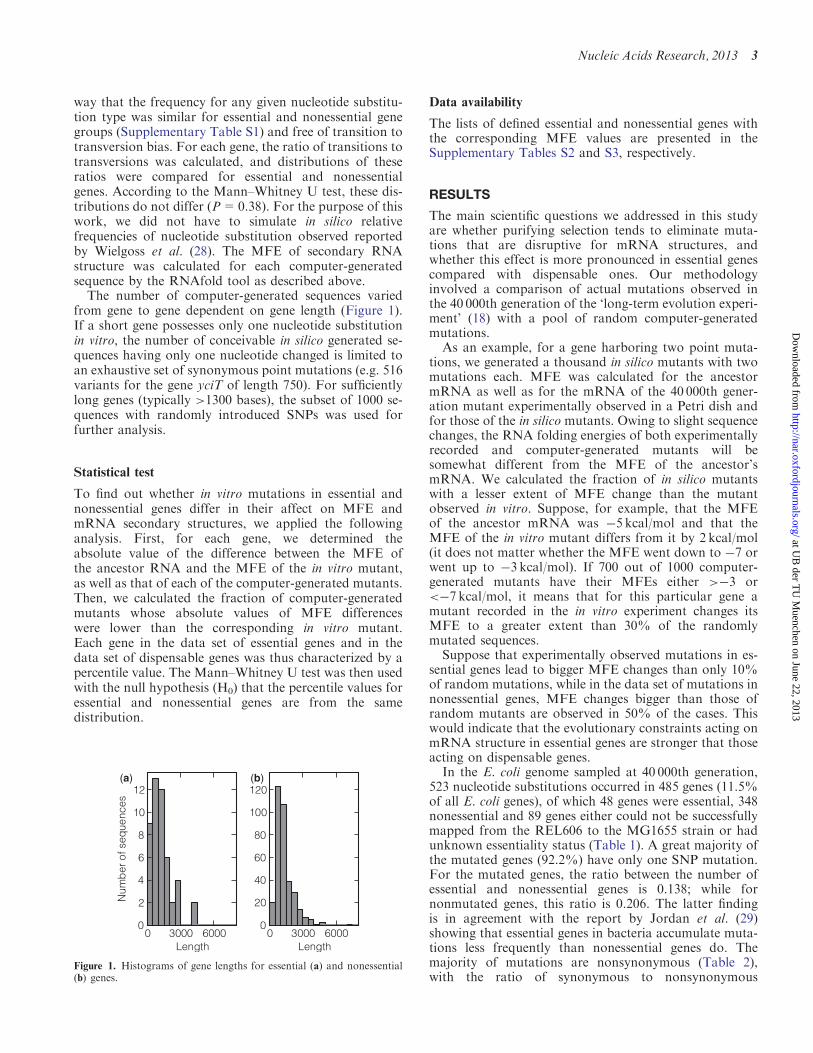
Transcript
TECHNISCHE UNIVERSITAT MUNCHEN
Fachgebiet fur Bioinformatik
Sequence-structure relationships in mRNAs
Andrey Chursov
Vollstandiger Abdruck der von der Fakultat Wissenschaftszentrum Weihenstephanfur Ernahrung, Landnutzung un Umwelt der Technischen Universitat Munchen zurErlangung des akademischen Grades eines
Doktors der Naturwissenschaften
genehmigten Dissertation.
Vorsitzender: Univ.-Prof. Dr. A. GierlPrufer der Dissertation:
1. Univ.-Prof. Dr. D. Frischmann2. Univ.-Prof. Dr. B. Rost
Die Dissertation wurde am 26.08.2013 bei der Technischen Universitat Munchen ein-gereicht und durch die Fakultat Wissenschaftszentrum Weihenstephan fur Ernahrung,Landnutzung un Umwelt am 12.11.2013 angenommen.
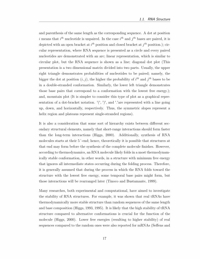
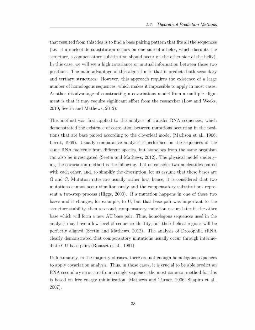
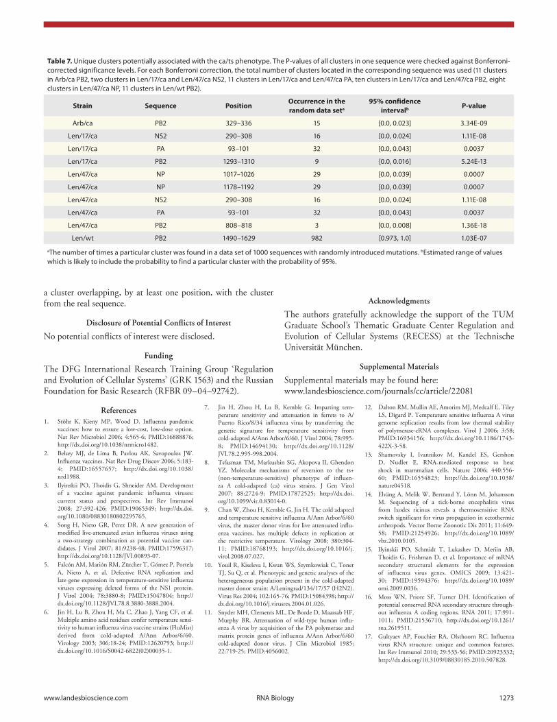
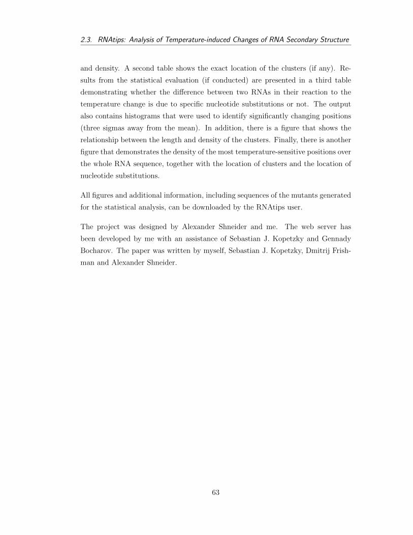
Plots were produced with VARNA visualiza7on tool (Darty -‐ VARNA. Interac7ve drawing and edi7ng of the RNA secondary structure) and RNA composer (Popenda -‐ Automated 3D structure composi7on for large RNAs).
a)
b)
d)
c)
e)
G C C C G G A U A G C U C A G U C G G U A G A G C A G G G G A U U G A A A A U C C C C G U G U C C U U G G U U C G A U U C C G A G U C C G G G C A C C A
1 10 20 30 40 50 60 70 76
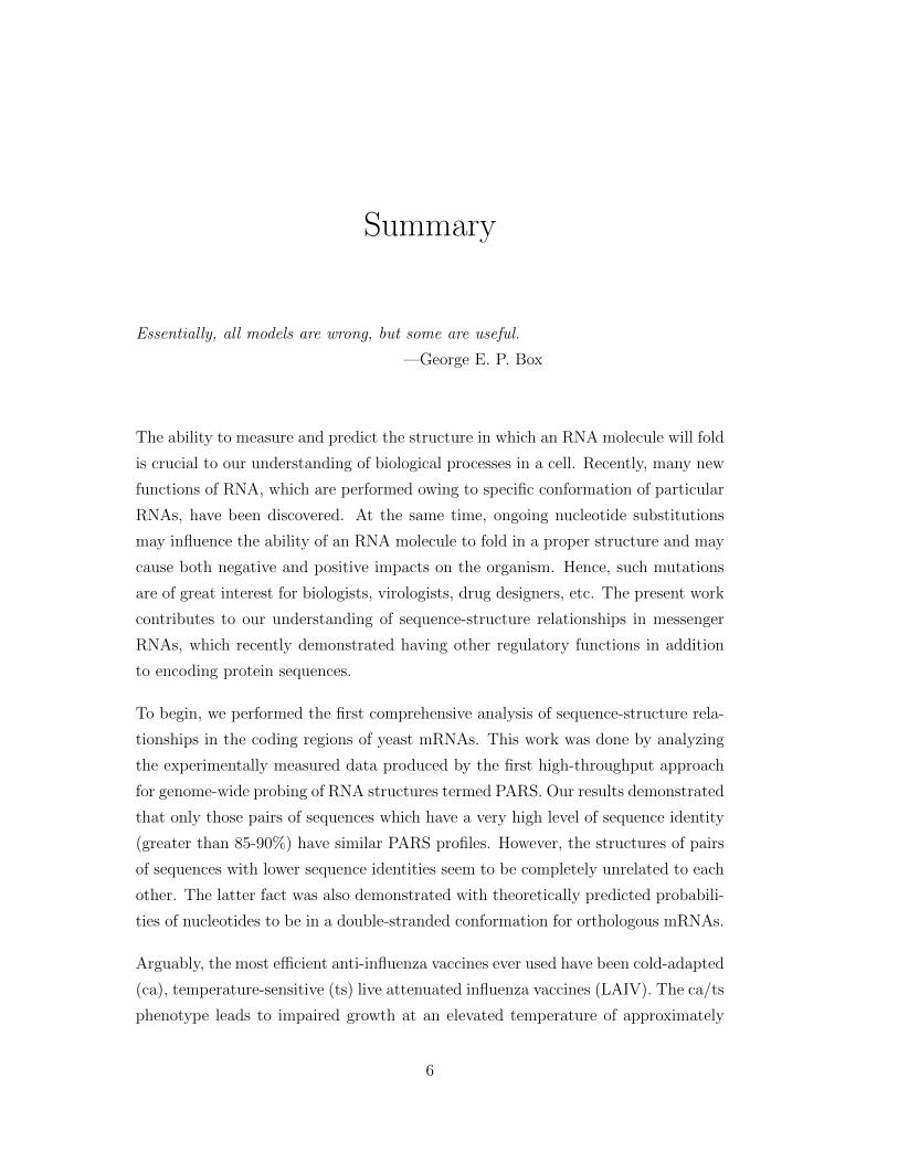
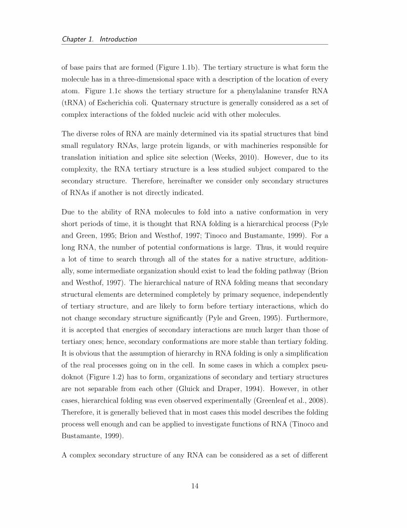
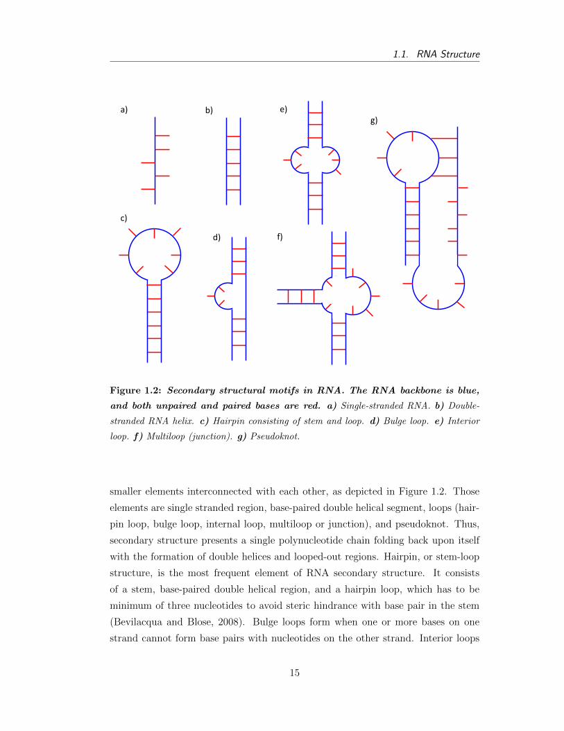
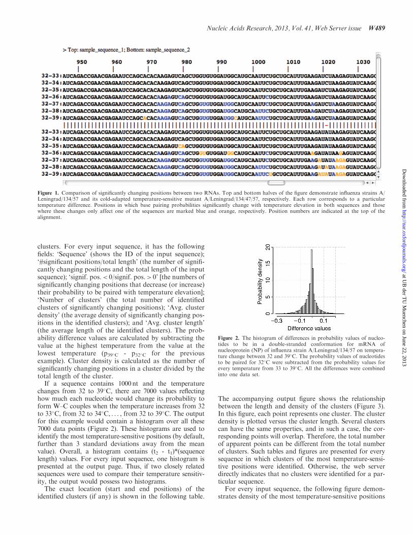
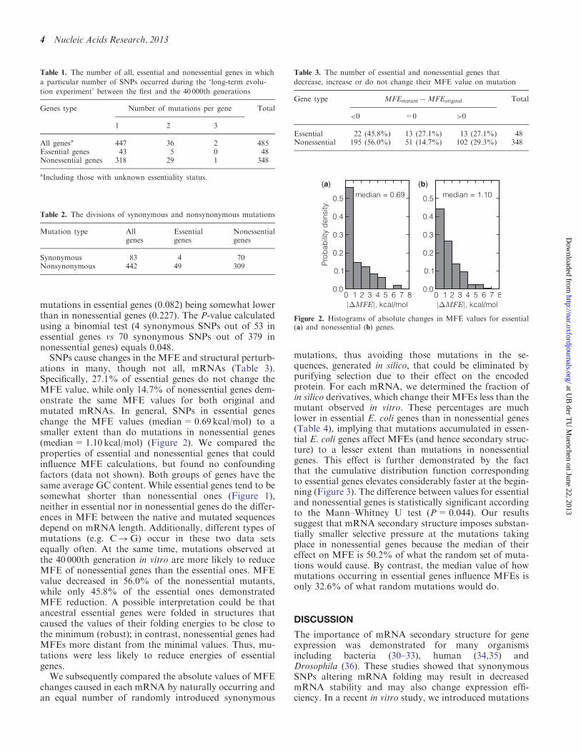
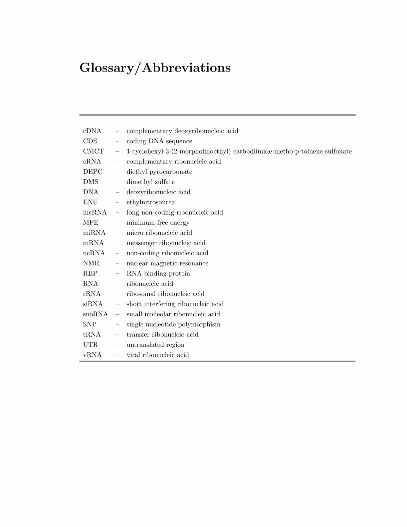
Figure 1.3: Different ways to represent graphically RNA secondary structure
include: a) Dot-bracket notation. b) Circular representation (created by sfold (Ding
et al., 2004)). c) Linear representation (created by VARNA visualization tool (Darty
et al., 2009)). d) Mountain plot (created by RNAfold (Gruber et al., 2008)). e) Energy
dot plot (created by mfold (Zuker, 2003)).
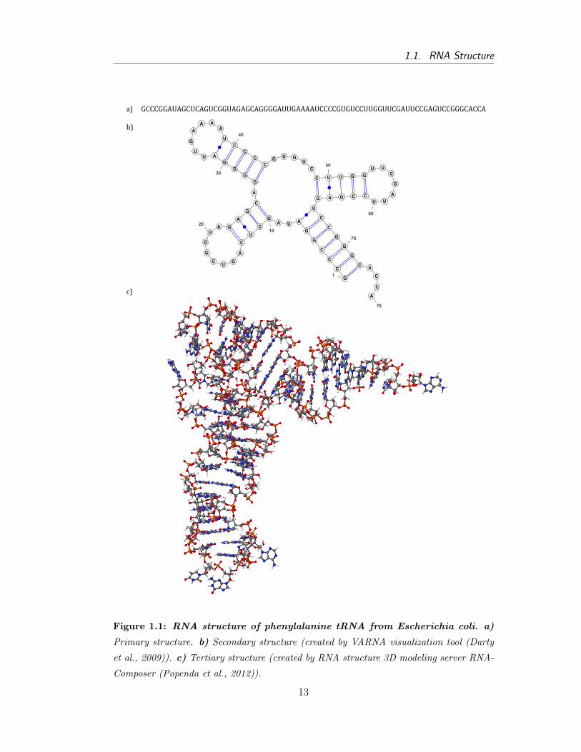
the most common approaches is to draw a simple planar plot similar to the one
shown on Figure 1.1b. Other ways include: dot-bracket notation (A string of dots
16
1.1. RNA Structure
and parenthesis of the same length as the corresponding sequence. A dot at position
i means that ith nucleotide is unpaired. In the case ith and jth bases are paired, it is
depicted with an open bracket at ith position and closed bracket at jth position.); cir-
cular representation, where RNA sequence is presented as a circle and every paired
nucleotides are demonstrated with an arc; linear representation, which is similar to
circular plot, but the RNA sequence is shown as a line; diagonal dot plot (This
presentation is a two dimensional matrix divided into two parts. Usually, the upper
right triangle demonstrates probabilities of nucleotides to be paired; namely, the
bigger the dot at position (i, j), the higher the probability of ith and jth bases to be
in a double-stranded conformation. Similarly, the lower left triangle demonstrates
those base pairs that correspond to a conformation with the lowest free energy.);
and, mountain plot (It is simpler to consider this type of plot as a graphical repre-
sentation of a dot-bracket notation. '(', ')', and '.'are represented with a line going
up, down, and horizontally, respectively. Thus, the symmetric slopes represent a
helix region and plateaus represent single-stranded regions).
It is also a consideration that some sort of hierarchy exists between different sec-
ondary structural elements, namely that short-range interactions should form faster
than the long-term interactions (Higgs, 2000). Additionally, synthesis of RNA
molecules starts at their 5´-end; hence, theoretically it is possible that structures at
that end may form before the synthesis of the complete molecule finishes. However,
according to thermodynamics, an RNA molecule likely folds in a most thermodynam-
ically stable conformation, in other words, in a structure with minimum free energy
that ignores all intermediate states occurring during the folding process. Therefore,
it is generally assumed that during the process in which the RNA folds toward the
structure with the lowest free energy, some temporal base pairs might form, but
those interactions will be rearranged later (Tinoco and Bustamante, 1999).
Many researches, both experimental and computational, have aimed to investigate
the stability of RNA structures. For example, it was shown that real tRNAs have
thermodynamically more stable structure than random sequences of the same length
and base composition (Higgs, 1993, 1995). It is likely that the high stability of tRNA
structure compared to alternative conformations is crucial for the function of the
molecule (Higgs, 2000). Lower free energies (resulting to higher stability) of real
sequences compared to the random ones were also reported for mRNAs (Seffens and
17
Chapter 1. Introduction
Digby, 1999). However, if shuffling is made with preserving dinucleotide frequen-
cies, then the difference between the minimum free energy (MFE) values of real and
random sequences is lower (Workman and Krogh, 1999). Researches of other long
RNAs, including rRNA, rRNase P and introns, showed the same inference about
higher stability of the natural sequences (Schultes et al., 1999). These facts suggest
the existence of thermodynamic constraints in the course of evolution.
RNA sequence can potentially fold into many RNA structures (Wan et al., 2011).
Thus, another interesting set of experiments was aimed to study the number of
possible suboptimal conformations of one sequence as a function of folding energy
(Higgs, 1993). Studying of tRNA demonstrated that there is an energy gap between
MFE structure and suboptimal conformations (Wuchty et al., 1999; Chen and Dill,
2000). However, several researchers demonstrated that large RNAs fold via rugged
energy landscape, and that during the folding process a molecule can get one of
the metastable conformations (Higgs, 2000; Weeks, 2010). Moreover, Hobartner
and Micura experimentally demonstrated that even very short RNA sequences in
thermodynamic equilibrium could fold into different structures with some ratio of
frequencies between them (Hobartner and Micura, 2003). From a theoretical point of
view, it resulted in the wide application of partition function to calculating the prob-
abilities of nucleotides to be in a double-stranded conformation. As was mentioned
above, at the equilibrium state an RNA molecule should be folded in a conforma-
tion with minimum free energy. However, if there are several possible conformations
with very close values of free energy, then, according to statistical mechanics, the
probability that an RNA molecule folds in each of them is proportional to the Boltz-
mann factor of the conformation. Nevertheless, the process of transition from one
structure to another is not currently well understood.
1.2 Functions of RNA
During the last couple of decades, many articles have been published, which reveal
different aspects of RNA functioning both as a protein-coding intermediate and as
a regulatory agent. Such dual functionality of RNA does not seem surprising from
the viewpoint of the RNA World hypothesis (Kloc et al., 2011). The list of known
roles of RNAs has grown up substantially. A large number of non-coding RNAs
18
1.2. Functions of RNA
(ncRNAs) have been discovered, which are transcribed and carry out essential struc-
tural, catalytic or regulatory functions within a cell, but do not encode for amino
acid sequences (Eddy, 2001). Those functions are influenced by RNA secondary
and tertiary conformations according to which RNA molecules can interact with
other RNAs, ligands and RNA-binding proteins (RBPs) (Wan et al., 2011). Also,
the existence of functional structural elements in untranslated and coding regions of
messenger RNAs has been presented in recent experimental and computational pa-
pers (Bevilacqua and Blose, 2008; Wan et al., 2011). Plenty of excellent reviews on
the different aspects of various functions that RNAs perform have been published;
therefore, only some examples will be given here.
The three most known types of ribonucleic acids are messenger RNA (mRNA), trans-
fer RNA (tRNA), and ribosomal RNA (rRNA). All of them participate in protein
synthesis, and their roles in this process are well documented. Messenger RNAs are
copies of genes, which are translated into their corresponding protein; tRNAs are
adaptor molecules, one end of which can read the triplet code in the mRNA, and an
amino acid is attached to another end; finally, rRNAs move sequentially along the
mRNA and catalyze the attachment of amino acids to the growing peptide chain.
The described role of tRNA in protein synthesis is the most known function of trans-
fer RNA. Meanwhile, tRNA is also involved in many other biological processes apart
from protein synthesis, such as regulating the transcription of mRNA for enzymes
associated with biosynthesis of its amino acid both in prokaryotic and eukaryotic
systems, participation in making a DNA copy of the viral RNA, and acting as an
enzyme inhibitor (Rich and RajBhandary, 1976).
After the first cases of discovering catalytic activity of RNA molecules (namely that
the RNA moiety of ribonuclease P from Escherichia coli cleaves precursors of the
transfer RNA molecules (Guerrier-Takada et al., 1983; Guerrier-Takada and Altman,
1984), and that ribosomal RNA of Tetrahymena whose ribosomal RNA contains a
self-splicing exon (Kruger et al., 1982; Zaug et al., 1983, 1984)), it became clear
that other catalytic functions might be performed by RNA as well. The range of
catalytic activity currently known to be fulfilled by RNAs is rather wide (Tarasow
and Eaton, 1998). For instance, priming of DNA synthesis in mitochondria (Wong
and Clayton, 1986), priming reverse transcription (Kikuchi et al., 1986), and others
(Greider and Blackburn, 1985; Fire et al., 1998).
19
Chapter 1. Introduction
Since the discovery of RNA catalytic functions, many cases in which RNA molecules
carry out other essential functions within a cell, apart from encoding proteins, have
been discovered. Most of those RNAs are newly identified non-coding RNAs that
participate in different biological processes, and the number of which constantly
grows (Eddy, 2001). In particular, there are several interesting cases in which an
RNA molecule performs its function almost without folding into any conformation.
For instance, microRNAs (miRNAs) have been discovered that bind to complemen-
tary regions of target messenger RNA and, thus, inhibit the translation of corre-
sponding protein (Matzke and Birchler, 2005; Bevilacqua and Blose, 2008). Another
example is small interfering RNAs (siRNAs) that form base pairs with complemen-
tary mRNA and target it for degradation (Matzke and Birchler, 2005). Such mech-
anism of targeting mRNAs for cleavage is called RNA interference (RNAi) (Matzke
and Birchler, 2005). The degradation of mRNA in this case occurs by RNA-induced
silencing complex (RISC), the core component of which is either Argonaute protein
or RNA-dependent RNA polymerase (Matzke and Birchler, 2005).
Although there are several RNAs, which act without forming specific RNA structure,
most RNA molecules have to fold into a particular shape or contain a certain struc-
tural element, such as hairpin or pseudoknot, to perform their function (Bevilacqua
and Blose, 2008). Regulatory RNAs termed riboswitches have been found in bacte-
rial messenger RNAs and appear to control gene expression (Nudler and Mironov,
2004; Serganov and Patel, 2007; Montange and Batey, 2008). Small RNAs (sRNAs),
identified in a wide range of bacteria, have been shown to regulate the expression of
proteins, and to modulate the activity and stability of messenger RNAs (Gottesman
and Storz, 2011). RNAs also play an important role in mechanisms of phage defense
in bacteria (Marraffini and Sontheimer, 2010).
An extremely interesting area of research is the investigation of roles that differ-
ent types of RNAs can play in different diseases. miRNAs, long non-coding RNAs
(lncRNAs), small nuclear RNAs (snoRNAs), large intergenic non-coding RNAs (lin-
cRNAs), and others have been associated with different disorders, which makes them
potential new therapeutic targets (Taft et al., 2010; Esteller, 2011). miRNAs have
been associated with cancer, neurological disorders, cardiovascular disorders, and
other diseases; snoRNAs have been associated with cancer (Esteller, 2011). Many
long non-coding RNAs (lncRNAs) have also been associated with different diseases
20
1.2. Functions of RNA
including cancer (Kung et al., 2013). For example, numerous lncRNAs demonstrate
different expression levels between normal and cancer cells (Huarte and Rinn, 2010).
Several lncRNAs have demonstrated inhibiting tumor suppressor functions in can-
cer cells (Gutschner and Diederichs, 2012). In addition, long non-coding RNAs have
been noted to play roles in metastasis formation (Gutschner and Diederichs, 2012).
Obviously, we are only beginning to understand all these molecular mechanisms and
there is still plenty to learn.
The biology of viruses and how it is affected by their RNA conformations is another
area of great significance. Numerous studies on viruses have revealed the importance
of RNA structures for virus assembly (Larson and McPherson, 2001; Schneemann,
2006; Hutchinson et al., 2010). One RNA of HIV-1 virus folds into two different
conformations promoting different functions (Lu et al., 2011). Locations of the most
stable predicted structural motifs correlate with regions were structures are thought
to play an important role in the genomes of the RNA picornavirus (Palmenberg
and Sgro, 1997). Translation initiation can occur in the middle of an mRNA se-
quence owing to the existence of internal ribosome entry sites (IRES) (Jackson and
Kaminski, 1995). Additionally, pseudoknots regulating gene expression and genome
replication have been identified in many viruses (Brierley et al., 2007). All these ex-
amples clearly demonstrate how significant RNA structures are for a diverse range
of viral activities.
For many years, it was thought that only non-synonymous nucleotide substitutions
in messenger RNAs, which lead to change in the function of the protein, might be
deleterious for an organism. Yet, recent studies have demonstrated that mRNAs,
both in eukaryotes and prokaryotes, have other hidden non-coding functions which
are performed by secondary structural elements and are unrelated to encoding for
correspondent proteins (Ulveling et al., 2011). Therefore, those mutations, which
result in a change in mRNA secondary structure, might affect some essential biolog-
ical processes due their affect on the ability of mRNA to interact with other RNAs
and proteins in a proper way.
Another generally accepted belief was that pre-mRNA is solely a passive transcript,
from which messages are produced by spliceosome, a large protein-RNA complex
that removes non-coding introns. Now, there are many examples that RNA struc-
21
Chapter 1. Introduction
tures are actively involved in this regulation and can either inhibit or aid splicing
(Warf and Berglund, 2010). Splicing can be regulated by structures in pre-mRNA
directly, or by proteins or small molecules, which bind RNA structural elements.
One genome-wide study also reported that the existence of predicted secondary
structure elements with low free energy near splice sites decreases the efficiency of
splicing compared to introns, which have structural elements with higher free energy
(Shepard and Hertel, 2008).
Structural elements in messenger RNAs can regulate gene expression by controlling
the efficiency of translation initiation. RNA structure can influence accessibility
of the start codon or other signals that should be recognized by a ribosome (Mc-
Carthy and Gualerzi, 1990). A pseudoknot in retroviral RNA is responsible for
ribosomal frameshifting, and results in the production of two proteins at particular
ratios, which in turn are required for viral propagation (Chamorro et al., 1992).
Frameshifting has been observed in other viruses and species as well (Giedroc et al.,
2000), including Rous sarcoma virus (Jacks and Varmus, 1985; Jacks et al., 1988),
coronaviruses (Bredenbeek et al., 1990), and bacteria (Tsuchihashi and Kornberg,
1990; Flower and McHenry, 1990).
One of the methods for regulating gene expression is promoting or preventing the
degradation of target mRNA (Emory et al., 1992). The range of mRNA half-lives
within a single cell is rather wide. For instance, in E. coli it can vary from seconds
up to nearly one hour (Emory et al., 1992). At the same time, in eukaryotic or-
ganisms mRNAs usually live longer than in prokaryotic species (Belasco and Chen,
1988). In many cases, the degradation rate of messenger RNAs is determined by the
stability of some hairpin in the structure of this molecule. For example, particular
hairpin structure near the 3´-end of the transcript was experimentally demonstrated
protecting mRNA from degradation in Rhodobacter capsulatus (Belasco and Chen,
1988). A stem-loop element in the 5´-untranslated region E. coli ompA mRNA slows
down its degradation, although the sequence in this region is relatively unimportant
(Emory et al., 1992). However, hairpins that may promote mRNA degradation have
been observed as well. For instance, it was shown that in Escherichia coli, during
the processing of the mRNA for ribosomal protein S20, a hairpin structure helps to
localize a cleavage site for a single-strand specific endonuclease RNase E (Mackie
and Genereaux, 1993; Bevilacqua and Blose, 2008).
22
1.2. Functions of RNA
Dual functionality of coding RNAs, both in eukaryotic and prokaryotic organisms,
has been recently observed in several cases (Kloc et al., 2011; Ulveling et al., 2011).
One very interesting example is a so called transfer-messenger RNA (tmRNA), ini-
tially known as 10Sa RNA and expressed from the SsrA gene, which combines fea-
tures of both mRNA and tRNA (Jentsch, 1996; Atkins and Gesteland, 1996). In all
eubacteria, tmRNA directs a process called trans-translation aimed to release stalled
ribosomes from a defective mRNA (lacking a stop codon) (Keiler et al., 1996; Gillet
and Felden, 2001). The structure of tmRNA from E. coli was determined experimen-
tally (Felden et al., 1997). It contains two domains: an mRNA-like and a tRNA-like,
which can be charged with alanine that will link to the truncated polypeptide chain.
Transfer-messenger RNA engages the stalled ribosome and mRNA-like domain re-
places the defective mRNA bound. Hence, translation switches to tmRNA from the
broken mRNA. The following translation of the tmRNA adds a peptide tag to the
nascent protein, which targets the polypeptide for rapid degradation (Keiler, 2008).
Thus, trans-translation is a quality control mechanism, which ensures that synthe-
sized proteins are correct (Keiler, 2008).
Another interesting case is the p53 tumor suppressor, mutations of which are found
in approximately 50% of human tumors (Soussi and Wiman, 2007). Due to this
fact, p53 is a fascinating research target for clinicians and researchers. Usually p53
protein is persistently degraded, but, upon stress, its activation eliminates tumor
cells (Farnebo et al., 2010; Ulveling et al., 2011). p53 tumor suppressor activity is
mainly regulated by the E3 ubiquitin ligase Mdm2, which binds p53 protein and tar-
gets it for degradation (Candeias et al., 2008; Farnebo et al., 2010). Recently, it has
been demonstrated that messenger RNA of p53 gene interacts directly with Mdm2
protein and thus restrains the Mdm2 activity of promoting p53 degradation (Can-
deias et al., 2008). Additionally, it was shown that synonymous single nucleotide
polymorphisms in the p53 mRNA can impair its interaction with Mdm2, and, as a
consequence, activity of p53 in this case will be decreased (Candeias et al., 2008).
Other known cases of messenger RNAs that can also fulfill some structural role in-
clude: VegT mRNA from Xenopus, which is involved in the cytokeratin network
of primordial germ; oskar mRNA from Drosophila melanogaster, which was shown
being responsible for the early oogenesis; and others (Kloc et al., 2011; Ulveling
et al., 2011).
23
Chapter 1. Introduction
Undoubtedly, functions of molecules within a cell depend on their three-dimensional
structures. Therefore, to understand the functions that RNA molecules may per-
form, it is crucial to be able to determine atomic coordinates nucleotides in a 3D
conformation and what residues are base-paired. Nevertheless, neither present ex-
perimental techniques nor theoretical predictions allow analyzing high-throughput
data on tertiary structures of RNAs. Moreover, it is extremely difficult to pre-
dict three-dimensional folding. As a result, the first step toward determining and
studying the RNA tertiary conformation is to identify its secondary structure (base
pairing interactions within a molecule).
1.3 Experimental Techniques
The first ever experimentally determined structure was a conformation of transfer
RNA (tRNA), which was crucial to the understanding of molecular mechanisms of
protein synthesis and other biological functions of tRNA (Sigler, 1975). Initially
an X-ray diffraction analysis was applied to yeast phenylalanine tRNA to measure
its three-dimensional folding (Kim et al., 1971, 1972, 1973). The electron density
maps from the experiments showed double helix regions connected to each other
with weaker regions of electron density. These were interpreted as a confirmation
of the idea first proposed by Holley and his collaborators (Holley et al., 1965) who
sequenced alanine transfer RNA from yeast and suggested that tRNAs could be
folded into a secondary structure widely known as cloverleaf (Figure 1b). Later, that
method was improved and it allowed measuring the structure first at 3 angstrom res-
olution (Suddath et al., 1974; Kim et al., 1974; Robertus et al., 1974b), and then at
2.5 angstrom resolution (Quigley et al., 1975; Ladner et al., 1975). Further research
enabled reciprocal space refinement and the ability to model precise atomic coor-
dinates of the entire tRNA molecule (Sussman et al., 1978; Hingerty et al., 1978).
Subsequently, the X-ray crystallography was used to determine the structure of an-
other elongator transfer RNA (Giege et al., 1977) and yeast aspartic acid tRNA
(Westhof et al., 1988b), confirming the proposed general tRNA structure. To an
RNA, other than tRNA, the X-ray crystallography was first applied in 1994, when a
structure of hammerhead ribozyme was determined at 2.6 angstrom resolution (Pley
et al., 1994). Further development of crystallographic methods is described in the
review by Holbrook (Holbrook and Kim, 1997).
24
1.3. Experimental Techniques
X-ray diffraction from crystals gives the highest accuracy, but does not say whether
the structure in the crystal is the same as the structure in solution, hence, other
methods had to be adopted to answer this question (Rich and RajBhandary, 1976;
Holbrook and Kim, 1997). The high-resolution NMR (nuclear magnetic resonance)
spectroscopy study was first applied to determine the structure of yeast phenylala-
nine tRNA (Kearns and Shulman, 1974). Next, high-resolution NMR spectra of
several other purified tRNAs was examined by different groups (Reid and Robil-
lard, 1975; Reid et al., 1975; Daniel and Cohn, 1975; Wong et al., 1975; Bolton
and Kearns, 1975). Results of those NMR studies supported the conclusion that
the structure of the tRNA molecule in solution is identical to the 3D conformation
determined in the crystal (Rich and RajBhandary, 1976). Such knowledge of the
three-dimensional structure of tRNA led to the better understanding of chemistry
and the role of transfer RNA in different biological processes (protein synthesis, tran-
scription of messenger RNA, reverse transcription, etc.) (Rich and RajBhandary,
1976). The size of the RNA that can be analyzed at atomic resolution by NMR
is continually increasing, but slowly. For many years the upper limit was approxi-
mately 100 nt (Tinoco and Bustamante, 1999). Only recently, a nuclear magnetic
resonance approach, which enables detection of structural elements within longer
sequences, has been developed and applied to investigation of HIV-1 5´-leader RNA
(Lu et al., 2011). In addition, both X-ray approach and NMR studies require of
large amounts of highly purified material (Ehresmann et al., 1987).
Another experimental approach to probe structure is the use of chemical modifi-
cations to test the reactivity of every nucleotide. An RNA of interest is modified
somehow by treating it with a specific chemical reagent in such a way that any two
modification events are independent from each other (Weeks, 2010). Some bases
will be reactive while others will react at a much slower rate. Such reactivity of
bases identifies which nucleotides are unpaired and which ones are paired. Two ap-
proaches of determining modified nucleotides are using end-labeled RNA molecules,
which allows the detection of scissions in the RNA chain, and primer extension, which
detects stops of transcription at modified sites (Ehresmann et al., 1987). What nu-
cleotides should react depends upon the reagent used (Rich and RajBhandary, 1976).
The list of reagents, which have been applied to examine the secondary and ter-
tiary structures of transfer ribonucleic acid, includes: β-ethoxy-α-ketobutyraldehyde
25
Chapter 1. Introduction
(kethoxal), which reacts with guanine (Litt, 1969); methoxyamine (Cashmore et al.,
1971; Robertus et al., 1974a; Chang, 1973) and hydrogen sulfide (Miura et al., 1982),
which react with cytosine; 1-cyclohexyl-3-(2-morpholinoethyl) carbodiimide metho-
p-toluene sulfonate (CMCT), which used to map unpaired uridines and guanosines
(Robertus et al., 1974a; Chang, 1973); ethylnitrosourea (ENU), a reagent ethylating
phosphates in nucleic acids (Vlassov et al., 1981, 1983; Garret et al., 1984a; Romby
et al., 1985); dimethyl sulfate (DMS), which reacts with the N1 of adenine, the N3
of cytosine and the N7 of guanine (Peattie and Gilbert, 1980; de Bruijn and Klug,
1983; Garret et al., 1984a; Romby et al., 1987); diethyl pyrocarbonate (DEPC),
which reacts with the N7 of adenosines (Peattie and Gilbert, 1980; de Bruijn and
Klug, 1983; Garret et al., 1984a; Romby et al., 1987); and others (Igo-Kemenes and
Zachau, 1969, 1971; Vary and Vournakis, 1984a; Brunel and Romby, 2000; Rocca-
Serra et al., 2011).
Following tRNA, different parts of ribosomal RNA became the subject of interest
and were studied with different chemical probes. For example, mouse 5S ribosomal
RNA was tested with kethoxal (Miura et al., 1983b) and hydrogen sulfide (Miura
et al., 1983a), E. coli 16S ribosomal RNA was analyzed with diethyl pyrocarbonate
(Van Stolk and Noller, 1984); investigation of the interaction of ribosomal protein
S4 with E. coli 16S rRNA was done with using of kethoxal and DMS (Stern et al.,
1986), binding of ribosomal protein S8 to 16S ribosomal RNA was studied with
DMS, CMCT, DEPC and ethylnitrosourea (Mougel et al., 1986); bisulfite, which
converts unpaired cytosine to uridine, was used to probe the RNA structure of 5S
rRNA from Spinacea oleracea (Pieler et al., 1983); and many others.
Another group of methods is similar to chemical probing and is based on using a
structure-specific enzymatic probe, which cleaves RNA at single- or double-stranded
regions. In fact, many groups combined data from chemical and enzymatic struc-
ture probes (Ehresmann et al., 1987). As with chemical reagents, there are several
enzymes, most of which cut the RNA within unpaired regions. First studies of RNA
base pairing were performed applying single-stranded-specific RNase T1, which cuts
unpaired guanosines, and S1 nuclease, which cleaves preferentially all single-stranded
nucleotides, to digest transfer RNAs from different organisms and at different envi-
ronment conditions (Wurst et al., 1978; Wrede et al., 1979b; Wrede and Rich, 1979;
Wrede et al., 1979a). The obtained results were consistent with previously deter-
26
1.3. Experimental Techniques
mined three-dimensional folding.
Other enzymatic reagents that are actively used to probe RNA structure include
RNase U2, which cuts unpaired adenines (Mougel et al., 1986; Baudin et al., 1987);
RNase Cl3, which cleaves unpaired cytidines, adenosines and uridines, but for the
latter two it requires longer incubation time and high concentration of enzyme (Flo-
rentz et al., 1982); RNase T2, which cuts single-stranded adenosine residues (Vary
and Vournakis, 1984b; Kean and Draper, 1985; Romaniuk, 1985; Christiansen et al.,
1987). There are also other enzymes used to probe RNA structure, which similar
to nuclease S1 cleave single-stranded RNA regions without being specific to a par-
ticular nucleotide. For example, RNase ONE, which cleaves all unpaired bases, was
used in studying structure elements of umbravirus and panicovirus (Wang et al.,
2009); RNase J1 from Bacillus subtilis was used to solve the structure of the hbs
mRNA (Daou-Chabo and Condon, 2009); Neurospora crassa nuclease was used to
study interactions between beef tryptophan transfer RNA and avian myeloblastosis
reverse transcriptase (Garret et al., 1984b).
RNase V1 from cobra venom is the only enzyme that cuts preferentially double-
stranded regions (Wan et al., 2011). This ribonuclease specifically cleaves RNA in
regions that are helical, indicating where the RNA is base paired. It was first ap-
plied to probe the structure of yeast phenylalanine and E. coli methionine tRNAs,
and results demonstrated that the V1 nuclease also recognizes non-canonical base
pairs and tertiary interactions, in addition to usual secondary helices (Lockard and
Kumar, 1981). Owing to this uncommon specificity, it is a widely used enzyme
for probing RNA structure (Favorova et al., 1981; Troutt et al., 1982; Lowman and
Draper, 1986).
The use of chemical modifications for testing RNA structure is very time consuming
and requires a lot of effort (Weeks, 2010). Although chemical probing with a variety
of structure-specific probes provides comprehensive information at the nucleotide
level, data obtained solely from chemical probing techniques do not show which nu-
cleotides are base pairing with each other (Ehresmann et al., 1987). However, such
data can be directly incorporated as folding constraints into dynamic programming
algorithms for secondary structure prediction (Mathews et al., 2004).
27
Chapter 1. Introduction
The current state of art in chemical probing techniques is the one termed Selective
2’-Hydroxyl Acylation analyzed by Primer Extension (SHAPE) and based on the
discovery that the nucleophilic reactivity of a ribose 2’-hydroxyl group is gated by
local nucleotide flexibility (Merino et al., 2005; Wilkinson et al., 2005, 2006). It was
first applied to reproduce the well-studied structure of aspartic acid transfer RNA in
yeast (Westhof et al., 1985, 1988b,a; Perret et al., 1990). Single nucleotide resolution
SHAPE chemistry computes nucleotide flexibility at all four ribonucleotides and dif-
ferentiates paired residues from flexible ones. Knowledge of such local flexibilities
of nucleotide positions allows determining the RNA secondary structure. However,
SHAPE chemistry is a rather slow technique. Original protocol required two days to
complete for RNA with only 100-200 nucleotides (Wilkinson et al., 2006). Later, a
new faster-acting reagent was designed to improve the SHAPE chemistry (Mortimer
and Weeks, 2007); nonetheless, the entire procedure still required a lot of time.
Further development of SHAPE technology allowed analyzing long RNAs in a sin-
gle experiment and measuring flexibility at more than 99% of the bases (Wilkinson
et al., 2008; Watts et al., 2009). Thus, SHAPE measurements yield comprehensive
information about what nucleotides are paired and what nucleotides are unpaired in
the RNA structure. Such improved SHAPE technology was used to assess the RNA
secondary structure of a complete HIV-1 genome and revealed many previously un-
recognized structural elements and long-range interactions (Wilkinson et al., 2008;
Watts et al., 2009). Additionally, as with other chemical probing techniques, ex-
perimental data produced by the SHAPE analysis can be coupled with computa-
tional prediction methods. This feature has been implemented in the RNAstructure
program (Mathews et al., 2004) and increases the accuracy of secondary structure
predictions dramatically (Deigan et al., 2009; Low and Weeks, 2010). For example,
taking into account SHAPE reactivity information in benchmarking was performed
on 16S ribosomal RNA of Escherichia coli, the crystal structure of which was ear-
lier solved at 3 angstrom resolution (Wimberly et al., 2000). The accuracy of the
structure, based solely on a thermodynamic model, was less than 50%; however, the
SHAPE-directed structure modeling of E. coli 16S rRNA demonstrated higher than
95% accuracy (Deigan et al., 2009).
The two main disadvantages of all the methods described above are that they are
limited to probing one RNA molecule at a time, and only a few hundred bases can
28
1.3. Experimental Techniques
be examined per experiment. Recently, several high-throughput methods of deter-
mination of the RNA structures have been suggested. The first technique, termed
Parallel Analysis of RNA Structures (PARS), was successfully applied to measure
the secondary structures of the messenger RNAs for over 3,000 distinct transcripts of
the Saccharomyces cerevisiae (Kertesz et al., 2010). The method is based on treating
mRNAs separately with S1 nuclease and RNase V1, nucleases, which cleave single-
and double-stranded RNA, respectively. Then, RNA is converted into a cDNA li-
brary. High-throughput sequencing of cDNA library enables identification of the
cleavage sites. Those nucleotides, whose RNase V1 cleavage number is higher than
S1 nuclease cleavage number, are considered base-paired. And the other way round,
the nucleotides, whose RNase V1 cleavage number is lower than S1 nuclease cleavage
number, are considered unpaired. The enzymatic footprinting takes about five days
to complete and subsequent sequencing and analysis requires six to eight days (Wan
et al., 2013).
Analysis of yeast structural profiles measured by PARS revealed that nucleotides
in the coding regions of mRNAs are prone to appear in double-stranded conforma-
tions more often than nucleotides in the untranslated regions (UTRs) (Kertesz et al.,
2010; Mauger and Weeks, 2010). A similar finding was reported for the HIV-1 virus
genome (Watts et al., 2009). Another detail demonstrated by PARS was that the ef-
ficiency of mRNA translation is anti-correlated to the probability of nucleotides near
the translation start site to be in a double-stranded conformation (Kertesz et al.,
2010; Mauger and Weeks, 2010). Lately, a new approach similar to PARS analysis,
termed Parallel Analysis of RNA structures with Temperature Elevation (PARTE),
was suggested by the same group (Wan et al., 2012). It was applied to probe yeast
RNA structures and different temperatures and it helped to identify thousands of
putative RNA thermometers (Wan et al., 2012).
Zheng et al. combined nuclease-based mapping with high-throughput sequencing
and applied this technique, which they called dsRNA-seq, to a genome-wide analy-
sis of Arabidopsis (Zheng et al., 2010). The approach was based on treating RNA
samples with a single-strand specific RNase One. It allowed them to identify highly
stable regions of secondary structure, and also to identify many new small RNAs.
Later, this approach evolved. A double-strand specific RNase V1 was added to this
methodology; and, it was applied to the analysis of Drosophila melanogaster and
29
Chapter 1. Introduction
Caenorhabditis elegans transcriptomes (Li et al., 2012). Interestingly, they found
that nucleotides both in the 5´- and 3´-untranslated regions have higher propensity
to be in a double stranded conformation than nucleotides in the coding regions.
This finding was interpreted as the existence of many regulatory signals or interac-
tion sites for RNA-binding proteins (Li et al., 2012).
Finally, an alternative technology, termed fragmentation sequencing (Frag-seq), was
used to probe for single-stranded regions of mouse transcriptome (Underwood et al.,
2010). The method relies on high-throughput sequencing of RNA fragments that
resulted from treating RNAs in solution with P1 endonuclease, which cleaves the
RNA of interest at single-stranded regions. A high number of cleaves at a particu-
lar position indicates that this nucleotide is unpaired. Through this method, known
structured regions in noncoding RNAs were validated and new, previously unprobed
RNAs, were tested.
Recently, SHAPE technology has also been paired with deep sequencing and was
termed SHAPE-Seq (Lucks et al., 2011). Compared with other high-throughput
methods of probing RNA structure, which use large nucleases, SHAPE-Seq uses
a small chemical probe. As a result, it has considerably higher accuracy of mea-
surement. This method was applied to probe the structure of the highly conserved
Bacillus subtilis RNase P and to identify changes in the structure resulting from
single nucleotide polymorphisms (SNPs). The method also can be further extended
to determine how the structure changes due to RNA-RNA or RNA-protein interac-
tions (Lucks et al., 2011).
It is interesting that PARS, dsRNA-seq and Frag-seq appeared almost simultane-
ously in research. This shows that high-throughput methods of RNA structure
mapping are of great interest and rapid further development of such techniques is
very likely. However, the biological importance of RNA and the long absence of
experimental techniques for measuring RNA structure have resulted in a fast grow-
ing number of works that are analyzing RNA functions based solely on theoretical
predictions.
30
1.4. Theoretical Prediction Methods
1.4 Theoretical Prediction Methods
To better understand of the biological functions of RNA molecules within a cell, it
is crucial to know their structures. Despite the fact that RNA structures play im-
portant roles in different biological processes, the experimental techniques to probe
RNA structure by high-throughput sequencing are only beginning to appear. There-
fore, the majority of researches connected to RNA structure is based on theoretical
predictions of secondary structures.
There are several problems that make the theoretical prediction of RNA structures
very complicated. First of all, RNA structures are dynamic, which means that RNA
conformation depends on surrounding conditions (such as temperature, salt con-
centrations, etc.) and on the functional role that an RNA molecule is supposed to
perform at the particular biological state (Weeks, 2010; Wan et al., 2011). Thus,
many of the processes that influence the conformation into which RNA folds (e.g.
folding kinetics, higher-order interactions, etc.), are too complex to be taken into
account to produce high accuracy results. Another problem is that the number of
theoretically possible conformations for an RNA sequence increases exponentially
with the length of the sequence, N (Zuker and Sankoff, 1984; Mathews, 2006):
Number of secondary structures ≈ (1.8)N
From the experimental results, it is also becoming clear that RNA can fold into many
stable states with energy somehow different from the global minimum (Hobartner
and Micura, 2003; Weeks, 2010). Therefore, the longer the sequence, the worse the
quality of prediction is. Several studies were aimed to assess the accuracy of theo-
retical predictions. For instance, Higgs demonstrated that 85% of tRNA structures
were correctly predicted (Higgs, 1995). However, the accuracy of predicting longer
sequences drops significantly (Zuker and Jacobson, 1995; Konings and Gutell, 1995;
Fields and Gutell, 1996; Doshi et al., 2004). For instance, even the most accurate
dynamic programming algorithm predicts correctly less than 50% of the base pairs
for 16S ribosomal RNA of E. coli (Deigan et al., 2009; Weeks, 2010).
Since the conformation of RNA with the lowest possible value of free energy is con-
sidered the most thermodynamically stable, one of the most common methods of
secondary structure prediction is based on searching for the MFE structure. But,
31
Chapter 1. Introduction
to incorporate such energy parameters into a prediction algorithm, they have to be
experimentally measured first. Thus, one potential explanations of poor accuracy is
incorrectness of the experimental energy parameters for base pairings (Doshi et al.,
2004). This factor can be especially important in the case that several alternative
structures, with similar values of free energy, do exist. Another reason is that more
than one structure may exist at equilibrium (Tinoco and Bustamante, 1999). Thus,
predicting only one structure for a long sequence may not show the entire picture.
A possible way to increase the quality of RNA secondary structure predictions,
especially for large RNA molecules, is to use data from experimental probing as
constraints into the prediction algorithms. Several works have demonstrated that in-
corporating chemical probing data into a dynamic programming algorithm improves
the accuracy of predictions dramatically (Mathews et al., 2004). For instance, indi-
cating those bases, which demonstrated high reactivity towards chemical probes, as
certainly unpaired helped to increase the accuracy of prediction from 50% to 72%
for 16S ribosomal RNA (Weeks, 2010).
Computer modeling of RNA molecules and computing of atomic coordinates, when
taking into account electrostatic interactions, are still extremely difficult tasks (Auffin-
ger and Westhof, 1998). Therefore, in those cases when it is absolutely necessary to
know atomic coordinates, the common solution is to use X-ray diffraction (Holbrook
and Kim, 1997). However, in most cases, we would like to know the structure well
enough to be able to understand the function it performs, instead of highest possible
resolution (Tinoco and Bustamante, 1999). Thus, predicting RNA structures can
be very useful either in interpreting experimental data concerning a particular RNA
function, or in suggesting new RNA regions that may be functionally important and
testing them experimentally (Seetin and Mathews, 2012).
It is generally accepted that the approach termed comparative sequence or covari-
ation analysis is the most reliable method of determining a secondary structure of
an RNA molecule (James et al., 1989; Pace et al., 1999; Weeks, 2010). The under-
lying assumption of this technique is that we would intuitively expect that if there
is a functionally important element of secondary structure then all the available
sequences must have this element of the structure (in other words, that structure
should be more conserved by evolution than sequence). Therefore, the main goal
32
1.4. Theoretical Prediction Methods
that resulted from this idea is to find a base pairing pattern that fits all the sequences
(i.e. if a nucleotide substitution occurs on one side of a helix, which disrupts the
structure, a compensatory substitution should occur on the other side of the helix).
In this case, we will see a high covariance or mutual information between those two
positions. The main advantage of this algorithm is that it predicts both secondary
and tertiary structures. However, this approach requires the existence of a large
number of homologous sequences, which makes it impossible to apply in most cases.
Another disadvantage of constructing a covariations model from a multiple align-
ment is that it may require significant effort from the researcher (Low and Weeks,
2010; Seetin and Mathews, 2012).
This method was first applied to the analysis of transfer RNA sequences, which
demonstrated the existence of correlation between mutations occurring in the posi-
tions that are base paired according to the cloverleaf model (Madison et al., 1966;
Levitt, 1969). Usually comparative analysis is performed on the sequences of the
same RNA molecule from different species, but homologs from the same organism
can also be investigated (Seetin and Mathews, 2012). The physical model underly-
ing the covariation method is the following. Let us consider two nucleotides paired
with each other, and, to simplify the description, let us assume that these bases are
G and C. Mutation rates are usually rather low; hence, it is considered that two
mutations cannot occur simultaneously and the compensatory substitutions repre-
sent a two-step process (Higgs, 2000). If a mutation happens in one of these two
bases and it changes, for example, to U, but that base pair was important to the
structure stability, then a second, compensatory mutation occurs later in the other
base which will form a new AU base pair. Thus, homologous sequences used in the
analysis may have a low level of sequence identity, but their helical regions will be
perfectly aligned (Seetin and Mathews, 2012). The analysis of Drosophila rRNA
clearly demonstrated that compensatory mutations usually occur through interme-
diate GU base pairs (Rousset et al., 1991).
Unfortunately, in the majority of cases, there are not enough homologous sequences
to apply covariation analysis. Thus, in those cases, it is crucial to be able predict an
RNA secondary structure from a single sequence; the most common method for this
is based on free energy minimization (Mathews and Turner, 2006; Shapiro et al.,
2007).
33
Chapter 1. Introduction
First attempts to estimate stability of RNA secondary structure by minimizing fold-
ing energy were done more than 40 years ago by Tinoco et al. (Tinoco et al., 1971,
1973). According to thermodynamics, at equilibrium state the structure with the
lowest free energy should dominate (Turner et al., 1988). Algorithms based on this
thermodynamic model usually find many possible structures and estimate a free
energy value for each of them. The main assumption is that the total energy of a
conformation is just a sum over energies of separate local structural components, like
stems and loops (Tinoco and Bustamante, 1999). Free energies of base paired regions
are negative, hence, more favorable; while, loops are usually taken into account with
free energy penalties because loops do not make the structure more stable and are
considered as unfavorable elements (SantaLucia and Turner, 1997). Additionally, the
energy of a double-stranded region depends on the sequence. Namely, the energy of
a helix depends not only on the type of base pairs in the helix, but also on the order
of base pairs, so called base stacking. Stability of loops depends on the sequence
as well (Mathews et al., 1999). Moreover, accuracy of free energy associated with
a loop is much lower than accuracy of helix parameters (SantaLucia and Turner,
1997). At the same time, the energy of a base pair is considered dependent only on
the types of adjacent base pairs. This is termed the nearest neighbor model (Tinoco
and Bustamante, 1999). Thus, to assess the free energy of a particular structure, it
is usually divided into elementary parts (energies of which are known or reasonably
estimated) and then energies of those simple parts are combined (Higgs, 2000). The
lower the free energy of a structure, the more stable this structure is considered.
The results of predictions made by free energy minimization algorithms strongly
depend on experimental thermodynamic data. As the accuracy of measuring free
energies of different interactions progresses, the quality of predictions improves as
well (Mathews et al., 1999). Usually optical melting studies are used to experi-
mentally determine energy parameters for the nearest neighbor model (Xia et al.,
1998; Mathews and Turner, 2002b; Mathews et al., 2004). The Nearest Neighbor
Database was created recently to summarize those parameters (Turner and Mathews,
2010). However, there are different methods of measuring the energy and different
models for the stacking free energy in helices (SantaLucia and Turner, 1997). For
example, there are pure computational approaches to estimate energy parameters
(Andronescu et al., 2007) and theoretical optimization of experimentally measured
34
1.4. Theoretical Prediction Methods
parameters (Mathews et al., 1999).
In 1978, Nussinov et al. presented the first dynamic programming algorithm to
find a particular folded form with the largest number of base pairs (Nussinov et al.,
1978). The biological underground idea is that every base pairing contributes to the
stability of the structure. Therefore, the more base pairings the structure has, the
more stable an RNA molecule is. Thus, the algorithm maximizing the number of
base pairings was developed. This algorithm included an important simplification,
however, it did not take pseudoknots into account. If we number all the nucleotides
in a sequence from 1 to N , then nucleotides ith and jth can form a base pair only
if they are complementary and at least three other bases exist between them. Now,
let us assume that ith nucleotide is paired with jth and kth nucleotide is paired with
lth. There are three possible variants of their mutual location: (i) one of the base
pairs is located aside of the other one (i < j < k < l); (ii) one pair is within
the other (i < k < l < j), so called nested base pairs; (iii) they are intersecting
(i < k < j < l). The latter case is called pseudoknot (Higgs, 2000). Elimi-
nating pseudoknots resulted in the fact that the original algorithm has O(n3) time
complexity and requires O(n2) memory; and hence, can be applied in most cases.
Later, several attempts were also made to accelerate this folding algorithm by using
graphics processing units (GPU) (Chang et al., 2010; Stojanovski et al., 2012; Su
et al., 2013).
Usually, the structure predicted by the original Nussinov’s algorithm is very different
from a conformation into which a real RNA folds. This is because the algorithm
takes into account only the number of possible base pairs and maximizes this num-
ber, which is not the best model from a thermodynamics point of view. It also
does not take into account different energy parameters for different base pairs, and
there are no penalties for the loops. However, a new version of their algorithm for
RNA structure predictions with incorporated energy parameters for base pairs was
presented two years later (Nussinov and Jacobson, 1980).
Nevertheless, energy rules for base stacking and destabilizing regions cannot be in-
corporated into the Nussinov’s algorithm. This problem was solved by Zuker and
Stiegler, who designed a new dynamic programming algorithm that allows taking
into account such energy parameters (Zuker and Stiegler, 1981). In many cases, the
35
Chapter 1. Introduction
structure with the minimum free energy was not consistent with those measured
experimentally, but could be observed among the structures with free energy close
to the minimum. Therefore, Zuker developed a new version of the algorithm, which
allows finding not only the MFE structure, but all suboptimal structures within a
particular range of free energy (Zuker, 1989). Theoretically, suboptimal structures
correspond to less stable conformations. Yet, they also can be considered as highly
probable alternate structures because of simplifications used in the algorithm and
errors in the energy parameters measurements. This algorithm became a basis for
a popular software package termed mfold (Zuker, 2003).
In 1990 McCaskill proposed a novel application of dynamic programming, namely
to calculate partition function instead of just the MFE structure (McCaskill, 1990).
The partition function describes the entire ensemble of secondary structures in ther-
modynamic equilibrium and is defined as a sum of Boltzmann factors over all the
possible conformations of a particular sequence:
Z =∑
qi
e−∆E(qi)
kBT
where −∆E(qi) represents the difference in free energies between a particular con-
formation, qi, and an unfolded state; kB is the Boltzmann constant; and T is the
temperature in kelvins. According to statistical physics, the probability of a given
conformation qi in the equilibrium can be assessed as:
Pi =e
−∆E(qi)
kBT
Z
From this formula it is obvious that the MFE structure of an RNA molecule, as
well as the weights of different conformations in the partition function, depends on
the temperature. Therefore, this approach enables investigating how an ensemble of
alternative structures alters upon the temperature change instead of studying the
most probable conformation. In addition, from the partition function, it is possible
to compute the probability for any two bases to be paired (McCaskill, 1990). Thus,
calculating a partition function reveals important information about the complete
ensemble of possible alternative conformations and enables assessing the power of
prediction algorithms (McCaskill, 1990). For instance, it was demonstrated that the
base pairs, which have high predicted probability to be paired, have higher chances
to be present in a real structure (Mathews et al., 2004). Also, it was shown that
36
1.4. Theoretical Prediction Methods
the probabilities of nucleotides to be paired are less sensitive to errors in free energy
parameters (Layton and Bundschuh, 2005).
Later, a dynamic programming algorithm by Zuker for free energy minimization and
computation of partition function was combined into a package termed the Vienna
RNA Package (Hofacker et al., 1994). This implementation was demonstrated to be
much faster than the original versions and became rather popular (Hofacker et al.,
1994).
Since RNAs, especially long ones, may fold into different co-existing conformations,
the partition function approach was extended by Ding and Lawrence to select a sta-
tistical representation of structures from the ensemble (Ding and Lawrence, 2003;
Ding, 2006). This approach allows for easily estimating the probabilities of partic-
ular secondary structural elements, instead of individual base pairs. Also, it was
demonstrated to be very useful in rational design of RNAs, which have to fold into
a particular structure (Ding, 2006).
The methods described above do not take into account the kinetic aspects of RNA
folding, rather, they try to estimate an equilibrium state. However, there is also
a class of algorithms that try to predict RNA structure by simulating the folding
process instead of simply optimizing free energy (Abrahams et al., 1990; WuJu and
JiaJin, 1998). For instance, specially adapted forms of Monte Carlo simulations
were applied (Fernandez, 1992; Schmitz and Steger, 1996). Also, several groups pro-
posed using genetic algorithms to solve the optimization problem of kinetic folding
(van Batenburg et al., 1995; Benedetti and Morosetti, 1995; Shapiro and Wu, 1996;
Shapiro et al., 2001). Nevertheless, all those algorithms have not achieved preva-
lence.
Since comparative analysis is considered the most accurate method, there have been
many attempts to automate this process and to combine comparative and thermo-
dynamics methods. Usually, such covariation models are built with using stochas-
tic context-free grammars (SCFG), which use formal language theory to develop a
grammar to describe RNA secondary structure, or hidden Markov models (Durbin,
1998; Dowell and Eddy, 2004; Do et al., 2006; Shapiro et al., 2007; Jossinet et al.,
2007). The first type of such techniques begins with constructing a multiple se-
37
Chapter 1. Introduction
quence alignment and then predicting the consensus structure for the alignment
(Hofacker et al., 2002; Bernhart et al., 2008; Bernhart and Hofacker, 2009; Mathews
et al., 2010). This approach is very fast, but the quality of the consensus structure
depends a lot on the quality of the alignment. Such methods, for example, were
applied to analyze genomes of a wide range of viruses, including HIV-1, hepatitis
C virus, hantavirus, and others (Luck et al., 1996; Tabaska et al., 1998; Hofacker
et al., 1998; Hofacker and Stadler, 1999). The second technique does the opposite.
They predict a set of suboptimal structures, which have a free energy value close
to the minimum, and then search for a structure, which is common to all sequences
in the alignment. This paradigm is implemented in the RNAshapes software tool
(Steffen et al., 2006), which is very fast (Mathews et al., 2010; Seetin and Mathews,
2012). However, authors of this approach try to find a common topology, termed
abstract shape (Giegerich et al., 2004), instead of analyzing real structures (Reeder
and Giegerich, 2005). Finally, the last approach is to fold and align the sequences
simultaneously (Gorodkin et al., 1997b,a; Mathews and Turner, 2002a). One of the
first such algorithms was proposed by Sankoff (Sankoff, 1985), but for the S input
sequences, it has a complexity of O(n3S) which makes it impractical for the majority
of cases. The algorithm suggested by Gorodkin et al. has a complexity of O(n4),
but does not take into account multi-branched loops (Gorodkin et al., 1997b). Ac-
counting for multi-branched loops increases the complexity to O(n6) and also makes
it impractical for most applications (Gorodkin et al., 1997b). As this is the most
expensive approach, it is usually applicable only to two sequences (Mathews et al.,
2010).
As was mentioned earlier, there are experimental evidences that pseudoknots can be
functionally important. Moreover, the number of known pseudoknots has been con-
stantly increasing and has resulted to creating a pseudoknot database (Taufer et al.,
2009). However, most of the developed algorithms for structure prediction do not
contain pseudoknots (Lyngsø and Pedersen, 2000). Predicting pseudoknots remains
a huge challenge and the accuracy of currently existing algorithms is still rather low.
The main complication is that RNA secondary structure prediction with pseudo-
knots was proved being NP-hard, which means that prediction of the structure for
a particular sequence can be performed computationally in a reasonable amount of
time only for very short RNAs (Lyngsø, 2004). Thus, some simplifications are used
38
1.5. Thesis Motivation and Outline of the Work
in attempts to predict pseudoknots.
Another problem of predicting pseudoknots, apart from the high complexity of
algorithms, is that although the melting behavior and thermodynamics data for
some types of pseudoknots have been obtained (Wyatt et al., 1990; Gregorian and
Crothers, 1995; Qiu et al., 1996; Theimer et al., 1998; Theimer and Giedroc, 1999;
Gonzalez Jr and Tinoco Jr, 1999), there is still limited experimental information
about energy parameters of pseudoknots. As a result, there have even been at-
tempts to estimate thermodynamic parameters for pseudoknots only from theory
(Gultyaev et al., 1999).
There are several groups, which have been working on developing dynamic program-
ming algorithms for predicting pseudoknots. For example, Rivas and Eddy suggested
a new dynamic programming algorithm that takes into account simple topologies of
pseudoknots (Rivas and Eddy, 1999). However, the algorithm complexity is O(n6),
which makes it impractical for applying to long sequences. A dynamic algorithm
with O(n4) complexity was suggested by Akutsu (Akutsu, 2000). However, this
algorithm is similar to the original algorithm by Nussinov in the sense that it just
maximizes the number of base pairs in the structure and does not take into ac-
count energy parameters. Stochastic modeling (Cai et al., 2003; Xayaphoummine
et al., 2003), heuristic algorithms (Ruan et al., 2004; Ren et al., 2005), and other
approaches also have been suggested to solve this problem. However, the accuracy
of those approaches is not very high yet.
1.5 Thesis Motivation and Outline of the Work
For many years it was considered that proteins and protein interactions control prac-
tically every biological process. More recently, however, an increasing number of sci-
entific papers have been published that describe a growing list of examples for when
RNA structures play important roles in cellular regulatory functioning. Therefore, a
deeper understanding of how RNA folds is required, including the kinetic aspect of
RNA folding, alternative conformations, etc. At the same time, indisputably, muta-
tions occur in RNA molecules. Hence, some very interesting questions are: What are
the relationships between sequence and structure? How big is the conformational
change of RNA resulting from occurred mutations? Can structure conservation be
39
Chapter 1. Introduction
a filter to mutations disrupting the structure? Can modifications in RNA structure
play a role in different diseases and/or infections? Answering these and many other
questions about RNAs will help us better understand the processes within a cell,
will result in producing better vaccines and medicine in the future, and will assist
in investigating predecessors of the RNA World and of the origin of life.
The following chapter presents the results that have been achieved from our con-
tributing efforts to investigate these questions. Four articles are presented. The
first paper describes an analysis of sequence-structure relationships in yeast mR-
NAs based on the first-ever published genome-wide measurements of base pairing
propensities in mRNA structures. The second article presents an attempt to explain
a molecular mechanism of the rather famous cold-adapted, temperature-sensitive
phenotype of influenza virus. It shows that alterations in secondary structures of
viral mRNAs upon temperature change may be a potential factor affecting the cold-
adapted, temperature-sensitive phenotype. To demonstrate this fact, we developed
a new computational method of determining highly temperature-sensitive regions of
RNA structure. Based on this methodology, we implemented a web server described
in the third paper. The fourth publication is aimed at assessing the importance of
mRNA secondary structures in bacteria and how those structures may be a poten-
tial factor of filtering out nucleotide substitutions occurring in E. coli during the
Long-term evolutionary experiment by Richard Lenski.
Finally, the last chapter briefly presents some conclusions and possible applications
of our findings.
40
Chapter 2Results
This chapter presents the results of our work which have been published as four
papers in peer-reviewed scientific journals.
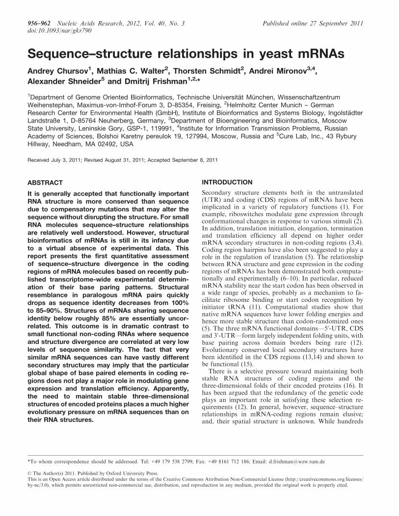
2.1 Sequence-structure Relationships in Yeast mR-
NAs
Andrey Chursov, Mathias C. Walter, Thorsten Schmidt, Andrei Mironov, Alexan-
der Shneider and Dmitrij Frishman
Nucleic Acids Res., 40(3):956-962, 2012
Structural bioinformatics of mRNAs is still in its infancy due to the absence of exper-
imentally known (rather than computationally predicted) structures. We provided
the first ever analysis of sequence-structure relationships in eukaryotic mRNAs based
on the experimental measurements of base pairing propensities published in 2010 by
Kertesz and colleagues in Nature (Kertesz et al., 2010). Our main finding is that
the relationship between sequence and structure divergence in mRNA molecules is
much weaker than in small RNAs, implying a high degree of evolutionary neutrality.
On a more general vein, the objective of our work was to analyze global structural
arrangements and their similarity as a function of sequence identity, similar in spirit
to the original work of Chothia and Lesk (Chothia and Lesk, 1986). In this work,
we focused on the comparison of experimentally determined as well as predicted
41
Chapter 2. Results
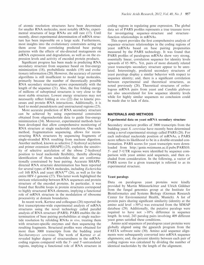
secondary structures for yeast mRNA sequences that encode paralogous proteins.
We considered potential correlations between sequences and mRNA structures. In
order to detect such correlations, we used large-scale experimental data on yeast
transcriptome RNA structure (Kertesz et al., 2010). We combined these data with
theoretical predictions and compared structural and sequence similarities for a num-
ber of yeast paralogous genes. The results demonstrated correlations for relatively
highly similar sequences (higher than 85-90%), and their absence for sequences with
lower similarity.
The result we obtained was not anticipated. To our surprise, we found that only
extremely similar sequences are folded into similar structures, while quite similar se-
quences, sharing as much as 80-85% identity, fold differently. Thanks to the Kertesz
et al. dataset, which was the first large-scale measurement of mRNA structures
ever published, we could then derive for the first time the quantitative dependence
between sequence and structure divergence in mRNAs. Such dependence was not
previously known.
The next obvious step was to compare mRNA structures for orthologous genes with
a similar distribution of nucleotide sequence identities to the distribution of similar-
ities between paralogs in S. cerevisiae. Does the structure diverge faster in paralogs
than in orthologs with comparable sequence similarity? For such a comparison, we
chose Candida glabrata, the closest organism with a completely sequenced genome,
and compared predicted structures in S. cerevisiae with C. glabrata.
In addition, we have made all sequence alignments, together with experimentally
determined and predicted structures, in FASTA format available as Supplementary
Files.
The research was designed by Dmitrij Frishman and me. I did the programming
and performed the research. The resulting data were analyzed by all the authors.
The paper was written by myself, Andrei Mironov and Dmitrij Frishman.
42
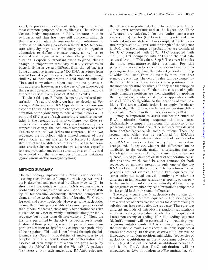
Sequence–structure relationships in yeast mRNAsAndrey Chursov1, Mathias C. Walter2, Thorsten Schmidt2, Andrei Mironov3,4,
Alexander Shneider5 and Dmitrij Frishman1,2,*
1Department of Genome Oriented Bioinformatics, Technische Universitat Munchen, WissenschaftzentrumWeihenstephan, Maximus-von-Imhof-Forum 3, D-85354, Freising, 2Helmholtz Center Munich – GermanResearch Center for Environmental Health (GmbH), Institute of Bioinformatics and Systems Biology, IngolstadterLandstraße 1, D-85764 Neuherberg, Germany, 3Department of Bioengineering and Bioinformatics, MoscowState University, Leninskie Gory, GSP-1, 119991, 4Institute for Information Transmission Problems, RussianAcademy of Sciences, Bolshoi Karetny pereulok 19, 127994, Moscow, Russia and 5Cure Lab, Inc., 43 RyburyHillway, Needham, MA 02492, USA
Received July 3, 2011; Revised August 31, 2011; Accepted September 8, 2011
ABSTRACT
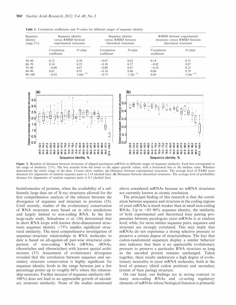
It is generally accepted that functionally importantRNA structure is more conserved than sequencedue to compensatory mutations that may alter thesequence without disrupting the structure. For smallRNA molecules sequence–structure relationshipsare relatively well understood. However, structuralbioinformatics of mRNAs is still in its infancy dueto a virtual absence of experimental data. Thisreport presents the first quantitative assessmentof sequence–structure divergence in the codingregions of mRNA molecules based on recently pub-lished transcriptome-wide experimental determin-ation of their base paring patterns. Structuralresemblance in paralogous mRNA pairs quicklydrops as sequence identity decreases from 100%to 85–90%. Structures of mRNAs sharing sequenceidentity below roughly 85% are essentially uncor-related. This outcome is in dramatic contrast tosmall functional non-coding RNAs where sequenceand structure divergence are correlated at very lowlevels of sequence similarity. The fact that verysimilar mRNA sequences can have vastly differentsecondary structures may imply that the particularglobal shape of base paired elements in coding re-gions does not play a major role in modulating geneexpression and translation efficiency. Apparently,the need to maintain stable three-dimensionalstructures of encoded proteins places a much higherevolutionary pressure on mRNA sequences than ontheir RNA structures.
INTRODUCTION
Secondary structure elements both in the untranslated(UTR) and coding (CDS) regions of mRNAs have beenimplicated in a variety of regulatory functions (1). Forexample, riboswitches modulate gene expression throughconformational changes in response to various stimuli (2).In addition, translation initiation, elongation, terminationand translation efficiency all depend on higher ordermRNA secondary structures in non-coding regions (3,4).Coding region hairpins have also been suggested to play arole in the regulation of translation (5). The relationshipbetween RNA structure and gene expression in the codingregions of mRNAs has been demonstrated both computa-tionally and experimentally (6–10). In particular, reducedmRNA stability near the start codon has been observed ina wide range of species, probably as a mechanism to fa-cilitate ribosome binding or start codon recognition byinitiator tRNA (11). Computational studies show thatnative mRNA sequences have lower folding energies andhence more stable structure than codon-randomized ones(5). The three mRNA functional domains—50-UTR, CDSand 30-UTR—form largely independent folding units, withbase pairing across domain borders being rare (12).Evolutionary conserved local secondary structures havebeen identified in the CDS regions (13,14) and shown tobe functional (15).
There is a selective pressure toward maintaining bothstable RNA structures of coding regions and thethree-dimensional folds of their encoded proteins (16). Ithas been argued that the redundancy of the genetic codeplays an important role in satisfying these selection re-quirements (12). In general, however, sequence–structurerelationships in mRNA-coding regions remain elusive;and, their spatial structure is unknown. While hundreds
*To whom correspondence should be addressed. Tel: +49 179 538 2799; Fax: +49 8161 712 186; Email: [email protected]
956–962 Nucleic Acids Research, 2012, Vol. 40, No. 3 Published online 27 September 2011doi:10.1093/nar/gkr790
� The Author(s) 2011. Published by Oxford University Press.This is an Open Access article distributed under the terms of the Creative Commons Attribution Non-Commercial License (http://creativecommons.org/licenses/by-nc/3.0), which permits unrestricted non-commercial use, distribution, and reproduction in any medium, provided the original work is properly cited.
of atomic resolution structures have been determinedfor smaller RNA molecules, most notably tRNAs, experi-mental structures of large RNAs are still rare (17). Untilrecently, direct experimental determination of mRNA struc-ture has been impossible on a large scale. Furthermore,most insights into the evolutionary constraints acting onthem arose from correlating predicted base paringpatterns with the effects of site-directed mutagenesis onmRNA expression and degradation, as well as on the ex-pression levels and activity of encoded protein products.
Significant progress has been made in predicting RNAsecondary structure from sequence based on free-energyminimization (18), probabilistic models (19) and evolu-tionary information (20). However, the accuracy of currentalgorithms is still insufficient to model large molecules,primarily because the number of theoretically possibleRNA secondary structures grows exponentially with thelength of the sequence (21). Also, the free folding energyof millions of suboptimal structures is very close to themost stable structure. Lowest energy structures may notnecessarily reflect folding in vivo (22) due to kinetic pro-cesses and protein–RNA interactions. Additionally, it ishard to model pseudoknots and unstructured regions (23).
More accurate prediction of RNA secondary structurecan be achieved by using experimental constraintsobtained from oligonucleotide data to guide free-energyminimization (24). Moreover, experimental methods havebeen developed that allow comprehensive monitoring ofRNA structure at single nucleotide resolution. One suchmethod, fragmentation sequencing, allows for recon-structing RNA structures by sequencing fragments ofsingle-stranded RNA resulting from nuclease digestion.Another method, known as selective 20-hydroxyl acylationand primer extension (SHAPE) (25), exploits the sensitiv-ity of selective acetylation of the ribose 20-hydroxylposition to local nucleotide flexibility, thereby allowingidentification of those nucleotides that are conforma-tionally constrained by base pairing. Accurate SHAPE-directed RNA structure determination has been reportedfor several types of RNA molecules, including Escherichiacoli 16S RNA and yeast tRNAasp (26), as well as for theentire HIV-1 genome (27). This latter work highlighted theintricate relationship between RNA sequences and proteinstructure of the encoded proteins. In particular, it wasfound that flexible loops in protein structures correspondto highly structured RNA elements, implying a functionalrole of mRNA structure in the modulation of ribosomeprocessivity at domain boundaries.
In recent work, Kertesz and colleagues (28) reported thefirst transcriptome-wide experimental analysis of mRNAstructures using the novel technology called parallelanalysis of RNA structure (PARS). PARS enables the de-termination of base pairing probabilities at single nucleo-tide resolution by refolding RNAs in vivo, treating themwith structure-specific enzymes and then sequencing theresulting fragments. Structural profiles were obtained formore than 3000 transcripts from the budding yeastSaccharomyces cerevisiae. The work of Kertesz et al.revealed higher degree of structuredness in the mRNA-coding regions compared with the 30- and 50-untranslatedregions, implying a functional role of RNA structure in
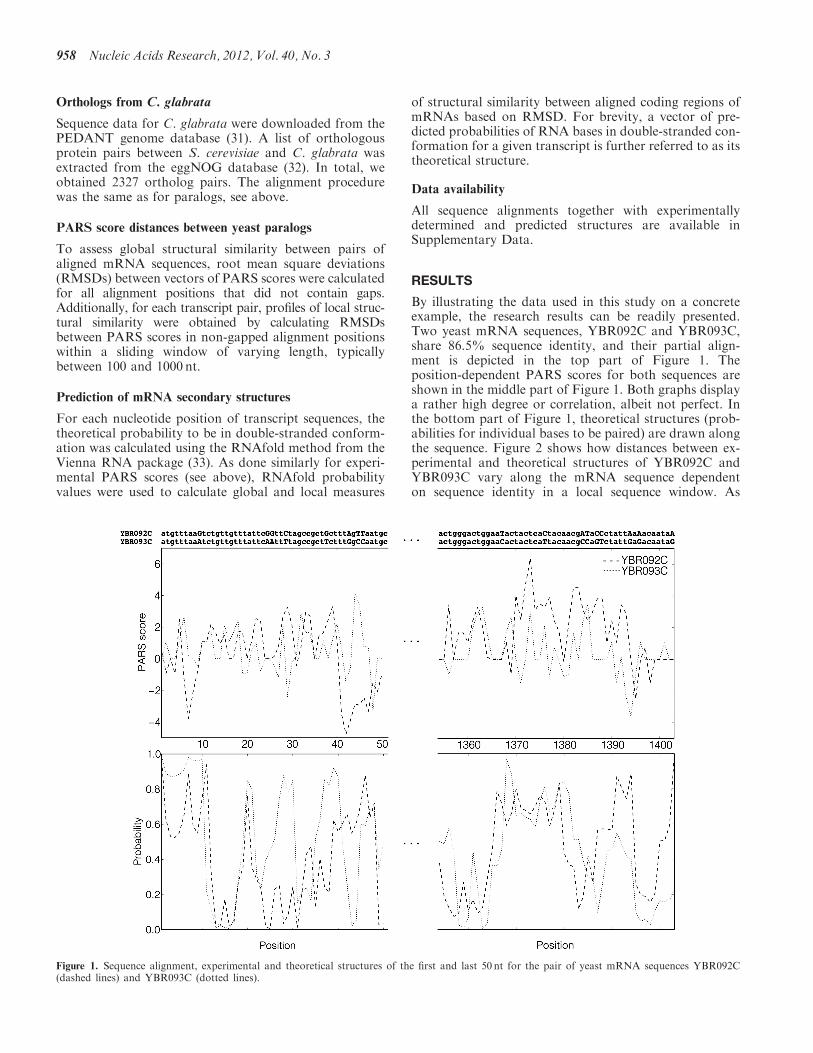
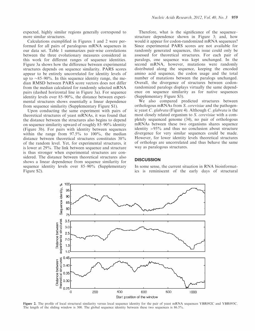
coding regions in regulating gene expression. The globaldata set of PARS profiles represents a true treasure trovefor investigating sequence–structure and structure–function relationships in mRNAs.This report provides the first comprehensive analysis of
sequence–structure relationships in the coding regions ofyeast mRNAs based on base pairing propensitiesmeasured by the PARS technology. It was found thatPARS profiles of paralogous mRNAs show very strong,essentially linear, correlation sequence for identity levelsupwards of 85–90%. Yet, pairs of more distantly relatedyeast transcripts secondary structure appear to be unre-lated. Interestingly, predicted secondary structures ofyeast paralogs display a similar behavior with respect tosequence identity; and, there is a significant correlationbetween experimental and theoretical structures, asnoted previously (28). Theoretical structures of ortho-logous mRNA pairs from yeast and Candida glabrataare also uncorrelated for low sequence identity levelswhile for highly similar sequences no conclusion couldbe made due to lack of data.
MATERIALS AND METHODS
Experimental data on yeast mRNA secondary structure
Secondary structure profiles of 3000 transcripts from thebudding yeast S. cerevisiae have recently been determinedusing a novel experimental strategy called PARS (28). Foreach individual nucleotide position of mRNAs, a PARSscore reflects its likelihood to be in a double-stranded con-formation. PARS scores for yeast transcripts were down-loaded from http://genie.weizmann.ac.il/pubs/PARS10.50- and 30-UTR regions were identified by sequence com-parison with yeast amino acid sequences, and then ex-cluded from consideration. In the following, a vector ofPARS scores for a given transcript is referred to as itsexperimental structure.
Yeast paralogs
Data on paralogous yeast proteins were kindlyprovided by Martin Munsterkotter and Ulrich Guldnerfrom the fungal genomics group at the Institute forBioinformatics and Systems Biology (German ResearchCenter for Environmental Health, Munich). A list ofprotein pairs sharing significant similarity (identity at theamino acid level >50%) was extracted from the SIMAPdatabase (29). Additionally, the putative paralogs wererequired to have not >10% difference in sequencelength. In total, 243 paralog pairs involving 409 differentyeast genes satisfied these conditions.Amino acid sequences of paralogous yeast proteins were
globally aligned using the ggsearch program from theFASTA software suite (30). Amino acid sequence align-ments were subsequently converted into mRNA sequencealignments; and, the percent identity between each pair ofcoding regions was calculated by dividing the number ofidentical nucleotides by the length of the alignment.
Nucleic Acids Research, 2012, Vol. 40, No. 3 957
Orthologs from C. glabrata
Sequence data for C. glabrata were downloaded from thePEDANT genome database (31). A list of orthologousprotein pairs between S. cerevisiae and C. glabrata wasextracted from the eggNOG database (32). In total, weobtained 2327 ortholog pairs. The alignment procedurewas the same as for paralogs, see above.
PARS score distances between yeast paralogs
To assess global structural similarity between pairs ofaligned mRNA sequences, root mean square deviations(RMSDs) between vectors of PARS scores were calculatedfor all alignment positions that did not contain gaps.Additionally, for each transcript pair, profiles of local struc-tural similarity were obtained by calculating RMSDsbetween PARS scores in non-gapped alignment positionswithin a sliding window of varying length, typicallybetween 100 and 1000 nt.
Prediction of mRNA secondary structures
For each nucleotide position of transcript sequences, thetheoretical probability to be in double-stranded conform-ation was calculated using the RNAfold method from theVienna RNA package (33). As done similarly for experi-mental PARS scores (see above), RNAfold probabilityvalues were used to calculate global and local measures
of structural similarity between aligned coding regions ofmRNAs based on RMSD. For brevity, a vector of pre-dicted probabilities of RNA bases in double-stranded con-formation for a given transcript is further referred to as itstheoretical structure.
Data availability
All sequence alignments together with experimentallydetermined and predicted structures are available inSupplementary Data.
RESULTS
By illustrating the data used in this study on a concreteexample, the research results can be readily presented.Two yeast mRNA sequences, YBR092C and YBR093C,share 86.5% sequence identity, and their partial align-ment is depicted in the top part of Figure 1. Theposition-dependent PARS scores for both sequences areshown in the middle part of Figure 1. Both graphs displaya rather high degree or correlation, albeit not perfect. Inthe bottom part of Figure 1, theoretical structures (prob-abilities for individual bases to be paired) are drawn alongthe sequence. Figure 2 shows how distances between ex-perimental and theoretical structures of YBR092C andYBR093C vary along the mRNA sequence dependenton sequence identity in a local sequence window. As
Figure 1. Sequence alignment, experimental and theoretical structures of the first and last 50 nt for the pair of yeast mRNA sequences YBR092C(dashed lines) and YBR093C (dotted lines).
958 Nucleic Acids Research, 2012, Vol. 40, No. 3
expected, highly similar regions generally correspond tomore similar structures.
Calculations exemplified in Figures 1 and 2 were per-formed for all pairs of paralogous mRNA sequences inour data set. Table 1 summarizes pair-wise correlationsbetween the three evolutionary measures considered inthis work for different ranges of sequence identities.Figure 3a shows how the difference between experimentalstructures depends on sequence similarity. PARS scoresappear to be entirely uncorrelated for identity levels ofup to �85–90%. In this sequence identity range, the me-dian RMSD between PARS score vectors does not differfrom the median calculated for randomly selected mRNApairs (dashed horizontal line in Figure 3a). For sequenceidentity levels over 85–90%, the distance between experi-mental structures shows essentially a linear dependencefrom sequence similarity (Supplementary Figure S1).
Upon conducting the same experiment with pairs oftheoretical structures of yeast mRNAs, it was found thatthe distance between the structures also begins to dependon sequence similarity upward of roughly 85–90% identity(Figure 3b). For pairs with identity between sequenceswithin the range from 97.5% to 100%, the mediandistance between theoretical structures constitutes 38%of the random level. Yet, for experimental structures, itis lower at 29%. The link between sequence and structureis thus stronger when experimental structures are con-sidered. The distance between theoretical structures alsoshows a linear dependence from sequence similarity forsequence identity levels over 85–90% (SupplementaryFigure S2).
Therefore, what is the significance of the sequence–structure dependence shown in Figure 3; and, howwould it appear for codon-randomized mRNA sequences?Since experimental PARS scores are not available forrandomly generated sequences, this issue could only beassessed for theoretical structures. For each pair ofparalogs, one sequence was kept unchanged. In thesecond mRNA, however, mutations were randomlydistributed along the sequence, keeping the encodedamino acid sequence, the codon usage and the totalnumber of mutations between the paralogs unchanged.Overall, the divergence of structures between codon-randomized paralogs displays virtually the same depend-ence on sequence similarity as for native sequences(Supplementary Figure S3).We also compared predicted structures between
orthologous mRNAs from S. cerevisiae and the pathogen-ic yeast C. glabrata (Figure 4). Although C. glabrata is themost closely related organism to S. cerevisiae with a com-pletely sequenced genome (34), no pair of orthologousmRNAs between these two organisms shares sequenceidentity >95% and thus no conclusion about structuredivergence for very similar sequences could be made.However, for lower identity levels theoretical structuresof orthologs are uncorrelated and thus behave the sameway as paralogous structures.
DISCUSSION
In some sense, the current situation in RNA bioinformat-ics is reminiscent of the early days of structural
Figure 2. The profile of local structural similarity versus local sequence identity for the pair of yeast mRNA sequences YBR092C and YBR093C.The length of the sliding window is 300. The global sequence identity between these two sequences is 86.5%.
Nucleic Acids Research, 2012, Vol. 40, No. 3 959
bioinformatics of proteins, when the availability of a suf-ficiently large data set of X-ray structures allowed for thefirst comprehensive analysis of the relation between thedivergence of sequence and structure in proteins (35).Until recently, studies of the evolutionary conservationof RNA structures were based on in silico predictionsand largely limited to non-coding RNA. In the firstlarge-scale study, Schudoma et al. (36) determined thatin short RNA loops with known three-dimensional struc-tures sequence identity >75% implies significant struc-tural similarity. The most comprehensive investigation ofsequence–structure relationships in RNA molecules todate is based on all-against-all pair-wise structural com-parison of non-coding RNAs (tRNAs, rRNAs,riboswitches and riboswitches) with known spatial archi-tectures (37). Assessment of evolutionary divergencerevealed that the correlation between sequence and sec-ondary structure conservation is highly significant forsequence identity levels in the range between just a fewpercentage points up to roughly 60% where this relation-ship saturates. Further increase of sequence similarity (60–100%) does not lead to an appreciable growth of second-ary structure similarity. None of the studies mentioned
above considered mRNAs because no mRNA structuresare currently known at atomic resolution.
The principal finding of this research is that the correl-ation between sequence and structure in the coding regionsof yeast mRNAs is much weaker than in small non-codingRNAs. Up to �85–90% sequence identity, the similarityof both experimental and theoretical base pairing pro-pensities between paralogous yeast mRNAs is at randomlevel; while, for more similar sequence pairs, sequence andstructure are strongly correlated. This may imply thatmRNAs do not experience a strong selective pressure topreserve a certain degree of structuredness. The fact thatcodon-randomized sequences display a similar behavioralso indicates that there is no appreciable evolutionarypressure to preserve a particular RNA structure as longas the encoded protein remains unchanged. Takentogether, these results underscore a high degree of evolu-tionary neutrality in yeast mRNA molecules, both at thelevel of primary (third codon position) and secondary(extent of base paring) structure.
On one hand, our findings are in strong contrast tomany non-coding RNAs and cis-acting regulatoryelements of mRNAs whose biological function is primarily
Figure 3. Boxplots of distances between structures of aligned paralogous mRNAs in different ranges of sequence similarity. Each box corresponds tothe range of similarity 2.5%. The box extends from the lower to the upper quartile values, with a horizontal line at the median value. Whiskersdemonstrate the entire range of the data. Crosses show outliers. (a) Distances between experimental structures. The average level of PARS scoredistances for alignments of random sequence pairs is 2.14 (dashed line). (b) Distances between theoretical structures. The average level of probabilitydistance for alignments of random sequence pairs is 0.5 (dashed line).
Table 1. Correlation coefficients and P-values for different ranges of sequence identity
mediated by their spatial architecture (38) stabilized bytertiary interactions, modified bases and interactionswith proteins and small ligands. On the other hand,sequence–structure relationships observed in this workare compatible with the notion that, in general, RNA mol-ecules do not have a single global structure. Instead, theyexist as a highly dynamic ensemble of alternative conform-ations (39,40) that are often capable of performing differ-ent functions (41). The extent of base pairing may play arole in the regulation of pre-mRNA splicing, translationand mRNA degradation. Both experimentally determinedPARS scores and computationally derived partition func-tions analyzed in this work are statistical measures thatreflect the propensity of each nucleotide to form a basepair across a large number of metastable structures.
This analysis has several important limitations. First,PARS probes RNA structures in vitro rather than in theliving cell and may not always reproduce functional RNAstructures (42). Second, even if the base paring informa-tion obtained by the PARS technology were perfectly cor-rect, it still merely represents a one-dimensional profile ofstructural propensities, a far cry from knowing the actualRNA secondary structure, let alone spatial architecture,for each individual molecule at any moment of time.Third, the findings do not rule out much strongersequence–structure correlations in certain local structuralelements of coding regions, such as reprogrammedgenetic-decoding signals (43) or mRNA localizationsignals. We also cannot rule out the possibility that thedegree of mRNA structuredness does have an importantfunctional role in spite of quick erosion of structural simi-larity between paralogs with diminishing sequence similar-ity, and that this erosion reflects functional differentiation.However, we consider such explanation unlikely becausethe same behavior is observed between orthologousmRNAs. Finally, only a small subset of the PARS dataconstituted by pairs of sequence similar yeast mRNAs(paralogs) was explored. As a next step, it will be exciting
to conduct comparative analyses of mRNA structuromes[the term coined by Westhof and Romby (44)], focusingon orthologous sequences from multiple organisms andtaking into account important genomic variables, suchas expression level and evolutionary rate. Given the cur-rent pace of high-throughput RNA analysis technologiesthere is no doubt that such data will become available inthe near future.
SUPPLEMENTARY DATA
Supplementary Data are available at NAR Online:Supplementary figures S1–S3.
ACKNOWLEDGEMENTS
We would like to thank Dmitry Ivankov and NatalyaBogatyreva for helpful discussions and Janusz Bujnickifor illuminating comments on the article.
FUNDING
The DFG International Research Training Group‘Regulation and Evolution of Cellular Systems’ (GRK1563) and by the Russian Foundation for BasicResearch (RFBR 09-04-92742). Funding for open accesscharge: German Research Center for EnvironmentalHealth, Munich.
Conflict of interest statement. None declared.
REFERENCES
1. Bevilacqua,P.C. and Blose,J.M. (2008) Structures, kinetics,thermodynamics, and biological functions of RNA hairpins.Annu. Rev. Phys. Chem., 59, 79–103.
2. Serganov,A. and Patel,D.J. (2007) Ribozymes, riboswitchesand beyond: regulation of gene expression without proteins.Nat. Rev. Genet., 8, 776–790.
3. Gray,N.K. and Hentze,M.W. (1994) Regulation of proteinsynthesis by mRNA structure. Mol. Biol. Rep., 19, 195–200.
4. Kozak,M. (2005) Regulation of translation via mRNA structurein prokaryotes and eukaryotes. Gene, 361, 13–37.
5. Katz,L. and Burge,C.B. (2003) Widespread selection for localRNA secondary structure in coding regions of bacterial genes.Genome Res., 13, 2042–2051.
6. Kudla,G., Murray,A.W., Tollervey,D. and Plotkin,J.B. (2009)Coding-sequence determinants of gene expression in Escherichiacoli. Science, 324, 255–258.
7. Duan,J., Wainwright,M.S., Comeron,J.M., Saitou,N.,Sanders,A.R., Gelernter,J. and Gejman,P.V. (2003) Synonymousmutations in the human dopamine receptor D2 (DRD2) affectmRNA stability and synthesis of the receptor. Hum. Mol. Genet.,12, 205–216.
8. Ilyinskii,P.O., Schmidt,T., Lukashev,D., Meriin,A.B., Thoidis,G.,Frishman,D. and Shneider,A.M. (2009) Importance of mRNAsecondary structural elements for the expression of influenza virusgenes. OMICS, 13, 421–430.
9. Carlini,D.B., Chen,Y. and Stephan,W. (2001) The relationshipbetween third-codon position nucleotide content, codon bias,mRNA secondary structure and gene expression in thedrosophilid alcohol dehydrogenase genes Adh and Adhr. Genetics,159, 623–633.
10. Nackley,A.G., Shabalina,S.A., Tchivileva,I.E., Satterfield,K.,Korchynskyi,O., Makarov,S.S., Maixner,W. and Diatchenko,L.(2006) Human catechol-O-methyltransferase haplotypes modulate
Figure 4. Boxplot of distances between theoretical structures of alignedorthologous mRNAs in different ranges of sequence similarity.Notation as in Figure 3.
Nucleic Acids Research, 2012, Vol. 40, No. 3 961
protein expression by altering mRNA secondary structure.Science, 314, 1930–1933.
11. Gu,W., Zhou,T. and Wilke,C.O. (2010) A universal trend ofreduced mRNA stability near the translation-initiation site inprokaryotes and eukaryotes. PLoS Comput. Biol., 6, e1000664.
12. Shabalina,S.A., Ogurtsov,A.Y. and Spiridonov,N.A. (2006) Aperiodic pattern of mRNA secondary structure created by thegenetic code. Nucleic Acids Res., 34, 2428–2437.
13. Meyer,I.M. and Miklos,I. (2005) Statistical evidence forconserved, local secondary structure in the coding regions ofeukaryotic mRNAs and pre-mRNAs. Nucleic Acids Res., 33,6338–6348.
14. Steigele,S., Huber,W., Stocsits,C., Stadler,P.F. and Nieselt,K.(2007) Comparative analysis of structured RNAs in S. cerevisiaeindicates a multitude of different functions. BMC Biol., 5, 25.
15. Olivier,C., Poirier,G., Gendron,P., Boisgontier,A., Major,F. andChartrand,P. (2005) Identification of a conserved RNA motifessential for She2p recognition and mRNA localization to theyeast bud. Mol. Cell. Biol., 25, 4752–4766.
16. White,H.B. III, Laux,B.E. and Dennis,D. (1972) Messenger RNAstructure: compatibility of hairpin loops with protein sequence.Science, 175, 1264–1266.
17. Holbrook,S.R. (2008) Structural principles from large RNAs.Annu. Rev. Biophys., 37, 445–464.
18. Mathews,D.H. and Turner,D.H. (2006) Prediction ofRNA secondary structure by free energy minimization.Curr. Opin. Struct. Biol., 16, 270–278.
19. Dowell,R.D. and Eddy,S.R. (2006) Efficient pairwise RNAstructure prediction and alignment using sequence alignmentconstraints. BMC Bioinformatics, 7, 400.
20. Bernhart,S.H. and Hofacker,I.L. (2009) From consensus structureprediction to RNA gene finding. Brief. Funct. Genomic Proteomic,8, 461–471.
21. Meyer,I.M. and Miklos,I. (2007) SimulFold: simultaneouslyinferring RNA structures including pseudoknots, alignments, andtrees using a Bayesian MCMC framework. PLoS Comput. Biol.,3, e149.
22. Mahen,E.M., Watson,P.Y., Cottrell,J.W. and Fedor,M.J. (2010)mRNA secondary structures fold sequentially but exchangerapidly in vivo. PLoS Biol., 8, e1000307.
23. Reeder,J. and Giegerich,R. (2004) Design, implementation andevaluation of a practical pseudoknot folding algorithm based onthermodynamics. BMC Bioinformatics, 5, 104.
24. Duan,S., Mathews,D.H. and Turner,D.H. (2006) Interpretingoligonucleotide microarray data to determine RNA secondarystructure: application to the 30 end of Bombyx mori R2 RNA.Biochemistry, 45, 9819–9832.
25. Merino,E.J., Wilkinson,K.A., Coughlan,J.L. and Weeks,K.M.(2005) RNA structure analysis at single nucleotide resolution byselective 20-hydroxyl acylation and primer extension (SHAPE).J. Am. Chem. Soc., 127, 4223–4231.
35. Chothia,C. and Lesk,A.M. (1986) The relation between thedivergence of sequence and structure in proteins. EMBO J., 5,823–826.
36. Schudoma,C., May,P., Nikiforova,V. and Walther,D. (2010)Sequence-structure relationships in RNA loops: establishing thebasis for loop homology modeling. Nucleic Acids Res., 38,970–980.
37. Capriotti,E. and Marti-Renom,M.A. (2010) Quantifying therelationship between sequence and three-dimensional structureconservation in RNA. BMC Bioinformatics, 11, 322.
38. Gruber,A.R., Bernhart,S.H., Hofacker,I.L. and Washietl,S. (2008)Strategies for measuring evolutionary conservation of RNAsecondary structures. BMC Bioinformatics, 9, 122.
39. Mironov,A.A., Dyakonova,L.P. and Kister,A.E. (1985) A kineticapproach to the prediction of RNA secondary structures.J. Biomol. Struct. Dyn., 2, 953–962.
40. Danilova,L.V., Pervouchine,D.D., Favorov,A.V. andMironov,A.A. (2006) RNAKinetics: a web server thatmodels secondary structure kinetics of an elongating RNA.J. Bioinform. Comput. Biol., 4, 589–596.
41. Zhao,P., Zhang,W.B. and Chen,S.J. (2010) Predicting secondarystructural folding kinetics for nucleic acids. Biophys. J., 98,1617–1625.
42. Mauger,D.M. and Weeks,K.M. (2010) Toward global RNAstructure analysis. Nat. Biotechnol., 28, 1178–1179.
43. Namy,O., Rousset,J.P., Napthine,S. and Brierley,I. (2004)Reprogrammed genetic decoding in cellular gene expression.Mol. Cell, 13, 157–168.
44. Westhof,E. and Romby,P. (2010) The RNA structurome:high-throughput probing. Nat. Methods, 7, 965–967.
962 Nucleic Acids Research, 2012, Vol. 40, No. 3
Chapter 2. Results
2.2 Specific Temperature-induced Perturbations
of Secondary mRNA Structures are Associ-
ated with the Cold-adapted Temperature-sensitive
Phenotype of Influenza A Virus
Andrey Chursov, Sebastian J. Kopetzky, Ignaty Leshchiner, Ivan Kondofersky,
Fabian J. Theis, Dmitrij Frishman and Alexander Shneider
RNA biology, 9(10):1266-1274, 2012
Influenza A viruses constitute a serious threat to human health and vaccines against
these viruses are of crucial importance. There are two types of vaccines against
influenza A viruses: inactivated viruses, which are widespread in Europe; and live
attenuated vaccines, which were developed in the USSR and are used in the USA.
Live attenuated vaccines are cold-adapted, temperature-sensitive (ca/ts) mutant in-
fluenza A viruses. Mutations in the three proteins constituting the viral polymerase
complex are thought to contribute to the ca/ts phenotype. However, several virus
studies have elegantly demonstrated that the thermodynamic stability of certain
RNA structures is critical for optimal virus replication (Berkhout et al., 1997; Mir-
momeni et al., 1997; Rowe et al., 2000). Hence, this article presents an interesting
new idea: cold-adapted, temperature-sensitive (ca/ts) phenotypes of influenza may
depend on the temperature dependence of the RNA secondary structure. To test
this hypothesis, we developed a new approach to predicting RNA sequences that
may be responsible for this effect.
In this work we addressed the question of the contribution of the viral mRNA struc-
tures to the cold-adapted, temperature-sensitive phenotype. We developed a new
bioinformatics tool to analyze the variations of the RNA structures when the tem-
perature is shifted from 32 to 39°C, and to compare these variations in the ca/ts viral
strains and in the parental viruses from which they are derived. We used RNAfold
to predict the probability of each nucleotide to be involved in base pairing (via
partition functions), rather than predicting secondary structures. We found that
the nucleotides, whose probability to be base-paired was most affected by temper-
50
2.2. Specific Temperature-induced Perturbations of Secondary mRNA Structures areAssociated with the Cold-adapted Temperature-sensitive Phenotype of Influenza A
Virus
ature, tend to cluster (organize into regions with several such changes). Moreover,
these clusters were different for wild type and ca/ts mutant viruses. By comparing
these viruses with a pool of artificial viral sequences in which synonymous muta-
tions were randomly introduced (wt sequences with same number of mutations as
in ca/ts sequences), we concluded that the existence of nine clusters in the ca/ts
mutant viruses, but not in the wild type viruses, statistically correlated with the
ca/ts phenotype. Additionally, the analysis revealed the existence of one cluster,
present in the wt strain but absent in the ca/ts mutant, which could be attributed
to introducing specific mutations causing the ca/ts phenotype.
It is worth noting that the length of the overlap between clusters (when we compare
the location of clusters from two different strains) affects the strength of the sta-
tistical conclusion concerning appearance/disappearance of clusters resulting from
randomly introduced mutations. The longer the overlap allowed, the easier it is to
conclude that two clusters are different. Thus, we have restricted ourselves to short
overlaps only. Any conclusion that two clusters are different, which was made based
on short overlaps, would remain to be true if one allows longer overlaps. By contrast,
a conclusion made on longer overlaps may not sustain a test with shorter overlaps.
Thus, our results present the lower limit.
We also tested if the statistically significant clusters we have observed co-locate
with those few RNA structures, which are already known for the influenza A virus.
None such overlaps were observed. However, a comprehensive analysis of influenza
RNA structures was never conducted. Several structures were discovered because
of their biological importance and/or viral impairment in case these structures are
disturbed. Still, relatively little is known about influenza virus RNA folding and its
influence on influenza virus replication (Gultyaev et al., 2010). Thus, our results
provide a rationale for testing experimentally whether RNA structures are indeed
present at the locations of the clusters of highly temperature-sensitive positions at
32 and 39°C.
The research was designed by Alexander Shneider and me. The programming and all
the computations were performed by me and Sebastian J. Kopetzky. The resulting
data were analyzed by all the authors. The paper was written by myself, Sebastian
J. Kopetzky, Dmitrij Frishman and Alexander Shneider.
*Correspondence to: Dmitrij Frishman and Alexander Shneider; Email: [email protected] and [email protected]: 07/17/12; Revised: 08/29/12; Accepted: 09/04/12http://dx.doi.org/10.4161/rna.22081
Introduction
Influenza vaccines have been a great public health priority1 and their future is man-made constructs created using molecular biology tools. Compared with other types of influenza vaccines, live attenuated influenza vaccines (LAIV) possess major advan-tages because of administration convenience and potency of the immune response.2 There are alternative approaches which can lead to viral attenuation and be utilized for LAIV design.3
Since the late 1960s, cold-adapted temperature-sensitive (ca/ts) LAIVs have become an important vaccination instrument in the USSR. The ca/ts phenotype leads to impaired growth at an elevated temperature of approximately 39°C4-9 while permitting viral growth at lower temperatures. Molecular mechanism(s) causing the ca/ts phenotype in influenza A viruses remain unclear. Significant effort was devoted to explaining temperature sensitivity through mutations in the coding regions and amino
For decades, cold-adapted, temperature-sensitive (ca/ts) strains of influenza A virus have been used as live attenuated vaccines. Due to their great public health importance it is crucial to understand the molecular mechanism(s) of cold adaptation and temperature sensitivity that are currently unknown. For instance, secondary RNA structures play important roles in influenza biology. Thus, we hypothesized that a relatively minor change in temperature (32–39°c) can lead to perturbations in influenza RNA structures and, that these structural perturbations may be different for mRNAs of the wild type (wt) and ca/ts strains. To test this hypothesis, we developed a novel in silico method that enables assessing whether two related RNA molecules would undergo (dis)similar structural perturbations upon temperature change. The proposed method allows identifying those areas within an RNA chain where dissimilarities of RNA secondary structures at two different temperatures are particularly pronounced, without knowing particular RNA shapes at either temperature. We identified such areas in the Ns2, pA, pB2 and Np mRNAs. however, these areas are not identical for the wt and ca/ts mutants. Differences in temperature-induced structural changes of wt and ca/ts mRNA structures may constitute a yet unappreciated molecular mechanism of the cold adaptation/temperature sensitivity phenomena.
Specific temperature-induced perturbations of secondary mRNA structures are associated with the cold-adapted temperature-sensitive
phenotype of influenza A virusAndrey chursov,1,† sebastian J. Kopetzky,1,† Ignaty Leshchiner,2,3 Ivan Kondofersky,4 Fabian J. Theis,4 Dmitrij Frishman1,4,‡,*
and Alexander shneider3,‡,*
1Department of Genome Oriented Bioinformatics; Technische Universität München; Wissenschaftzentrum Weihenstephan; Freising, Germany; 2Genetics Division; Brigham and Women’s hospital; harvard Medical school; Boston, MA UsA; 3cure Lab, Inc.; Needham, MA UsA; 4helmholtz center Munich - German Research center for environmental
health (Gmbh); Institute of Bioinformatics and systems Biology; Neuherberg, Germany
†These authors contributed equally to this work and should be regarded as joint first authors. ‡These authors contributed equally to this work and should be regarded as joint last authors.
acid changes. Jin et al. found that certain non-silent mutations in PB1, PB2 and NP might lead to temperature-sensitivity when induced in A/Ann Arbor/6/60.6 According to Song et al., three non-silent mutations in PB1 and one non-silent mutation in PB2 might lead to the ts phenotype.4 Youil et al. investigated several A/Leningrad/134/17/57 subclones and found that the most tem-perature-sensitive one had amino acid changes in the PB1, PA and NS1 genes.10 Furthermore, Snyder et al. found that it can be sufficient to induce the temperature-sensitive phenotype by replacing the two segments of coding for PA and M1/M2 of a wild type virus with those of A/Ann Arbor/6/60.11 Interestingly, in all these cases at least one subunit of the viral polymerase (PA, PB1 and PB2) is affected.
In addition to the attempts to explain the ca/ts phenotype through mutations in viral proteins, there were also reports implicating RNAs in temperature sensitivity. A promising find-ing was made by Dalton et al.,12 suggesting that, at an elevated
www.landesbioscience.com RNA Biology 1267
ReseARch pApeR ReseARch pApeR
Thurner et al.26 to investigate secondary structure conservation in Picornaviridae and Flaviviridae, respectively, and by Chursov et al.27 for elucidating sequence-structure relationships in yeast mRNAs. However, so far, partition functions have not been used to assess and compare structural RNA perturbations caused by temperature elevation.
Based on partition functions, we have developed a technique to identify RNA sequence regions where probabilities of nucleo-tide coupling change the most with temperature elevation. We demonstrate that dense areas of altered nucleotide coupling are not identical for closely related wt and ca/ts RNAs. Thus, although, we cannot predict the exact RNA structures, we know that these structures are changing differently with temperature elevation.
Results
The propensity of nucleotides to appear in double-stranded con-formations depends on temperature. As seen in Figure 3, all nucleotides change their base-pairing probabilities upon tem-perature elevation from 32°C to 39°C, with transitions from a double-stranded to a single-stranded conformation being expect-edly more frequent (see Table 2). Between 62.8% and 75.2% of positions in each mRNA change their probability to be coupled to a lower value. Furthermore, between 3.9% and 10.9% of nucleotides in each mRNA change their base-pairing probabili-ties significantly (more than three standard deviations below or above the mean over all seven temperature increments between 33–39°C and 32°C (see the Materials and Methods section and Table 3). In all but one mRNAs, the majority of significantly changing positions (between 52% and 88.6%) shows a decrease in their base-pairing probability, whereas this percentage is some-what lower (42.1%) for NS2 Arb/ca.
For each mRNA, we computed a density plot of signifi-cantly changing positions along the sequence as described in the Materials and Methods section (Fig. 2; Figs. S1–19). From these plots, it becomes immediately apparent that strongly tempera-ture-sensitive positions are not evenly or randomly distributed along the sequence but rather aggregate in clusters. The num-bers of clusters defined by the density-based algorithm for each mRNA are presented in Table 4. The only mRNA where no clus-ters were detected is NS2 Len/wt. The average length of clusters varies between 15.9 and 61.0 positions (Table 5) and the average density of significantly changing positions in the clusters is in the range of 22% to 53% (Table 6). Overall, very short clusters are required by the DBSCAN algorithm to have a very high density while the density in longer clusters can be as low as 21% (Fig. 4).
Furthermore, we found that patterns of cluster occurrence exhibit substantial differences between the wild type strains and their cold-adapted, temperature-sensitive mutants, as exemplified in Figure 1 for a subsequence of the PA mRNA. In this case, a cluster of significantly changing positions is observed in Len/17/ca but not in Len/wt. This figure demonstrates that a perturba-tion of mRNA structure begins at a temperature of approximately 37°C. Out of 218 clusters of temperature-sensitive positions, 126 clusters are present in both wt and ca/ts strains, 38 clusters are
temperature, viral polymerase tends to dissociate from the cRNA-promoter, thereby leading to a decreased vRNA synthesis while the synthesis of cRNA and mRNA remains approximately constant. A decrease in the synthesis of vRNA related to temper-ature sensitivity, which also maintained mRNA synthesis, was described by Chan et al.9 In a more general vein, RNAs can serve as intracellular thermometers.13 For example, a thermosensitive RNA switch was implicated in the propagation of tick-borne encephalitis virus.14 Recent publications suggest that, apart from RNA abundance, RNA structures may play a comparably impor-tant role. The importance of mRNA secondary structures for expression of influenza virus genes was recently demonstrated by Ilyinskii et al.15 Therefore, identification of previously unknown influenza RNA structures16 and the analysis of their functional roles are areas of increasing interest.17-19
We hypothesized that changing temperature causes pertur-bations in mRNA secondary structures, which contributes to the cold-adapted, temperature-sensitive phenotype. To test this hypothesis, we have developed a new in silico method of analysis to reveal if the structures of two closely related RNA molecules would react differently to temperature elevation. Unfortunately, it is not possible to reliably calculate exact structures of each RNA molecule at two temperatures, compare the differences between the two structures, and then evaluate whether or not these differ-ences are identical for two RNAs. First of all, at each particular temperature an RNA molecule may have different co-existing structures. Furthermore, since the number of possible structures increases rapidly with the length of the input sequence, the preci-sion of RNA structure predictions suffers. Another limitation of RNA secondary structure predictions is that taking pseudoknots into account makes the task non-deterministic polynomial-time hard (NP-hard).20 In this particular case NP-hard means that growth of RNA length elevates time necessary for computation to a restrictive duration. However, in support of our hypothesis, one does not need to know the exact structures before and after perturbation to conclude that the two structures have reacted differently. For example, if two windows are broken into a dif-ferent number of pieces by soccer balls, we need to know nei-ther the shapes of the windows and nor the exact forms of the pieces to conclude that the perturbations of the two glasses are not identical.
An ensemble of RNA structures can be represented via a parti-tion function,21,22 which is a sum of Boltzmann factors over every possible secondary structure. In using partition functions, one can calculate the probability for each nucleotide to be coupled within a double-stranded conformation.23,24 An advantage of partition functions is that they take into account not just the minimum free energy structure, but rather an ensemble of ener-getically favorable structures. Thus, if one adenine would be bound to a particular uracil within a single highly likely struc-ture, while another adenine would couple with ten uracils within ten less likely structures, parameters for these two adenines may be the same. Although partition functions are not precisely accurate, they are much more accurate than in silico predictions of the actual RNA structures. Partition functions were used instead of actual structures, for example, by Witwer et al.25 and
1268 RNA Biology Volume 9 Issue 10
length of these ca/ts associated clusters is in the range of 8 to 19 positions and the density of significantly changing positions in them varies between 32% and 80% (Table 7). All the clus-ters that can be associated with ca/ts phenotype are indicated in Figure 4D. The existence of such clusters suggests that the ca/ts phenotype may be associated with specific perturbations in mRNA secondary structures. Importantly, in all three ca/ts mutants, there are clusters located in the polymerase genes (PA or PB2), in line with previous reports where polymerase genes were consistently associated with the temperature-sensitive phenotype.4,6,10,11
Discussion
Temperature-sensitive mutants were reported for a variety of viruses.39-42 Several studies have demonstrated that thermody-namic stability of certain RNA structures is critical for virus rep-lication.43-45 Temperature-sensitive, anti-viral and anti-bacterial vaccines remain to be promising public health instruments.46-48 So far, cold-adapted temperature-sensitive anti-influenza vaccines have arguably made the largest contribution to the prevention of this infection around the world. Still, molecular mechanism(s) underlining the ca/ts influenza phenotype is poorly understood. Here, we have explored the hypothesis that ca/ts properties of known influenza strains can be (at least partially) explained by temperature-induced perturbations of mRNA structure.
It was, therefore, our intention to compare mRNAs at each of the temperatures of interest. However, despite the fact that sig-nificant attempts have been made toward theoretical predictions of RNA structure based on energy calculations24,34,49 and co-vari-ation analysis,50,51 it is still not possible to calculate secondary structures of mRNAs accurately using currently available algo-rithms. At the same time, experimental technologies to deter-mine RNA structures are only beginning to emerge52,53 and are barely available for a broad spectrum of research projects. Thus, we had to develop an indirect computational method aimed to assess if two RNA molecules change their shapes differently in response to temperature elevation.
At each temperature, we calculate probability vectors that contain, for each nucleotide position, the probability to be coupled with another nucleotide within the same RNA, form-ing a double-helix structure. Apparently, this coupling is tem-perature-sensitive, with increasing temperature generally leading to a reduced likelihood of “weak” structures. Thus, (1) differ-ent structures may constitute an ensemble for the same RNA at different temperatures, and/or (2) at different temperatures the same structures may be present with different abundance. Both of these options are valid and may coexist because, in each given cell, multiple copies of the same RNA molecules may be distrib-uted between alternative shapes.
The fact that the base-paring probability at each position within a probability vector changes with temperature elevation does not necessarily indicate that structural perturbations (or re-distribution of alternative RNA structures) equally involve each nucleotide. Thus, we selected only those nucleotide positions that exhibited the most significant changes of their coupling
present in wt strains but absent in ca/ts mutants, and 54 clus-ters are present in ca/ts mutants but absent in the wt counterpart (Fig. 4 and Supplemental Data).
The existence of clusters unique for ca/ts strains raises the question whether such clusters are associated with the mutations inducing the ca/ts phenotype or whether random mutations unrelated to the ca/ts phenotype would be as likely to induce these clusters. Likewise, one can ask whether the disappearance of some clusters present in wt strains from ca/ts mutants may be caused by particular ca/ts associated mutations. The best way to approach this problem would be to test whether or not the same pattern of cluster occurrence would be observed while compar-ing the wt strains investigated here with a high number of natu-rally occurring influenza virus strains as similar to the wt strains as their ca/ts mutants. However, there are currently not enough naturally occurring strains with the same extent of similarity to the wt as possessed by the ca/ts mutants.
We therefore compared wt sequences with computer-gen-erated mutants possessing random synonymous mutations unrelated to the phenotype of interest. This analysis revealed existence of only one cluster that is present in wt strain (Len/wt) but absent in ca/ts mutant and could be attributed to introduc-ing specific mutations causing the ca/ts phenotype (Table 7). The length of this cluster is 140 nucleotides and the density of significantly changing positions in it equals 38%. At the same time, there are nine clusters (one in Arb/ca, three in Len/17/ca, and five in Len/47/ca) present in ca/ts mutants, and not present in wt, that cannot be observed in the pool of in silico gener-ated random mutants with statistically significant P-values. The
Figure 1. comparison of significantly changing positions between the pA mRNA of Len/wt (upper 7 rows) and Len/17/ca (lower 7 rows). each row corresponds to a difference vector v32–33, …, v32–39 containing changes of base pairing probabilities between 32°c and a particular higher temperature. positions in which base paring probabilities signifi-cantly change with temperature elevation in both sequences and those where these changes only affect one of the phenotypes are marked blue and orange, respectively. Only the first 40 bases of each sequence are shown; position numbers of the coding sequence are indicated at the top of the alignment.
www.landesbioscience.com RNA Biology 1269
of mutations, one could conclude that the cluster disappearance and ca/ts behavior are associated. For all such clusters except one, a high number of computer-generated mutants, which are extremely unlikely to be ca/ts, also demonstrate disappearance of the same clusters. Thus, these clusters may simply correspond to temperature-sensitive regions within particular influenza mRNAs unrelated to ca/ts phenotype. Nevertheless, we did observe one cluster, which is associated with the ca/ts phenotype with statistically significant P-value. This cluster is present in the wt strain. It disappears specifically in the ca/ts mutant, but remains in the computer-generated mutants possessing the same number of mutations as the ca/ts one.
probabilities. We do not assert that if in two closely related RNA molecules the most temperature-sensitive positions coincide; these two RNA molecules undergo identical temperature-induced structural RNA perturbations. However, it is probably safe to assume that if two RNA variants manifest different nucleotide positions as the most temperature-sensitive ones within the prob-ability vector, temperature elevation influences the structures of these RNA molecules in a different way. Thus, we have proposed here a new technique aimed at identifying mutations that influ-ence temperature-dependent RNA behavior. The central finding upon which our approach is based is that temperature-sensitive positions are not randomly distributed along the length of RNA but rather form distinct clusters. We speculate that such clusters of temperature-sensitive positions may be located within RNA domains that change their shapes particularly strongly with tem-perature elevation. Although developed for a particular purpose, our method can be applied for studying the role of RNA struc-ture perturbations in a wide range of temperature-related bio-logical phenomena, such as the evolution of warm-bloodedness, thermophilic adaptation of prokaryptic organisms, or suscepti-bility of parasites and pathogens to increases in host temperature.
Differences in clusters of temperature-sensitive positions are a potential indicator that RNA structures of mutants react dif-ferently to temperature change. This raises the question whether these differences can be a causative factor for (or, at least, associ-ated with) the unique ca/ts behavior of the particular influenza virus strains under study. We identified three types of clusters of temperature-sensitive positions that are (1) present in both wt and ca/ts mutants, (2) present in wt, but absent in ca/ts mutants, and (3) absent in wt, but appear in the ca/ts mutants. We, there-fore, first tested whether the disappearance of some clusters in the mutants can indicate that they are causative for a rare pheno-type, ca/ts. If these clusters would disappear in ca/ts mutants but remain in non-ca/ts RNA variants possessing the same number
Figure 2. Distributions of significantly changing positions along the pB2 mRNAs of Arb/wt and Arb/ca. A sliding window of size 20 was moved in steps of 1 position over the vector v32–39 and the percentage of significantly changing positions in the window was calculated for each possible starting position. The resulting density plots are depicted in Figure 2A and Figure 2C. Location of clusters of significantly changing positions identified by the DBscAN algorithm are depicted in Figure 2B and Figure 2D with gray color. synonymous and non-synonymous mutations are depicted in Figure 2B and Figure 2D with red and blue vertical lines, respectively.
Figure 3. The histogram of differences of the probability values of nucleotides to be in a double-stranded conformation for pB1 Arb/wt upon temperature change between 32°c and 39°c. The vector of prob-abilities for 32°c was subtracted from the vector for 39°c.
1270 RNA Biology Volume 9 Issue 10
underlying mechanism of the ca/ts behavior of influenza virus. Further research in this direction might contribute to the rational design of live-attenuated influenza vaccines.
Materials and Methods
Sequences. In our analysis, we have used the cold-adapted, temperature-sensitive mutants A/Ann Arbor/6/60 (Arb/ca) stemming from the wild type (Arb/wt) with the same name and the two mutants A/Leningrad/134/17/57 (Len/17/ca) and A/Leningrad/134/47/57 (Len/47/ca) stemming from the wild type (wt) A/Leningrad/134/57 (Len/wt). Since information on the location of UTRs was not available, only coding regions were used for the analysis. Information on the locations and sequences of coding regions was retrieved from EMBL-ENA (European Nucleotide Archive).28 However, these sequences were adapted according to the publications where they originally were reported29,30 since the mutations annotated in the database were not in agreement with those papers, and no further references were given. The files containing final sequences, used in the cur-rent analysis, are presented in the Supplementary Data.
The influenza A genome is composed of eight segments encod-ing 12 proteins: three polymerase subunits (PB1, PB2, and PA),
Applying the same computational approach, we then tested if appearance of clusters of temperature-sensitive positions in ca/ts mutants, which are lacking in wt, is a phenotype-specific phenomenon. Based on comparisons with computer-generated mutants, we have demonstrated that nine particular clusters are unlikely to appear in mutants other than ca/ts. Thus, we hypoth-esize that changes in RNA structure caused by raising tempera-ture could be a potential factor contributing to the molecular mechanisms of the temperature-sensitive and/or cold-adapted phenotype in influenza A.
Direct experimental evidence both on secondary structures of mRNAs and their interactions partners will be required to eluci-date the exact role of temperature-induced structural changes in the acquisition of the ca/ts phenotype. For example, it is conceiv-able that conformational changes of influenza mRNA may play a role through altering the RNA ability to associate/dissociate with proteins and other molecules. Also, it cannot be ruled out that temperature-induced structural changes in the untranslated regions, which we have not considered in our analysis, contrib-ute to the ca/ts phenotype. The current scarcity of sequence data for temperature-sensitive strains and their wild type coun-terparts notwithstanding, we here propose the hypothesis that temperature-induced structural RNA perturbations may be an
Figure 4. Density of significantly changing positions in determined clusters vs. length. (A) clusters that occur only in wt mRNAs but are not statistically significant (37). (B) clusters occurring in both wt and ca/ts mRNAs (126). (C) clusters that occur only in ca/ts mutants but are not statistically significant (45). (D) statistically significant clusters (9 of them occur only in ca/ts mutants and 1 of them occurs only in wt mRNA). Different colors show different numbers of clusters that have identical values of length and density. Black, one cluster; red, two clusters; green, three clusters; blue, four clusters.
www.landesbioscience.com RNA Biology 1271
it appeared at some temperature and remained to be such at all higher temperatures.
Comparison of significantly changing positions between wt and ca/ts strains. To test whether significant temperature-induced structural changes in secondary RNA structures are the same or different for wild type strains and their cold-adapted, temperature-sensitive counterparts, we designed a visualization method allowing simultaneous comparison of temperature-induced changes for two RNAs. For example, Figure 1 depicts a comparison of significantly changing positions between Len/wt and Len/17/ca for a subsequence of the PA mRNA.
Visualization of significantly changing positions demon-strated that such positions are not evenly distributed along the sequences but rather have a tendency to aggregate into clusters, i.e. regions with a high density of significantly changing posi-tions. As a tool to analyze such clusters, we employed density plots obtained by sliding a 20-base long window over the vec-tor v
32–39 and calculating the percentage of significantly changing
positions in the window for each possible starting position. For example, Figures 2A and 2C depict density plots for the PB2 mRNAs of Arb/wt and Arb/ca, respectively.
Identification of clusters of temperature-sensitive positions. We further sought to provide a definition of clusters of changing positions for each RNA, focusing on the difference vectors v
32–39.
To these difference vectors, we applied the density-based spatial clustering of applications with noise (DBSCAN) algorithm.36,37 This algorithm needs two parameters as input, a distance thresh-old r and a density threshold MinPts. For a given set of points D (in our case the set of significantly changing positions in mRNA according to the difference vector v
32–39), the density of every
point pi from D is calculated as the number of points q
i that are
within a radius r around pi. If q
i > MinPts, then the point p
i is
classified as a core point. If the distance between two points is less than r, then they are said to be directly-connected. Two points are considered density-connected if they are connected to core points and these core points are, in turn, density-connected. A cluster is constructed as a maximally connected component of the set of points that have a distance of smaller than r to some core point. We used the implementation of DBSCAN from the scikit-learn Python module38 with a distance threshold r equal to 11 and a density threshold MinPts equals 4.
Generation of randomly mutated mRNAs. In order to assess whether the appearance of clusters of temperature-sensitive posi-tions is specific for mutations inducing the ca/ts phenotype or whether random mutations unrelated to ca/ts phenotype would be as likely to induce these clusters, we adopted an approach similar to that employed in our previous paper.27 For each wt mRNA, a data set consisting of 1000 mutant sequences was generated in silico. Each in silico generated variant contained the same number of mutations as the respective ca/ts mutant. All computer-generated mutations were synonymous ones and introduced into the sequences randomly. It is safe to assume that none (or extremely few) of the randomly generated in silico mutants would possess the ca/ts phenotype if tested in vitro and/or in vivo. Significantly changing positions in the sequences
a small proapoptotic mitochondrial protein (PB1-F2), hemagglu-tinin (HA), neuraminidase (NA), the nucleoprotein (NP), the matrix protein M1, an integral membrane protein M2, and the two nonstructural proteins NS1 and NS2.31 Recently, Wise et al. showed that PB1 gene segment also encodes a twelfth gene prod-uct, N-terminally truncated version on the polypeptide, N40.32 Sequences for NA and HA were not taken into consideration since these segments do not stem from attenuated viruses in the reassortant live vaccines, and thus cannot be associated with the temperature-sensitive phenotype. For all other genes, the num-bers of single nucleotide polymorphisms (SNPs) in the coding sequences of the ca/ts mutants compared with their wild type counterparts are presented in Table 1.
Identification of significantly changing positions. For the first step, we wanted to identify those nucleotides within each mRNA that are the most prone to changing their coupling pat-tern with temperature elevation. These nucleotides would cor-respond to the most temperature labile positions within RNA chains. To achieve this goal, we proposed and implemented a new technique as discussed here.
At each particular temperature, an RNA sequence consisting of N nucleotides can be presented by a vector of probabilities (hereinafter referred to as “probability vector”) for each nucleo-tide to be in a double-stranded conformation at this tempera-ture. Thus, we substitute a sequence of N ribonucleotides with a sequence of N real numbers between 0.0 and 1.0. Then, we calcu-late the probability vectors for each of the influenza mRNAs for the temperatures 32°C up to 39°C (in increments of 1°C) using the RNAfold tool from the Vienna RNA package (v.1.8.5)23,24,33-
35 with the command line option –noLP that disallows base pairs that can only occur as helices of length 1. Performing the above described procedure, eight probability vectors were gener-ated for each mRNA. Seven difference vectors v
32–33, …, v
32–39
were calculated from the probability vectors for 33°C to 39°C for the same RNA and the vector at 32°C, containing the set of differences between the value for each position of the prob-ability vector at higher temperature and the value for the same position at lower temperature. These positions in difference vec-tors of each mRNA that possess values more than three standard deviations apart from the mean calculated over all values of the seven difference vectors were considered temperature-sensitive. Such “significantly changing” positions are presumed to result from perturbations in secondary RNA structures due to the tem-perature elevation. Furthermore, to filter out possible calculation artifacts, we considered a position temperature-sensitive only if
Table 1. The number of sNps in the coding sequences of the ca/ts mu-tants compared with their wild type counterparts.
Strain M1 M2 NP NS1 NS2 PA PB1 PB2
Arb/ca 0 1 2 1 1 3 7 7
Len/17/ca 1 2 0 0 1 3 3 1
Len/47/ca 1 2 1 0 1 3 4 3
The sequences of the ca/ts mutants of the M1, M2, Ns2 and pA genes in Len/17/ca and Len/47/ca are identical.
1272 RNA Biology Volume 9 Issue 10
from the artificial data sets were determined as described above and used to calculate clusters of changing positions by apply-ing the DBSCAN algorithm. Clusters from computer-generated sequences were compared with the clusters from naturally occur-ring wt and ca/ts mutants.
Statistical tests. For each particular cluster of interest identi-fied in wt and/or the ca/ts mutants, the frequency of its occurrence in the in silico generated mutants was calculated. Using these fre-quencies we conducted a statistical analysis to test if occurrence/disappearance of a particular cluster is associated with the ca/ts phenotype. For each cluster, which we observed in a ca/ts mutant but not in the wt, we tested the null hypothesis (H
0) that the
probability to observe this cluster among the computer generated sequences was 5% or higher. Conversely, for each cluster, which was observed in the wt but not in ca/ts strain, the null hypoth-esis (H
0) was that the probability to observe this cluster was less
than 95%. In other words, a low frequency means that a cluster, which we observe in naturally occurring ca/ts strain although it is absent in the wt, is unlikely to occur by chance. Thus, the appearance of this cluster is likely to be associated with the ca/ts phenotype. Similarly, the fact that a cluster was present in the wt but disappeared in the ca/ts mutant can only be explained by the ca/ts phenotype if the probability to observe this cluster in the random mutants is 95% or higher.
To that end, we used one-sided binomial tests. The signifi-cance level for the test was Bonferroni-corrected by dividing the significance level of 5% by the total number of clusters in that sequence. H
0 was rejected for P-values lower than the adjusted
significance level. For these calculations, a cluster was considered to be ‘present’ in an artificial sequence if that sequence contained
Table 3. The number of nucleotides in each mRNA where the base pairing probability decreases (increases) significantly (more than three standard deviations from the mean over all temperature differences between 33°c to 39°c compared with 32°c) upon temperature change between 32°c and 39°c compared with other nucleotides in the same mRNA
Table 2. The number of positions in each mRNA where the probability of nucleotides to be in a double-stranded conformation decreases (increases) upon temperature elevation from 32°c to 39°c
There are no positions at which the probability to be paired upon temperature change between 32°c and 39°c remains unchanged. here, and in all subsequent tables for those mRNAs that were not considered in the analysis due to the absence of mutations, values are not shown.
Table 4. The number of clusters in each mRNA as determined by the DBscAN algorithm
Strain M1 M2 NP NS1 NS2 PA PB1 PB2
Arb/wt - 1 7 5 3 5 12 9
Arb/ca - 1 8 4 1 10 9 11
Len/wt 4 1 6 - 0 10 15 11
Len/17/ca 5 1 - - 2 11 9 10
Len/47/ca 5 1 8 - 2 11 10 10
Table 5. Average cluster length in each mRNA
Strain M1 M2 NP NS1 NS2 PA PB1 PB2
Arb/wt - 42.0 24.9 27.0 18.3 19.4 37.8 58.1
Arb/ca - 58.0 15.9 21.5 45.0 28.6 34.9 34.9
Len/wt 22.5 42.0 26.5 - - 25.0 34.9 39.9
Len/17/ca 19.8 61.0 - - 23.0 21.5 38.9 33.3
Len/47/ca 19.8 61.0 19.5 - 23.0 21.5 35.0 29.0
Table 6. Average density of significantly changing positions inside clusters in each mRNA
Strain M1 M2 NP NS1 NS2 PA PB1 PB2
Arb/wt - 0.43 0.39 0.38 0.45 0.37 0.44 0.39
Arb/ca - 0.38 0.53 0.36 0.22 0.34 0.43 0.42
Len/wt 0.41 0.40 0.36 - - 0.38 0.37 0.41
Len/17/ca 0.46 0.38 - - 0.26 0.48 0.44 0.41
Len/47/ca 0.46 0.38 0.46 - 0.26 0.48 0.44 0.44
www.landesbioscience.com RNA Biology 1273
Acknowledgments
The authors gratefully acknowledge the support of the TUM Graduate School’s Thematic Graduate Center Regulation and Evolution of Cellular Systems (RECESS) at the Technische Universität München.
Supplemental Materials
Supplemental materials may be found here: www.landesbioscience.com/journals/cc/article/22081
a cluster overlapping, by at least one position, with the cluster from the real sequence.
Disclosure of Potential Conflicts of Interest
No potential conflicts of interest were disclosed.
Funding
The DFG International Research Training Group ‘Regulation and Evolution of Cellular Systems’ (GRK 1563) and the Russian Foundation for Basic Research (RFBR 09–04–92742).
References1. Stöhr K, Kieny MP, Wood D. Influenza pandemic
vaccines: how to ensure a low-cost, low-dose option. Nat Rev Microbiol 2006; 4:565-6; PMID:16888876; http://dx.doi.org/10.1038/nrmicro1482.
2. Belsey MJ, de Lima B, Pavlou AK, Savopoulos JW. Influenza vaccines. Nat Rev Drug Discov 2006; 5:183-4; PMID:16557657; http://dx.doi.org/10.1038/nrd1988.
3. Ilyinskii PO, Thoidis G, Shneider AM. Development of a vaccine against pandemic influenza viruses: current status and perspectives. Int Rev Immunol 2008; 27:392-426; PMID:19065349; http://dx.doi.org/10.1080/08830180802295765.
4. Song H, Nieto GR, Perez DR. A new generation of modified live-attenuated avian influenza viruses using a two-strategy combination as potential vaccine can-didates. J Virol 2007; 81:9238-48; PMID:17596317; http://dx.doi.org/10.1128/JVI.00893-07.
5. Falcón AM, Marión RM, Zürcher T, Gómez P, Portela A, Nieto A, et al. Defective RNA replication and late gene expression in temperature-sensitive influenza viruses expressing deleted forms of the NS1 protein. J Virol 2004; 78:3880-8; PMID:15047804; http://dx.doi.org/10.1128/JVI.78.8.3880-3888.2004.
6. Jin H, Lu B, Zhou H, Ma C, Zhao J, Yang CF, et al. Multiple amino acid residues confer temperature sensi-tivity to human influenza virus vaccine strains (FluMist) derived from cold-adapted A/Ann Arbor/6/60. Virology 2003; 306:18-24; PMID:12620793; http://dx.doi.org/10.1016/S0042-6822(02)00035-1.
7. Jin H, Zhou H, Lu B, Kemble G. Imparting tem-perature sensitivity and attenuation in ferrets to A/Puerto Rico/8/34 influenza virus by transferring the genetic signature for temperature sensitivity from cold-adapted A/Ann Arbor/6/60. J Virol 2004; 78:995-8; PMID:14694130; http://dx.doi.org/10.1128/JVI.78.2.995-998.2004.
8. Tsfasman TM, Markushin SG, Akopova II, Ghendon YZ. Molecular mechanisms of reversion to the ts+ (non-temperature-sensitive) phenotype of influen-za A cold-adapted (ca) virus strains. J Gen Virol 2007; 88:2724-9; PMID:17872525; http://dx.doi.org/10.1099/vir.0.83014-0.
9. Chan W, Zhou H, Kemble G, Jin H. The cold adapted and temperature sensitive influenza A/Ann Arbor/6/60 virus, the master donor virus for live attenuated influ-enza vaccines, has multiple defects in replication at the restrictive temperature. Virology 2008; 380:304-11; PMID:18768193; http://dx.doi.org/10.1016/j.virol.2008.07.027.
10. Youil R, Kiseleva I, Kwan WS, Szymkowiak C, Toner TJ, Su Q, et al. Phenotypic and genetic analyses of the heterogeneous population present in the cold-adapted master donor strain: A/Leningrad/134/17/57 (H2N2). Virus Res 2004; 102:165-76; PMID:15084398; http://dx.doi.org/10.1016/j.virusres.2004.01.026.
11. Snyder MH, Clements ML, De Borde D, Maassab HF, Murphy BR. Attenuation of wild-type human influ-enza A virus by acquisition of the PA polymerase and matrix protein genes of influenza A/Ann Arbor/6/60 cold-adapted donor virus. J Clin Microbiol 1985; 22:719-25; PMID:4056002.
12. Dalton RM, Mullin AE, Amorim MJ, Medcalf E, Tiley LS, Digard P. Temperature sensitive influenza A virus genome replication results from low thermal stability of polymerase-cRNA complexes. Virol J 2006; 3:58; PMID:16934156; http://dx.doi.org/10.1186/1743-422X-3-58.
13. Shamovsky I, Ivannikov M, Kandel ES, Gershon D, Nudler E. RNA-mediated response to heat shock in mammalian cells. Nature 2006; 440:556-60; PMID:16554823; http://dx.doi.org/10.1038/nature04518.
14. Elväng A, Melik W, Bertrand Y, Lönn M, Johansson M. Sequencing of a tick-borne encephalitis virus from Ixodes ricinus reveals a thermosensitive RNA switch significant for virus propagation in ectothermic arthropods. Vector Borne Zoonotic Dis 2011; 11:649-58; PMID:21254926; http://dx.doi.org/10.1089/vbz.2010.0105.
15. Ilyinskii PO, Schmidt T, Lukashev D, Meriin AB, Thoidis G, Frishman D, et al. Importance of mRNA secondary structural elements for the expression of influenza virus genes. OMICS 2009; 13:421-30; PMID:19594376; http://dx.doi.org/10.1089/omi.2009.0036.
17. Gultyaev AP, Fouchier RA, Olsthoorn RC. Influenza virus RNA structure: unique and common features. Int Rev Immunol 2010; 29:533-56; PMID:20923332; http://dx.doi.org/10.3109/08830185.2010.507828.
Table 7. Unique clusters potentially associated with the ca/ts phenotype. The p-values of all clusters in one sequence were checked against Bonferroni-corrected significance levels. For each Bonferroni correction, the total number of clusters located in the corresponding sequence was used (11 clusters in Arb/ca pB2, two clusters in Len/17/ca and Len/47/ca Ns2, 11 clusters in Len/17/ca and Len/47/ca pA, ten clusters in Len/17/ca and Len/47/ca pB2, eight clusters in Len/47/ca Np, 11 clusters in Len/wt pB2).
Strain Sequence PositionOccurrence in the random data seta
95% confidence intervalb P-value
Arb/ca pB2 329–336 15 [0.0, 0.023] 3.34e-09
Len/17/ca Ns2 290–308 16 [0.0, 0.024] 1.11e-08
Len/17/ca pA 93–101 32 [0.0, 0.043] 0.0037
Len/17/ca pB2 1293–1310 9 [0.0, 0.016] 5.24e-13
Len/47/ca Np 1017–1026 29 [0.0, 0.039] 0.0007
Len/47/ca Np 1178–1192 29 [0.0, 0.039] 0.0007
Len/47/ca Ns2 290–308 16 [0.0, 0.024] 1.11e-08
Len/47/ca pA 93–101 32 [0.0, 0.043] 0.0037
Len/47/ca pB2 808–818 3 [0.0, 0.008] 1.36e-18
Len/wt pB2 1490–1629 982 [0.973, 1.0] 1.03e-07
aThe number of times a particular cluster was found in a data set of 1000 sequences with randomly introduced mutations. bestimated range of values which is likely to include the probability to find a particular cluster with the probability of 95%.
1274 RNA Biology Volume 9 Issue 10
43. Berkhout B, Klaver B, Das AT. Forced evolution of a regulatory RNA helix in the HIV-1 genome. Nucleic Acids Res 1997; 25:940-7; PMID:9023102; http://dx.doi.org/10.1093/nar/25.5.940.
44. Mirmomeni MH, Hughes PJ, Stanway G. An RNA tertiary structure in the 3' untranslated region of enteroviruses is necessary for efficient replication. J Virol 1997; 71:2363-70; PMID:9032373.
45. Rowe A, Ferguson GL, Minor PD, Macadam AJ. Coding changes in the poliovirus protease 2A compen-sate for 5'NCR domain V disruptions in a cell-specific manner. Virology 2000; 269:284-93; PMID:10753707; http://dx.doi.org/10.1006/viro.2000.0244.
46. Sugimoto M, Yamanouchi K. Characteristics of an attenuated vaccinia virus strain, LC16m0, and its recombinant virus vaccines. Vaccine 1994; 12:675-81; PMID:8091843; http://dx.doi.org/10.1016/0264-410X(94)90215-1.
47. White MD, Bosio CM, Duplantis BN, Nano FE. Human body temperature and new approaches to constructing temperature-sensitive bacterial vaccines. Cell Mol Life Sci 2011; 68:3019-31; PMID:21626408; http://dx.doi.org/10.1007/s00018-011-0734-2.
48. Collins PL, Murphy BR. New generation live vaccines against human respiratory syncytial virus designed by reverse genetics. Proc Am Thorac Soc 2005; 2:166-73; PMID:16113487; http://dx.doi.org/10.1513/pats.200501-011AW.
49. Reuter JS, Mathews DH. RNAstructure: software for RNA secondary structure prediction and analysis. BMC Bioinformatics 2010; 11:129; PMID:20230624; http://dx.doi.org/10.1186/1471-2105-11-129.
50. Knudsen B, Hein J. Pfold: RNA secondary structure prediction using stochastic context-free grammars. Nucleic Acids Res 2003; 31:3423-8; PMID:12824339; http://dx.doi.org/10.1093/nar/gkg614.
51. Bernhart SH, Hofacker IL, Will S, Gruber AR, Stadler PF. RNAalifold: improved consensus structure pre-diction for RNA alignments. BMC Bioinformatics 2008; 9:474; PMID:19014431; http://dx.doi.org/10.1186/1471-2105-9-474.
52. Kertesz M, Wan Y, Mazor E, Rinn JL, Nutter RC, Chang HY, et al. Genome-wide measurement of RNA secondary structure in yeast. Nature 2010; 467:103-7; PMID:20811459; http://dx.doi.org/10.1038/nature09322.
53. Watts JM, Dang KK, Gorelick RJ, Leonard CW, Bess JW Jr., Swanstrom R, et al. Architecture and secondary structure of an entire HIV-1 RNA genome. Nature 2009; 460:711-6; PMID:19661910; http://dx.doi.org/10.1038/nature08237.
31. Nelson MI, Holmes EC. The evolution of epi-demic influenza. Nat Rev Genet 2007; 8:196-205; PMID:17262054; http://dx.doi.org/10.1038/nrg2053.
32. Wise HM, Foeglein A, Sun J, Dalton RM, Patel S, Howard W, et al. A complicated message: Identification of a novel PB1-related protein trans-lated from influenza A virus segment 2 mRNA. J Virol 2009; 83:8021-31; PMID:19494001; http://dx.doi.org/10.1128/JVI.00826-09.
33. Zuker M, Stiegler P. Optimal computer folding of large RNA sequences using thermodynamics and aux-iliary information. Nucleic Acids Res 1981; 9:133-48; PMID:6163133; http://dx.doi.org/10.1093/nar/9.1.133.
34. Hofacker IL, Fontana W, Stadler PF, Bonhoeffer LS, Tacker M, Schuster P. Fast Folding and Comparison of Rna Secondary Structures. Monatsh Chem 1994; 125:167-88; http://dx.doi.org/10.1007/BF00818163.
35. Bompfünewerer AF, Backofen R, Bernhart SH, Hertel J, Hofacker IL, Stadler PF, et al. Variations on RNA folding and alignment: lessons from Benasque. J Math Biol 2008; 56:129-44; PMID:17611759; http://dx.doi.org/10.1007/s00285-007-0107-5.
36. Ester M, Kriegel H-P, Sander J, Xu X. A density-based algorithm for discovering clusters in large spatial data-bases with noise. Proceedings of the 2nd International Conference on Knowledge Discovery and Data MIning 1996:226-31.
37. Kriegel H-P, Kröger P, Sander J, Zimek A. Density-based clustering. Wiley Interdisciplinary Reviews: Data Mining and Knowledge Discovery 2011; 1:231-40; http://dx.doi.org/10.1002/widm.30.
38. Pedregosa F, Varoquaux G, Gramfort A, Michel V, Thirion B, Grisel O, et al. Scikit-learn: Machine Learning in Python. J Mach Learn Res 2011; 12:2825-30.
39. Kung YH, Huang SW, Kuo PH, Kiang D, Ho MS, Liu CC, et al. Introduction of a strong temperature-sensi-tive phenotype into enterovirus 71 by altering an amino acid of virus 3D polymerase. Virology 2010; 396:1-9; PMID:19906393; http://dx.doi.org/10.1016/j.virol.2009.10.017.
40. Lulla V, Sawicki DL, Sawicki SG, Lulla A, Merits A, Ahola T. Molecular defects caused by temperature-sensitive mutations in Semliki Forest virus nsP1. J Virol 2008; 82:9236-44; PMID:18596091; http://dx.doi.org/10.1128/JVI.00711-08.
41. Fan Y, Zhao Q, Zhao Y, Wang Q, Ning Y, Zhang Z. Complete genome sequence of attenuated low-temperature Thiverval strain of classical swine fever virus. Virus Genes 2008; 36:531-8; PMID:18401695; http://dx.doi.org/10.1007/s11262-008-0229-x.
42. Sparks JS, Donaldson EF, Lu X, Baric RS, Denison MR. A novel mutation in murine hepatitis virus nsp5, the viral 3C-like proteinase, causes temperature-sensitive defects in viral growth and protein processing. J Virol 2008; 82:5999-6008; PMID:18385240; http://dx.doi.org/10.1128/JVI.00203-08.
18. Priore SF, Moss WN, Turner DH. Influenza A virus coding regions exhibit host-specific global ordered RNA structure. PLoS ONE 2012; 7:e35989; PMID:22558296; http://dx.doi.org/10.1371/journal.pone.0035989.
19. Motard J, Rouxel R, Paun A, von Messling V, Bisaillon M, Perreault JP. A novel ribozyme-based prophy-laxis inhibits influenza A virus replication and protects from severe disease. PLoS ONE 2011; 6:e27327; PMID:22110627; http://dx.doi.org/10.1371/journal.pone.0027327.
20. Lyngso RB. Complexity of pseudoknot prediction in simple models. Lect Notes Comput Sci 2004; 3142:919-31; http://dx.doi.org/10.1007/978-3-540-27836-8_77.
21. Pipas JM, McMahon JE. Method for predicting RNA secondary structure. Proc Natl Acad Sci USA 1975; 72:2017-21; PMID:1056009; http://dx.doi.org/10.1073/pnas.72.6.2017.
22. Zuker M, Sankoff D. Rna Secondary Structures and Their Prediction. Bull Math Biol 1984; 46:591-621.
23. McCaskill JS. The equilibrium partition function and base pair binding probabilities for RNA secondary struc-ture. Biopolymers 1990; 29:1105-19; PMID:1695107; http://dx.doi.org/10.1002/bip.360290621.
24. Mathews DH. Using an RNA secondary structure partition function to determine confidence in base pairs predicted by free energy minimization. RNA 2004; 10:1178-90; PMID:15272118; http://dx.doi.org/10.1261/rna.7650904.
25. Witwer C, Rauscher S, Hofacker IL, Stadler PF. Conserved RNA secondary structures in Picornaviridae genomes. Nucleic Acids Res 2001; 29:5079-89; PMID:11812840; http://dx.doi.org/10.1093/nar/29.24.5079.
26. Thurner C, Witwer C, Hofacker IL, Stadler PF. Conserved RNA secondary structures in Flaviviridae genomes. J Gen Virol 2004; 85:1113-24; PMID:15105528; http://dx.doi.org/10.1099/vir.0.19462-0.
27. Chursov A, Walter MC, Schmidt T, Mironov A, Shneider A, Frishman D. Sequence-structure relation-ships in yeast mRNAs. Nucleic Acids Res 2012; 40:956-62; PMID:21954438; http://dx.doi.org/10.1093/nar/gkr790.
28. Kanz C, Aldebert P, Althorpe N, Baker W, Baldwin A, Bates K, et al. The EMBL Nucleotide Sequence Database. Nucleic Acids Res 2005; 33(Database issue):D29-33; PMID:15608199; http://dx.doi.org/10.1093/nar/gki098.
29. Cox NJ, Kitame F, Kendal AP, Maassab HF, Naeve C. Identification of sequence changes in the cold-adapted, live attenuated influenza vaccine strain, A/Ann Arbor/6/60 (H2N2). Virology 1988; 167:554-67; PMID:2974219.
30. Klimov AI, Cox NJ, Yotov WV, Rocha E, Alexandrova GI, Kendal AP. Sequence changes in the live attenuated, cold-adapted variants of influ-enza A/Leningrad/134/57 (H2N2) virus. Virology 1992; 186:795-7; PMID:1733114; http://dx.doi.org/10.1016/0042-6822(92)90050-Y.
2.3. RNAtips: Analysis of Temperature-induced Changes of RNA Secondary Structure
2.3 RNAtips: Analysis of Temperature-induced
Changes of RNA Secondary Structure
Andrey Chursov, Sebastian J. Kopetzky, Gennady Bocharov, Dmitrij Frishman
and Alexander Shneider
Nucleic Acids Res., 41(W1):W486-W491, 2013
This article describes a web server for predicting temperature-induced changes in
RNA secondary structure. A wide range of biological phenomena (e.g. elevation
of body temperature due to an illness, adaptation to environmental temperature
conditions, biology of cold-blooded vs. warm-blooded organisms) is related to the
temperature effect on the organism. Although, thousands of papers have been pub-
lished attempting to explain temperature effects through perturbations of protein
structures, it has been almost impossible to assess if changes in RNA structures may
contribute to these effects. The web server we have developed closes this gap and
enables researchers to study temperature-induced perturbation of RNA structures.
Perturbations of secondary RNA structures may play an important role in an organ-
ism’s reaction to temperature change. The RNAtips (temperature-induced perturbation
of structure) web server can be used to predict regions of RNA secondary structures
that are likely to undergo structural changes when prompted by a change in tem-
perature. For a single RNA sequence, RNAtips identifies those nucleotides that
have the greatest reaction to temperature change and the temperature-sensitive
clusters of such nucleotides. When the research goal is to compare two RNA se-
quences and identify if they react to a temperature change differently, the locations
of temperature-sensitive clusters within the two RNAs are compared. If the two
sequences are homologs with a limited number of codon substitutions, an analysis
can be performed to demonstrate if the difference in the location of temperature-
sensitive clusters between the two sequences is specific to these particular nucleotide
substitutions, or if it could be achieved with the same number of random mutations
(synonymous or non-synonymous). The type of random computer-introduced mu-
tations depends on whether the input sequences are coding or non-coding.
For input into RNAtips, one or two RNA sequences of the same length must be
61
Chapter 2. Results
provided in FASTA-format (the header can be omitted). These sequences can ei-
ther be uploaded as a text file (each file may only contain one sequence) or directly
pasted into an input field. Sequence length is restricted to 9999 nucleotides. Ad-
ditionally, a user has to select a temperature range for which the RNA structural
perturbation should be calculated (the default is 32°C – 39°C). The minimal and
the maximal temperatures allowed are 0°C and 99°C, respectively. Furthermore, the
maximal temperature difference (i.e. tmax− tmin + 1) is restricted to 20°C (e.g. 30°C– 49°C). The advanced options allow the setting of the following: (i) a significance
threshold (see the following paragraph on processing), (ii) the DBSCAN parameters
ε and MinPts (the defaults are 11 for ε and 5 for MinPts), and (iii) parameters for
RNAfold in order to decide if GU-pairs should be allowed.
In addition, to avoid incorrect attribution of coding or non-coding input sequences,
we implemented a checkbox where the user can directly indicate that the input se-
quence(s) is (are) non-coding. If a user does not set this checkbox, then by default
the server assumes that the input sequence(s) is (are) coding ones. Also, if a dataset
of random mutants is generated, we indicate directly what type of mutations was
introduced at the link for downloading this dataset. In the case of coding sequences,
it says: “only synonymous mutations were introduced”; otherwise, it says: “random
mutations were introduced.”
Processing is based on the method described in the previous paper. For each temper-
ature in the given range, the probabilities of each nucleotide to be coupled within a
double-stranded conformation are calculated with the RNAfold tool (Hofacker et al.,
1994). For a temperature range [tmin : tmax], the probability-differences for each nu-
cleotide position for (tmin +1)− tmin, ..., tmax− tmin are then calculated. A threshold
is applied (the default is three standard deviations) to the resulting distribution in
order to identify the most temperature-sensitive positions. By using the DBSCAN
algorithm (Ester et al., 1996) on the vector tmax − tmin, clusters of significantly
changing positions are identified. P-values are obtained by applying one-sided bino-
mial tests to evaluate the occurrence of identified clusters in the mutant dataset.
The HTML output provides colored visualization of temperature-sensitive positions
and clusters. In addition, a table displays information on identified significant po-
sitions and clusters, including their total number as well as their average length
62
2.3. RNAtips: Analysis of Temperature-induced Changes of RNA Secondary Structure
and density. A second table shows the exact location of the clusters (if any). Re-
sults from the statistical evaluation (if conducted) are presented in a third table
demonstrating whether the difference between two RNAs in their reaction to the
temperature change is due to specific nucleotide substitutions or not. The output
also contains histograms that were used to identify significantly changing positions
(three sigmas away from the mean). In addition, there is a figure that shows the
relationship between the length and density of the clusters. Finally, there is another
figure that demonstrates the density of the most temperature-sensitive positions over
the whole RNA sequence, together with the location of clusters and the location of
nucleotide substitutions.
All figures and additional information, including sequences of the mutants generated
for the statistical analysis, can be downloaded by the RNAtips user.
The project was designed by Alexander Shneider and me. The web server has
been developed by me with an assistance of Sebastian J. Kopetzky and Gennady
Bocharov. The paper was written by myself, Sebastian J. Kopetzky, Dmitrij Frish-
man and Alexander Shneider.
63
RNAtips: analysis of temperature-induced changesof RNA secondary structureAndrey Chursov1, Sebastian J. Kopetzky1, Gennady Bocharov2, Dmitrij Frishman1,3,* and
Alexander Shneider4,*
1Department of Genome Oriented Bioinformatics, Technische Universitat Munchen, WissenschaftzentrumWeihenstephan, Maximus-von-Imhof-Forum 3, D-85354 Freising, Germany, 2Institute of Numerical Mathematics,Russian Academy of Sciences, Gubkina str. 8, 119333 Moscow, Russia, 3Helmholtz Center Munich-GermanResearch Center for Environmental Health (GmbH), Institute of Bioinformatics and Systems Biology, IngolstadterLandstraße 1, D-85764 Neuherberg, Germany and 4Cure Lab, Inc., 43 Rybury Hillway, Needham, MA 02492,USA
Received March 6, 2013; Revised May 1, 2013; Accepted May 11, 2013
ABSTRACT
Although multiple biological phenomena are relatedto temperature (e.g. elevation of body temperaturedue to an illness, adaptation to environmental tem-perature conditions, biology of coldblooded versuswarm-blooded organisms), the molecular mechan-isms of these processes remain to be understood.Perturbations of secondary RNA structures mayplay an important role in an organism’s reaction totemperature change—in all organisms from virusesand bacteria to humans. Here, we present RNAtips(temperature-induced perturbation of structure)web server, which can be used to predict regionsof RNA secondary structures that are likely toundergo structural alterations prompted by tem-perature change. The server can also be used to:(i) detect those regions in two homologous RNA se-quences that undergo different structural perturb-ations due to temperature change and (ii) testwhether these differences are specific to the par-ticular nucleotide substitutions distinguishing thesequences. The RNAtips web server is freely ac-cessible without any login requirement at http://rnatips.org.
INTRODUCTION
Structural perturbations in RNA molecules induced bytemperature change may have important biological impli-cations. For instance, the stability of mRNA structural
elements in 50-untranslated regions correlates with thetranslation rate in Saccharomyces cerevisiae (1). Anotherexample is the temperature-sensitivity of cold-adaptedinfluenza vaccine strains. For decades, it was a conundrumwhy wild-type influenza strains react differently toelevated temperature than their cold-adapted tempera-ture-sensitive counterparts. Recently, it has been demo-nstrated that this difference in temperature sensitivitymay be due to the difference in temperature-induced per-turbations in mRNA secondary structures (2). Perhaps,the most widely known example is RNA thermometers,which at a particular temperature alter their structure, andregulate translation of heat-shock, cold-shock and viru-lence genes (3–8). Usually, RNA thermometers arelocated in 50-untranslated regions, and their structuresmelt at an elevated temperature thereby permitting ribo-somes to initiate the translation process.
There are several experimental approaches to measuringthe melting temperature of an RNA structure (9),including ultraviolet absorbance (10,11), Fuorescence-based techniques (12,13) and thermal gradient electro-phoresis (14–16). Recently, temperature stability ofRNA structural elements was assessed on a genome-wide basis (17). The Parallel Analysis of RNAStructures with Temperature Elevation technique wasapplied to the yeast transcriptome, and relative meltingtemperatures for RNA structures were obtained byprobing RNA structures at different temperatures from23 to 75�C. As a result of this assessment, thousands ofpotential RNA thermometers and highly temperature-stable structures were identified.
Temperature-induced perturbations of RNA structuresmay play crucial, and yet unknown, biological role(s) in a
*To whom correspondence should be addressed. Tel: +1 609 841 1201; Fax: +1 310 446 9814; Email: [email protected] may also be addressed to Dmitrij Frishman. Tel: +49 8161 712134; Fax: +49 8161 71 2186; Email: [email protected]
The authors wish it to be known that, in their opinion, the first two authors should be regarded as joint First Authors.
The RNAtips web server can be accessed at http://rnatips.org. This website is free and open to all users, and there is no login requirement.
W486–W491 Nucleic Acids Research, 2013, Vol. 41, Web Server issue Published online 12 June 2013doi:10.1093/nar/gkt486
� The Author(s) 2013. Published by Oxford University Press.This is an Open Access article distributed under the terms of the Creative Commons Attribution License (http://creativecommons.org/licenses/by/3.0/), whichpermits unrestricted reuse, distribution, and reproduction in any medium, provided the original work is properly cited.
at UB
der TU
Muenchen on June 22, 2013
http://nar.oxfordjournals.org/D
ownloaded from
variety of processes. Elevation of body temperature is themost common symptom of many illnesses. The effects ofelevated body temperature on RNA structures both inpathogens and their hosts are still unknown, althoughthey may constitute a defense mechanism. Additionally,it would be interesting to assess whether RNA tempera-ture sensitivity plays an evolutionary role in organismadaptation to different climate zones, as well as toseasonal and day–night temperature change. The latterquestion is especially important owing to global climatechange. Is temperature sensitivity of RNA structures inbacteria living in geysers different from that of bacterialiving at negative temperatures? Do RNA structures fromwarm-blooded organisms react to the temperature changesimilarly to their counterparts in cold-blooded animals?These and many other questions could not be systematic-ally addressed, however, as (to the best of our knowledge)there is no convenient instrument to identify and comparetemperature-sensitive regions of RNA molecules.
To close this gap, RNAtips (temperature-induced per-turbation of structure) web server has been developed. Fora single RNA sequence, RNAtips identifies (i) those nu-cleotides for which temperature change causes appreciablealteration of the probability to formWatson–Crick (W–C)pairs and (ii) clusters of such temperature-sensitive nucleo-tides. If the research goal is to compare two RNA se-quences and identify whether they react differently to atemperature change, the locations of temperature-sensitiveclusters within the two RNAs are compared. If the twosequences are homologs with a limited number of basesubstitutions, an analysis can be performed to demon-strate whether the difference in location of the tempera-ture-sensitive clusters between the two sequences is specificto these particular nucleotide substitutions, or if it couldbe achieved with the same number of random mutations(synonymous and/or non-synonymous).
METHOD SUMMARY
The methodology implemented in RNAtips web server forassessing such impacts of temperature change was previ-ously described and published by Chursov et al. (2). Inshort, each nucleotide within an RNA sequence has aprobability of being paired via W–C bonds. This probabil-ity is temperature dependent; therefore, temperaturechanges influence the probability of forming W–C pairsfor each and every nucleotide. However, some nucleotideschange their pairing probabilities to a much greater extentthan others. Moreover, these highly temperature-sensitivenucleotides may not be evenly distributed along the RNAsequence but rather form distinct clusters (2). Thus, thefirst task performed by the RNAtips web server is identi-fication of those positions, which are prone through tem-perature elevation to significantly change their probabilityof being paired. This task is performed through the fol-lowing steps. Step 1: Probabilities of nucleotides to becoupled within a double-stranded conformation areassessed at each temperature within the given range byusing the RNAfold tool of the ViennaRNA package(18). Step 2: For each nucleotide, RNAtips calculates
the difference in probability for it to be in a paired stateat the lower temperature and at the higher one. Thesedifferences are calculated for the entire temperaturerange (t1 : t2) [i.e. for (t1+1) � t1, . . . , t2� t1] and thencombined into one data set. For example, if the tempera-ture range is set to 32–39�C and the length of the sequenceis 1000, then the changes of probabilities are consideredfor 33�C compared with 32�C, 34�C compared with32�C, . . . , 39�C compared with 32�C, and the final dataset would contain 7000 values. Step 3: The server identifiesthe most temperature-sensitive positions. For thispurpose, the server selects those values (and their corres-ponding nucleotides) from the data set generated in Step2, which are distant from the mean by more than threestandard deviations (the default value can be changed bythe user). The server then considers these positions to bethe most temperature-sensitive, and they are then mappedon the original sequence. Furthermore, clusters of signifi-cantly changing positions are then identified by applyingthe density-based spatial clustering of applications withnoise (DBSCAN) algorithm to the locations of such pos-itions. The server default action is to apply the clusteranalysis algorithm only to the highest temperature differ-ences t2-t1, (32–39
�C in the previous example) (19,20).It may be important to assess whether structures of
RNA molecules sharing sequence similarity react(dis)similarly to temperature change. For simplicity of ex-planation, assume that one RNA sequence was derivedfrom another sequence via some mutations. Then, thesecond task, which can be performed by RNAtipsserver, is to identify whether structures of two homolo-gous RNA sequences react differently to the temperaturechange and, if they do, whether this difference can beattributed to the specific mutations separating the twohomologous sequences. Thus, if a user inputs two se-quences, RNAtips identifies clusters of temperature-sensi-tive positions, which could be either common for bothsequences or uniquely present in only one of the twoRNA molecules. If the clusters of temperature-sensitivepositions are not identical for the two sequences, theserver offers statistical analysis identifying whether thedifference in temperature sensitivity is specific to the par-ticular nucleotide substitutions naturally differentiatingthe sequences or whether any set of mutations comparablein size could lead to the same difference.Therefore, assume that N nucleotide substitutions dif-
ferentiate sequence A from sequence B. The server gener-ates a data set of derivative sequences for A introducing Nsubstitutions into each derivative sequence. There are twodifferent methods of introducing random substitutionsinto a sequence(s) depending on whether the sequence(s)is(are) non-coding or coding. If A is a coding sequence(default), mutants will be generated by introducing syn-onymous mutations only. If A is a non-coding sequence,the user should mark a checkbox: ‘The input sequence(s)is(are) non-coding’. In this case, in silico mutations will beintroduced at random positions mimicking frequencies ofnucleotide substitutions naturally occurring between Aand B (e.g. if 25% of nucleotide substitutions between Aand B are T->C, then T->C substitutions will beintroduced in 25% of random in silico mutations). For
Nucleic Acids Research, 2013, Vol. 41, Web Server issue W487
at UB
der TU
Muenchen on June 22, 2013
http://nar.oxfordjournals.org/D
ownloaded from
each computer-generated sequence, the server will calcu-late its clusters of the temperature-sensitive positions asdescribed earlier in the text. If sequence B has asequence-specific cluster of temperature-sensitive positionsnot present in A, some of in silico derivatives of A maypossess clusters overlapping with the sequence-specificcluster observed in B. Let us assume that 1% or less ofcomputer-generated sequences possess such clustersoverlapping with the sequence-specific cluster in B. Thismeans that 99% of random mutation sets did not lead tothe appearance of this sequence-specific cluster of tem-perature sensitivity specific for sequence B, but not forA. Thus, one can conclude that the RNA structure ofsequence B reacts to the temperature change differentlythan the structure of sequence A because it possesses aspecific set of mutations as opposed to just N non-specific mutations. The RNAtips server performs a statis-tical analysis calculating a P-value for every sequence-specific cluster by performing a one-sided binomial test.For sequence-specific clusters occurring in the firstsequence but not in the second one, the null hypothesis(H0) is that the probability to observe this cluster is<95%. Consequently, a small P-value shows that thecluster is unlikely to disappear in the second sequence bychance. For sequence-specific clusters occurring in thesecond sequence but not in the first one, the null hypoth-esis (H0) is that the probability to observe this clusteramongst the mutants generated in silico is �5%.Therefore, a small P-value shows that the cluster isunlikely to appear in the second sequence by chance.
WEB SERVER
Input data
The input for RNAtips consists of either one or two RNAsequences of the same length that should be provided inFASTA-format (the header can be omitted). The se-quences can either be uploaded as text files (each filemay contain only one sequence), or the sequences maybe directly pasted into an input field. The sequences maycontain the characters A, C, G, U and T (for further com-putations, all Thymidines will be replaced with Uracilsautomatically). The maximal length of the sequences islimited to 9999 nt. To see an example of possible inputsequences, the user can click on the ‘sample’ link on theStart page. Influenza strains A/Leningrad/134/57 and itscold-adapted temperature-sensitive mutant A/Leningrad/134/47/57 are used as sample sequences.Additionally, a user has to specify two temperatures t1
and t2 (in�C) to define the temperature range (t1 : t2) for
which the RNA structural perturbation should becalculated (the default range is 32–39�C). t2 is the tempera-ture for which the actual cluster identification will be per-formed. The minimal allowed temperature is 0�C, and themaximal allowed temperature is 99�C. Furthermore, themaximal allowed temperature difference (i.e. t2 - t1+1) isrestricted to 20�C (e.g. 30–49�C).If a user inputs two sequences, two options are avail-
able. The default option is to perform a statistical analysisof the sequence-specific clusters identified in each of the
two sequences and to test whether these sequence-specificclusters result from the particular set of mutations distin-guishing the sequences. However, this analysis takes sometime and may be unnecessary for the particular user. Inthis case, the user can choose the checkbox for the ‘Don’tCreate a Mutant Dataset’ option (this option is onlyrelevant for two input sequences).
An advanced user can deviate from this default setupand input his parameters of choice. It was described in the‘Method Summary’ section that the statistical thresholdfor identifying significantly changing positions is 3.0standard deviations. However, in a custom calculation, auser may also choose a threshold level other than 3.0. Thenext two advanced parameters, e and MinPts, are bothparameters for the clustering algorithm called DBSCAN.As the first step, the algorithm randomly selects one sig-nificantly changing position. MinPts specifies the minimalnumber of significantly changing positions in a cluster. especifies the distance from the chosen nucleotide. If thenumber of significant positions specified by MinPts islocated within distance e, the sequence segment is thenconsidered part of a cluster. The default values for e andMinPts are 11 and 5, respectively.
Finally, a user can then select checkbox options: ‘Don’tallow GU pairs at the end of helices’ and/or ‘Don’t allowGU pairs’. These selections instruct the server whether GUpairs should be considered in calculations when theprobabilities of nucleotides to be in a double-stranded con-firmation at any given temperature are calculated. Thesetwo checkboxes are converted into the –noCloseGU and–noGU parameters of RNAfold during the calculations ofprobabilities of nucleotides to be coupled.
Server output
At the top of the results page from RNAtips, the HTMLoutput provides colored visual representation of identifiedtemperature-sensitive positions (Figure 1). The leftcolumn contains values for each temperature within thetemperature range (t1 : t2). The right column presents theinput sequence with those nucleotides—which are themost temperature-sensitive at this temperature—markedin either blue or orange color. The header line presentsthe FASTA header of the sequence(s). Position numbersare indicated under the header line. In the case of twoinput sequences, a line between the results for both se-quences indicated matching positions with ‘j’ andmismatches (mutations) with ‘-’. Additionally, significantpositions that are sequence specific to one of the two se-quences only are displayed in orange color. Positions thatchange their probabilities to be paired significantly in bothsequences are displayed in blue color. If only one sequenceis used as an input, then all positions demonstrating thehighest potency to change their likelihood of forming W–C bonds are displayed in orange color. In addition, theHTML output demonstrates the temperature initiating aperturbation of the RNA structure.
All tables and figures presenting more detailed resultsare shown in the lower part of the page and described inthe following paragraphs. The first table displays generalinformation on identified significant positions and
W488 Nucleic Acids Research, 2013, Vol. 41, Web Server issue
at UB
der TU
Muenchen on June 22, 2013
http://nar.oxfordjournals.org/D
ownloaded from
clusters. For every input sequence, it has the followingfields: ‘Sequence’ (shows the ID of the input sequence);‘#significant positions/total length’ (the number of signifi-cantly changing positions and the total length of the inputsequence); ‘signif. pos.< 0/signif. pos.> 0’ [the numbers ofsignificantly changing positions that decrease (or increase)their probability to be paired with temperature elevation];‘Number of clusters’ (the total number of identifiedclusters of significantly changing positions); ‘Avg. clusterdensity’ (the average density of significantly changing pos-itions in the identified clusters); and ‘Avg. cluster length’(the average length of the identified clusters). The prob-ability difference values are calculated by subtracting thevalue at the highest temperature from the value at thelowest temperature (p39�C - p32�C for the previousexample). Cluster density is calculated as the number ofsignificantly changing positions in a cluster divided by thetotal length of the cluster.
If a sequence contains 1000nt and the temperaturechanges from 32 to 39�C, there are 7000 values reflectinghow much each nucleotide would change its probability toform W–C couples when the temperature increases from 32to 33�C, from 32 to 34�C, . . . , from 32 to 39�C. The outputfor this example would contain a histogram over all these7000 data points (Figure 2). These histograms are used toidentify the most temperature-sensitive positions (by default,further than 3 standard deviations away from the meanvalue). Overall, a histogram contains (t2 - t1)*(sequencelength) values. For every input sequence, one histogram ispresented at the output page. Thus, if two closely relatedsequences were used to compare their temperature sensitiv-ity, the output would possess two histograms.
The exact location (start and end positions) of theidentified clusters (if any) is shown in the following table.
The accompanying output figure shows the relationshipbetween the length and density of the clusters (Figure 3).In this figure, each point represents one cluster. The clusterdensity is plotted versus the cluster length. Several clusterscan have the same properties, and in such a case, the cor-responding points will overlap. Therefore, the total numberof apparent points can be different from the total numberof clusters. Such tables and figures are presented for everysequence in which clusters of the most temperature-sensi-tive positions were identified. Otherwise, the web serverdirectly indicates that no clusters were identified for a par-ticular sequence.For every input sequence, the following figure demon-
strates density of the most temperature-sensitive positions
Figure 1. Comparison of significantly changing positions between two RNAs. Top and bottom halves of the figure demonstrate influenza strains A/Leningrad/134/57 and its cold-adapted temperature-sensitive mutant A/Leningrad/134/47/57, respectively. Each row corresponds to a particulartemperature difference. Positions in which base pairing probabilities significantly change with temperature elevation in both sequences and thosewhere these changes only affect one of the sequences are marked blue and orange, respectively. Position numbers are indicated at the top of thealignment.
Figure 2. The histogram of differences in probability values of nucleo-tides to be in a double-stranded conformation for mRNA ofnucleoprotein (NP) of influenza strain A/Leningrad/134/57 on tempera-ture change between 32 and 39�C. The probability values of nucleotidesto be paired for 32�C were subtracted from the probability values forevery temperature from 33 to 39�C. All the differences were combinedinto one data set.
Nucleic Acids Research, 2013, Vol. 41, Web Server issue W489
at UB
der TU
Muenchen on June 22, 2013
http://nar.oxfordjournals.org/D
ownloaded from
over the whole RNA sequence together with localizationof clusters and localization of nucleotide substitutions(if any) (Figure 4). The upper part of this figure iscreated by moving a sliding window of size 2*e+1 overthe corresponding sequence and determining the densityof significantly changing positions within it. The lowerpart shows the localization of clusters and mutation siteson the sequence.As described earlier in the text, if two homologous
RNA sequences constitute an input, one of the sequencesmay possess clusters of temperature-sensitive nucleotides,which are not present in the other RNA molecule (i.e.clusters that can be found for the given DBSCAN param-eters in one RNA, and they do not overlap with anyclusters from the other RNA). Appearance of thesesequence-specific clusters may be a specific consequenceof the particular nucleotide substitutions differentiatingthe RNAs. Alternatively, the clusters could result from ahigh number of non-specific mutations. Results of the stat-istical analysis presented in the last table (if conducted)demonstrate whether a sequence-specific temperature-sen-sitive cluster observed in one RNA but not in another isdue to specific nucleotide substitutions taking place in thesequences. In other words, these data demonstratewhether such a specific difference between the twoRNAs can be achieved by introducing the same numberof random mutations. The server generates a data set ofin silico mutants for the first RNA as described in the‘Method Summary’ section. Some of these in silicomutants may possess temperature-sensitive clusters,which are not present in the original RNA sequence.The table shows positions of sequence-specific clustersobserved in the RNA sequence, the frequency for eachsequence-specific cluster to be overlapping with a clusterin the computer-generated mutants (at least, by one pos-ition), the P-value and 95% confidence interval calculatedfrom the binomial test for each sequence-specific cluster tobe a result of a random mutation set introduced into theoriginal RNA.All figures and additional information can be down-
loaded by RNAtips users. The results page enables auser to download a zip-file of all sequences of thein silico mutants (if generated). Results of every job will
be stored on the server for at least 3 days. Every submittedjob receives a unique URL and a user can browse theresults during this period.
Implementation
RNAtips web server has a user-friendly interface and runsunder the Linux operating system. The server’s back-end,including the core part of computations as well as imple-mentation of the DBSCAN algorithm, is written inPython. Statistical tests and generation of plots are imple-mented in R programming language. Calculation ofprobabilities of nucleotides to be paired in a double-stranded conformation is performed by using theRNAfold tool of the ViennaRNA package. The front-end part of the web server is implemented in HTMLmarkup language with dynamic parts written inJavaScript programming language. A MySQL databaseis used to store the input parameters and results of thecomputations. The server contains a help page withdetailed explanation of its functionality.
DISCUSSION
Before this presentation of RNAtips web server, re-searchers did not have a simple and feasible way toevaluate the affect of temperature change on secondaryRNA structure. RNAtips is based on the analysisproposed and described by Chursov et al. (2). The nameRNAtips stands for ‘temperature-induced perturbation ofstructure’. This server can be used to analyze localizationof temperature-induced changes in the secondary struc-tures of RNA and to compare such changes betweentwo sequences of the same length. There are at leastthree advantages of using RNAtips web server instead ofsimply calculating the probabilities of nucleotides to bepaired at two different temperatures and then comparingthose probabilities. First, RNAtips deciphers those
Figure 4. The upper panel demonstrates a density plot of significantlychanging positions along the input sequence. A sliding window of size2*e+1 is moved in steps of 1 position over the sequence with thehighest temperature difference t2� t1. The percentage of significantlychanging positions in the window is calculated for each possiblestarting position. The bottom panel shows location of clusters of sig-nificantly changing positions identified by the DBSCAN algorithmdepicted with gray color, and mutation is depicted with red verticalline. The depicted mutation corresponds to the nucleotide differencebetween influenza strains A/Leningrad/134/57 and its cold-adaptedtemperature-sensitive mutant A/Leningrad/134/47/57.
Figure 3. Density of significantly changing positions in determinedclusters versus length of those clusters. Clusters were identified formRNA of nucleoprotein (NP) of influenza strain A/Leningrad/134/57by applying default parameters of the web server.
W490 Nucleic Acids Research, 2013, Vol. 41, Web Server issue
at UB
der TU
Muenchen on June 22, 2013
http://nar.oxfordjournals.org/D
ownloaded from
nucleotides within the RNA sequence, which change themost in their probability to form W–C bonds in responseto a given temperature change. The web server demon-strates clusters of these positions within a sequence,which constitute the most temperature-sensitive structuralregions. The second major benefit of RNAtips is the tool itprovides to compare whether RNA structures of twoclosely related sequences would react (dis)similarly to atemperature change. If two RNA molecules possess differ-ent clusters of temperature-sensitive positions, their RNAstructures react to the temperature change differently.Furthermore, if two RNA sequences are distinct in somenucleotide substitutions, RNAtips can be used to analyzewhether either the difference in temperature sensitiveclusters is specific to these particular nucleotide substitu-tions or whether it was likely to be caused by a similarnumber of non-specific nucleotide substitutions. Finally,the top RNAtips’ results page is an HTML output thatpresents the temperature initiating a perturbation of sec-ondary structure in a particular temperature-sensitiveregion. To the best of our knowledge, no other serverprovides these options. RNAtips web server can beapplied to a broad spectrum of research topics such asdrug development, molecular diagnostic and disease prog-nosis, evolutionary mechanisms, ecology, investigation ofclimate change effects and many more. In addition, cur-rently, we are preparing a downloadable version of thesource code for local usage.
FUNDING
DFG International Research Training Group ‘Regulationand Evolution of Cellular Systems’ [GRK 1563]; RussianFoundation for Basic Research [RFBR 09-04-92742].Funding for open access charge: DFG InternationalResearch Training Group ‘Regulation and Evolution ofCellular Systems’ [GRK 1563].
Conflict of interest statement. None declared.
REFERENCES
1. Ringner,M. and Krogh,M. (2005) Folding free energies of 50-UTRs impact post-transcriptional regulation on a genomic scalein yeast. PLoS Comput. Biol., 1, e72.
2. Chursov,A., Kopetzky,S.J., Leshchiner,I., Kondofersky,I.,Theis,F.J., Frishman,D. and Shneider,A. (2012) Specifictemperature-induced perturbations of secondary mRNA structures
are associated with the cold-adapted temperature-sensitivephenotype of influenza A virus. RNA Biol., 9, 1266–1274.
Temperature-controlled structural alterations of an RNAthermometer. J. Biol. Chem., 278, 47915–47921.
9. Mergny,J.L. and Lacroix,L. (2003) Analysis of thermal meltingcurves. Oligonucleotides, 13, 515–537.
10. Marmur,J. and Doty,P. (1959) Heterogeneity indeoxyribonucleic acids. I. Dependence on composition of theconfigurational stability of deoxyribonucleic acids. Nature, 183,1427–1429.
11. Marmur,J. and Doty,P. (1962) Determination of the basecomposition of deoxyribonucleic acid from its thermaldenaturation temperature. J. Mol. Biol., 5, 109–118.
12. Reed,G.H., Kent,J.O. and Wittwer,C.T. (2007) High-resolutionDNA melting analysis for simple and efficient moleculardiagnostics. Pharmacogenomics, 8, 597–608.
13. Baaske,P., Duhr,S. and Braun,D. (2007) Melting curve analysis ina snapshot. Appl. Phys. Lett., 91, 133901.
14. Thatcher,D.R. and Hodson,B. (1981) Denaturation of proteinsand nucleic acids by thermal-gradient electrophoresis. Biochem. J.,197, 105–109.
15. Rosenbaum,V. and Riesner,D. (1987) Temperature-gradient gelelectrophoresis. Thermodynamic analysis of nucleic acids andproteins in purified form and in cellular extracts. Biophys. Chem.,26, 235–246.
16. Wienken,C.J., Baaske,P., Duhr,S. and Braun,D. (2011)Thermophoretic melting curves quantify the conformation andstability of RNA and DNA. Nucleic Acids Res., 39, e52.
17. Wan,Y., Qu,K., Ouyang,Z., Kertesz,M., Li,J., Tibshirani,R.,Makino,D.L., Nutter,R.C., Segal,E. and Chang,H.Y. (2012)Genome-wide measurement of RNA folding energies. Mol. Cell,48, 169–181.
18. Hofacker,I.L., Fontana,W., Stadler,P.F., Bonhoeffer,L.S.,Tacker,M. and Schuster,P. (1994) Fast folding andcomparison of RNA secondary structures. Monatsh. Chem., 125,167–188.
19. Ester,M., Kriegel,H.-P., Sander,J. and Xu,X. (1996) A density-based algorithm for discovering clusters in large spatialdatabases with noise. In: Proceedings of the 2nd InternationalConference on Knowledge Discovery and Data MIning. Associationfor the Advancement of Artificial Intelligence, Portland, Oregon,pp. 226–231.
20. Kriegel,H.-P., Kroger,P., Sander,J. and Zimek,A. (2011) Density-based clustering. Wiley Interdiscip. Rev. Data. Min. Knowl.Discov., 1, 231–240.
Nucleic Acids Research, 2013, Vol. 41, Web Server issue W491
at UB
der TU
Muenchen on June 22, 2013
http://nar.oxfordjournals.org/D
ownloaded from
Chapter 2. Results
2.4 Conservation of mRNA Secondary Structures
May Filter Out Mutations in Escherichia coli
Evolution
Andrey Chursov, Dmitrij Frishman and Alexander Shneider
Nucleic Acids Res., 41(16):7854-7860, 2013
In this paper, we stipulate and address the hypothesis stating that conservation of
mRNA structures and mRNA minimum folding energy serves as a previously un-
known factor of bacterial evolution. Our main finding is that purifying selection
tends to eliminate those mutations in essential genes that lead to greater changes of
MFE values and, therefore, may be more disruptive for the corresponding mRNA
secondary structures. This effect implies that mutations disrupting mRNA sec-
ondary structures may directly affect the fitness of the organism. Thus, our results
support the hypothesis of the paper and imply conservation of mRNA structures as
a previously unknown factor of bacterial evolution.
The present research was devoted to testing one single hypothesis: “Does disruption
of mRNA structure in bacteria serve as a functional gene knockout?” This ques-
tion is a direct continuation of a previous work by Ilyinski et al. (Ilyinskii et al.,
2009) who have demonstrated that perturbation of mRNA structure serves as a
functional gene knockout in a non-bacterial system. Thus, we have hypothesized
that the same effect may take place in bacteria as well. If this hypothesis is correct,
then mutations that are disruptive of mRNA structures would be “filtered out” only
if they took place in essential genes. By definition, knockout of an essential gene
renders bacteria nonviable. Alternatively, if a mutation perturbs mRNA structure
of a nonessential gene making the gene non-functional, this mutation would not be
eliminated. Following this logic and conducting a hypothesis-driven research, we
have focused on essential vs. nonessential genes only. Our enthusiasm in testing this
hypothesis was further encouraged by discussions with Richard Lenski and other
bacteriologists considering this particular question of high biological relevance.
Additionally, it is important to note that we originally selected the essentiality data
70
2.4. Conservation of mRNA Secondary Structures May Filter Out Mutations inEscherichia coli Evolution
produced by Gerdes et al. (Gerdes et al., 2003) instead of the Keio dataset (Baba
et al., 2006; Yamamoto et al., 2009) based on a biological reason. These two datasets
utilize alternative definitions of essentiality both of which make biological sense. To
produce the Keio collection, Mori’s group have knocked out genes one by one and
observed if the particular clone can grow by itself without assistance and/or interac-
tion with other clones. In contrast, Gerdes and colleagues have analyzed the ability
to grow bacteria lacking a gene, while the bacteria was surrounded with other bac-
terial cells possessing this particular gene. Obviously, the system used by Gerdes
et al. is more similar to the situation of Lenski’s experiment. Additionally, it is
known, that some bacteria lacking a gene could not grow by itself, but can maintain
growth in the presence of a helper strain, for example the report by D’Onofrio et al.
(D’Onofrio et al., 2010). Thus, utilizing the Keio collection as a proxy for Lenski’s
experimental design, we would be at a higher risk to identify some genes as false-
positive and/or false-negative than with Gerdes dataset.
The research was designed by Alexander Shneider and me. The research was per-
formed by myself. All the authors participated in analyzing the resulting data and
in writing the paper.
71
Conservation of mRNA secondary structures mayfilter out mutations in Escherichia coli evolutionAndrey Chursov1, Dmitrij Frishman1,2,* and Alexander Shneider3,*
1Department of Genome Oriented Bioinformatics, Technische Universitat Munchen, WissenschaftzentrumWeihenstephan, Maximus-von-Imhof-Forum 3, D-85354, Freising, Germany, 2Helmholtz Center Munich—GermanResearch Center for Environmental Health (GmbH), Institute of Bioinformatics and Systems Biology, IngolstadterLandstraße 1, D-85764 Neuherberg, Germany and 3Cure Lab, Inc., 43 Rybury Hillway, Needham, MA 02492, USA
Received January 11, 2013; Revised April 29, 2013; Accepted May 1, 2013
ABSTRACT
Recent reports indicate that mutations in viralgenomes tend to preserve RNA secondary structure,and those mutations that disrupt secondary struc-tural elements may reduce gene expression levels,thereby serving as a functional knockout. In thisarticle, we explore the conservation of secondarystructures of mRNA coding regions, a previouslyunknown factor in bacterial evolution, by comparingthe structural consequences of mutations in essentialand nonessential Escherichia coli genes accumulatedover 40 000 generations in the course of the ‘long-term evolution experiment’. We monitored theextent to which mutations influence minimum freeenergy (MFE) values, assuming that a substantialchange in MFE is indicative of structural perturbation.Our principal finding is that purifying selection tendsto eliminate those mutations in essential genes thatlead to greater changes of MFE values and, therefore,may be more disruptive for the corresponding mRNAsecondary structures. This effect implies that syn-onymous mutations disrupting mRNA secondarystructures may directly affect the fitness of the organ-ism. These results demonstrate that the need tomaintain intact mRNA structures imposes additionalevolutionary constraints on bacterial genomes, whichgo beyond preservation of structure and function ofthe encoded proteins.
INTRODUCTION
Increasing experimental (1) and computational (2,3)evidence points to the existence of extensive RNA struc-tures in the coding regions of mRNA molecules. RNAsecondary structures have been implicated in regulation
of translation initiation, elongation and termination inboth prokaryotes and eukaryotes (4,5). In particular, theanti-correlation between translation efficiency and thethermodynamic stability of local secondary structure inthe vicinity of the translation initiation site has been thor-oughly documented (6). RNA hairpins are thought tobe involved in controlling mRNA decay (7), localization(8–10) and interaction with other molecules (11). Overall,the mRNA coding regions appear to be more structuredthan the untranslated regions (1) and have lowerminimum folding free energies. Hence, the mRNAcoding regions appear to have more stable structuresthan codon-randomized sequences (12). Owing to theneed to simultaneously preserve both the function andstructure of the encoded protein, as well as the structuralelements of the RNA molecule itself, mRNA codingregions are subject to dual selection pressure.Using a mammalian system, we have recently shown
that mutations altering secondary structures of influenzamRNAs may serve as a functional knockout of the cor-responding genes (13). More recently, Moss et al. (14) es-tablished a direct connection between mutation patterns inthe influenza virus genome and the hydrogen-bondingpatterns shaping RNA structures. Thus, preservation ofviral RNA structures and elimination of mutations disrup-tive for RNA structures may be a previously unknownmechanism of viral evolution. In the present article, weput forward the hypothesis that conservation of RNAstructures may also play a role in bacterial evolution. Toexamine this hypothesis, we compared the genomes ofparental and progeny Escherichia coli clones standing40 000 generations apart. The ‘long-term evolution experi-ment’ (15–18) tracking genetic changes in 12 populationsof E. coli was started by Richard Lenski in February 1988.All 12 replicate populations have originated from a singlecell of the baseline strain, which was an E. coli B clone,and have been propagated at 37�C in liquid culture. Every500 generations, samples for each population were frozen
*To whom correspondence should be addressed. Tel: +49 179 538 2799; Fax: +49 8161 712 186; Email: [email protected] may also be addressed to Alexander Shneider. Tel: +1 609 841 1201; Fax: +1 260 572 3405; Email: [email protected]
� The Author(s) 2013. Published by Oxford University Press.This is an Open Access article distributed under the terms of the Creative Commons Attribution License (http://creativecommons.org/licenses/by/3.0/), whichpermits unrestricted reuse, distribution, and reproduction in any medium, provided the original work is properly cited.
Nucleic Acids Research Advance Access published June 19, 2013 at U
B der T
U M
uenchen on June 22, 2013http://nar.oxfordjournals.org/
Dow
nloaded from
away at �80�C and retained for sequencing and compari-son with their predecessors.If our hypothesis is correct, mutations in essential genes
that disrupt mRNA secondary structures would lead toinsufficient gene expression and, due to the essentiality ofthose genes, such mutations would be filtered out as lethal.By contrast, selection against mutations disrupting mRNAsecondary structures of nonessential genes would beexpected to be less pronounced because an altered expres-sion level of nonessential genes would not influence bacter-ial propagation. Supplementary Figure S1 exemplifiespredicted structural perturbations induced by mutationsaltering minimal free energy of an E. coli mRNA.To demonstrate this effect, one would ideally need to
calculate exact secondary structures for both the originaland the mutated mRNAs, compare them and make aninference about the changes in the RNA structurecaused by mutations. However, a single RNA moleculemay fold into more than one conformation (19,20). Withincreasing sequence length, the number of possible struc-tures that an RNA molecule can adopt with similar (inmany cases even the same) values of folding energy in-creases as well (21), thereby resulting in diminished pre-diction accuracy. Another well-known complication isthat predicting secondary structures with pseudoknots isan NP-hard problem (22), which necessitates using ap-proximations in structure prediction algorithms.Therefore, instead of calculating explicit secondary struc-ture shapes for mRNAs, we pursued an indirect method ofassessing whether mutation(s) affect secondary structuresby quantifying minimum free energy (MFE) change.While different RNA structures may have exactly thesame MFE, different MFE values are guaranteed to cor-respond to different structures. Despite the fact that amutation did not change MFE does not mean that theRNA structure remained the same, yet, an opposite situ-ation is reliably conclusive. The more mutations changethe MFE, the greater affect on a secondary RNA structurethey have.Using this approach, we investigated how mutations
observed in essential and nonessential genes influence theMFE values of mRNA structures. This article presentsevidence that mutations in essential genes of E. coli thatoccurred during the ‘long-term evolution experiment’changed the MFE of mRNA secondary structures to alesser extent than mutations in nonessential genes. Weemphasize that we focus exclusively on the conservationof secondary structures of mRNA coding regions and donot consider noncoding RNAs. This finding supports ourhypothesis that mutations disrupting the mRNA structureof essential genes are filtered out during the course ofbacterial evolution.
MATERIALS AND METHODS
Experimental data on evolutionary mutations in E. coli
In our analysis, we used data on genetic polymorphisms inE. coli accumulated in the course of the ‘long-term evolu-tion experiment’ (16–18). Specifically, mutations in the40 000th generation of one of the populations (Ara-1),
with the ancestral strain REL606 (GenBank accessionnumber NC_012967.1), were investigated. In this40 000th clone, 627 single-nucleotide polymorphisms(SNPs) and 26 deletions, insertions and other polymorph-isms were detected. Hereinafter, we take into account onlySNPs. Ninety-two mutations occurring in intergenicregions as well as six mutations in pseudogenes and onemutation in an insertion sequence element were excludedfrom consideration. We also ignored one SNP owing to aninconsistency between the mutated nucleic acid, asreported in (18) and the nucleic acid occurring at thisposition in the complete genome sequence. Two geneswith available SNP data were not considered: one withan inconsistency between its nucleotide and amino acidsequences, and another that had one of the reported mu-tations in its start codon. Our final data set contained 523mutations involving 485 genes.
Data on essential and nonessential genes of E. coli
There is no essentiality data available for the B strain ofE. coli, but it is closely related to the well-studied E. coliK-12 MG1655 (23,24). For this latter strain (GenBankaccession number U00096.2) Gerdes et al. (25) experimen-tally identified 620 genes as essential and 3126 genes asdispensable using a genetic footprinting technique.Because of the numerous discrepancies between the genenames, we conducted similarity-based transfer of essenti-ality data from the MG1655 strain to the REL606 strain,using the bidirectional best hit strategy to identifyorthologous genes. Using blastp (26), we aligned allmutated genes from the REL606 genome against allgenes from the MG1655 genome and vice versa. Genesfrom the two genomes were considered orthologous ifthey were the best hits for each other, with amino acidsequence identity >75% and e-value <10�25. This proced-ure enabled us to map 456 out of the 485 mutated genes inREL606 to the MG1655 strain, of which 48 were essential,348 dispensable and 60 had undefined essentiality accord-ing to the MG1655 annotation.
MFE values of RNA secondary structures
For each of the 48 essential and 348 nonessential mutatedgenes, we calculated MFE values of secondary RNAstructures for both the original ancestral mRNAs andtheir 40 000th generation counterparts. We used theRNAfold tool from the Vienna RNA Package with thecommand line option noLP, which disallowed base pairsthat can only occur as helices of length 1 (27).
Generation of randomly mutated mRNAs
For each gene reported in (18) as possessing mutation(s) inthe 40 000th generation, we produced an in silico family ofrandom counterparts. Synonymous random mutationswere introduced into ancestral mRNAs. When comparedwith the ancestral strain, each computer-generated RNAsequence had the same number of point mutations as therespective 40 000th generation mutant. There are six typesof possible nucleotide substitutions: C:G!A:U;A:U!C:G; A:U!U:A; C:G!G:C; C:G!U:A;A:U!G:C. We introduced random mutations in such a
2 Nucleic Acids Research, 2013
at UB
der TU
Muenchen on June 22, 2013
http://nar.oxfordjournals.org/D
ownloaded from
way that the frequency for any given nucleotide substitu-tion type was similar for essential and nonessential genegroups (Supplementary Table S1) and free of transition totransversion bias. For each gene, the ratio of transitions totransversions was calculated, and distributions of theseratios were compared for essential and nonessentialgenes. According to the Mann–Whitney U test, these dis-tributions do not differ (P=0.38). For the purpose of thiswork, we did not have to simulate in silico relativefrequencies of nucleotide substitution observed reportedby Wielgoss et al. (28). The MFE of secondary RNAstructure was calculated for each computer-generatedsequence by the RNAfold tool as described above.
The number of computer-generated sequences variedfrom gene to gene dependent on gene length (Figure 1).If a short gene possesses only one nucleotide substitutionin vitro, the number of conceivable in silico generated se-quences having only one nucleotide changed is limited toan exhaustive set of synonymous point mutations (e.g. 516variants for the gene yciT of length 750). For sufficientlylong genes (typically >1300 bases), the subset of 1000 se-quences with randomly introduced SNPs was used forfurther analysis.
Statistical test
To find out whether in vitro mutations in essential andnonessential genes differ in their affect on MFE andmRNA secondary structures, we applied the followinganalysis. First, for each gene, we determined theabsolute value of the difference between the MFE ofthe ancestor RNA and the MFE of the in vitro mutant,as well as that of each of the computer-generated mutants.Then, we calculated the fraction of computer-generatedmutants whose absolute values of MFE differenceswere lower than the corresponding in vitro mutant.Each gene in the data set of essential genes and in thedata set of dispensable genes was thus characterized by apercentile value. The Mann–Whitney U test was then usedwith the null hypothesis (H0) that the percentile values foressential and nonessential genes are from the samedistribution.
Data availability
The lists of defined essential and nonessential genes withthe corresponding MFE values are presented in theSupplementary Tables S2 and S3, respectively.
RESULTS
The main scientific questions we addressed in this studyare whether purifying selection tends to eliminate muta-tions that are disruptive for mRNA structures, andwhether this effect is more pronounced in essential genescompared with dispensable ones. Our methodologyinvolved a comparison of actual mutations observed inthe 40 000th generation of the ‘long-term evolution experi-ment’ (18) with a pool of random computer-generatedmutations.As an example, for a gene harboring two point muta-
tions, we generated a thousand in silico mutants with twomutations each. MFE was calculated for the ancestormRNA as well as for the mRNA of the 40 000th gener-ation mutant experimentally observed in a Petri dish andfor those of the in silico mutants. Owing to slight sequencechanges, the RNA folding energies of both experimentallyrecorded and computer-generated mutants will besomewhat different from the MFE of the ancestor’smRNA. We calculated the fraction of in silico mutantswith a lesser extent of MFE change than the mutantobserved in vitro. Suppose, for example, that the MFEof the ancestor mRNA was �5 kcal/mol and that theMFE of the in vitro mutant differs from it by 2 kcal/mol(it does not matter whether the MFE went down to �7 orwent up to �3 kcal/mol). If 700 out of 1000 computer-generated mutants have their MFEs either >�3 or<�7 kcal/mol, it means that for this particular gene amutant recorded in the in vitro experiment changes itsMFE to a greater extent than 30% of the randomlymutated sequences.Suppose that experimentally observed mutations in es-
sential genes lead to bigger MFE changes than only 10%of random mutations, while in the data set of mutations innonessential genes, MFE changes bigger than those ofrandom mutants are observed in 50% of the cases. Thiswould indicate that the evolutionary constraints acting onmRNA structure in essential genes are stronger that thoseacting on dispensable genes.In the E. coli genome sampled at 40 000th generation,
523 nucleotide substitutions occurred in 485 genes (11.5%of all E. coli genes), of which 48 genes were essential, 348nonessential and 89 genes either could not be successfullymapped from the REL606 to the MG1655 strain or hadunknown essentiality status (Table 1). A great majority ofthe mutated genes (92.2%) have only one SNP mutation.For the mutated genes, the ratio between the number ofessential and nonessential genes is 0.138; while fornonmutated genes, this ratio is 0.206. The latter findingis in agreement with the report by Jordan et al. (29)showing that essential genes in bacteria accumulate muta-tions less frequently than nonessential genes do. Themajority of mutations are nonsynonymous (Table 2),with the ratio of synonymous to nonsynonymous
(a) (b)
Figure 1. Histograms of gene lengths for essential (a) and nonessential(b) genes.
Nucleic Acids Research, 2013 3
at UB
der TU
Muenchen on June 22, 2013
http://nar.oxfordjournals.org/D
ownloaded from
mutations in essential genes (0.082) being somewhat lowerthan in nonessential genes (0.227). The P-value calculatedusing a binomial test (4 synonymous SNPs out of 53 inessential genes vs 70 synonymous SNPs out of 379 innonessential genes) equals 0.048.SNPs cause changes in the MFE and structural perturb-
ations in many, though not all, mRNAs (Table 3).Specifically, 27.1% of essential genes do not change theMFE value, while only 14.7% of nonessential genes dem-onstrate the same MFE values for both original andmutated mRNAs. In general, SNPs in essential geneschange the MFE values (median=0.69 kcal/mol) to asmaller extent than do mutations in nonessential genes(median=1.10 kcal/mol) (Figure 2). We compared theproperties of essential and nonessential genes that couldinfluence MFE calculations, but found no confoundingfactors (data not shown). Both groups of genes have thesame average GC content. While essential genes tend to besomewhat shorter than nonessential ones (Figure 1),neither in essential nor in nonessential genes do the differ-ences in MFE between the native and mutated sequencesdepend on mRNA length. Additionally, different types ofmutations (e.g. C!G) occur in these two data setsequally often. At the same time, mutations observed atthe 40 000th generation in vitro are more likely to reduceMFE of nonessential genes than the essential ones. MFEvalue decreased in 56.0% of the nonessential mutants,while only 45.8% of the essential ones demonstratedMFE reduction. A possible interpretation could be thatancestral essential genes were folded in structures thatcaused the values of their folding energies to be close tothe minimum (robust); in contrast, nonessential genes hadMFEs more distant from the minimal values. Thus, mu-tations were less likely to reduce energies of essentialgenes.We subsequently compared the absolute values of MFE
changes caused in each mRNA by naturally occurring andan equal number of randomly introduced synonymous
mutations, thus avoiding those mutations in the se-quences, generated in silico, that could be eliminated bypurifying selection due to their effect on the encodedprotein. For each mRNA, we determined the fraction ofin silico derivatives, which change their MFEs less than themutant observed in vitro. These percentages are muchlower in essential E. coli genes than in nonessential genes(Table 4), implying that mutations accumulated in essen-tial E. coli genes affect MFEs (and hence secondary struc-ture) to a lesser extent than mutations in nonessentialgenes. This effect is further demonstrated by the factthat the cumulative distribution function correspondingto essential genes elevates considerably faster at the begin-ning (Figure 3). The difference between values for essentialand nonessential genes is statistically significant accordingto the Mann–Whitney U test (P=0.044). Our resultssuggest that mRNA secondary structure imposes substan-tially smaller selective pressure at the mutations takingplace in nonessential genes because the median of theireffect on MFE is 50.2% of what the random set of muta-tions would cause. By contrast, the median value of howmutations occurring in essential genes influence MFEs isonly 32.6% of what random mutations would do.
DISCUSSION
The importance of mRNA secondary structure for geneexpression was demonstrated for many organismsincluding bacteria (30–33), human (34,35) andDrosophila (36). These studies showed that synonymousSNPs altering mRNA folding may result in decreasedmRNA stability and may also change expression effi-ciency. In a recent in vitro study, we introduced mutations
(a) (b)
Figure 2. Histograms of absolute changes in MFE values for essential(a) and nonessential (b) genes.
Table 1. The number of all, essential and nonessential genes in which
a particular number of SNPs occurred during the ‘long-term evolu-
tion experiment’ between the first and the 40 000th generations
Table 2. The divisions of synonymous and nonsynonymous mutations
Mutation type Allgenes
Essentialgenes
Nonessentialgenes
Synonymous 83 4 70Nonsynonymous 442 49 309
4 Nucleic Acids Research, 2013
at UB
der TU
Muenchen on June 22, 2013
http://nar.oxfordjournals.org/D
ownloaded from
into an influenza gene, particularly into a region of thegene encoding for a functionally important proteindomain (13). As a result of the perturbations in theRNA structure caused by these mutations, gene expres-sion was significantly reduced. Mutations altering RNAstructures thus had a functional knockout effect. Wealso demonstrated that mutations disruptive to RNAstructure may impair transcription without facilitatingmRNA degradation. Thus, a new mechanism of viral evo-lution was proposed (13). We hypothesized that mutationsdisruptive to RNA structures would likely be eliminatedto preserve the gene regions encoding for functionally im-portant sites of viral proteins. Following this line ofthought, the goal of the present study was to examinewhether preservation of mRNA structures is implicatedin the evolution of bacteria.
An important evolutionary characteristic of bacterialgenes is their essentiality for organism survival, whichcan be experimentally assessed based on absence ofgrowth on knockout. We hypothesized that if some ofthe mutations causing perturbations in mRNA structuresalso result in reduction in expression levels of bacterialgenes, these mutations are more likely to be eliminatedby purifying selection if they take place in essentialrather than nonessential genes. Indeed, we found thatmRNA secondary structures of essential genes are moreconserved than those of nonessential genes in bacteria.
Previous work revealed that essential bacterial genes aremore evolutionarily conserved than nonessential ones(29,37). It was shown that in the E. coli genome pairedDNA bases have lower propensities to mutate thanunpaired bases (38,39). Based on the comparison of theE. coli and Salmonella typhi genomes, it was concludedthat homologous RNAs of polycistronic genes in bothorganisms have significantly higher folding potentialthan randomized sequences, which is a sign that naturalselection is acting to preserve RNA secondary structure inthe coding regions of polycistronic genes (7). However, tothe best of our knowledge, preservation of intact mRNAstructures of individual genes has not yet been assessed asa potential constraint on the evolution of bacterial genes.As the best available proxy for E. coli B essential genes
we used experimentally determined the essentiality statusof genes in the closely related E. coli K-12 genome (25).Such homology mapping may not always be accurate,even between similar organisms, owing to possible differ-ences in gene regulation, posttranslational modificationsand other cellular processes. An additional factor poten-tially masking the true magnitude of the effect is that weused changes in MFE as an indirect indication that thesecondary structure of the mRNA has changed. However,changes in RNA sequence and the resulting perturbationsof its structure may in fact take place without causingMFE changes (Table 3). Owing to these obvious limita-tions, we believe that our results represent a conservativeestimate of the role played by mRNA structure in con-straining mutations.Our results point to the preservation of coding mRNA
structures as a previously unappreciated factor influencingbacterial evolution. Until now, selective pressure in codingregions was thought to primarily act against mutationsthat either impair protein function and stability or affectrobustness against mistranslations (40). In particular, se-lection against mistranslation-induced protein misfoldingis currently considered to be the major factor determiningthe strong dependence of protein evolutionary rate on thelevel of expression (41). The bulk of this research has thusbeen devoted to the ‘protein half of the equation’—trans-lation, folding and function. In the past few years, atten-tion is being increasingly focused on the noncodingselective pressure in coding regions, which is manifestedby the presence of synonymous constraint (42–44). Suchnoncoding selective pressure may be caused, on one hand,by the presence of various functional elements, such asmicroRNA binding sites, transcription factor bindingsites and splicing enhancers in eukaryotic mRNAs, and,on the other hand, by the formation of RNA structuralelements playing a role in mRNA localization, degrad-ation and interactions with other molecules. This articlepresents the first statistical evidence linking mRNAfolding to bacterial evolution. Our principal finding isthat purifying selection tends to eliminate those mutationsin essential genes that lead to greater changes of MFEvalues and, therefore, may be more disruptive for the cor-responding mRNA secondary structures. This effect isimplying that synonymous mutations disrupting mRNAsecondary structures may directly affect the fitness of theorganism.
Figure 3. Cumulative distribution functions of the percentages ofrandomly introduced in silico mutations that change the MFE valuesless than the mutations occurring in vitro for essential genes (solid line)and nonessential genes (dashed line). Each curve gives the probabilitythat the MFE change in a particular gene due to an actual mutationwill be higher than MFE changes observed in a given percentage ofgenes with randomly introduced mutations.
Table 4. Summary of MFE changes in mRNA secondary structures
Lower quartile, median and upper quartile values are presented for thedistributions of percentages of computer-generated mutants withrandomly introduced mutations that change the MFE less than thenaturally occurring mutations.
Nucleic Acids Research, 2013 5
at UB
der TU
Muenchen on June 22, 2013
http://nar.oxfordjournals.org/D
ownloaded from
SUPPLEMENTARY DATA
Supplementary Data are available at NAR Online:Supplementary Tables 1–3 and Supplementary Figure 1.
ACKNOWLEDGEMENTS
The authors would like to thank Prof. Richard Lenski forproductive and stimulating discussions, Dr OlivierTenaillon for illuminating comments on the article andProf. Andrei Osterman for helpful comments on the es-sentiality data. The authors gratefully acknowledge thesupport of the TUM Graduate School’s ThematicGraduate Center Regulation and Evolution of CellularSystems (RECESS) at the Technische UniversitatMunchen.
FUNDING
DFG International Research Training Group ‘Regulationand Evolution of Cellular Systems’ [GRK 1563]; RussianFoundation for Basic Research [RFBR 09-04-92742].Funding for open access charge: DFG InternationalResearch Training Group ‘Regulation and Evolution ofCellular Systems’ [GRK 1563].
Conflict of interest statement. None declared.
REFERENCES
1. Wan,Y., Kertesz,M., Spitale,R.C., Segal,E. and Chang,H.Y.(2011) Understanding the transcriptome through RNA structure.Nat. Rev. Genet., 12, 641–655.
2. Meyer,I.M. and Miklos,I. (2005) Statistical evidence forconserved, local secondary structure in the coding regions ofeukaryotic mRNAs and pre-mRNAs. Nucleic Acids Res., 33,6338–6348.
4. Gray,N.K. and Hentze,M.W. (1994) Regulation of proteinsynthesis by mRNA structure. Mol. Biol. Rep., 19, 195–200.
5. Kozak,M. (2005) Regulation of translation via mRNA structurein prokaryotes and eukaryotes. Gene, 361, 13–37.
6. Gu,W., Zhou,T. and Wilke,C.O. (2010) A universal trend ofreduced mRNA stability near the translation-initiation site inprokaryotes and eukaryotes. PLoS Comput. Biol., 6, e1000664.
7. Katz,L. and Burge,C.B. (2003) Widespread selection for localRNA secondary structure in coding regions of bacterial genes.Genome Res., 13, 2042–2051.
8. Gonzalez,I., Buonomo,S.B., Nasmyth,K. and von Ahsen,U.(1999) ASH1 mRNA localization in yeast involves multiplesecondary structural elements and Ash1 protein translation. Curr.Biol., 9, 337–340.
9. Chartrand,P., Meng,X.H., Singer,R.H. and Long,R.M. (1999)Structural elements required for the localization of ASH1 mRNAand of a green fluorescent protein reporter particle in vivo. Curr.Biol., 9, 333–336.
10. Olivier,C., Poirier,G., Gendron,P., Boisgontier,A., Major,F. andChartrand,P. (2005) Identification of a conserved RNA motifessential for She2p recognition and mRNA localization to theyeast bud. Mol. Cell Biol., 25, 4752–4766.
11. Anko,M.L. and Neugebauer,K.M. (2012) RNA-proteininteractions in vivo: global gets specific. Trends Biochem. Sci., 37,255–262.
12. Seffens,W. and Digby,D. (1999) mRNAs have greater negativefolding free energies than shuffled or codon choice randomizedsequences. Nucleic Acids Res., 27, 1578–1584.
13. Ilyinskii,P.O., Schmidt,T., Lukashev,D., Meriin,A.B., Thoidis,G.,Frishman,D. and Shneider,A.M. (2009) Importance of mRNAsecondary structural elements for the expression of influenza virusgenes. Omics, 13, 421–430.
14. Moss,W.N., Priore,S.F. and Turner,D.H. (2011) Identification ofpotential conserved RNA secondary structure throughoutinfluenza A coding regions. RNA, 17, 991–1011.
15. Lenski,R.E., Rose,M.R., Simpson,S.C. and Tadler,S.C. (1991)long-term experimental evolution in Escherichia coli .I.Adaptation and divergence during 2,000 generations. Am. Nat.,138, 1315–1341.
16. Cooper,T.F., Rozen,D.E. and Lenski,R.E. (2003) Parallel changesin gene expression after 20,000 generations of evolution inEscherichia coli. Proc. Natl Acad. Sci. USA, 100, 1072–1077.
17. Blount,Z.D., Borland,C.Z. and Lenski,R.E. (2008) Historicalcontingency and the evolution of a key innovation in anexperimental population of Escherichia coli. Proc. Natl Acad. Sci.USA, 105, 7899–7906.
18. Barrick,J.E., Yu,D.S., Yoon,S.H., Jeong,H., Oh,T.K.,Schneider,D., Lenski,R.E. and Kim,J.F. (2009) Genome evolutionand adaptation in a long-term experiment with Escherichia coli.Nature, 461, 1243–1247.
19. Schultes,E.A. and Bartel,D.P. (2000) One sequence, tworibozymes: implications for the emergence of new ribozyme folds.Science, 289, 448–452.
20. Hobartner,C. and Micura,R. (2003) Bistable secondary structuresof small RNAs and their structural probing by comparativeimino proton NMR spectroscopy. J. Mol. Biol., 325, 421–431.
21. Tinoco,I. Jr and Bustamante,C. (1999) How RNA folds. J. Mol.Biol., 293, 271–281.
23. Jeong,H., Barbe,V., Lee,C.H., Vallenet,D., Yu,D.S., Choi,S.H.,Couloux,A., Lee,S.W., Yoon,S.H., Cattolico,L. et al. (2009)Genome sequences of Escherichia coli B strains REL606 andBL21(DE3). J. Mol. Biol., 394, 644–652.
24. Studier,F.W., Daegelen,P., Lenski,R.E., Maslov,S. and Kim,J.F.(2009) Understanding the differences between genome sequencesof Escherichia coli B Strains REL606 and BL21(DE3) andcomparison of the E. coli B and K-12 genomes. J. Mol. Biol.,394, 653–680.
25. Gerdes,S.Y., Scholle,M.D., Campbell,J.W., Balazsi,G., Ravasz,E.,Daugherty,M.D., Somera,A.L., Kyrpides,N.C., Anderson,I.,Gelfand,M.S. et al. (2003) Experimental determination and systemlevel analysis of essential genes in Escherichia coli MG1655.J. Bacteriol., 185, 5673–5684.
26. Altschul,S.F., Gish,W., Miller,W., Myers,E.W. and Lipman,D.J.(1990) Basic local alignment search tool. J. Mol. Biol., 215,403–410.
27. Hofacker,I.L., Fontana,W., Stadler,P.F., Bonhoeffer,L.S.,Tacker,M. and Schuster,P. (1994) Fast folding and comparison ofRNA secondary structures. Monatsh. Chem., 125, 167–188.
28. Wielgoss,S., Barrick,J.E., Tenaillon,O., Wiser,M.J., Dittmar,W.J.,Cruveiller,S., Chane-Woon-Ming,B., Medigue,C., Lenski,R.E. andSchneider,D. (2013) Mutation rate dynamics in a bacterialpopulation reflect tension between adaptation and genetic load.Proc. Natl Acad. Sci. USA, 110, 222–227.
29. Jordan,I.K., Rogozin,I.B., Wolf,Y.I. and Koonin,E.V. (2002)Essential genes are more evolutionarily conserved than arenonessential genes in bacteria. Genome Res., 12, 962–968.
30. Kudla,G., Murray,A.W., Tollervey,D. and Plotkin,J.B. (2009)Coding-sequence determinants of gene expression in Escherichiacoli. Science, 324, 255–258.
31. Hall,M.N., Gabay,J., Debarbouille,M. and Schwartz,M. (1982) Arole for mRNA secondary structure in the control of translationinitiation. Nature, 295, 616–618.
32. Qing,G., Xia,B. and Inouye,M. (2003) Enhancement oftranslation initiation by A/T-rich sequences downstream of theinitiation codon in Escherichia coli. J. Mol. Microbiol. Biotechnol.,
6, 133–144.
6 Nucleic Acids Research, 2013
at UB
der TU
Muenchen on June 22, 2013
http://nar.oxfordjournals.org/D
ownloaded from
33. Griswold,K.E., Mahmood,N.A., Iverson,B.L. and Georgiou,G.(2003) Effects of codon usage versus putative 50-mRNA structureon the expression of Fusarium solani cutinase in the Escherichiacoli cytoplasm. Protein Expr. Purif., 27, 134–142.
34. Nackley,A.G., Shabalina,S.A., Tchivileva,I.E., Satterfield,K.,Korchynskyi,O., Makarov,S.S., Maixner,W. and Diatchenko,L.(2006) Human catechol-O-methyltransferase haplotypes modulateprotein expression by altering mRNA secondary structure.Science, 314, 1930–1933.
35. Duan,J., Wainwright,M.S., Comeron,J.M., Saitou,N.,Sanders,A.R., Gelernter,J. and Gejman,P.V. (2003) Synonymousmutations in the human dopamine receptor D2 (DRD2) affectmRNA stability and synthesis of the receptor. Hum. Mol. Genet.,12, 205–216.
36. Carlini,D.B., Chen,Y. and Stephan,W. (2001) The relationshipbetween third-codon position nucleotide content, codon bias,mRNA secondary structure and gene expression in thedrosophilid alcohol dehydrogenase genes Adh and Adhr. Genetics,159, 623–633.
38. Wright,B.E., Reschke,D.K., Schmidt,K.H., Reimers,J.M. andKnight,W. (2003) Predicting mutation frequencies in stem-loopstructures of derepressed genes: implications for evolution. Mol.Microbiol., 48, 429–441.
39. Hoede,C., Denamur,E. and Tenaillon,O. (2006) Selection acts onDNA secondary structures to decrease transcriptionalmutagenesis. PLoS Genet., 2, e176.
40. Pal,C., Papp,B. and Lercher,M.J. (2006) An integrated view ofprotein evolution. Nat. Rev. Genet., 7, 337–348.
41. Drummond,D.A. and Wilke,C.O. (2008) Mistranslation-inducedprotein misfolding as a dominant constraint on coding-sequenceevolution. Cell, 134, 341–352.
42. Chen,H. and Blanchette,M. (2007) Detecting non-coding selectivepressure in coding regions. BMC Evol. Biol., 7(Suppl.1), S9.
44. Chamary,J.V. and Hurst,L.D. (2005) Evidence for selection onsynonymous mutations affecting stability of mRNA secondarystructure in mammals. Genome Biol., 6, R75.
Nucleic Acids Research, 2013 7
at UB
der TU
Muenchen on June 22, 2013
http://nar.oxfordjournals.org/D
ownloaded from
Chapter 3Discussion
A large number of papers, both theoretical and experimental, have demonstrated
the importance of the different conformations in which non-coding RNAs have to
fold for the regulation of different cellular processes (Eddy, 2001; Kung et al., 2013).
Furthermore, secondary structures of unstranslated regions of messenger RNAs have
been shown to play a role in translation, degradation, stability and others (Mignone
et al., 2002). Recently, functionally important secondary structural elements in cod-
ing regions of mRNAs have also been reported for different living organisms and
viruses. Nonetheless, a comprehensive analysis of the diversity and functional roles
of the structures of mRNAs is still needed.
One of the main obstacles in the analysis of the functional roles of RNA structures is
the lack of well developed experimental methods for probing RNA structures. As a
result, most of the publications on RNA structures published so far have been based
on analysis of theoretical predictions, which are subjected to various limitations
and are thus characterized by low accuracy. One standard approach for predicting
RNA secondary structure is based on free energy minimization, but the free energy
nearest-neighbor model used in it is incomplete (Lu et al., 2006). However, it is
worth noting that the model is continuously improved by integration of new mea-
surements. Another limitation of the theoretical approaches is caused by the fact
that the number of possible conformations for an RNA sequence rises very fast with
the length of the sequence. Moreover, taking pseudoknots into consideration even
makes the problem of predicting RNA structure NP-hard. That is why all prediction
algorithms have to use some simplifications and approximations. Furthermore, sev-
79
Discussion
eral experimental works have confirmed that an RNA may have different co-existing
conformations that will be important for fulfilling different functions (Hobartner and
Micura, 2003; Schultes and Bartel, 2000). Thus, predicting a single optimal struc-
ture for an RNA sequence may not accurately reflect the reality.
To alleviate some of these problems, alternative approaches have been developed,
one of which for example is based on the prediction not of a structure but of proba-
bilities of nucleotides to be in a double-stranded conformation. This can be done by
computing a partition function, which is the sum of Boltzmann factors over all possi-
ble conformations of an RNA molecule. Then the probability of every conformation
can be assessed as a ratio of the Boltzmann factor for this particular conformation
over the partition function. Finally, the probability of every nucleotide to be paired
can be assessed as a sum of the probabilities corresponding to those structures in
which that nucleotide is in a double-stranded conformation. This approach is widely
used because instead of trying to predict one optimal structure, it considers the en-
tire ensemble of possible conformations, and it was actively applied in the course of
the present thesis.
The main goal of this thesis was to increase the understanding of the mechanisms
and role of secondary structure of messenger RNAs. One of the underlying basis for
this thesis was the paper by Ilyinsky and co-authors (Ilyinskii et al., 2009), which
showed that disruption of a particular secondary structural element in coding re-
gions of influenza mRNAs leads to a significant decrease in gene expression level.
Mutations altering RNA structures thus had a functional knockout effect.
In the first publication of this thesis the first ever transcriptome-wide measurement
of mRNA secondary structures was analyzed. Based on that experimental data and
on theoretical predictions we wanted to examine the hypothesis that structure (and
potentially, as a consequence, function) is more conserved than sequence in mRNAs.
We compared probabilities of nucleotides to be in a double-stranded conformation
between mRNAs of homologous genes in yeasts. Our results showed that the prob-
abilities of nucleotides to be paired are unrelated for those sequences, the sequence
identity level of which is below 85-90%. When the sequence identity level was in the
range of 90100%, the distance between the structures (evaluated as root mean square
deviation between vectors of probabilities) and the sequence similarity followed lin-
80
Discussion
ear correlation. Our findings demonstrate that the structures of coding regions of
mRNAs are less evolutionary conserved than those of non-coding RNAs. Thus,
from the evolutionary point of view, it is more important to eliminate misfoldings of
proteins than to maintain global structures of messenger RNAs. In the second publi-
cation of the thesis, we suggested a computational method of finding those regions in
RNA molecule, the secondary structure of which is most likely to alter with changes
in temperature. With this technique, cold-adapted (ca) temperature-sensitive (ts)
influenza strains, which are widely used as live attenuated influenza vaccines, were
analyzed and compared to wild type (wt) influenza strains. We showed that there
are temperature-sensitive regions in influenza mRNAs, in which nucleotides are the
most prone to changing their probability to be paired with temperature elevation.
These regions differed between the wt strains and their ca/ts counterparts. To as-
sess the statistical significance of such differences, mutants were generated in silico
by introducing the same number of single nucleotide polymorphisms as there is be-
tween the wt and the real ca/ts strains. The conducted statistical analysis revealed
the existence of ten regions, the difference in which is likely to be associated with
the ca/ts phenotype. Nine of those temperature-sensitive areas were not observed
in the wt strains, but were detected in the ca/ts mutants. The tenth region had
the opposite behavior, it was present in the wt strain, but was absent in the ca/ts
mutant. Thus, based on the developed computational method, we demonstrated
that changes in mRNA secondary structures caused by temperature elevation may
potentially determine temperature-sensitivity of cold-adapted influenza strains.
Next, based on the suggested indirect methodology for identification of temperature-
sensitive regions in RNA secondary structures, the publicly available web service
RNAtips (temperature-induced perturbations of structure) was implemented. Prior
to our development, researchers studying broad range of temperature-related biolog-
ical phenomena did not have a simple way of analyzing the consequences of changes
in temperature on RNA conformations. The service provides every scientist with
an instrument for evaluation of temperature-induced perturbations in the secondary
structure of RNAs. RNAtips has a user-friendly graphical interface, allows cus-
tomization of different parameters of interest, and generates high-resolution output
plots. Additionally, if a researcher is interested in comparing the areas of tempera-
ture sensitivity between two homologous sequences, RNAtips is capable of conduct-
81
Discussion
ing statistical analysis of the difference in the location of the temperature-sensitive
regions to determine if that difference is specific to a particular set of nucleotide sub-
stitutions. For this, a dataset of random mutants will be automatically generated
by introducing into one of the input sequences the same number of mutations as
there is between the original sequences. Also, the original algorithm was upgraded
as initially we worked with coding regions of mRNAs and hence for building datasets
of random mutants only synonymous mutations were inserted. However, for those
scientists, who are interested in non-coding RNAs, RNAtips would generate datasets
of random mutants by introducing any random mutations. The service is freely ac-
cessible to all users and may be applied in studying multiple biological phenomena
related to temperature.
Last but not least, we demonstrated that RNA structures may play a role in bacte-
rial evolution. Mutations in E. coli occurring in the long-term evolution experiment
being conducted by Richard Lenski and their influence on the minimum free energy
values of messenger RNAs were analyzed. According to the lows of thermodynam-
ics, every RNA molecule tends to fold into a structure with the lowest possible free
energy. Therefore, we calculated minimal free energy (MFE) values for the original
mRNAs and for the mutated mRNAs from the 40,000th generation and assessed how
it has changed. It is important to note that if ancestral and progeny RNA sequences
have the same MFE value, it does not mean that their structures are identical.
However, different MFE values would indicate that the structure has changed in
the course of evolution. Comparing changes in the minimum free energy values
of messenger RNAs of essential and nonessential genes in E. coli, we determined
that in general mutations in essential genes tend to change the MFE value to a
lesser extent compared to mutations in nonessential genes. Next, we compared the
changes in MFE values attributed to the mutations that were observed between the
first and the 40,000th generations to the MFE changes that could be caused by the
mutations that were filtered out or could have potentially taken place. To do that,
in silico mutants were generated by introducing the same number of synonymous
mutations as observed in the real experiment mutant to the ancestral mRNAs of
each gene. We calculated the difference in the MFE values between the original E.
coli mRNAs and the computer-generated mutants and computed the percentage of
those in silico mutants which changed the MFE value to a lesser extent than the real
82
Discussion
mutant did. Cumulative distribution functions for those percentage values for es-
sential and nonessential genes clearly demonstrated that such percentages are much
lower in essential genes compared to nonessential ones. Our results clearly show
that mutations accumulated in essential E. coli genes affect the minimum free en-
ergy values (and, hence, the secondary structure of mRNAs) to a lesser extent than
those in nonessential genes. Thus, we can conclude that mutations, which disrupt
the secondary structures of mRNAs of essential genes may be filtered out during the
evolution. This in turn suggests that mRNA secondary structure imposes selective
pressure on single nucleotide polymorphisms taking place in essential genes, whereas
its effect is much weaker in nonessential genes.
So far, studying RNA structures has been largely hampered by the absence of ex-
perimentally measured data. However, genome-scale methods for measuring RNA
structures are becoming increasingly available. Without any doubts, further techni-
cal improvements will allow for measuring RNA conformations more accurately and
faster, and hence, will greatly contribute to increasing our notion of the plethora of
functions associated with the different RNA structures. The structural RNA field is
still marked by a large number of unanswered questions. For instance, investigating
the dynamics of RNA conformations will be crucial for understanding how structure
alters and what functions the same molecule may perform at different moments in
time. Another unexplored area that would be beneficial for both experimental mea-
surements and theoretical predictions is concerned with measuring or taking into
consideration potential interactions between the thousands of different molecules
within the cell.
The work done in this thesis contributes to understanding the great potential that
the field of structural RNA holds and in particular demonstrates the power of com-
putational tools for unraveling important functional aspects of RNA structures.
83
Chapter 4Conclusions and outlook
Until recently it had been considered that proteins accomplish most of the regu-
latory functions in living organisms. Results from the ENCODE (ENCyclopedia
Of DNA Elements) project (Feingold et al., 2004; Thomas et al., 2007; ENCODE
Project Consortium, 2011) aimed to identify all functional elements encoded in the
human genome revealed that more than 80% of the genome have some biochemi-
cal functions. There are over 20,000 protein-coding genes in the human genome,
but they cover only a tiny portion of the genome. Much of this 80% is transcribed
to non-protein-coding RNAs performing some regulatory functions (Dunham et al.,
2012; Djebali et al., 2012; Banfai et al., 2012). Therefore, as we can see there are
many more very important functions that RNA molecules perform within modern
cells and there is a lot to learn about RNA.
Experimental techniques of measuring RNA structures have been developed to the
point that large RNAs can be probed. Hence, it is likely that more RNA genomes
will be probed and, due to the obvious reasons, viral genomes are of the greatest
interest. Information about RNA conformations of different viruses will help to un-
derstand viral biology or pathogenicity (Wan et al., 2011). At the same time, as the
amount of data generated by next-generation technologies for probing the structure
of RNA molecules increases, the importance of applying bioinformatics approaches
to analyzing the data properly and to accurately interpreting the results will in-
crease as well. In addition, further improvements of experimental techniques likely
will allow accurate determining how different RNAs interact with each other and
with RBPs. Such data will also contribute significantly to our understanding of the
84
Conclusions and outlook
different biological processes and will be very important for drugs design.
I also believe that accurate bioinformatics analysis may indicate specific things that
should be tested experimentally. Our research has demonstrated that further stud-
ies aimed to identify exact mRNA sequences (including UTRs) of influenza strains
are needed. 5´- as well as 3´-untranslated regions might have a major impact on
structural elements in the coding region. However, at the present time, the only
available information we possess is cRNA/vRNAs and CDSs. Obviously, 5´- as well
as 3´-untranslated regions have important RNA structures. These structures have
to be preserved due to their crucial biological functions.
It would be interesting to test experimentally if the clusters, which we predicted in
our work, are indeed the regions of the viral mRNAs in which the RNA structure
is most sensitive to temperature. This would be the first ever experimental work of
its kind (after all, the very concept of temperature-sensitive clusters is proposed in
our paper). The experimental analysis, demonstrating that mRNA structures are
the most temperature-sensitive in the regions corresponding to the clusters we have
discovered, would require a comprehensive experimental analysis of influenza mR-
NAs at two temperatures and a comparison of structural RNA perturbations both
within and outside of the clusters.
Further research of secondary structures of mRNAs in Escherichia coli is needed
as well. Experiments aimed to measure expression levels of essential and nonessen-
tial genes, which can support our hypothesis, are of potential interest. Difference
in the expression levels between essential and nonessential genes, if it is observed,
can serve as an experimental evidence of selective pressure on the essential genes in
Escherichia coli.
Currently, we are observing only the tip of the iceberg. It is very likely that many
more classes of RNAs, as well as new unexpected functions of RNAs, will be dis-
covered. This in turn will require development of new bioinformatics tools and
algorithms for data analysis and predictions. The present work is a small step to-
ward our understanding of biological roles of RNAs, yet it can serve as a basis for
the further hypotheses.
85
Bibliography
Abrahams, J. P., van den Berg, M., van Batenburg, E., and Pleij, C. (1990). Predictionof rna secondary structure, including pseudoknotting, by computer simulation. NucleicAcids Res, 18(10):3035–3044.
Akutsu, T. (2000). Dynamic programming algorithms for rna secondary structure predic-tion with pseudoknots. Discrete Applied Mathematics, 104(1):45–62.
Andronescu, M., Condon, A., Hoos, H. H., Mathews, D. H., and Murphy, K. P. (2007).Efficient parameter estimation for rna secondary structure prediction. Bioinformatics,23(13):i19–i28.
Atkins, J. F. and Gesteland, R. F. (1996). A case for trans translation. Nature,379(6568):769–771.
Auffinger, P. and Westhof, E. (1998). Simulations of the molecular dynamics of nucleicacids. Curr Opin Struct Biol, 8(2):227–236.
Baba, T., Ara, T., Hasegawa, M., Takai, Y., Okumura, Y., Baba, M., Datsenko, K. A.,Tomita, M., Wanner, B. L., and Mori, H. (2006). Construction of escherichia coli k-12in-frame, single-gene knockout mutants: the keio collection. Molecular systems biology,2(1).
Banfai, B., Jia, H., Khatun, J., Wood, E., Risk, B., Gundling, Jr, W. E., Kundaje, A.,Gunawardena, H. P., Yu, Y., Xie, L., Krajewski, K., Strahl, B. D., Chen, X., Bickel,P., Giddings, M. C., Brown, J. B., and Lipovich, L. (2012). Long noncoding rnas arerarely translated in two human cell lines. Genome Res, 22(9):1646–1657.
Baudin, F., Ehresmann, C., Romby, P., Mougel, M., Colin, J., Lempereur, L., Bachel-lerie, J.-P., Ebel, J.-P., and Ehresmann, B. (1987). Higher-order structure of domainiii in escherichia coli 16s ribosomal rna, 30s subunit and 70s ribosome. Biochimie,69(10):1081–1096.
Belasco, J. G. and Chen, C. Y. (1988). Mechanism of puf mrna degradation: the role ofan intercistronic stem-loop structure. Gene, 72(1-2):109–117.
Benedetti, G. and Morosetti, S. (1995). A genetic algorithm to search for optimal andsuboptimal rna secondary structures. Biophys Chem, 55(3):253–259.
86
Bibliography
Berkhout, B., Klaver, B., and Das, A. T. (1997). Forced evolution of a regulatory rnahelix in the hiv-1 genome. Nucleic acids research, 25(5):940–947.
Bernhart, S. H. and Hofacker, I. L. (2009). From consensus structure prediction to rnagene finding. Brief Funct Genomic Proteomic, 8(6):461–471.
Bernhart, S. H., Hofacker, I. L., Will, S., Gruber, A. R., and Stadler, P. F. (2008). Rnaali-fold: improved consensus structure prediction for rna alignments. BMC Bioinformatics,9:474.
Bevilacqua, P. C. and Blose, J. M. (2008). Structures, kinetics, thermodynamics, andbiological functions of rna hairpins. Annu Rev Phys Chem, 59:79–103.
Bolton, P. H. and Kearns, D. R. (1975). Nmr evidence for common tertiary structure basepairs in yeast and e. coli trna. Nature.
Bredenbeek, P. J., Pachuk, C. J., Noten, A. F., Charite, J., Luytjes, W., Weiss, S. R.,and Spaan, W. J. (1990). The primary structure and expression of the second openreading frame of the polymerase gene of the coronavirus mhv-a59; a highly conservedpolymerase is expressed by an efficient ribosomal frameshifting mechanism. NucleicAcids Res, 18(7):1825–1832.
Brierley, I., Pennell, S., and Gilbert, R. J. C. (2007). Viral rna pseudoknots: versatilemotifs in gene expression and replication. Nat Rev Microbiol, 5(8):598–610.
Brion, P. and Westhof, E. (1997). Hierarchy and dynamics of rna folding. Annu RevBiophys Biomol Struct, 26:113–137.
Brunel, C. and Romby, P. (2000). Probing rna structure and rna-ligand complexes withchemical probes. Methods Enzymol, 318:3–21.
Cai, L., Malmberg, R. L., and Wu, Y. (2003). Stochastic modeling of rna pseudoknottedstructures: a grammatical approach. Bioinformatics, 19 Suppl 1:i66–i73.
Candeias, M. M., Malbert-Colas, L., Powell, D. J., Daskalogianni, C., Maslon, M. M.,Naski, N., Bourougaa, K., Calvo, F., and Fahraeus, R. (2008). P53 mrna controls p53activity by managing mdm2 functions. Nat Cell Biol, 10(9):1098–1105.
Cashmore, A. R., Brown, D. M., and Smith, J. D. (1971). Selective reaction ofmethoxyamine with cytosine bases in tyrosine transfer ribonucleic acid. J Mol Biol,59(2):359–373.
Cech, T. R. (1986). The generality of self-splicing rna: relationship to nuclear mrnasplicing. Cell, 44(2):207–210.
Chamorro, M., Parkin, N., and Varmus, H. E. (1992). An rna pseudoknot and an opti-mal heptameric shift site are required for highly efficient ribosomal frameshifting on aretroviral messenger rna. Proc Natl Acad Sci U S A, 89(2):713–717.
Chang, D.-J., Kimmer, C., and Ouyang, M. (2010). Accelerating the nussinov rna foldingalgorithm with cuda/gpu. In Signal Processing and Information Technology (ISSPIT),2010 IEEE International Symposium on, pages 120–125. IEEE.
87
Bibliography
Chang, S. E. (1973). Selective modification of cytidine and uridine residues in escherichiacoli formylmethionine transfer ribonucleic acid. J Mol Biol, 75(3):533–547.
Chen, S. J. and Dill, K. A. (2000). Rna folding energy landscapes. Proc Natl Acad Sci US A, 97(2):646–651.
Chothia, C. and Lesk, A. M. (1986). The relation between the divergence of sequence andstructure in proteins. The EMBO journal, 5(4):823.
Christiansen, J., Brown, R. S., Sproat, B. S., and Garrett, R. A. (1987). Xenopus transcrip-tion factor iiia binds primarily at junctions between double helical stems and internalloops in oocyte 5s rna. EMBO J, 6(2):453–460.
Crick, F. (1962). Towards the genetic code.
Crick, F. et al. (1970). Central dogma of molecular biology. Nature, 227(5258):561–563.
Crick, F. H. (1958). On protein synthesis. In Symposia of the Society for ExperimentalBiology, volume 12, page 138.
Crick, F. H. (1968). The origin of the genetic code. J Mol Biol, 38(3):367–379.
Daniel, Jr, W. and Cohn, M. (1975). Proton nuclear magnetic resonance of spin-labeledescherichia coli trnaf1met. Proc Natl Acad Sci U S A, 72(7):2582–2586.
Daou-Chabo, R. and Condon, C. (2009). Rnase j1 endonuclease activity as a probe of rnasecondary structure. RNA, 15(7):1417–1425.
Darty, K., Denise, A., and Ponty, Y. (2009). Varna: Interactive drawing and editing ofthe rna secondary structure. Bioinformatics, 25(15):1974–1975.
de Bruijn, M. H. and Klug, A. (1983). A model for the tertiary structure of mammalianmitochondrial transfer rnas lacking the entire ’dihydrouridine’ loop and stem. EMBOJ, 2(8):1309–1321.
Deigan, K. E., Li, T. W., Mathews, D. H., and Weeks, K. M. (2009). Accurate shape-directed rna structure determination. Proc Natl Acad Sci U S A, 106(1):97–102.
Ding, Y. (2006). Statistical and bayesian approaches to rna secondary structure prediction.RNA, 12(3):323–331.
Ding, Y., Chan, C. Y., and Lawrence, C. E. (2004). Sfold web server for statistical foldingand rational design of nucleic acids. Nucleic Acids Res, 32(Web Server issue):W135–W141.
Ding, Y. and Lawrence, C. E. (2003). A statistical sampling algorithm for rna secondarystructure prediction. Nucleic Acids Res, 31(24):7280–7301.
88
Bibliography
Djebali, S., Davis, C. A., Merkel, A., Dobin, A., Lassmann, T., Mortazavi, A., Tanzer, A.,Lagarde, J., Lin, W., Schlesinger, F., Xue, C., Marinov, G. K., Khatun, J., Williams,B. A., Zaleski, C., Rozowsky, J., Roder, M., Kokocinski, F., Abdelhamid, R. F., Alioto,T., Antoshechkin, I., Baer, M. T., Bar, N. S., Batut, P., Bell, K., Bell, I., Chakrabortty,S., Chen, X., Chrast, J., Curado, J., Derrien, T., Drenkow, J., Dumais, E., Dumais,J., Duttagupta, R., Falconnet, E., Fastuca, M., Fejes-Toth, K., Ferreira, P., Foissac, S.,Fullwood, M. J., Gao, H., Gonzalez, D., Gordon, A., Gunawardena, H., Howald, C., Jha,S., Johnson, R., Kapranov, P., King, B., Kingswood, C., Luo, O. J., Park, E., Persaud,K., Preall, J. B., Ribeca, P., Risk, B., Robyr, D., Sammeth, M., Schaffer, L., See, L.-H.,Shahab, A., Skancke, J., Suzuki, A. M., Takahashi, H., Tilgner, H., Trout, D., Walters,N., Wang, H., Wrobel, J., Yu, Y., Ruan, X., Hayashizaki, Y., Harrow, J., Gerstein,M., Hubbard, T., Reymond, A., Antonarakis, S. E., Hannon, G., Giddings, M. C.,Ruan, Y., Wold, B., Carninci, P., Guigo, R., and Gingeras, T. R. (2012). Landscape oftranscription in human cells. Nature, 489(7414):101–108.
Do, C. B., Woods, D. A., and Batzoglou, S. (2006). Contrafold: Rna secondary structureprediction without physics-based models. Bioinformatics, 22(14):e90–e98.
D’Onofrio, A., Crawford, J. M., Stewart, E. J., Witt, K., Gavrish, E., Epstein, S., Clardy,J., and Lewis, K. (2010). Siderophores from neighboring organisms promote the growthof uncultured bacteria. Chemistry & biology, 17(3):254–264.
Doshi, K. J., Cannone, J. J., Cobaugh, C. W., and Gutell, R. R. (2004). Evaluation ofthe suitability of free-energy minimization using nearest-neighbor energy parameters forrna secondary structure prediction. BMC Bioinformatics, 5:105.
Dowell, R. D. and Eddy, S. R. (2004). Evaluation of several lightweight stochastic context-free grammars for rna secondary structure prediction. BMC Bioinformatics, 5:71.
Dunham, I., Kundaje, A., Aldred, S. F., Collins, P. J., Davis, C. A., Doyle, F., Epstein,C. B., Frietze, S., Harrow, J., Kaul, R., Khatun, J., Lajoie, B. R., Landt, S. G., Lee,B.-K., Pauli, F., Rosenbloom, K. R., Sabo, P., Safi, A., Sanyal, A., Shoresh, N., Simon,J. M., Song, L., Trinklein, N. D., Altshuler, R. C., Birney, E., Brown, J. B., Cheng,C., Djebali, S., Dong, X., Dunham, I., Ernst, J., Furey, T. S., Gerstein, M., Giardine,B., Greven, M., Hardison, R. C., Harris, R. S., Herrero, J., Hoffman, M. M., Iyer, S.,Kelllis, M., Khatun, J., Kheradpour, P., Kundaje, A., Lassmann, T., Li, Q., Lin, X.,Marinov, G. K., Merkel, A., Mortazavi, A., Parker, S. C. J., Reddy, T. E., Rozowsky,J., Schlesinger, F., Thurman, R. E., Wang, J., Ward, L. D., Whitfield, T. W., Wilder,S. P., Wu, W., Xi, H. S., Yip, K. Y., Zhuang, J., Bernstein, B. E., Birney, E., Dunham,I., Green, E. D., Gunter, C., Snyder, M., Pazin, M. J., Lowdon, R. F., Dillon, L. A. L.,Adams, L. B., Kelly, C. J., Zhang, J., Wexler, J. R., Green, E. D., Good, P. J., Feingold,E. A., Bernstein, B. E., Birney, E., Crawford, G. E., Dekker, J., Elinitski, L., Farnham,P. J., Gerstein, M., Giddings, M. C., Gingeras, T. R., Green, E. D., Guigo, R., Hardison,R. C., Hubbard, T. J., Kellis, M., Kent, W. J., Lieb, J. D., Margulies, E. H., Myers,R. M., Snyder, M., Starnatoyannopoulos, J. A., Tennebaum, S. A., Weng, Z., White,K. P., Wold, B., Khatun, J., Yu, Y., Wrobel, J., Risk, B. A., Gunawardena, H. P.,Kuiper, H. C., Maier, C. W., Xie, L., Chen, X., Giddings, M. C., Bernstein, B. E.,Epstein, C. B., Shoresh, N., Ernst, J., Kheradpour, P., Mikkelsen, T. S., Gillespie, S.,
89
Bibliography
Goren, A., Ram, O., Zhang, X., Wang, L., Issner, R., Coyne, M. J., Durham, T., Ku, M.,Truong, T., Ward, L. D., Altshuler, R. C., Eaton, M. L., Kellis, M., Djebali, S., Davis,C. A., Merkel, A., Dobin, A., Lassmann, T., Mortazavi, A., Tanzer, A., Lagarde, J., Lin,W., Schlesinger, F., Xue, C., Marinov, G. K., Khatun, J., Williams, B. A., Zaleski, C.,Rozowsky, J., Roder, M., Kokocinski, F., Abdelhamid, R. F., Alioto, T., Antoshechkin,I., Baer, M. T., Batut, P., Bell, I., Bell, K., Chakrabortty, S., Chen, X., Chrast, J.,Curado, J., Derrien, T., Drenkow, J., Dumais, E., Dumais, J., Duttagupta, R., Fastuca,M., Fejes-Toth, K., Ferreira, P., Foissac, S., Fullwood, M. J., Gao, H., Gonzalez, D.,Gordon, A., Gunawardena, H. P., Howald, C., Jha, S., Johnson, R., Kapranov, P., King,B., Kingswood, C., Li, G., Luo, O. J., Park, E., Preall, J. B., Presaud, K., Ribeca, P.,Risk, B. A., Robyr, D., Ruan, X., Sammeth, M., Sandu, K. S., Schaeffer, L., See, L.-H.,Shahab, A., Skancke, J., Suzuki, A. M., Takahashi, H., Tilgner, H., Trout, D., Walters,N., Wang, H., Wrobel, J., Yu, Y., Hayashizaki, Y., Harrow, J., Gerstein, M., Hubbard,T. J., Reymond, A., Antonarakis, S. E., Hannon, G. J., Giddings, M. C., Ruan, Y.,Wold, B., Carninci, P., Guigo, R., Gingeras, T. R., Rosenbloom, K. R., Sloan, C. A.,Learned, K., Malladi, V. S., Wong, M. C., Barber, G. P., Cline, M. S., Dreszer, T. R.,Heitner, S. G., Karolchik, D., Kent, W. J., Kirkup, V. M., Meyer, L. R., Long, J. C.,Maddren, M., Raney, B. J., Furey, T. S., Song, L., Grasfeder, L. L., Giresi, P. G., Lee,B.-K., Battenhouse, A., Sheffield, N. C., Simon, J. M., Showers, K. A., Safi, A., London,D., Bhinge, A. A., Shestak, C., Schaner, M. R., Kim, S. K., Zhang, Z. Z., Mieczkowski,P. A., Mieczkowska, J. O., Liu, Z., McDaniell, R. M., Ni, Y., Rashid, N. U., Kim,M. J., Adar, S., Zhang, Z., Wang, T., Winter, D., Keefe, D., Birney, E., Iyer, V. R.,Lieb, J. D., Crawford, G. E., Li, G., Sandhu, K. S., Zheng, M., Wang, P., Luo, O. J.,Shahab, A., Fullwood, M. J., Ruan, X., Ruan, Y., Myers, R. M., Pauli, F., Williams,B. A., Gertz, J., Marinov, G. K., Reddy, T. E., Vielmetter, J., Partridge, E. C., Trout,D., Varley, K. E., Gasper, C., Bansal, A., Pepke, S., Jain, P., Amrhein, H., Bowling,K. M., Anaya, M., Cross, M. K., King, B., Muratet, M. A., Antoshechkin, I., Newberry,K. M., McCue, K., Nesmith, A. S., Fisher-Aylor, K. I., Pusey, B., DeSalvo, G., Parker,S. L., Balasubramanian, S., Davis, N. S., Meadows, S. K., Eggleston, T., Gunter, C.,Newberry, J. S., Levy, S. E., Absher, D. M., Mortazavi, A., Wong, W. H., Wold, B.,Blow, M. J., Visel, A., Pennachio, L. A., Elnitski, L., Margulies, E. H., Parker, S. C. J.,Petrykowska, H. M., Abyzov, A., Aken, B., Barrell, D., Barson, G., Berry, A., Bignell,A., Boychenko, V., Bussotti, G., Chrast, J., Davidson, C., Derrien, T., Despacio-Reyes,G., Diekhans, M., Ezkurdia, I., Frankish, A., Gilbert, J., Gonzalez, J. M., Griffiths, E.,Harte, R., Hendrix, D. A., Howald, C., Hunt, T., Jungreis, I., Kay, M., Khurana, E.,Kokocinski, F., Leng, J., Lin, M. F., Loveland, J., Lu, Z., Manthravadi, D., Mariotti,M., Mudge, J., Mukherjee, G., Notredame, C., Pei, B., Rodriguez, J. M., Saunders,G., Sboner, A., Searle, S., Sisu, C., Snow, C., Steward, C., Tanzer, A., Tapanari,E., Tress, M. L., van Baren, M. J., Walters, N., Washieti, S., Wilming, L., Zadissa,A., Zhengdong, Z., Brent, M., Haussler, D., Kellis, M., Valencia, A., Gerstein, M.,Raymond, A., Guigo, R., Harrow, J., Hubbard, T. J., Landt, S. G., Frietze, S., Abyzov,A., Addleman, N., Alexander, R. P., Auerbach, R. K., Balasubramanian, S., Bettinger,K., Bhardwaj, N., Boyle, A. P., Cao, A. R., Cayting, P., Charos, A., Cheng, Y., Cheng,C., Eastman, C., Euskirchen, G., Fleming, J. D., Grubert, F., Habegger, L., Hariharan,M., Harmanci, A., Iyenger, S., Jin, V. X., Karczewski, K. J., Kasowski, M., Lacroute, P.,Lam, H., Larnarre-Vincent, N., Leng, J., Lian, J., Lindahl-Allen, M., Min, R., Miotto,B., Monahan, H., Moqtaderi, Z., Mu, X. J., O’Geen, H., Ouyang, Z., Patacsil, D., Pei,
90
Bibliography
B., Raha, D., Ramirez, L., Reed, B., Rozowsky, J., Sboner, A., Shi, M., Sisu, C., Slifer,T., Witt, H., Wu, L., Xu, X., Yan, K.-K., Yang, X., Yip, K. Y., Zhang, Z., Struhl,K., Weissman, S. M., Gerstein, M., Farnham, P. J., Snyder, M., Tenebaum, S. A.,Penalva, L. O., Doyle, F., Karmakar, S., Landt, S. G., Bhanvadia, R. R., Choudhury,A., Domanus, M., Ma, L., Moran, J., Patacsil, D., Slifer, T., Victorsen, A., Yang, X.,Snyder, M., White, K. P., Auer, T., Centarin, L., Eichenlaub, M., Gruhl, F., Heerman,S., Hoeckendorf, B., Inoue, D., Kellner, T., Kirchmaier, S., Mueller, C., Reinhardt, R.,Schertel, L., Schneider, S., Sinn, R., Wittbrodt, B., Wittbrodt, J., Weng, Z., Whitfield,T. W., Wang, J., Collins, P. J., Aldred, S. F., Trinklein, N. D., Partridge, E. C., Myers,R. M., Dekker, J., Jain, G., Lajoie, B. R., Sanyal, A., Balasundaram, G., Bates, D. L.,Byron, R., Canfield, T. K., Diegel, M. J., Dunn, D., Ebersol, A. K., Ebersol, A. K.,Frum, T., Garg, K., Gist, E., Hansen, R. S., Boatman, L., Haugen, E., Humbert,R., Jain, G., Johnson, A. K., Johnson, E. M., Kutyavin, T. M., Lajoie, B. R., Lee, K.,Lotakis, D., Maurano, M. T., Neph, S. J., Neri, F. V., Nguyen, E. D., Qu, H., Reynolds,A. P., Roach, V., Rynes, E., Sabo, P., Sanchez, M. E., Sandstrom, R. S., Sanyal, A.,Shafer, A. O., Stergachis, A. B., Thomas, S., Thurman, R. E., Vernot, B., Vierstra,J., Vong, S., Wang, H., Weaver, M. A., Yan, Y., Zhang, M., Akey, J. A., Bender, M.,Dorschner, M. O., Groudine, M., MacCoss, M. J., Navas, P., Stamatoyannopoulos, G.,Kaul, R., Dekker, J., Stamatoyannopoulos, J. A., Dunham, I., Beal, K., Brazma, A.,Flicek, P., Herrero, J., Johnson, N., Keefe, D., Lukk, M., Luscombe, N. M., Sobral, D.,Vaquerizas, J. M., Wilder, S. P., Batzoglou, S., Sidow, A., Hussami, N., Kyriazopoulou-Panagiotopoulou, S., Libbrecht, M. W., Schaub, M. A., Kundaje, A., Hardison, R. C.,Miller, W., Giardine, B., Harris, R. S., Wu, W., Bickel, P. J., Banfai, B., Boley, N. P.,Brown, J. B., Huang, H., Li, Q., Li, J. J., Noble, W. S., Bilmes, J. A., Buske, O. J.,Hoffman, M. M., Sahu, A. O., Kharchenko, P. V., Park, P. J., Baker, D., Taylor,J., Weng, Z., Iyer, S., Dong, X., Greven, M., Lin, X., Wang, J., Xi, H. S., Zhuang,J., Gerstein, M., Alexander, R. P., Balasubramanian, S., Cheng, C., Harmanci, A.,Lochovsky, L., Min, R., Mu, X. J., Rozowsky, J., Yan, K.-K., Yip, K. Y., and Birney,E. (2012). An integrated encyclopedia of dna elements in the human genome. Nature,489(7414):57–74.
Durbin, R. (1998). Biological sequence analysis: probabilistic models of proteins and nucleicacids. Cambridge university press.
Eddy, S. R. (2001). Non-coding rna genes and the modern rna world. Nat Rev Genet,2(12):919–929.
Ehresmann, C., Baudin, F., Mougel, M., Romby, P., Ebel, J.-P., and Ehresmann, B. (1987).Probing the structure of rnas in solution. Nucleic Acids Research, 15(22):9109–9128.
Emory, S. A., Bouvet, P., and Belasco, J. G. (1992). A 5’-terminal stem-loop structurecan stabilize mrna in escherichia coli. Genes Dev, 6(1):135–148.
ENCODE Project Consortium (2011). A user’s guide to the encyclopedia of dna elements(encode). PLoS Biol, 9(4):e1001046.
Esteller, M. (2011). Non-coding rnas in human disease. Nat Rev Genet, 12(12):861–874.
91
Bibliography
Ester, M., Kriegel, H.-P., Sander, J., and Xu, X. (1996). A density-based algorithm fordiscovering clusters in large spatial databases with noise. In Proceedings of the 2ndInternational Conference on Knowledge Discovery and Data MIning. Kdd.
Farnebo, M., Bykov, V. J. N., and Wiman, K. G. (2010). The p53 tumor suppressor: amaster regulator of diverse cellular processes and therapeutic target in cancer. BiochemBiophys Res Commun, 396(1):85–89.
Favorova, O. O., Fasiolo, F., Keith, G., Vassilenko, S. K., and Ebel, J. P. (1981). Partialdigestion of trna–aminoacyl-trna synthetase complexes with cobra venom ribonuclease.Biochemistry, 20(4):1006–1011.
Feingold, E., Good, P., Guyer, M., Kamholz, S., Liefer, L., Wetterstrand, K., Collins, F.,Gingeras, T., Kampa, D., Sekinger, E., et al. (2004). The encode (encyclopedia of dnaelements) project. Science, 306(5696):636–640.
Felden, B., Himeno, H., Muto, A., McCutcheon, J. P., Atkins, J. F., and Gesteland,R. F. (1997). Probing the structure of the escherichia coli 10sa rna (tmrna). RNA,3(1):89–103.
Fernandez, A. (1992). A parallel computation revealing the role of the in vivo environ-ment in shaping the catalytic structure of a mitochondrial rna transcript. Journal oftheoretical biology, 157(4):487–503.
Fields, D. S. and Gutell, R. R. (1996). An analysis of large rrna sequences folded by athermodynamic method. Fold Des, 1(6):419–430.
Fire, A., Xu, S., Montgomery, M. K., Kostas, S. A., Driver, S. E., and Mello, C. C.(1998). Potent and specific genetic interference by double-stranded rna in caenorhabditiselegans. Nature, 391(6669):806–811.
Florentz, C., Briand, J. P., Romby, P., Hirth, L., Ebel, J. P., and Glege, R. (1982). Thetrna-like structure of turnip yellow mosaic virus rna: structural organization of the last159 nucleotides from the 3’ oh terminus. EMBO J, 1(2):269–276.
Flower, A. M. and McHenry, C. S. (1990). The gamma subunit of dna polymerase iiiholoenzyme of escherichia coli is produced by ribosomal frameshifting. Proc Natl AcadSci U S A, 87(10):3713–3717.
Garret, M., Labouesse, B., Litvak, S., Romby, P., Ebel, J. P., and Giege, R. (1984a).Tertiary structure of animal trnatrp in solution and interaction of trnatrp withtryptophanyl-trna synthetase. Eur J Biochem, 138(1):67–75.
Garret, M., Romby, P., Giege, R., and Litvak, S. (1984b). Interactions between avianmyeloblastosis reverse transcriptase and trnatrp. mapping of complexed trna with chem-icals and nucleases. Nucleic Acids Res, 12(5):2259–2271.
Gerdes, S., Scholle, M., Campbell, J., Balazsi, G., Ravasz, E., Daugherty, M., Somera,A., Kyrpides, N., Anderson, I., Gelfand, M., et al. (2003). Experimental determinationand system level analysis of essential genes in escherichia coli mg1655. Journal ofbacteriology, 185(19):5673–5684.
92
Bibliography
Giedroc, D. P., Theimer, C. A., and Nixon, P. L. (2000). Structure, stability and func-tion of rna pseudoknots involved in stimulating ribosomal frameshifting. J Mol Biol,298(2):167–185.
Giege, R., Moras, D., and Thierry, J. (1977). Yeast transfer rna¡ sup¿ asp¡/sup¿: A newhigh-resolution x-ray diffracting crystal form of a transfer rna. Journal of molecularbiology, 115(1):91–96.
Giegerich, R., Voss, B., and Rehmsmeier, M. (2004). Abstract shapes of rna. NucleicAcids Res, 32(16):4843–4851.
Gilbert, W. (1986). Origin of life: The rna world. Nature, 319(6055).
Gillet, R. and Felden, B. (2001). Emerging views on tmrna-mediated protein tagging andribosome rescue. Mol Microbiol, 42(4):879–885.
Gluick, T. C. and Draper, D. E. (1994). Thermodynamics of folding a pseudoknottedmrna fragment. J Mol Biol, 241(2):246–262.
Gonzalez Jr, R. L. and Tinoco Jr, I. (1999). Solution structure and thermodynamics ofa divalent metal ion binding site in an rna pseudoknot. Journal of molecular biology,289(5):1267–1282.
Gorodkin, J., Heyer, L. J., Brunak, S., and Stormo, G. D. (1997a). Displaying the infor-mation contents of structural rna alignments: the structure logos. Comput Appl Biosci,13(6):583–586.
Gorodkin, J., Heyer, L. J., and Stormo, G. D. (1997b). Finding the most significantcommon sequence and structure motifs in a set of rna sequences. Nucleic Acids Research,25(18):3724–3732.
Gottesman, S. and Storz, G. (2011). Bacterial small rna regulators: versatile roles andrapidly evolving variations. Cold Spring Harb Perspect Biol, 3(12).
Greenleaf, W. J., Frieda, K. L., Foster, D. A. N., Woodside, M. T., and Block, S. M.(2008). Direct observation of hierarchical folding in single riboswitch aptamers. Science,319(5863):630–633.
Gregorian, Jr, R. and Crothers, D. M. (1995). Determinants of rna hairpin loop-loopcomplex stability. J Mol Biol, 248(5):968–984.
Greider, C. W. and Blackburn, E. H. (1985). Identification of a specific telomere terminaltransferase activity in tetrahymena extracts. Cell, 43(2):405–413.
Gruber, A. R., Lorenz, R., Bernhart, S. H., Neubck, R., and Hofacker, I. L. (2008). Thevienna rna websuite. Nucleic Acids Res, 36(Web Server issue):W70–W74.
Guerrier-Takada, C. and Altman, S. (1984). Catalytic activity of an rna molecule preparedby transcription in vitro. Science, 223(4633):285–286.
Guerrier-Takada, C., Gardiner, K., Marsh, T., Pace, N., and Altman, S. (1983). The rnamoiety of ribonuclease p is the catalytic subunit of the enzyme. Cell, 35(3 Pt 2):849–857.
93
Bibliography
Gultyaev, A. P., Fouchier, R. A., and Olsthoorn, R. C. (2010). Influenza virus rna struc-ture: unique and common features. International reviews of immunology, 29(6):533–556.
Gultyaev, A. P., van Batenburg, F. H., and Pleij, C. W. (1999). An approximation of loopfree energy values of rna h-pseudoknots. RNA, 5(5):609–617.
Gutschner, T. and Diederichs, S. (2012). The hallmarks of cancer: a long non-coding rnapoint of view. RNA Biol, 9(6):703–719.
Hendrix, D. K., Brenner, S. E., Holbrook, S. R., et al. (2005). Rna structural motifs:building blocks of a modular biomolecule. Quarterly reviews of biophysics, 38(3):221–244.
Higgs, P. G. (1993). Rna secondary structure: a comparison of real and random sequences.Journal de Physique I, 3(1):43–59.
Higgs, P. G. (1995). Thermodynamic properties of transfer rna: a computational study.J. Chem. Soc., Faraday Trans., 91(16):2531–2540.
Higgs, P. G. (2000). Rna secondary structure: physical and computational aspects. Quar-terly reviews of Biophysics, 33(03):199–253.
Hingerty, B., Brown, R., and Jack, A. (1978). Further refinement of the structure of yeasttrna¡ sup¿ phe¡/sup¿. Journal of molecular biology, 124(3):523–534.
Hobartner, C. and Micura, R. (2003). Bistable secondary structures of small rnas andtheir structural probing by comparative imino proton nmr spectroscopy. J Mol Biol,325(3):421–431.
Hofacker, I. L., Fekete, M., Flamm, C., Huynen, M. A., Rauscher, S., Stolorz, P. E.,and Stadler, P. F. (1998). Automatic detection of conserved rna structure elements incomplete rna virus genomes. Nucleic Acids Res, 26(16):3825–3836.
Hofacker, I. L., Fekete, M., and Stadler, P. F. (2002). Secondary structure prediction foraligned rna sequences. J Mol Biol, 319(5):1059–1066.
Hofacker, I. L., Fontana, W., Stadler, P. F., Bonhoeffer, L. S., Tacker, M., and Schuster,P. (1994). Fast folding and comparison of rna secondary structures. Monatshefte furChemie/Chemical Monthly, 125(2):167–188.
Hofacker, I. L. and Stadler, P. F. (1999). Automatic detection of conserved base pairingpatterns in rna virus genomes. Comput Chem, 23(3-4):401–414.
Holbrook, S. R. and Kim, S.-H. (1997). Rna crystallography. Biopolymers, 44(1):3–21.
Holley, R. W., Apgar, J., Everett, G. A., Madison, J. T., Marquisee, M., Merrill, S. H.,Penswick, J. R., and Zamir, A. (1965). Structure of a ribonucleic acid. Science,147(3664):1462–1465.
Huarte, M. and Rinn, J. L. (2010). Large non-coding rnas: missing links in cancer? HumMol Genet, 19(R2):R152–R161.
94
Bibliography
Hutchinson, E. C., von Kirchbach, J. C., Gog, J. R., and Digard, P. (2010). Genomepackaging in influenza a virus. J Gen Virol, 91(Pt 2):313–328.
Igo-Kemenes, T. and Zachau, H. G. (1969). On the specificity of the reduction of transferribonucleic acids with sodium borohydride. Eur J Biochem, 10(3):549–556.
Igo-Kemenes, T. and Zachau, H. G. (1971). Involvement of 1-methyladenosine and 7-methylguanosine in the three-dimensional structure of (yeast)trnaphe. Eur J Biochem,18(2):292–298.
Ilyinskii, P. O., Schmidt, T., Lukashev, D., Meriin, A. B., Thoidis, G., Frishman, D.,and Shneider, A. M. (2009). Importance of mrna secondary structural elements for theexpression of influenza virus genes. OMICS A Journal of Integrative Biology, 13(5):421–430.
Jacks, T., Madhani, H. D., Masiarz, F. R., and Varmus, H. E. (1988). Signals for ribosomalframeshifting in the rous sarcoma virus gag-pol region. Cell, 55(3):447–458.
Jacks, T. and Varmus, H. E. (1985). Expression of the rous sarcoma virus pol gene byribosomal frameshifting. Science, 230(4731):1237–1242.
Jackson, R. J. and Kaminski, A. (1995). Internal initiation of translation in eukaryotes:the picornavirus paradigm and beyond. RNA, 1(10):985–1000.
James, B. D., Olsen, G. J., and Pace, N. R. (1989). Phylogenetic comparative analysis ofrna secondary structure. Methods Enzymol, 180:227–239.
Jentsch, S. (1996). When proteins receive deadly messages at birth. Science,271(5251):955–956.
Jossinet, F., Ludwig, T. E., and Westhof, E. (2007). Rna structure: bioinformatic analysis.Curr Opin Microbiol, 10(3):279–285.
Kean, J. M. and Draper, D. E. (1985). Secondary structure of a 345-base rna fragmentcovering the s8/s15 protein binding domain of escherichia coli 16 s ribosomal rna. Bio-chemistry, 24(19):5052–5061.
Kearns, D. R. and Shulman, R. G. (1974). High-resolution nuclear magnetic resonancestudies of the structure of transfer ribonucleic acid and other polynucleotides in solution.Accounts of Chemical Research, 7(2):33–39.
Keiler, K. C. (2008). Biology of trans-translation. Annu Rev Microbiol, 62:133–151.
Keiler, K. C., Waller, P. R., and Sauer, R. T. (1996). Role of a peptide tagging systemin degradation of proteins synthesized from damaged messenger rna. SCIENCE-NEWYORK THEN WASHINGTON-, pages 990–993.
Kertesz, M., Wan, Y., Mazor, E., Rinn, J. L., Nutter, R. C., Chang, H. Y., and Segal,E. (2010). Genome-wide measurement of rna secondary structure in yeast. Nature,467(7311):103–107.
95
Bibliography
Kikuchi, Y., Ando, Y., and Shiba, T. (1986). Unusual priming mechanism of rna-directeddna synthesis in copia retrovirus-like particles of drosophila. Nature, 323(6091):824–826.
Kim, S. H., Quigley, G., Suddath, F. L., McPherson, A., Sneden, D., Kim, J. J., Weinzierl,J., Blattmann, P., and Rich, A. (1972). The three-dimensional structure of yeast pheny-lalanine transfer rna: shape of the molecule at 5.5-a resolution. Proc Natl Acad Sci US A, 69(12):3746–3750.
Kim, S. H., Quigley, G., Suddath, F. L., and Rich, A. (1971). High-resolution x-raydiffraction patterns of crystalline transfer rna that show helical regions. Proc Natl AcadSci U S A, 68(4):841–845.
Kim, S. H., Quigley, G. J., Suddath, F. L., McPherson, A., Sneden, D., Kim, J. J.,Weinzierl, J., and Rich, A. (1973). Three-dimensional structure of yeast phenylalaninetransfer rna: folding of the polynucleotide chain. Science, 179(4070):285–288.
Kim, S. H., Suddath, F. L., Quigley, G. J., McPherson, A., Sussman, J. L., Wang, A. H.,Seeman, N. C., and Rich, A. (1974). Three-dimensional tertiary structure of yeastphenylalanine transfer rna. Science, 185(4149):435–440.
Kloc, M., Foreman, V., and Reddy, S. A. (2011). Binary function of mrna. Biochimie,93(11):1955–1961.
Konings, D. A. and Gutell, R. R. (1995). A comparison of thermodynamic foldings withcomparatively derived structures of 16s and 16s-like rrnas. RNA, 1(6):559–574.
Kruger, K., Grabowski, P. J., Zaug, A. J., Sands, J., Gottschling, D. E., and Cech,T. R. (1982). Self-splicing rna: autoexcision and autocyclization of the ribosomal rnaintervening sequence of tetrahymena. Cell, 31(1):147–157.
Kung, J. T. Y., Colognori, D., and Lee, J. T. (2013). Long noncoding rnas: past, present,and future. Genetics, 193(3):651–669.
Ladner, J. E., Jack, A., Robertus, J. D., Brown, R. S., Rhodes, D., Clark, B. F., andKlug, A. (1975). Atomic co-ordinates for yeast phenylalanine trna. Nucleic Acids Res,2(9):1629–1637.
Larson, S. B. and McPherson, A. (2001). Satellite tobacco mosaic virus rna: structureand implications for assembly. Curr Opin Struct Biol, 11(1):59–65.
Layton, D. M. and Bundschuh, R. (2005). A statistical analysis of rna folding algorithmsthrough thermodynamic parameter perturbation. Nucleic Acids Res, 33(2):519–524.
Levitt, M. (1969). Detailed molecular model for transfer ribonucleic acid. Nature,224(5221):759–763.
Li, F., Zheng, Q., Ryvkin, P., Dragomir, I., Desai, Y., Aiyer, S., Valladares, O., Yang,J., Bambina, S., Sabin, L. R., Murray, J. I., Lamitina, T., Raj, A., Cherry, S., Wang,L.-S., and Gregory, B. D. (2012). Global analysis of rna secondary structure in twometazoans. Cell Rep, 1(1):69–82.
96
Bibliography
Litt, M. (1969). Structural studies on transfer ribonucleic acid. i. labeling of exposed gua-nine sites in yeast phenylalanine transfer ribonucleic acid with kethoxal. Biochemistry,8(8):3249–3253.
Lockard, R. E. and Kumar, A. (1981). Mapping trna structure in solution using double-strand-specific ribonuclease v1 from cobra venom. Nucleic Acids Res, 9(19):5125–5140.
Low, J. T. and Weeks, K. M. (2010). Shape-directed rna secondary structure prediction.Methods, 52(2):150–158.
Lowman, H. B. and Draper, D. E. (1986). On the recognition of helical rna by cobravenom v1 nuclease. J Biol Chem, 261(12):5396–5403.
Lu, K., Heng, X., Garyu, L., Monti, S., Garcia, E. L., Kharytonchyk, S., Dorjsuren, B.,Kulandaivel, G., Jones, S., Hiremath, A., Divakaruni, S. S., LaCotti, C., Barton, S.,Tummillo, D., Hosic, A., Edme, K., Albrecht, S., Telesnitsky, A., and Summers, M. F.(2011). Nmr detection of structures in the hiv-1 5’-leader rna that regulate genomepackaging. Science, 334(6053):242–245.
Lu, Z. J., Turner, D. H., and Mathews, D. H. (2006). A set of nearest neighbor parametersfor predicting the enthalpy change of rna secondary structure formation. Nucleic AcidsRes, 34(17):4912–4924.
Luck, R., Steger, G., and Riesner, D. (1996). Thermodynamic prediction of conservedsecondary structure: application to the rre element of hiv, the trna-like element of cmvand the mrna of prion protein. J Mol Biol, 258(5):813–826.
Lucks, J. B., Mortimer, S. A., Trapnell, C., Luo, S., Aviran, S., Schroth, G. P., Pachter,L., Doudna, J. A., and Arkin, A. P. (2011). Multiplexed rna structure characterizationwith selective 2’-hydroxyl acylation analyzed by primer extension sequencing (shape-seq). Proc Natl Acad Sci U S A, 108(27):11063–11068.
Lyngsø, R. B. (2004). Complexity of pseudoknot prediction in simple models. In Automata,Languages and Programming, pages 919–931. Springer.
Lyngsø, R. B. and Pedersen, C. N. (2000). Rna pseudoknot prediction in energy-basedmodels. Journal of computational biology, 7(3-4):409–427.
Mackie, G. A. and Genereaux, J. L. (1993). The role of rna structure in determining rnasee-dependent cleavage sites in the mrna for ribosomal protein s20 in vitro. J Mol Biol,234(4):998–1012.
Madison, J., Everett, G., and Kung, H. (1966). Nucleotide sequence of a yeast tyrosinetransfer rna. Science, 153(3735):531–534.
Marraffini, L. A. and Sontheimer, E. J. (2010). Crispr interference: Rna-directed adaptiveimmunity in bacteria and archaea. Nat Rev Genet, 11(3):181–190.
Mathews, D. H. (2006). Revolutions in rna secondary structure prediction. J Mol Biol,359(3):526–532.
97
Bibliography
Mathews, D. H., Disney, M. D., Childs, J. L., Schroeder, S. J., Zuker, M., and Turner,D. H. (2004). Incorporating chemical modification constraints into a dynamic program-ming algorithm for prediction of rna secondary structure. Proc Natl Acad Sci U S A,101(19):7287–7292.
Mathews, D. H., Moss, W. N., and Turner, D. H. (2010). Folding and finding rna secondarystructure. Cold Spring Harb Perspect Biol, 2(12):a003665.
Mathews, D. H., Sabina, J., Zuker, M., and Turner, D. H. (1999). Expanded sequence de-pendence of thermodynamic parameters improves prediction of rna secondary structure.J Mol Biol, 288(5):911–940.
Mathews, D. H. and Turner, D. H. (2002a). Dynalign: an algorithm for finding thesecondary structure common to two rna sequences. J Mol Biol, 317(2):191–203.
Mathews, D. H. and Turner, D. H. (2002b). Experimentally derived nearest-neighborparameters for the stability of rna three- and four-way multibranch loops. Biochemistry,41(3):869–880.
Mathews, D. H. and Turner, D. H. (2006). Prediction of rna secondary structure by freeenergy minimization. Curr Opin Struct Biol, 16(3):270–278.
Matzke, M. A. and Birchler, J. A. (2005). Rnai-mediated pathways in the nucleus. NatRev Genet, 6(1):24–35.
Mauger, D. M. and Weeks, K. M. (2010). Toward global rna structure analysis. Naturebiotechnology, 28(11):1178–1179.
McCarthy, J. E. and Gualerzi, C. (1990). Translational control of prokaryotic gene expres-sion. Trends Genet, 6(3):78–85.
McCaskill, J. S. (1990). The equilibrium partition function and base pair binding proba-bilities for rna secondary structure. Biopolymers, 29(6-7):1105–1119.
Merino, E. J., Wilkinson, K. A., Coughlan, J. L., and Weeks, K. M. (2005). Rna structureanalysis at single nucleotide resolution by selective 2’-hydroxyl acylation and primerextension (shape). Journal of the American Chemical Society, 127(12):4223–4231.
Mignone, F., Gissi, C., Liuni, S., and Pesole, G. (2002). Untranslated regions of mrnas.Genome Biol, 3(3):REVIEWS0004.
Mirmomeni, M. H., Hughes, P. J., and Stanway, G. (1997). An rna tertiary structure inthe 3’untranslated region of enteroviruses is necessary for efficient replication. Journalof virology, 71(3):2363–2370.
Miura, K., Iwano, T., Tsuda, S., Ueda, T., Harada, F., and Kato, N. (1982). Chemi-cal modification of cytosine residues of trnava1 with hydrogen sulfide (nucleosides andnucleotides. xl). Chem Pharm Bull (Tokyo), 30(11):4126–4133.
Miura, K., Tsuda, S., Iwano, T., Ueda, T., Harada, F., and Kato, N. (1983a). Chemicalmodification of cytosine residues of mouse 5 s ribosomal rna with hydrogen sulfide.(nucleosides and nucleotides 43). Biochim Biophys Acta, 739(2):181–189.
98
Bibliography
Miura, K., Tsuda, S., Ueda, T., Harada, F., and Kato, N. (1983b). Chemical modifica-tion of guanine residues of mouse 5 s ribosomal rna with kethoxal. (nucleosides andnucleotides 46). Biochim Biophys Acta, 739(3):281–285.
Montange, R. K. and Batey, R. T. (2008). Riboswitches: emerging themes in rna structureand function. Annu Rev Biophys, 37:117–133.
Mortimer, S. A. and Weeks, K. M. (2007). A fast-acting reagent for accurate analysis of rnasecondary and tertiary structure by shape chemistry. J Am Chem Soc, 129(14):4144–4145.
Mougel, M., Ehresmann, B., and Ehresmann, C. (1986). Binding of escherichia coli riboso-mal protein s8 to 16s rrna: kinetic and thermodynamic characterization. Biochemistry,25(10):2756–2765.
Nudler, E. and Mironov, A. S. (2004). The riboswitch control of bacterial metabolism.Trends Biochem Sci, 29(1):11–17.
Nussinov, R. and Jacobson, A. B. (1980). Fast algorithm for predicting the secondarystructure of single-stranded rna. Proc Natl Acad Sci U S A, 77(11):6309–6313.
Nussinov, R., Pieczenik, G., Griggs, J. R., and Kleitman, D. J. (1978). Algorithms forloop matchings. SIAM Journal on Applied mathematics, 35(1):68–82.
Orgel, L. E. (1968). Evolution of the genetic apparatus. J Mol Biol, 38(3):381–393.
Pace, N. R., Thomas, B. C., and Woese, C. R. (1999). Probing rna structure, function, andhistory by comparative analysis. COLD SPRING HARBOR MONOGRAPH SERIES,37:113–142.
Palmenberg, A. C. and Sgro, J.-Y. (1997). Topological organization of picornaviralgenomes: statistical prediction of rna structural signals. In Seminars in VIROLOGY,volume 8, pages 231–241. Elsevier.
Peattie, D. A. and Gilbert, W. (1980). Chemical probes for higher-order structure in rna.Proc Natl Acad Sci U S A, 77(8):4679–4682.
Perret, V., Garcia, A., Puglisi, J., Grosjean, H., Ebel, J. P., Florentz, C., and Giege, R.(1990). Conformation in solution of yeast trna(asp) transcripts deprived of modifiednucleotides. Biochimie, 72(10):735–743.
Pieler, T., Digweed, M., Bartsch, M., and Erdmann, V. A. (1983). Comparative structuralanalysis of cytoplasmic and chloroplastic 5s rrna from spinach. Nucleic Acids Res,11(3):591–604.
Pley, H. W., Flaherty, K. M., and McKay, D. B. (1994). Three-dimensional structure of ahammerhead ribozyme. Nature, 372(6501):68–74.
Popenda, M., Szachniuk, M., Antczak, M., Purzycka, K. J., Lukasiak, P., Bartol, N.,Blazewicz, J., and Adamiak, R. W. (2012). Automated 3d structure composition forlarge rnas. Nucleic Acids Res, 40(14):e112.
99
Bibliography
Pyle, A. M. and Green, J. B. (1995). Rna folding. Curr Opin Struct Biol, 5(3):303–310.
Qiu, H., Kaluarachchi, K., Du, Z., Hoffman, D. W., and Giedroc, D. P. (1996). Thermo-dynamics of folding of the rna pseudoknot of the t4 gene 32 autoregulatory messengerrna. Biochemistry, 35(13):4176–4186.
Quigley, G. J., Wang, A. H., Seeman, N. C., Suddath, F. L., Rich, A., Sussman, J. L., andKim, S. H. (1975). Hydrogen bonding in yeast phenylalanine transfer rna. Proc NatlAcad Sci U S A, 72(12):4866–4870.
Reeder, J. and Giegerich, R. (2005). Consensus shapes: an alternative to the sankoffalgorithm for rna consensus structure prediction. Bioinformatics, 21(17):3516–3523.
Reid, B. R., Ribeiro, N. S., Gould, G., Robillard, G., Hilbers, C. W., and Shulman, R. G.(1975). Tertiary hydrogen bonds in the solution structure of transfer rna. Proc NatlAcad Sci U S A, 72(6):2049–2053.
Reid, B. R. and Robillard, G. T. (1975). Demonstration and origin of six tertiary basepair resonances in the nmr spectrum of e. coli trna1val. Nature, 257(5524):287–291.
Ren, J., Rastegari, B., Condon, A., and Hoos, H. H. (2005). Hotknots: heuristic predictionof rna secondary structures including pseudoknots. RNA, 11(10):1494–1504.
Rich, A. and RajBhandary, U. (1976). Transfer rna: molecular structure, sequence, andproperties. Annual review of biochemistry, 45(1):805–860.
Rivas, E. and Eddy, S. R. (1999). A dynamic programming algorithm for rna structureprediction including pseudoknots. J Mol Biol, 285(5):2053–2068.
Robertson, M. P. and Joyce, G. F. (2012). The origins of the rna world. Cold Spring HarbPerspect Biol, 4(5).
Robertus, J. D., Ladner, J. E., Finch, J. T., Rhodes, D., Brown, R. S., Clark, B. F.,and Klug, A. (1974a). Correlation between three-dimensional structure and chemicalreactivity of transfer rna. Nucleic Acids Res, 1(7):927–932.
Robertus, J. D., Ladner, J. E., Finch, J. T., Rhodes, D., Brown, R. S., Clark, B. F.,and Klug, A. (1974b). Structure of yeast phenylalanine trna at 3 a resolution. Nature,250(467):546–551.
Rocca-Serra, P., Bellaousov, S., Birmingham, A., Chen, C., Cordero, P., Das, R., Davis-Neulander, L., Duncan, C. D. S., Halvorsen, M., Knight, R., Leontis, N. B., Mathews,D. H., Ritz, J., Stombaugh, J., Weeks, K. M., Zirbel, C. L., and Laederach, A. (2011).Sharing and archiving nucleic acid structure mapping data. RNA, 17(7):1204–1212.
Romaniuk, P. J. (1985). Characterization of the rna binding properties of transcriptionfactor iiia of xenopus laevis oocytes. Nucleic acids research, 13(14):5369–5387.
100
Bibliography
Romby, P., Moras, D., Bergdoll, M., Dumas, P., Vlassov, V. V., Westhof, E., Ebel, J. P.,and Giege, R. (1985). Yeast trnaasp tertiary structure in solution and areas of in-teraction of the trna with aspartyl-trna synthetase. a comparative study of the yeastphenylalanine system by phosphate alkylation experiments with ethylnitrosourea. J MolBiol, 184(3):455–471.
Romby, P., Moras, D., Dumas, P., Ebel, J. P., and Giege, R. (1987). Comparison of thetertiary structure of yeast trna¡ sup¿ asp¡/sup¿ and trna¡ sup¿ phe¡/sup¿ in solution:Chemical modification study of the bases. Journal of molecular biology, 195(1):193–204.
Rousset, F., Pelandakis, M., and Solignac, M. (1991). Evolution of compensatory substi-tutions through gu intermediate state in drosophila rrna. Proceedings of the NationalAcademy of Sciences, 88(22):10032–10036.
Rowe, A., Ferguson, G. L., Minor, P. D., and Macadam, A. J. (2000). Coding changes inthe poliovirus protease 2a compensate for 5 ncr domain v disruptions in a cell-specificmanner. Virology, 269(2):284–293.
Ruan, J., Stormo, G. D., and Zhang, W. (2004). An iterated loop matching approach to theprediction of rna secondary structures with pseudoknots. Bioinformatics, 20(1):58–66.
Sankoff, D. (1985). Simultaneous solution of the rna folding, alignment and protosequenceproblems. SIAM Journal on Applied Mathematics, 45(5):810–825.
SantaLucia, Jr, J. and Turner, D. H. (1997). Measuring the thermodynamics of rnasecondary structure formation. Biopolymers, 44(3):309–319.
Schmitz, M. and Steger, G. (1996). Description of rna folding by ”simulated annealing”.J Mol Biol, 255(1):254–266.
Schneemann, A. (2006). The structural and functional role of rna in icosahedral virusassembly. Annu Rev Microbiol, 60:51–67.
Schultes, E. A. and Bartel, D. P. (2000). One sequence, two ribozymes: implications forthe emergence of new ribozyme folds. Science, 289(5478):448–452.
Schultes, E. A., Hraber, P. T., and LaBean, T. H. (1999). Estimating the contributions ofselection and self-organization in rna secondary structure. J Mol Evol, 49(1):76–83.
Seetin, M. G. and Mathews, D. H. (2012). Rna structure prediction: an overview ofmethods. Methods Mol Biol, 905:99–122.
Seffens, W. and Digby, D. (1999). mrnas have greater negative folding free energies thanshuffled or codon choice randomized sequences. Nucleic Acids Res, 27(7):1578–1584.
Serganov, A. and Patel, D. J. (2007). Ribozymes, riboswitches and beyond: regulation ofgene expression without proteins. Nat Rev Genet, 8(10):776–790.
Shapiro, B. A. and Wu, J. C. (1996). An annealing mutation operator in the geneticalgorithms for rna folding. Comput Appl Biosci, 12(3):171–180.
101
Bibliography
Shapiro, B. A., Wu, J. C., Bengali, D., and Potts, M. J. (2001). The massively paral-lel genetic algorithm for rna folding: Mimd implementation and population variation.Bioinformatics, 17(2):137–148.
Shapiro, B. A., Yingling, Y. G., Kasprzak, W., and Bindewald, E. (2007). Bridging thegap in rna structure prediction. Curr Opin Struct Biol, 17(2):157–165.
Shepard, P. J. and Hertel, K. J. (2008). Conserved rna secondary structures promotealternative splicing. RNA, 14(8):1463–1469.
Sigler, P. B. (1975). An analysis of the structure of trna. Annual review of biophysics andbioengineering, 4(1):477–527.
Soussi, T. and Wiman, K. G. (2007). Shaping genetic alterations in human cancer: thep53 mutation paradigm. Cancer Cell, 12(4):303–312.
Steffen, P., Voss, B., Rehmsmeier, M., Reeder, J., and Giegerich, R. (2006). Rnashapes:an integrated rna analysis package based on abstract shapes. Bioinformatics, 22(4):500–503.
Stern, S., Wilson, R. C., and Noller, H. F. (1986). Localization of the binding site for pro-tein s4 on 16 s ribosomal rna by chemical and enzymatic probing and primer extension.J Mol Biol, 192(1):101–110.
Stojanovski, M. Z., Gjorgjevikj, D., and Madjarov, G. (2012). Parallelization of dynamicprogramming in nussinov rna folding algorithm on the cuda gpu. In ICT Innovations2011, pages 279–289. Springer.
Su, Q., Jiang, J., and Fu, Y. (2013). A hardware implementation of nussinov rna foldingalgorithm. In Computer Engineering and Technology, pages 84–91. Springer.
Suddath, F. L., Quigley, G. J., McPherson, A., Sneden, D., Kim, J. J., Kim, S. H., andRich, A. (1974). Three-dimensional structure of yeast phenylalanine transfer rna at3.0angstroms resolution. Nature, 248(443):20–24.
Sussman, J. L., Holbrook, S. R., Warrant, R. W., Church, G. M., and Kim, S. H. (1978).Crystal structure of yeast phenylalanine transfer rna. i. crystallographic refinement. JMol Biol, 123(4):607–630.
Tabaska, J. E., Cary, R. B., Gabow, H. N., and Stormo, G. D. (1998). An rna foldingmethod capable of identifying pseudoknots and base triples. Bioinformatics, 14(8):691–699.
Taft, R. J., Pang, K. C., Mercer, T. R., Dinger, M., and Mattick, J. S. (2010). Non-codingrnas: regulators of disease. J Pathol, 220(2):126–139.
Tarasow, T. M. and Eaton, B. E. (1998). Dressed for success: realizing the catalyticpotential of rna. Biopolymers, 48(1):29–37.
102
Bibliography
Taufer, M., Licon, A., Araiza, R., Mireles, D., van Batenburg, F. H. D., Gultyaev, A. P.,and Leung, M.-Y. (2009). Pseudobase++: an extension of pseudobase for easy search-ing, formatting and visualization of pseudoknots. Nucleic Acids Res, 37(Databaseissue):D127–D135.
Theimer, C. A. and Giedroc, D. P. (1999). Equilibrium unfolding pathway of an h-typerna pseudoknot which promotes programmed -1 ribosomal frameshifting. J Mol Biol,289(5):1283–1299.
Theimer, C. A., Wang, Y., Hoffman, D. W., Krisch, H. M., and Giedroc, D. P. (1998).Non-nearest neighbor effects on the thermodynamics of unfolding of a model mrnapseudoknot. Journal of molecular biology, 279(3):545–564.
Thomas, D. J., Rosenbloom, K. R., Clawson, H., Hinrichs, A. S., Trumbower, H., Raney,B. J., Karolchik, D., Barber, G. P., Harte, R. A., Hillman-Jackson, J., et al. (2007).The encode project at uc santa cruz. Nucleic acids research, 35(suppl 1):D663–D667.
Tinoco, Jr, I., Borer, P. N., Dengler, B., Levin, M. D., Uhlenbeck, O. C., Crothers, D. M.,and Bralla, J. (1973). Improved estimation of secondary structure in ribonucleic acids.Nat New Biol, 246(150):40–41.
Tinoco, Jr, I. and Bustamante, C. (1999). How rna folds. J Mol Biol, 293(2):271–281.
Tinoco, Jr, I., Uhlenbeck, O. C., and Levine, M. D. (1971). Estimation of secondarystructure in ribonucleic acids. Nature, 230(5293):362–367.
Troutt, A., Savin, T. J., Curtiss, W. C., Celentano, J., and Vournakis, J. N. (1982).Secondary structure of bombyx mori and dictyostelium discoideum 5s rrna from s1nuclease and cobra venom ribonuclease susceptibility, and computer assisted analysis.Nucleic Acids Res, 10(2):653–664.
Tsuchihashi, Z. and Kornberg, A. (1990). Translational frameshifting generates the gammasubunit of dna polymerase iii holoenzyme. Proc Natl Acad Sci U S A, 87(7):2516–2520.
Turner, D. H. and Mathews, D. H. (2010). Nndb: the nearest neighbor parameterdatabase for predicting stability of nucleic acid secondary structure. Nucleic AcidsRes, 38(Database issue):D280–D282.
Turner, D. H., Sugimoto, N., and Freier, S. M. (1988). Rna structure prediction. AnnuRev Biophys Biophys Chem, 17:167–192.
Ulveling, D., Francastel, C., and Hube, F. (2011). When one is better than two: Rna withdual functions. Biochimie, 93(4):633–644.
Underwood, J. G., Uzilov, A. V., Katzman, S., Onodera, C. S., Mainzer, J. E., Mathews,D. H., Lowe, T. M., Salama, S. R., and Haussler, D. (2010). Fragseq: transcriptome-widerna structure probing using high-throughput sequencing. Nat Methods, 7(12):995–1001.
van Batenburg, F. H., Gultyaev, A. P., and Pleij, C. W. (1995). An apl-programmed ge-netic algorithm for the prediction of rna secondary structure. J Theor Biol, 174(3):269–280.
103
Bibliography
Van Stolk, B. J. and Noller, H. F. (1984). Chemical probing of conformation in largerna molecules. analysis of 16 s ribosomal rna using diethylpyrocarbonate. J Mol Biol,180(1):151–177.
Vary, C. P. and Vournakis, J. N. (1984a). Rna structure analysis using methidiumpropyl-edta.fe(ii): a base-pair-specific rna structure probe. Proc Natl Acad Sci U S A,81(22):6978–6982.
Vary, C. P. and Vournakis, J. N. (1984b). Rna structure analysis using t2 ribonuclease:detection of ph and metal ion induced conformational changes in yeast trnaphe. Nucleicacids research, 12(17):6763–6778.
Vlassov, V. V., Giege, R., and Ebel, J. P. (1981). Tertiary structure of trnas in solu-tion monitored by phosphodiester modification with ethylnitrosourea. Eur J Biochem,119(1):51–59.
Vlassov, V. V., Kern, D., Romby, P., Giege, R., and Ebel, J. P. (1983). Interaction oftrnaphe and trnaval with aminoacyl-trna synthetases. a chemical modification study.Eur J Biochem, 132(3):537–544.
Wan, Y., Kertesz, M., Spitale, R. C., Segal, E., and Chang, H. Y. (2011). Understandingthe transcriptome through rna structure. Nat Rev Genet, 12(9):641–655.
Wan, Y., Qu, K., Ouyang, Z., and Chang, H. Y. (2013). Genome-wide mapping ofrna structure using nuclease digestion and high-throughput sequencing. Nat Protoc,8(5):849–869.
Wan, Y., Qu, K., Ouyang, Z., Kertesz, M., Li, J., Tibshirani, R., Makino, D. L., Nutter,R. C., Segal, E., and Chang, H. Y. (2012). Genome-wide measurement of rna foldingenergies. Mol Cell, 48(2):169–181.
Wang, Z., Treder, K., and Miller, W. A. (2009). Structure of a viral cap-independenttranslation element that functions via high affinity binding to the eif4e subunit of eif4f.Journal of Biological Chemistry, 284(21):14189–14202.
Warf, M. B. and Berglund, J. A. (2010). Role of rna structure in regulating pre-mrnasplicing. Trends Biochem Sci, 35(3):169–178.
Watson, J. D. and Crick, F. H. (1953a). Genetical implications of the structure of deoxyri-bonucleic acid. Nature, 171(4361):964–967.
Watson, J. D. and Crick, F. H. (1953b). Molecular structure of nucleic acids; a structurefor deoxyribose nucleic acid. Nature, 171(4356):737–738.
Watts, J. M., Dang, K. K., Gorelick, R. J., Leonard, C. W., Bess, Jr, J. W., Swanstrom,R., Burch, C. L., and Weeks, K. M. (2009). Architecture and secondary structure of anentire hiv-1 rna genome. Nature, 460(7256):711–716.
Weeks, K. M. (2010). Advances in rna structure analysis by chemical probing. Curr OpinStruct Biol, 20(3):295–304.
104
Bibliography
Westhof, E., Dumas, P., and Moras, D. (1985). Crystallographic refinement of yeastaspartic acid transfer rna. J Mol Biol, 184(1):119–145.
Westhof, E., Dumas, P., and Moras, D. (1988a). Hydration of transfer rna molecules: acrystallographic study. Biochimie, 70(2):145–165.
Westhof, E., Dumas, P., and Moras, D. (1988b). Restrained refinement of two crystallineforms of yeast aspartic acid and phenylalanine transfer rna crystals. Acta CrystallogrA, 44 ( Pt 2):112–123.
Wilkinson, K. A., Gorelick, R. J., Vasa, S. M., Guex, N., Rein, A., Mathews, D. H.,Giddings, M. C., and Weeks, K. M. (2008). High-throughput shape analysis revealsstructures in hiv-1 genomic rna strongly conserved across distinct biological states. PLoSBiol, 6(4):e96.
Wilkinson, K. A., Merino, E. J., and Weeks, K. M. (2005). Rna shape chemistry revealsnonhierarchical interactions dominate equilibrium structural transitions in trna(asp)transcripts. J Am Chem Soc, 127(13):4659–4667.
Wilkinson, K. A., Merino, E. J., and Weeks, K. M. (2006). Selective 2’-hydroxyl acylationanalyzed by primer extension (shape): quantitative rna structure analysis at singlenucleotide resolution. Nat Protoc, 1(3):1610–1616.
Wimberly, B. T., Brodersen, D. E., Clemons, Jr, W., Morgan-Warren, R. J., Carter, A. P.,Vonrhein, C., Hartsch, T., and Ramakrishnan, V. (2000). Structure of the 30s ribosomalsubunit. Nature, 407(6802):327–339.
Woese, C. R. (1967). The genetic code: the molecular basis for genetic expression. Harper& Row New York.
Wong, K., Bolton, P., and Kearns, D. (1975). Tertiary structure in e. coli trna arg andtrna val. Biochimica et biophysica acta, 383(4):446.
Wong, T. W. and Clayton, D. A. (1986). Dna primase of human mitochondria is associatedwith structural rna that is essential for enzymatic activity. Cell, 45(6):817–825.
Workman, C. and Krogh, A. (1999). No evidence that mrnas have lower folding freeenergies than random sequences with the same dinucleotide distribution. Nucleic AcidsRes, 27(24):4816–4822.
Wrede, P. and Rich, A. (1979). Stability of the unique anticodon loop conformation ofe.coli trnafmet. Nucleic Acids Res, 7(6):1457–1467.
Wrede, P., Woo, N. H., and Rich, A. (1979a). Initiator trnas have a unique anticodon loopconformation. Proc Natl Acad Sci U S A, 76(7):3289–3293.
Wrede, P., Wurst, R., Vournakis, J., and Rich, A. (1979b). Conformational changes ofyeast trnaphe and e. coli trna2glu as indicated by different nuclease digestion patterns.Journal of Biological Chemistry, 254(19):9608–9616.
105
Bibliography
Wuchty, S., Fontana, W., Hofacker, I. L., and Schuster, P. (1999). Complete suboptimalfolding of rna and the stability of secondary structures. Biopolymers, 49(2):145–165.
WuJu, L. and JiaJin, W. (1998). Prediction of rna secondary structure based on helicalregions distribution. Bioinformatics, 14(8):700–706.
Wurst, R. M., Vournakis, J. N., and Maxam, A. M. (1978). Structure mapping of 5’-32p-labeled rna with s1 nuclease. Biochemistry, 17(21):4493–4499.
Wyatt, J. R., Puglisi, J. D., and Tinoco, Jr, I. (1990). Rna pseudoknots. stability andloop size requirements. J Mol Biol, 214(2):455–470.
Xayaphoummine, A., Bucher, T., Thalmann, F., and Isambert, H. (2003). Prediction andstatistics of pseudoknots in rna structures using exactly clustered stochastic simulations.Proc Natl Acad Sci U S A, 100(26):15310–15315.
Xia, T., SantaLucia, Jr, J., Burkard, M. E., Kierzek, R., Schroeder, S. J., Jiao, X., Cox,C., and Turner, D. H. (1998). Thermodynamic parameters for an expanded nearest-neighbor model for formation of rna duplexes with watson-crick base pairs. Biochem-istry, 37(42):14719–14735.
Yamamoto, N., Nakahigashi, K., Nakamichi, T., Yoshino, M., Takai, Y., Touda, Y., Fu-rubayashi, A., Kinjyo, S., Dose, H., Hasegawa, M., et al. (2009). Update on the keiocollection of escherichia coli single-gene deletion mutants. Molecular systems biology,5(1).
Zaug, A. J., Grabowski, P. J., and Cech, T. R. (1983). Autocatalytic cyclization of anexcised intervening sequence rna is a cleavage-ligation reaction. Nature, 301(5901):578–583.
Zaug, A. J., Kent, J. R., and Cech, T. R. (1984). A labile phosphodiester bond at theligation junction in a circular intervening sequence rna. Science, 224(4649):574–578.
Zheng, Q., Ryvkin, P., Li, F., Dragomir, I., Valladares, O., Yang, J., Cao, K., Wang, L.-S.,and Gregory, B. D. (2010). Genome-wide double-stranded rna sequencing reveals thefunctional significance of base-paired rnas in arabidopsis. PLoS Genet, 6(9):e1001141.
Zuker, M. (1989). On finding all suboptimal foldings of an rna molecule. Science,244(4900):48–52.
Zuker, M. (2003). Mfold web server for nucleic acid folding and hybridization prediction.Nucleic Acids Res, 31(13):3406–3415.
Zuker, M. and Jacobson, A. B. (1995). ”well-determined” regions in rna secondary struc-ture prediction: analysis of small subunit ribosomal rna. Nucleic Acids Res, 23(14):2791–2798.
Zuker, M. and Sankoff, D. (1984). Rna secondary structures and their prediction. Bulletinof Mathematical Biology, 46(4):591–621.
Zuker, M. and Stiegler, P. (1981). Optimal computer folding of large rna sequences usingthermodynamics and auxiliary information. Nucleic Acids Res, 9(1):133–148.
106
Acknowledgements
The present thesis is a result of over three years of research and cooperation with
a lot of people. I owe all of them a debt of gratitude for their help, stimulating
discussions and great advice.
First of all, I would like to thank Prof. Dmitrij Frishman for giving me the oppor-
tunity to work in his group. I appreciate very much the freedom that I have been
given, the productive discussions, the exchange of ideas with him and especially his
open-mindedness.
Equally, I want to thank Dr. Alexander Shneider for all the lessons he has taught me
about the different aspects of biology and management, for his wonderful support
and for being a great source of inspiration to me. In addition, I am thankful to Prof.
Ilya Muchnik who, in fact, played one of the most crucial roles in order for this PhD
to happen.
I am also very grateful to the Dean of the Wissenschaftszentrum Weihenstephan
Prof. Alfons Gierl who kindly agreed to be the Chair of my examining committee.
I would like to express my gratitude to my colleagues, who have also become very
close friends of mine. Thanks to Dr. Dmitry Ivankov and Dr. Natalia Bogatireva
who have always supported me in many different ways since the moment I came
to Germany. Thanks to Florian Goebels for many interesting conversations during
coffee breaks. And special thanks go to Stefka Tyanova for constantly providing me
with opportunities to practice my acumen.
I would also like to thank Claudia Luksch and Leonie Corry for all their excellent
administrative help and for always being positive during my years in the lab. Also,
thanks go to Sebastian Toepel and Jonathan Hoser for the constant maintaining of
our servers and the IT infrastructure, and to Elizabeth Hamzi-Schmidt for the great
impact on my personal development.
Thanks to Sebastian Kopetzky, and Yanping Zhang, who nicely agreed to collabo-
rate with me. It was a great pleasure to have the opportunity to work with them.
Acknowledgements
Hopefully, this collaboration was as useful and helpful to them as it was to me. Also
many thanks go to the other RECESS members from Germany and from Russia for
all the nice times and the great fun we had during our RECESS retreats.
I want to thank Prof. Andrey Mironov, Prof. Oleg Kiselev, and Prof. Alexander
Gultyaev, who enthusiastically participated in discussing the projects I have been
working on, for their delightful comments and scientific input.
And last but not least, I would like to thank my family and numerous friends who
have supported me in many different ways along this route to successful completion
of my PhD thesis.
108
Andrey Chursov
Studiumseit 2010 Technische Universität München, Freising, Deutschland.
Promotion in Bioinformatik, Spezialisierung auf bioinformatischer Analyse von RNASekundärstruktur
2008–2009 Moskauer Institut für Physik und Technologie, Moskau, Russland.Masterabschluss in Management mit Spezialisierung auf Unternehmensführung
2007–2009 Yandex School of Data Analysis, Moskau, Russland.Zweijähriges Masterstudium im Bereich der Datenanalyse, Maschinelles Lernen, Algo-rithmus, etc.
2007–2009 Moskauer Institut für Physik und Technologie, Moskau, Russland.Masterabschluss in angewandte Mathematik und Physik mit Spezialisierung auf Infor-matik und Datenanalyse
2003–2007 Moskauer Institut für Physik und Technologie, Moskau, Russland.Bachelorstudium in angewandte Mathematik und Physik mit Spezialisierung auf Infor-matik
Entwicklung von Algorithmen und Überwachungssystemen für die Entdeckung von fastidentischen Dokumenten in Suchergebnissen. Entwicklung eines Systems zur Analysevon Benutzer-Reaktionen auf Suchergebnisse.
2006–2007 Software Ingenieur, Moscow Center of SPARC Technologies, Moskau,Russland.Entwicklung von Gerätetreibern für Linux- und Solaris-Betriebssysteme sowie derenLeistungstest
KenntnisseComputerkenntnisse Windows, Linux, Mac OS, Microsoft Office, LATEXProgrammiersprachenC, C++, Java, Python, mySQL, MATLAB, R, HTML, Javascript
Publikationen◦ Andrey Chursov, Mathias C. Walter, Thorsten Schmidt, Andrei Mironov,
Alexander Shneider and Dmitrij Frishman. Sequence-structure relationships inyeast mRNAs. Nucleic Acids Res., 40(3):956-962, 2012.
◦ Andrey Chursov, Sebastian J. Kopetzky, Ignaty Leshchiner, Ivan Kondofersky,Fabian J. Theis, Dmitrij Frishman and Alexander Shneider. Specific temperature-induced perturbations of secondary mRNA structures are associated with thecold-adapted temperature-sensitive phenotype of influenza A virus. RNA biology,9(10):1266-1274, 2012.
◦ Andrey Chursov, Sebastian J. Kopetzky, Gennady Bocharov, Dmitrij Frish-man and Alexander Shneider. RNAtips: analysis of temperature-induced pertur-bations of RNA secondary structures. Nucleic Acids Res., 41(W1):W486-W491,2013.
◦ Andrey Chursov, Dmitrij Frishman and Alexander Shneider. Conservationof mRNA secondary structures may filter out mutations in Escherichia colievolution. Nucleic Acids Res., 41(16):7854-7860, 2013.