1Fazekas B, et al. BMJ Case Rep 2021;14:e241963. doi:10.1136/bcr-2021-241963
Preseptal cellulitis and infraorbital abscess as a complication of a routine COVID-19 swabBalazs Fazekas,1 Bence Fazekas,2 Eyad Darraj,3 Delicia Jayakumar1
Case report
To cite: Fazekas B, Fazekas B, Darraj E, et al. BMJ Case Rep 2021;14:e241963. doi:10.1136/bcr-2021-241963
1Ophthalmology, Sherwood Forest Hospitals NHS Foundation Trust, Sutton- In- Ashfield, UK2Emergency Medicine, Sherwood Forest Hospitals NHS Foundation Trust, Sutton- In- Ashfield, UK3Otorhinolaryngology, Dubai Health Authority, Dubai, UAE
Correspondence toBalazs Fazekas; balazs. fazekas@ nhs. net
SUMMARYThis case report describes a significant complication of a routine COVID-19 swab in a previously fit and well young patient who developed preseptal cellulitis and an infraorbital abscess as a consequence of the mentioned nasal swabbing. Other authors have previously reported various complications in connection with the use of nasal swabs, including retained swab fragments, epistaxis and cerebrospinal fluid leakage. To our knowledge, to date, this is the first reported case of an abscess as a consequence of COVID-19 swabbing. There has been a clear growth in the use of nasal swabbing worldwide over the last 9 months and many healthcare workers involved in COVID-19 prevention may not be aware of the potential risks of nasopharyngeal swabbing. The presented case highlights the need for better awareness of the complications of these routine tests and we hope that it will also lead to their safer implementation.
BACKGROUNDThe COVID-19 pandemic has resulted in a substan-tial rise in the number of nasopharyngeal and deep nasal swab tests carried out worldwide. The possible complications of such frequent testing are often overlooked. It is of high importance to ensure correct implementation of swabbing in order to minimise the risk to those involved.
CASE PRESENTATIONA 35- year- old young woman presented to accident and emergency (A&E) of a district hospital with right eye periorbital swelling, redness and pain. It turned out that her symptoms had developed 1 week after having had a routine COVID-19 nasal and pharyngeal swab. The COVID-19 test was performed as a safety measure because she works as a care worker and in this regard her workplace regularly screens their employees every 5 days. She had undergone multiple swabs in the last 5 months, which had all returned negative, and at the time of presentation she had no symptoms suggestive of COVID-19 infection either. She described the swab-bing procedure in her right nostril 1 week prior to admission to have been particularly painful. Three days after this test, she noticed worsening redness, swelling and pain around her right eye associated with fever. The vision in the right eye also became blurry, without diplopia. Her general practitioner had commenced oral co- amoxiclav 1 day prior to presenting to A&E. She denied any recent coryzal or dental symptoms. She was previously fit and well and had no medical or ophthalmological
co- morbidities. In particular, she did not suffer from Diabetes Mellitus.
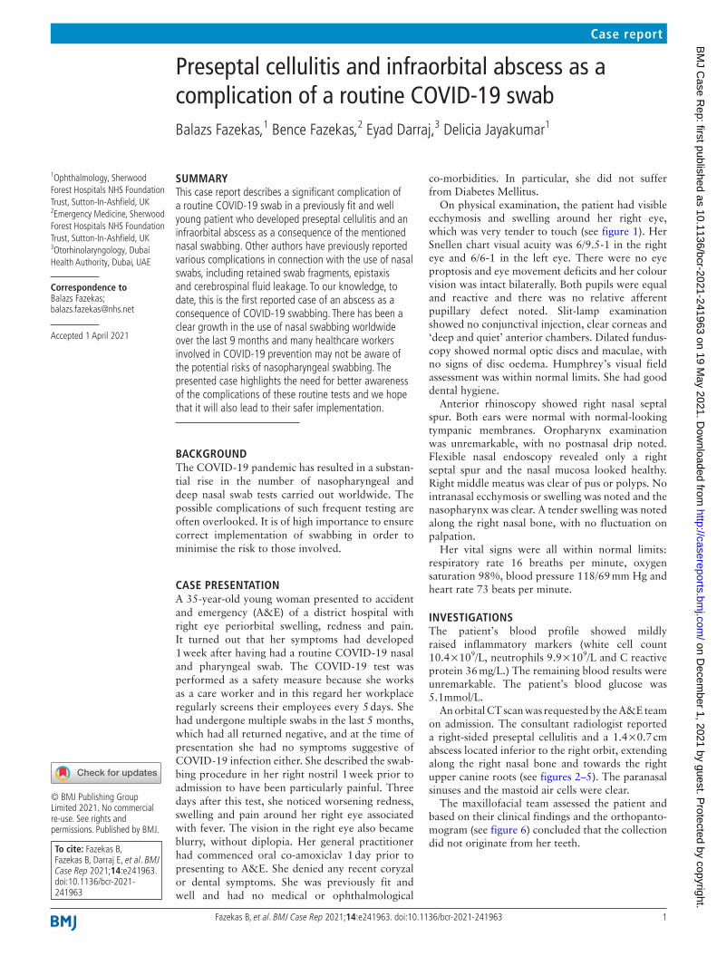
On physical examination, the patient had visible ecchymosis and swelling around her right eye, which was very tender to touch (see figure 1). Her Snellen chart visual acuity was 6/9.5-1 in the right eye and 6/6-1 in the left eye. There were no eye proptosis and eye movement deficits and her colour vision was intact bilaterally. Both pupils were equal and reactive and there was no relative afferent pupillary defect noted. Slit- lamp examination showed no conjunctival injection, clear corneas and ‘deep and quiet’ anterior chambers. Dilated fundus-copy showed normal optic discs and maculae, with no signs of disc oedema. Humphrey’s visual field assessment was within normal limits. She had good dental hygiene.
Anterior rhinoscopy showed right nasal septal spur. Both ears were normal with normal- looking tympanic membranes. Oropharynx examination was unremarkable, with no postnasal drip noted. Flexible nasal endoscopy revealed only a right septal spur and the nasal mucosa looked healthy. Right middle meatus was clear of pus or polyps. No intranasal ecchymosis or swelling was noted and the nasopharynx was clear. A tender swelling was noted along the right nasal bone, with no fluctuation on palpation.
Her vital signs were all within normal limits: respiratory rate 16 breaths per minute, oxygen saturation 98%, blood pressure 118/69 mm Hg and heart rate 73 beats per minute.
INVESTIGATIONSThe patient’s blood profile showed mildly raised inflammatory markers (white cell count 10.4×10⁹/L, neutrophils 9.9×10⁹/L and C reactive protein 36 mg/L.) The remaining blood results were unremarkable. The patient’s blood glucose was 5.1mmol/L.
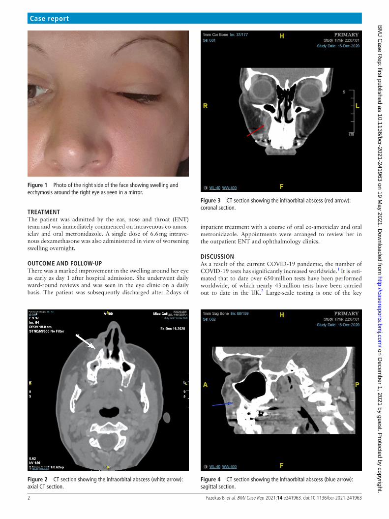
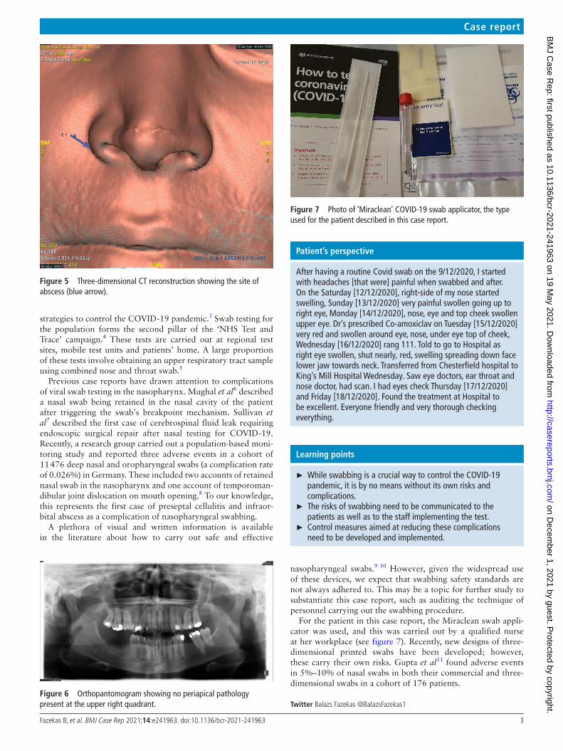
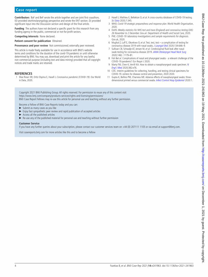
An orbital CT scan was requested by the A&E team on admission. The consultant radiologist reported a right- sided preseptal cellulitis and a 1.4×0.7 cm abscess located inferior to the right orbit, extending along the right nasal bone and towards the right upper canine roots (see figures 2–5). The paranasal sinuses and the mastoid air cells were clear.
The maxillofacial team assessed the patient and based on their clinical findings and the orthopanto-mogram (see figure 6) concluded that the collection did not originate from her teeth.
on Decem
ber 1, 2021 by guest. Protected by copyright.
http://casereports.bmj.com
/B
MJ C
ase Rep: first published as 10.1136/bcr-2021-241963 on 19 M
2 Fazekas B, et al. BMJ Case Rep 2021;14:e241963. doi:10.1136/bcr-2021-241963
Case report
TREATMENTThe patient was admitted by the ear, nose and throat (ENT) team and was immediately commenced on intravenous co- amox-iclav and oral metronidazole. A single dose of 6.6 mg intrave-nous dexamethasone was also administered in view of worsening swelling overnight.
OUTCOME AND FOLLOW-UPThere was a marked improvement in the swelling around her eye as early as day 1 after hospital admission. She underwent daily ward- round reviews and was seen in the eye clinic on a daily basis. The patient was subsequently discharged after 2 days of
inpatient treatment with a course of oral co- amoxiclav and oral metronidazole. Appointments were arranged to review her in the outpatient ENT and ophthalmology clinics.
DISCUSSIONAs a result of the current COVID-19 pandemic, the number of COVID-19 tests has significantly increased worldwide.1 It is esti-mated that to date over 650 million tests have been performed worldwide, of which nearly 43 million tests have been carried out to date in the UK.2 Large- scale testing is one of the key
Figure 1 Photo of the right side of the face showing swelling and ecchymosis around the right eye as seen in a mirror.
3Fazekas B, et al. BMJ Case Rep 2021;14:e241963. doi:10.1136/bcr-2021-241963
Case report
strategies to control the COVID-19 pandemic.3 Swab testing for the population forms the second pillar of the ‘NHS Test and Trace’ campaign.4 These tests are carried out at regional test sites, mobile test units and patients’ home. A large proportion of these tests involve obtaining an upper respiratory tract sample using combined nose and throat swab.5
Previous case reports have drawn attention to complications of viral swab testing in the nasopharynx. Mughal et al6 described a nasal swab being retained in the nasal cavity of the patient after triggering the swab’s breakpoint mechanism. Sullivan et al7 described the first case of cerebrospinal fluid leak requiring endoscopic surgical repair after nasal testing for COVID-19. Recently, a research group carried out a population- based moni-toring study and reported three adverse events in a cohort of 11 476 deep nasal and oropharyngeal swabs (a complication rate of 0.026%) in Germany. These included two accounts of retained nasal swab in the nasopharynx and one account of temporoman-dibular joint dislocation on mouth opening.8 To our knowledge, this represents the first case of preseptal cellulitis and infraor-bital abscess as a complication of nasopharyngeal swabbing.
A plethora of visual and written information is available in the literature about how to carry out safe and effective
nasopharyngeal swabs.9 10 However, given the widespread use of these devices, we expect that swabbing safety standards are not always adhered to. This may be a topic for further study to substantiate this case report, such as auditing the technique of personnel carrying out the swabbing procedure.
For the patient in this case report, the Miraclean swab appli-cator was used, and this was carried out by a qualified nurse at her workplace (see figure 7). Recently, new designs of three- dimensional printed swabs have been developed; however, these carry their own risks. Gupta et al11 found adverse events in 5%–10% of nasal swabs in both their commercial and three- dimensional swabs in a cohort of 176 patients.
Twitter Balazs Fazekas @BalazsFazekas1
Figure 5 Three- dimensional CT reconstruction showing the site of abscess (blue arrow).
Figure 6 Orthopantomogram showing no periapical pathology present at the upper right quadrant.
Figure 7 Photo of ‘Miraclean’ COVID-19 swab applicator, the type used for the patient described in this case report.
Patient’s perspective
After having a routine Covid swab on the 9/12/2020, I started with headaches [that were] painful when swabbed and after. On the Saturday [12/12/2020], right- side of my nose started swelling, Sunday [13/12/2020] very painful swollen going up to right eye, Monday [14/12/2020], nose, eye and top cheek swollen upper eye. Dr’s prescribed Co- amoxiclav on Tuesday [15/12/2020] very red and swollen around eye, nose, under eye top of cheek, Wednesday [16/12/2020] rang 111. Told to go to Hospital as right eye swollen, shut nearly, red, swelling spreading down face lower jaw towards neck. Transferred from Chesterfield hospital to King’s Mill Hospital Wednesday. Saw eye doctors, ear throat and nose doctor, had scan. I had eyes check Thursday [17/12/2020] and Friday [18/12/2020]. Found the treatment at Hospital to be excellent. Everyone friendly and very thorough checking everything.
Learning points
► While swabbing is a crucial way to control the COVID-19 pandemic, it is by no means without its own risks and complications.
► The risks of swabbing need to be communicated to the patients as well as to the staff implementing the test.
► Control measures aimed at reducing these complications need to be developed and implemented.
on Decem
ber 1, 2021 by guest. Protected by copyright.
http://casereports.bmj.com
/B
MJ C
ase Rep: first published as 10.1136/bcr-2021-241963 on 19 M
4 Fazekas B, et al. BMJ Case Rep 2021;14:e241963. doi:10.1136/bcr-2021-241963
Case report
Contributors BaF and BeF wrote the article together and are joint first coauthors. ED provided otorhinolaryngology perspective and wrote the ENT section. DJ provided significant input into the Discussion section and design of the final article.
Funding The authors have not declared a specific grant for this research from any funding agency in the public, commercial or not- for- profit sectors.
Competing interests None declared.
Patient consent for publication Obtained.
Provenance and peer review Not commissioned; externally peer reviewed.
This article is made freely available for use in accordance with BMJ’s website terms and conditions for the duration of the covid-19 pandemic or until otherwise determined by BMJ. You may use, download and print the article for any lawful, non- commercial purpose (including text and data mining) provided that all copyright notices and trade marks are retained.
REFERENCES 1 Max Roser HR, Ortiz- Ospina E, Hasell J. Coronavirus pandemic (COVID-19). Our World
in Data, 2020.
2 Hasell J, Mathieu E, Beltekian D, et al. A cross- country database of COVID-19 testing. Sci Data 2020;7:345.
3 WHO. Covid-19 strategic preparedness and response plan. World Health Organisation, 2020.
4 DoHS. Weekly statistics for NHS test and trace (England) and coronavirus testing (UK): 26 November to 2 December. Gov. uk. Department of Health and Social Care, 2020.
5 PHE. COVID-19: laboratory investigations and sample requirements for diagnosis. Gov. uk, 2020.
6 Mughal Z, Luff E, Okonkwo O, et al. Test, test, test – a complication of testing for coronavirus disease 2019 with nasal swabs. J Laryngol Otol 2020;134:646–9.
7 Sullivan CB, Schwalje AT, Jensen M, et al. Cerebrospinal fluid leak after nasal swab testing for coronavirus disease 2019. JAMA Otolaryngol Head Neck Surg 2020;146): :1179–81.
8 Foh Bet al. Complications of nasal and pharyngeal swabs - a relevant challenge of the COVID-19 pandemic? Eur Respir J 2020.
9 Marty FM, Chen K, Verrill KA. How to obtain a nasopharyngeal swab specimen. N Engl J Med 2020;382:e76.
10 CDC. Interim guidelines for collecting, handling, and testing clinical specimens for COVID-19. centers for disease control and prevention, 2020 2020.
11 Gupta K, Bellino PM, Charness ME. Adverse effects of nasopharyngeal swabs: three- dimensional printed versus commercial swabs. Infect Control Hosp Epidemiol 2020:1.
Copyright 2021 BMJ Publishing Group. All rights reserved. For permission to reuse any of this content visithttps://www.bmj.com/company/products-services/rights-and-licensing/permissions/BMJ Case Report Fellows may re-use this article for personal use and teaching without any further permission.
Become a Fellow of BMJ Case Reports today and you can: ► Submit as many cases as you like ► Enjoy fast sympathetic peer review and rapid publication of accepted articles ► Access all the published articles ► Re-use any of the published material for personal use and teaching without further permission
Customer ServiceIf you have any further queries about your subscription, please contact our customer services team on +44 (0) 207111 1105 or via email at [email protected].
Visit casereports.bmj.com for more articles like this and to become a Fellow
on Decem
ber 1, 2021 by guest. Protected by copyright.
http://casereports.bmj.com
/B
MJ C
ase Rep: first published as 10.1136/bcr-2021-241963 on 19 M