46
Mitteilungen Mitteilungen 2019
MitteilungenMitteilungen2 0 1 9
Mitteilungen 2019
Prof. Dr. med. Robert MlynskiUniversitätsmedizin Rostock
Klinik und Poliklinik für Hals-Nasen-Ohrenheilkunde,
Kopf- und Halschirurgie „Otto Körner“

Norddeutsche Gesellschaft
für Otorhinolaryngologie und zervikofaziale Chirurgie
Schriftleitung:Prof. Dr. med. Robert MlynskiUniversitätsmedizin Rostock
Klinik und Poliklinik für Hals-Nasen-Ohrenheilkunde,
Kopf- und Halschirurgie „Otto Körner“
Doberaner Straße 137-139D-18057 Rostock
Manuskripte:erbeten an die Schriftleitung
Die in dieser Broschüre veröffentlichten Beiträge sind urheberechtlich geschützt.
Alle Rechte vorbehalten, insbesondere für Nachdruck, Vervielfältigungen jeder Art, Übersetzungen, Vortrag, Funk,
Tonträger- und Fernsehsendungen, Mikroverfilmungen sowie die Einspeicherung und Verarbeitung in elektronischen Systemen, auch einzelner Teile.
Verlag und Anzeigen:Verlag Matthias Oehmke
Drosselweg 1, D-18184 RoggentinTel. (038204) 12328, Fax (038204) 14052
eMail: [email protected]
Herstellung:Verlag Matthias Oehmke
ISSN 1866-7392
6
Vorstand der Norddeutschen Gesellschaft für Otorhinolaryngologie und zervikofaziale Chirurgie . . . . . . . . . . . . . . 7
Grußwort . . . . . . . . . . . . . . . . . . . . . . . . . . . . . . 10
Einladung zur 19. Jahrestagung der Norddeutschen Gesellschaft für Otorhinolaryngologie und zervikofaziale Chirugie nach Stralsund vom 3. bis 4. Mai 2019 . . . . . . . . . . . . . . . . . 11
Kongressankündigung zur 90. Jahres-versammlung der Deutschen Gesellschaft für Hals-Nasen-Ohren-Heilkunde, Kopf- und Hals-Chirurgie e.V. vom 29. Mai bis 1. Juni 2019 in Berlin . . . . 14
Otto-Körner-Preis . . . . . . . . . . . . . . . . . . . . . . . 17
Otto-Körner-Preis 2018:
Uhl, BerndThe Endothelial Glycocalyx Controls Interactions of Quantum Dots with the Endothelium and Their Translocation across the Blood-Tissue Border. . . . . . . . . . . . 20
Protokoll über die Mitgliederversammlung der Norddeutschen Gesellschaft für Otorhinolaryngologie und zervikofaziale Chirurgie vom 23. Juni 2018 in Oldenburg . . . . . . . . . . . . . . . . . . . . . . . . . . . 37
Bericht über die 18. Jahrestagung der Nord deutschen Gesellschaft fürOtorhinolaryn gologie und zervikofazialeChirurgie vom 22. bis 23. Juni 2018 in Oldenburg . . . . . . . . . . . . . . . . . . . . . . . . . . . 39
Ankündigungen: Festveranstaltungen 2019 . . . . . . . . . . . . . . . . 42
Informationen des Schatzmeisters . . . . . . . . . 43
Beitrittserklärung. . . . . . . . . . . . . . . . . . . . . . . . 44
Beitragsbescheinigung . . . . . . . . . . . . . . . . . . . 45
Ehrenpräsidenten und Ehrenmitglieder . . . . 46
Mitgliederverzeichnis . . . . . . . . . . . . . . . . . . . . 22
Inhalt
Inhalt
Vorstand
7
1. Vorsitzender:Prof. Dr. med. Carsten BödekerFacharzt für HNO-HeilkundeHNO-Klinik StralsundGroße Parower Straße 47-53, D-18435 StralsundTel. (03831) 352400Fax (03831) 352405eMail: [email protected]
2. Vorsitzender:Prof. Dr. med. Randolf RiemannFacharzt für Hals-Nasen-Ohren-Heilkunde, Kopf-Hals-Chirurgie, Allergologie, Plastische Operationen, SchlafmedizinHNO-Klinik, Elbe Kliniken StadeBremervörder Straße 111, D-21682 StadeTel. (04141) 971301Fax (04141) 972757eMail: [email protected]
Schatzmeister:Dr. med. Thomas GünzelFacharzt für HNO-Heilkunde,Plastische Operationen, Stimm- und SprachstörungenHNO-Praxis Leer Steinburgsgang 4, D-26789 LeerTel. (0491) 65840Fax (0491) 65859eMail: [email protected]
Schriftführer:Prof. Dr. med. Christoph ArensHNO-Universitätsklinik MagdeburgLeipziger Straße 44, D-39120 MagdeburgTel. (0391) 6713800Fax (0391) 6713806eMail: [email protected]
Beisitzer:Prof. Dr. med. Robert MlynskiKlinik und Poliklinik für HNO-Heilkunde, Kopf- und Halschirurgie „Otto Körner“ Universitätsmedizin RostockDoberaner Straße 137-139, D-18057 RostockTel. (0381) 4948301Fax (0381) 4948302eMail: [email protected]
Beisitzer:Dr. med. Peter ImmerFacharzt für HNO-HeilkundeHals-Nasen-Ohren-PraxisBahnhofstraße 61, D-03046 CottbusTel. (0355) 700744eMail: [email protected]
Beisitzer:Dr. med. Jörg SilberzahnFacharzt für HNO-HeilkundePraxis WittmundDohuserweg 14, D-26499 WittmundTel. (04462) 923671Fax (04462) 923672eMail: [email protected]
Berater/Erweiterter Vorstand:Prof. Dr. med. Thomas Eichhorn, CottbusProf. Dr. med. Markus JungehülsingProf. Dr. med. Ercole Di Martino, BremenProf. Dr. med. Jens Meier, HamburgProf. Dr. med. Dr. med. dent. Hans-Jürgen Welkoborsky, Hannover
Vorstand
der Norddeutschen Gesellschaft für Otorhinolaryngologie und zervikofaziale Chirurgie, gewählt am 23. Juni 2018 in Oldenburg
Vorstand
8
Kassenprüfer:Prof. Dr. med. Burkhard KrampKlinik und Poliklinik für HNO-Heilkunde, Kopf- und Halschirurgie „Otto Körner“ Universitätsmedizin RostockDoberaner Straße 137-139, D-18057 RostockTel. (0381) 4948321Fax (0381) 4948302eMail: [email protected]
Dr. med. Henning WiegelsFacharzt für HNO-HeilkundeKlinik für Hals-, Nasen- und Ohrenkrankheiten,Plastische OperationenHelios Klinik SchwerinWismarsche Straße 393-397, D-19055 SchwerinTel. (0385) 5572946eMail: [email protected]
Grußwort
Liebe Kolleginnen und Kollegen,
wir hoffen, Sie hatten alle einen guten Start in dasneue Jahr 2019. Der Vorstand der NorddeutschenGesellschaft für Otorhinolaryngologie und zervi-kofaziale Chirurgie wünscht Ihnen und uns allenzufriedenstellenden Erfolg und ein ausgewogenesJahr 2019.
Die politische Debatte über unser ärztliches Han-deln wird in diesem Jahr u. a. vom Gesundheits-ausschuss des Deutschen Bundestages über dasTerminservice- und Versorgungsgesetz geprägt.Die Herausforderungen für unseren Berufsver-band und den Spitzenverband der FachärzteDeutschlands sowie der Fachgesellschaft sindgroß, um unsere Interessen bei der HNO-ärzt -lichen Versorgung der Bevölkerung für die Po -litik verständlich zu machen. Das Interessenge-menge hat außergewöhnliche Ausmaße erhalten,in der man gut beraten ist, einen kühlen Kopfzu bewahren und die Interessen unserer Patien-ten sachlich und begründet in den Vordergrundzu stellen.
Am 3. und 4. Mai findet unser Jahreskongress inStralsund statt. Professor Bödeker lädt ganz herz-lich in diese wunderschöne Stadt ein und hat einspannendes Programm für uns vorbereitet. AmFreitag werden Themen rund um das Ohr so-wie die Schlafmedizin im Vordergrund stehen.Am Samstag wird der Schwerpunkt auf Neubil-dungen im HNO-Fachgebiet liegen mit besonde-rer Betonung der HPV-assoziierten Malignome.Außerdem möchten wir interdisziplinär das The-ma Tinnitus bearbeiten.
Die Jahrestagung der Deutschen Gesellschaft fürHals-Nasen-Ohrenheilkunde, Kopf- und Hals -chirurgie wird erstmalig in mehreren aufeinanderfolgenden Jahren im Hotel „Estrel“ in Berlin statt-finden. Professor Dazert (Bochum) lädt ein undhat der Jahrestagung Themen der Digitalisierungspeziell für die HNO-Heilkunde zugeordnet.
Hinweise zu diesen Veranstaltungen finden Sie in diesem Heft sowie auf unserer neu gestalte-ten Homepage der Norddeutschen Gesellschaftfür Otorhinolaryngologie und zervikofaziale Chi -rurgie (http://www.ndg-hno.de), zu deren Besuchwir Sie herzlich einladen.
Im Namen des Vorstandes
Ihr
Prof. Dr. med. Robert Mlynski
Grußwort
10
Einladung
11
Liebe Kolleginnen und Kollegen,
die Norddeutsche Gesellschaft für Otorhinolaryn-gologie und zervikofaziale Chirurgie lädt ein zurtraditionellen 19. Jahrestagung am 3. und 4. Mai2019 ins Störtebeker Brauquartier in Stralsund.Als Schirmherr konnte Oberbürgermeister Dr. Badrow gewonnen werden.
Am Freitag möchten wir in diesem Jahr Themenrund um das Ohr sowie die Schlafmedizin in denVordergrund stellen. Am Samstag wird der Schwer-punkt auf Neubildungen des Fachgebietes liegenmit besonderer Betonung der HPV-assoziiertenMalignome. Außerdem möchten wir interdiszi-plinär des Thema Tinnitus bearbeiten und freu-en uns über einen Vortrag auf Einladung zur Be-gutachtung im Fachgebiet.
Wir sind sehr froh, Ihnen zu den aufgeführten The-men ausgesprochen namhafte Referenten aus denIn- und Ausland präsentieren zu können. Abge-rundet wird das wissenschaftliche Programm durchfreie Vorträge und Posterpräsentationen sowie Se-minare sowohl für Fachkollegen als auch für me-dizinische Fachangestellte. Für die Abendveran-staltung dürfen wir Sie am Freitagabend in dasMeeresmuseum in Stralsund einladen. Dieses liegtdirekt in der historischen Altstadt unserer altehr-würdigen Hansestadt. An beiden Kongresstagenkönnen wir Ihnen eine breitgefächerte Industrieaus-stellung präsentieren und sicherlich weitere viel-fältige Möglichkeiten für eine intensive Kommu-nikation zum Austausch im Kollegenkreis.
Die einzelnen Themen, die Referenten und Uhrzei-ten entnehmen Sie bitte dem Programmflyer. DieÄrztekammer Mecklenburg-Vorpommern hat un-sere Jahrestagung bereits mit 14 Fortbildungspunk-ten zertifiziert.
Ich würde mich sehr freuen, Sie in Namen derNorddeutschen Gesellschaft für Otorhinolaryngo-logie und zervikofaziale Chirurgie im Mai 2019 inStralsund begrüßen zu dürfen.
Mit freundlichen kollegialen Grüßen
Prof. Dr. med. Carsten C. Bödeker
19. Jahrestagung:3. und 4. Mai 2019 in Stralsund
Tagungsort: Störtebeker BraumanufakturGreifswalder Chaussee 84/85D-18439 Stralsund
Veranstalter: Norddeutsche Gesellschaft für Otorhino-laryngologie und zervikofaziale Chirurgie
Wissenschaftliche Leitung: Prof. Dr. med. Carsten Christof BödekerKlinik für HNO Heilkunde, Kopf- und HalschirurgieHelios Hanseklinikum Stralsund GmbHGroße Parower Straße 47-53, D-18435 StralsundSekretariat Gabriele Lau: Tel. (03831) 35-2400eMail: [email protected]
Organisation:ProFile Elke HähleMarkstraße 9, D-03046 CottbusTel. 0171-5310083eMail: elke@profile!pragentur.de
Einladung zur 19. Jahrestagung der Norddeutschen Gesellschaftfür Otorhinolaryngologie und zervikofaziale Chirurgie
Einladung
12
Programm:
Freitag, 3. Mai 2019
12.00-13.30 Uhr • Begrüßung und Grußwort • Die hörprothetische Versorgung einseitig
Tauber (Prof. Dr. med. Dr. h.c. Laszig) • Möglichkeiten und Grenzen der Hörgeräte-
versorgung – Indikationen im Wandel der Zeit(Univ.-Prof. Dr. med. Zorowka)
• Freie Vorträge
14.30-16.00 Uhr • Aspekte der Qualitätssicherung in der
rekonstruktiven Mittelohrchirurgie (Prof. Dr. med. Offergeld)
• Vollimplantierbare Hörsysteme – Stand derTechnik, klinische Ergebnisse und Ausblick(Prof. Dr. med. habil. Dr. h.c. Zahnert)
• Möglichkeiten und Grenzen der Tympano plastik für den Hörerhalt (Prof. Dr. med. Mlynski)
• Freie Vorträge
16.20-17.30 Uhr • Fortschritte in der Schlafmedizin
(Prof. Dr. med. Hörmann)• Sialadenoskopie der großen Kopfspeichel -
drüsen (Dr. med. Granitzka)• Freie Vorträge
19.00 Uhr• Abendveranstaltung im Meeresmuseum
Stralsund
Samstag, 4. Mai 2019
9.00-10.00 Uhr • Paragangliome: Bedeutung der Genetischen
Klassifikation (Prof. Dr. med. Dr. h.c. mult. Neumann)
• Kopf- und Hals-Paragangliome (Prof. Dr. med Bödeker)
• Aktuelles zur Insektengift-Therapie (Dr. med. Panzer)
10.20-11.50 Uhr • Stationäre Reha bei Tinnitus
(Prof. Dr. med. Weber)• Tinnitus und Kiefer (Dr. med. Kannengießer)• Tinnitus und Psychosomatik
(Dr. med. Klauer)• BK 2301 und? – Was der HNO-Arzt noch
begutachten kann (Prof. Dr. habil. Meister)
• Freier Vortrag
12.30-13.00 Uhr• Mitgliederversammlung
13.00-15.00 Uhr • Humane Papillomviren beim Oropharynx -
karzinom in der 8. Ausgabe der TNM- Klassifikation: Was wir wissen müssen (Prof. Dr. med. Hoffmann)
• Elektrochemotherapie bei Kopf-Halstumoren:Update (Prof. Dr. med. Riemann)
• Freier Vortrag: HPV-Schnelltest• Freie Vorträge
Seminare/Kurse für Ärzte:
Freitag, 3. Mai 2019, 8.00-12.00 Uhr1. Hörsysteme: Innovationen/Nutzen
(GN Resound)2. Audiologie: Messen und Auswerten
(Hörgerätezentrum Gabriele Jütz GmbH)3. Sialendoskopie
(bess Medizintechnik GmbH)
Seminare/Kurse für MFA:
Freitag, 3. Mai 2019, 13.00-17.00 Uhr1. Hörsysteme: Innovationen/Nutzen
(GN Resound)2. Audiologie: Messen und Auswerten
(Hörgerätezentrum Gabriele Jütz GmbH)3. Brandschutztraining
Kongressankündigung
14
Präsident: Prof. Dr. med. Stefan Dazert, Bochum
Ehrenpräsident: Prof. Dr. med. Dr. h.c. Jan Helms, ehem. Würzburg
90. Jahresversammlung der Deutschen Gesellschaft für Hals-Nasen-Ohren-Heilkunde, Kopf- und Hals-Chirurgie e.V. (2019)Mittwoch, 29. Mai bis Samstag, 1. Juni 2019 Estrel Congress Center Berlin
Kongressmotto:„Digitalisierung in der HNO-Heilkunde“
zugleich
18. Jahrestagung der Deutschen Akademie für Hals-Nasen-Ohrenheilkunde, Kopf- und Hals-Chirurgie e.V.Donnerstag, 30. Mai bis Samstag, 1. Juni 2019
und
19. HNO-PflegetagFreitag, 31. Mai 2019
Wissenschaftliches Program:
Referatethema: • Für die Zukunft der HNO-Heilkunde
relevante Digitalisierungsthemen
Hauptthemen: • Digitale Entwicklungen in Diagnostik und
Therapie • Molekulare Therapieansätze bei Kopf-
Hals-Tumoren• Geriatrische HNO-Erkrankungen• Digitale Strategien in der Aus- und
Weiter bildung• Intersektorale Netzwerkbildung • Radiologie – modernes Datenmanagement • Robotic in der HNO-Heilkunde• Biologicals in der Rhinologie• Aktuelle Aspekte in der Allergologie
Wullstein-Lecture• Molecular mechanisms of hair cell damage,
protection and repair (Allen F. Ryan, San Diego/USA)
13th International Forum May 30 – June 1, 2019Joint Meeting of the American Academy ofOtolaryngology-Head and Neck Surgery Foun-dation (AAO-HNSF) with the German Societyof Otorhinolaryngology-Head and Neck Surgery (DGHNO-KHC) • Endoscopic Ear Surgery • Digital Rehabilitation of Hearing • Immunotherapy in Head and Neck Cancer • International Scientific and Clinical
Cooperation
Deutsche Gesellschaft für Hals-Nasen-Ohren-Heilkunde, Kopf- und Hals-Chirurgie e.V., Bonn
Tag der Praxis Samstag, 1. Juni 2019 • Behandlung der rezidivierend akuten
Ton sillitis • Vor- und Nachbehandlung von
HNO-Operationen• Einordnung von Leitlinien für die praktische
Arbeit• Einsatzmöglichkeiten der endoskopischen
Ohrchirurgie • Digitale Vernetzung zwischen Klinik und
Praxis• Aktuelle Aspekte in der Tinnitus-Behandlung
18. Jahrestagung der Deutschen HNO-AkademieDonnerstag, 30. Mai bis Samstag, 1. Juni 2019 • Workshops zur ärztlichen Fort- und
Weiter bildung
19. HNO-PflegetagFreitag, 31. Mai 2019 • Wie kann die Attraktivität des Pflegeberufes
gesteigert werden? • HNO-relevante Pflegequalifikationen• IT-Anwendungen in der Pflege• Beteiligung an klinischen Studien
Kongressankündigung
15
Ausblick:
91. Jahresversammlung 202020. bis 23. Mai 2020 Estrel Congress Center Berlin
92. Jahresversammlung 202112. bis 15. Mai 2021 Estrel Congress Center Berlin
Auskunft/Information:
Deutsche Gesellschaft für Hals-Nasen-Ohren-Heilkunde, Kopf- und Hals-Chirurgie e.V.Geschäftsstelle: Frau Ulrike FischerFriedrich-Wilhelm-Straße 2, D-53113 BonnTel. (0228) 923922-0Fax (0228) 923922-10eMail: [email protected]
Congress Organisation Claudia SchäferCOCS GmbHAnsprechpartnerin: Martina WiederkrantzRosenheimer Straße 145c, D-81671 MünchenTel. (089) 890677-0Fax (089) 890677-77eMail: [email protected]
Otto-Körner-Preis
17
Mit dem Otto-Körner-Preis der Norddeutschen Ge-sellschaft für Otorhinolaryngologie und zerviko -faziale Chirurgie soll der Verfasser einer herausra-genden und zukunftsweisenden Publikation bzw.Promotion gewürdigt werden, die innerhalb derletzten zwei Jahre vor der jeweiligen Jahresver-sammlung in einer international anerkannten Zeit-schrift erschienen ist bzw. an einer Universität imEinzugsbereich der Regionalgesellschaft angefer-tigt und erfolgreich von einer Medizinischen Fa-kultät (einem medizinischen Fachbereich akzep-tiert) benotet wurde. Die Arbeiten sollen Problemedes Faches der HNO-Heilkunde thematisieren.
Zu der bewertenden Publikation sind der wissen-schaftliche Werdegang des Autors/der Autorin,sein/ihr bisheriges Publikationsverzeichnis und ggf.ein Begleitschreiben des Klinikleiters, bei dem dieArbeit angefertigt wurde, den Unterlagen beizu -fügen. Der zu bewertenden Dissertation sind diePromotionsurkunde, ein Lebenslauf und ggf. ein Begleitschreiben des wissenschaftlichen Betreuers(„Doktorvaters“) beizulegen. Die Unterlagen sindin einer elektronischen Version bereitzustellen. Die
Bewerbung um den ausgeschriebenen Preis solltemit einer Vortragsanmeldung für die Jahresver-sammlung einhergehen, in der auf die Thematikder publizierten Arbeit bzw. der Dissertation ein-gegangen wird.
Der Preis wird jährlich im Rahmen der Eröffnungoder aber der Mitgliederversammlung des Kongres-ses vergeben und beläuft sich auf eine Geldsummevon 1000 €. Über die Vergabe des Preises entschei-det der jeweilige Vorstand der Norddeutschen Gesellschaft für Otorhinolaryngologie und zerviko -faziale Chirurgie. Ausnahmsweise kann der Preisauch geteilt werden. Vorschläge für die Vergabe desPreises sollten bis zum 28. Februar des Kongress -jahres bei dem/der amtierenden Vorsitzenden derFachgesellschaft vom Verfasser der Arbeit bzw. derPromotion selbst oder von einem Mitglied derNorddeutschen Gesellschaft für Otorhinolaryn -gologie und zervikofaziale Chirurgie eingereichtwerden.
Zum Leben und Wirken Otto Körners: siehe „Mitteilungen 2011“
Otto-Körner-Preis
18
Otto-Körner-Preis
1995: O. Kaschke, H.-J. Gerhardt, K. Böhm,M. Wenzel, H. Planck; Berlin
1997: A. Haisch, O. Schultz, C. Perka, V. Jahnke, G. R. Burmester, M. Sittinger; Berlin
1998: Sabine Penk; MagdeburgKatrin Kracik; Leipzig
1999: C. Matthias, V. Jahnke, P. Hand, A. A. Fryer, R. C. Strange; BerlinM. Fuchs; Leipzig
2000: J. Oeken, A. Lenk, F. Bootz; Leipzig
2001: St. Dommerich; Rostock
2002: Saskia Rohrbach; GöttingenJ. Rudolf; Magdeburg
2003: Constanze Ondruschka; MagdeburgT. Stöver; HannoverD. Beutner; Dresden
2004: Christiane Motsch; MagdeburgJ. Ebmeyer; La Jolla (Kalifornien)
2005: St. Lang; LübeckM. Diensthuber; HannoverS. Ertongur, B. Mack, K. Wosikowski,B. Mühlenweg, O. Gires; München
2006: M. Hoffmann; KielM. Sanchez-Hanke; Hamburg
2007: M. Ellies; GöttingenT. Just; Rostock
2008: T. Görögh; KielM. Neudert; Dresden
2009: M. Herzog; GreifswaldA. Herrmann; Rostock
2010: Anke Leichtle; LübeckC. Bergmann; Essen M. Diensthuber; Frankfurt am Main
2011: M. Laudien; KielA. Meyer; Göttingen
2012: M. Canis; GöttingenMarja Loderstedt; Magdeburg
2013: nicht vergeben
Bisherige Johannes-Zange- und Dissertationspreisträger:
2014: M. Schimmer; Frankfurt (Oder)
2015: Susanne Voigt-Zimmermann; Magde burgN. Abram; Hamburg
2016: A. Böttcher; Hamburg
2017: J. Völker; Würzburg
2018: B. Uhl; München
Otto-Körner-Preisträger:
Otto-Körner-Preis 2018
20
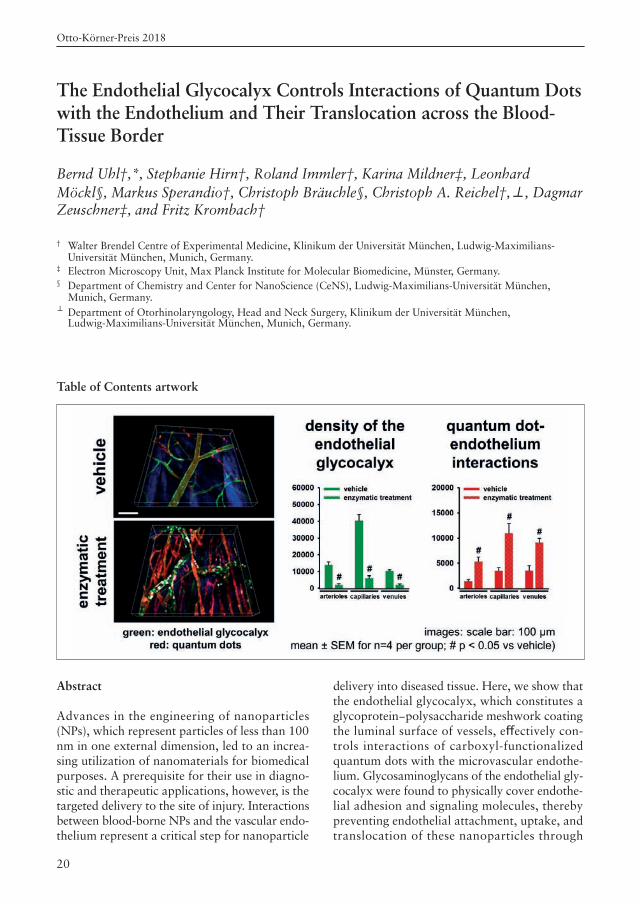
Table of Contents artwork
Abstract
Advances in the engineering of nanoparticles(NPs), which represent particles of less than 100nm in one external dimension, led to an increa-sing utilization of nanomaterials for biomedicalpurposes. A prerequisite for their use in diagno-stic and therapeutic applications, however, is thetargeted delivery to the site of injury. Interactionsbetween blood-borne NPs and the vascular endo-thelium represent a critical step for nanoparticle
The Endothelial Glycocalyx Controls Interactions of Quantum Dotswith the Endothelium and Their Translocation across the Blood-Tissue Border
Bernd Uhl†,*, Stephanie Hirn†, Roland Immler†, Karina Mildner‡, LeonhardMöckl§, Markus Sperandio†, Christoph Bräuchle§, Christoph A. Reichel†,⊥, DagmarZeuschner‡, and Fritz Krombach†
† Walter Brendel Centre of Experimental Medicine, Klinikum der Universität München, Ludwig-Maximilians-Universität München, Munich, Germany.
‡ Electron Microscopy Unit, Max Planck Institute for Molecular Biomedicine, Münster, Germany.§ Department of Chemistry and Center for NanoScience (CeNS), Ludwig-Maximilians-Universität München,
Munich, Germany.⊥ Department of Otorhinolaryngology, Head and Neck Surgery, Klinikum der Universität München,
Ludwig-Maximilians-Universität München, Munich, Germany.
delivery into diseased tissue. Here, we show thatthe endothelial glycocalyx, which constitutes aglycoprotein−polysaccharide meshwork coatingthe luminal surface of vessels, effectively con-trols interactions of carboxyl-functionalizedquantum dots with the microvascular endothe-lium. Glycosaminoglycans of the endothelial gly-cocalyx were found to physically cover endothe-lial adhesion and signaling molecules, therebypreventing endothelial attachment, uptake, andtranslocation of these nanoparticles through
Otto-Körner-Preis 2018
21
different layers of the vessel wall. Conversely,degradation of the endothelial glycocalyx pro-moted interactions of these nanoparticles withmicrovascular endothelial cells under the patho-logic condition of ischemia−reperfusion, thusidentifying the injured endothelial glycocalyx asan essential element of the blood−tissue borderfacilitating the targeted delivery of nanomateri-als to diseased tissue.
Keywordsnanoparticles, endothelial glycocalyx, nanopar-ticle−endothelium interactions, translocation,multiphoton in vivo microscopy, quantum dots,blood−tissue border
IntroductionThe endothelial glycocalyx (eGCX) covers theluminal side of the endothelium throughout theentire vasculature. In addition to glycoproteinsand proteoglycans serving as membrane an-chors, the eGCX predominantly consists of gly-cosaminoglycans (GAGs). Heparan sulfate (HS;>50%) and hyaluronic acid (HA; >40%) are themost abundant GAGs in the eGCX. Under phy-siological conditions, the GAGs are of negativecharge due to the presence of carboxyl and sul-fate groups. Besides, molecules from the blood-stream integrate into this mesh-like structure,collectively controlling a variety of biologicalprocesses such as vascular permeability, hemost-asis, or immune responses.1−5
Consequently, the loss of an intact eGCXcontributes to the initiation of different patho-logical conditions including ischemia−reperfusi-on (I/R) injury, sepsis, atherosclerosis, diabetes,and cancer.1,6−10 Nanoparticles (NPs) are charac-terized as materials with at least one external di-mension in the size range of 1−100 nm.11 Advan-ces in the synthesis of NPs have led to thedevelopment of numerous biomedical tools fordiagnostic and therapeutic applications that arealready utilized under experimental conditi-ons.12,13 Quantum dots (QDs) represent a subsetof engineered NPs that exhibit an intense andphotostable fluorescent signal. Due to these par-ticular functional properties, this nanomaterialhas been employed as an elegant tool for ima-ging approaches in various pathologic conditi-ons.14−19 The targeted delivery of NPs to the siteof injury is a prerequisite for its use in diagno-
stic and therapeutic applications. In this context,interactions of NPs with the vascular endotheli-um initiate the translocation of blood-borne NPsfrom the bloodstream into diseased tissue.20 Sin-ce the eGCX is critical for the maintenance ofthe microvascular integrity and has been impli-cated in the regulation of endothelial NP upta-ke in vitro,21,22 we hypothesized that this vascu-lar structure regulates the delivery of NPs to thesite of injury.
In this study, we demonstrate that the eGCXrepresents a barrier that effectively controls in-teractions of carboxylfunctionalized QDs withthe microvascular endothelium. GAGs of theeGCX were found to physically cover endothe-lial adhesion and signaling molecules, therebypreventing the endothelial attachment, uptake,and translocation of these NPs through the ves-sel wall. Conversely, injury of the eGCX enabledinteractions of these NPs with the microvascu-lar endothelium under the pathologic conditionof I/R, thus providing an essential element of theblood−tissue border, which might serve as a tar-get structure for the delivery of diagnostic andtherapeutic nanomaterials.
Results
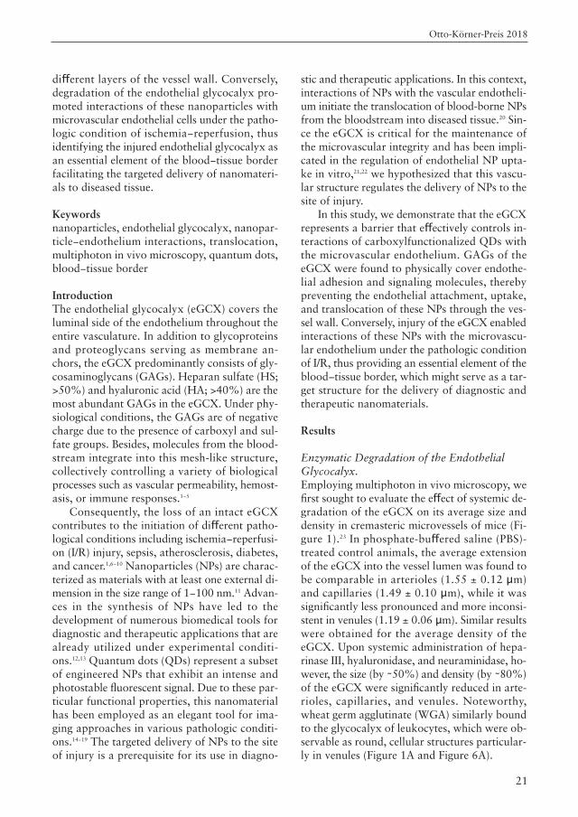
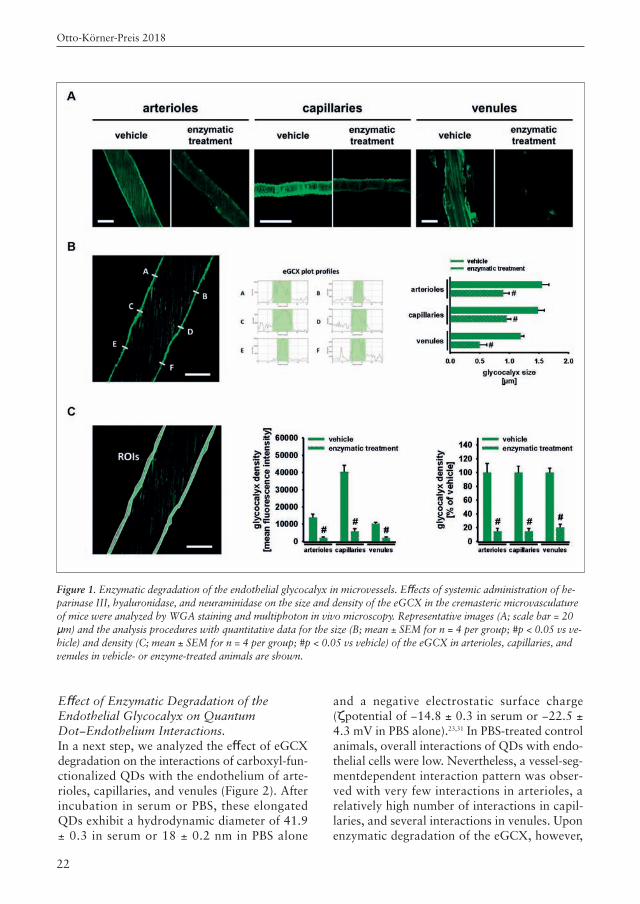
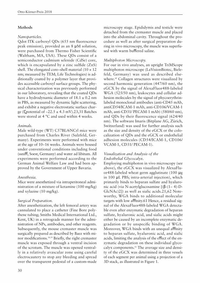
Enzymatic Degradation of the Endothelial Glycocalyx.Employing multiphoton in vivo microscopy, wefirst sought to evaluate the effect of systemic de-gradation of the eGCX on its average size anddensity in cremasteric microvessels of mice (Fi-gure 1).23 In phosphate-buffered saline (PBS)-treated control animals, the average extensionof the eGCX into the vessel lumen was found tobe comparable in arterioles (1.55 ± 0.12 µm)and capillaries (1.49 ± 0.10 µm), while it wassignificantly less pronounced and more inconsi-stent in venules (1.19 ± 0.06 µm). Similar resultswere obtained for the average density of theeGCX. Upon systemic administration of hepa-rinase III, hyaluronidase, and neuraminidase, ho-wever, the size (by ∼50%) and density (by ∼80%)of the eGCX were significantly reduced in arte-rioles, capillaries, and venules. Noteworthy,wheat germ agglutinate (WGA) similarly boundto the glycocalyx of leukocytes, which were ob-servable as round, cellular structures particular-ly in venules (Figure 1A and Figure 6A).
Otto-Körner-Preis 2018
22
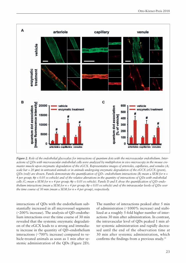
Effect of Enzymatic Degradation of the Endothelial Glycocalyx on QuantumDot−Endothelium Interactions.In a next step, we analyzed the effect of eGCXdegradation on the interactions of carboxyl-fun-ctionalized QDs with the endothelium of arte-rioles, capillaries, and venules (Figure 2). Afterincubation in serum or PBS, these elongatedQDs exhibit a hydrodynamic diameter of 41.9± 0.3 in serum or 18 ± 0.2 nm in PBS alone
and a negative electrostatic surface charge(ζpotential of −14.8 ± 0.3 in serum or −22.5 ±4.3 mV in PBS alone).23,31 In PBS-treated controlanimals, overall interactions of QDs with endo-thelial cells were low. Nevertheless, a vessel-seg-mentdependent interaction pattern was obser-ved with very few interactions in arterioles, arelatively high number of interactions in capil-laries, and several interactions in venules. Uponenzymatic degradation of the eGCX, however,
Figure 1. Enzymatic degradation of the endothelial glycocalyx in microvessels. Effects of systemic administration of he-parinase III, hyaluronidase, and neuraminidase on the size and density of the eGCX in the cremasteric microvasculatureof mice were analyzed by WGA staining and multiphoton in vivo microscopy. Representative images (A; scale bar = 20µm) and the analysis procedures with quantitative data for the size (B; mean ± SEM for n = 4 per group; #p < 0.05 vs ve-hicle) and density (C; mean ± SEM for n = 4 per group; #p < 0.05 vs vehicle) of the eGCX in arterioles, capillaries, andvenules in vehicle- or enzyme-treated animals are shown.
Otto-Körner-Preis 2018
23
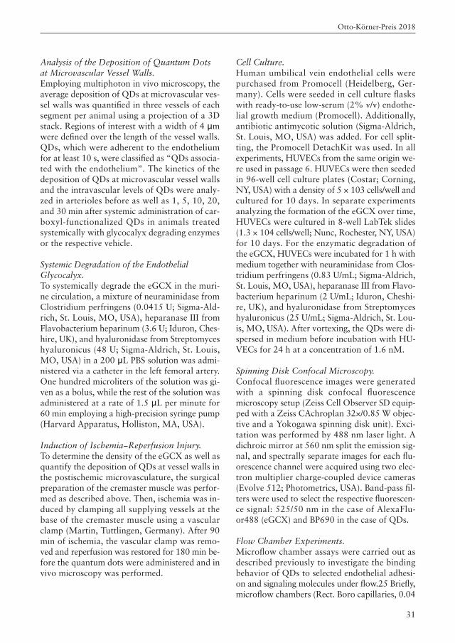
interactions of QDs with the endothelium sub-stantially increased in all microvessel segments(∼200% increase). The analysis of QD−endothe-lium interactions over the time course of 30 minrevealed that the systemic enzymatic degradati-on of the eGCX leads to a strong and immedia-te increase in the quantity of QD−endotheliuminteractions (∼700% increase) compared to ve-hicle-treated animals as soon as 1 min after sy-stemic administration of the QDs (Figure 2D).
The number of interactions peaked after 5 minof administration (∼1000% increase) and stabi-lized at a roughly 5-fold higher number of inter-actions 30 min after administration. In contrast,the intravascular level of QDs peaked 1 min af-ter systemic administration and rapidly decrea-sed until the end of the observation time at 30 min after systemic administration, whichconfirms the findings from a previous study.24
Figure 2. Role of the endothelial glycocalyx for interactions of quantum dots with the microvascular endothelium. Inter-actions of QDs with microvascular endothelial cells were analyzed by multiphoton in vivo microscopy in the mouse cre-master muscle upon enzymatic degradation of the eGCX. Representative images of arterioles, capillaries, and venules (A;scale bar = 20 µm) in untreated animals or in animals undergoing enzymatic degradation of the eGCX (eGCX (green),QDs (red)) are shown. Panels demonstrate the quantification of QD− endothelium interactions (B; mean ± SEM for n =4 per group; #p < 0.05 vs vehicle) and of the relative alterations in the quantity of interactions of QDs with endothelialcells (C; mean ± SEM for n = 4 per group; #p < 0.05 vs vehicle). Panels D and E show the quantification of QD−endo-thelium interactions (mean ± SEM for n = 4 per group; #p < 0.05 vs vehicle) and of the intravascular levels of QDs overthe time course of 30 min (mean ± SEM for n = 4 per group), respectively.
Otto-Körner-Preis 2018
24
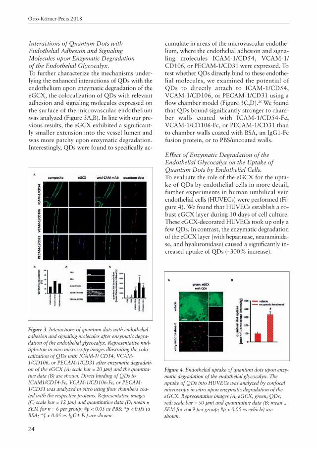
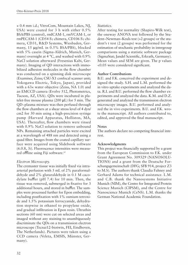
Interactions of Quantum Dots with Endothelial Adhesion and Signaling Molecules upon Enzymatic Degradation of the Endothelial Glycocalyx.To further characterize the mechanisms under-lying the enhanced interactions of QDs with theendothelium upon enzymatic degradation of theeGCX, the colocalization of QDs with relevantadhesion and signaling molecules expressed onthe surface of the microvascular endotheliumwas analyzed (Figure 3A,B). In line with our pre-vious results, the eGCX exhibited a significant-ly smaller extension into the vessel lumen andwas more patchy upon enzymatic degradation.Interestingly, QDs were found to specifically ac-
cumulate in areas of the microvascular endothe-lium, where the endothelial adhesion and signa-ling molecules ICAM-1/CD54, VCAM-1/CD106, or PECAM-1/CD31 were expressed. Totest whether QDs directly bind to these endothe-lial molecules, we examined the potential ofQDs to directly attach to ICAM-1/CD54,VCAM-1/CD106, or PECAM-1/CD31 using aflow chamber model (Figure 3C,D).25 We foundthat QDs bound significantly stronger to cham-ber walls coated with ICAM-1/CD54-Fc,VCAM-1/CD106-Fc, or PECAM-1/CD31 thanto chamber walls coated with BSA, an IgG1-Fcfusion protein, or to PBS/uncoated walls.
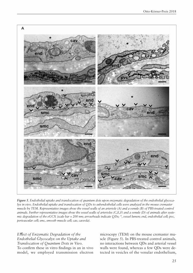
Effect of Enzymatic Degradation of the Endothelial Glycocalyx on the Uptake ofQuantum Dots by Endothelial Cells.To evaluate the role of the eGCX for the upta-ke of QDs by endothelial cells in more detail,further experiments in human umbilical veinendothelial cells (HUVECs) were performed (Fi-gure 4). We found that HUVECs establish a ro-bust eGCX layer during 10 days of cell culture.These eGCX-decorated HUVECs took up only afew QDs. In contrast, the enzymatic degradationof the eGCX layer (with heparinase, neuraminida-se, and hyaluronidase) caused a significantly in-creased uptake of QDs (∼300% increase).
Figure 3. Interactions of quantum dots with endothelialadhesion and signaling molecules after enzymatic degra-dation of the endothelial glycocalyx. Representative mul-tiphoton in vivo microscopy images illustrating the colo-calization of QDs with ICAM-1/ CD54, VCAM-1/CD106, or PECAM-1/CD31 after enzymatic degradati-on of the eGCX (A; scale bar = 20 µm) and the quantita-tive data (B) are shown. Direct binding of QDs toICAM1/CD54-Fc, VCAM-1/CD106-Fc, or PECAM-1/CD31 was analyzed in vitro using flow chambers coa-ted with the respective proteins. Representative images(C; scale bar = 12 µm) and quantitative data (D; mean ±SEM for n = 6 per group; #p < 0.05 vs PBS; *p < 0.05 vsBSA; *§ < 0.05 vs IgG1-Fc) are shown.
Figure 4. Endothelial uptake of quantum dots upon enzy-matic degradation of the endothelial glycocalyx. Theuptake of QDs into HUVECs was analyzed by confocalmicroscopy in vitro upon enzymatic degradation of theeGCX. Representative images (A; eGCX, green; QDs,red; scale bar = 50 µm) and quantitative data (B; mean ±SEM for n = 9 per group; #p < 0.05 vs vehicle) are shown.
Otto-Körner-Preis 2018
25
Effect of Enzymatic Degradation of the Endothelial Glycocalyx on the Uptake andTranslocation of Quantum Dots in Vivo.To confirm these in vitro findings in an in vivomodel, we employed transmission electron
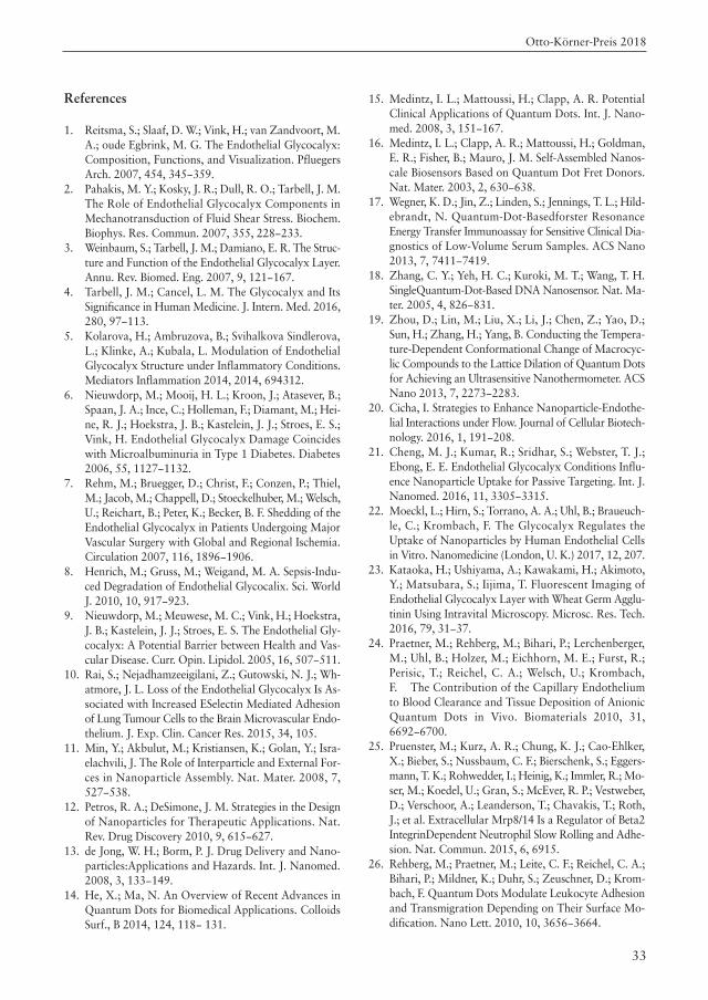
microcopy (TEM) on the mouse cremaster mu-scle (Figure 5). In PBS-treated control animals,no interactions between QDs and arterial vesselwalls were found, whereas a few QDs were de-tected in vesicles of the venular endothelium,
Figure 5. Endothelial uptake and translocation of quantum dots upon enzymatic degradation of the endothelial glycoca-lyx in vivo. Endothelial uptake and translocation of QDs to subendothelial cells were analyzed in the mouse cremastermuscle by TEM. Representative images show the vessel walls of an arteriole (A) and a venule (B) of PBS-treated controlanimals. Further representative images show the vessel walls of arterioles (C,E,F) and a venule (D) of animals after syste-mic degradation of the eGCX (scale bar = 200 nm; arrowheads indicate QDs; *, vessel lumen; end, endothelial cell; pvc,perivascular cell; smc, smooth muscle cell; cav, caveola).
Otto-Körner-Preis 2018
26
most likely in caveolae as described previously.26
Upon enzymatic degradation of the eGCX, ho-wever, the QDs were detected in the endotheliallayer as well as in the subendothelial tissue of themicrovasculature. In arterial vessel walls, we fo-und cluster-like accumulation of these QDs. In-terestingly, the QDs were not located in endothe-lial vesicles but between endothelial cells or incaveolae of subendothelial smooth muscle cells.In venules, however, the eGCX degradation par-ticularly led to considerably increased numbersof QDs taken up in endothelial vesicles as com-pared to PBS-treated control animals.
Integrity of the Endothelial Glycocalyx andMicrovascular Quantum Dot−Endothelium Interactions upon Ischemia−Reperfusion.Finally, we analyzed the effect of I/R injury onthe structural properties of the eGCX as well ason interactions of QDs with the microvascularendothelium (Figure 6). Upon I/R, both the ex-tension of the eGCX into the lumen and its den-sity were significantly reduced in arterioles, ca-pillaries, and venules as compared toshamoperated animals (by ∼50%). Conversely,I/R led to a significant elevation of QD−endothe-lium interactions in all vessel segments (∼400%increase).
DiscussionIn recent years, nanoparticles were increasinglyemployed for experimental diagnosis and treat-ment of various pathologic conditions.6−11 Theexact identification of target structures in disea-sed tissues is mandatory for an effective deliveryof NPs to the site of injury and, thus, their use inbiomedical applications. The endothelial glyco-calyx is a glycoprotein−polysaccharide mes-hwork, which coats the luminal surface of theendothelium in the entire vasculature.1−3 Sincethe eGCX is the initial contact structure forblood-borne NPs in the vasculature, we hypothe-sized that this layer critically controls NP−endo-thelium interactions in the microcirculation. Toprove this hypothesis, we first established an ex-perimental approach that allowed us to selective-ly degrade GAGs and sialic acids (SAs) of theeGCX in vivo using heparinase III, hyaluronida-se, and neuraminidase. In vivo multiphotonmicroscopy was employed on the mouse crema-ster muscle to visualize the size and density of the
eGCX by fluorescently labeled WGA. In linewith previous studies investigating the averagesize and density of the eGCX in the microvascu-lature, we observed a thick and continuous lay-er of this vascular structure in arterioles and ca-pillaries, while it was thinner and moreinconsistent in venules.27−30 The enzymatic treat-ment with heparinase, hyaluronidase, and neu-raminidase, however, significantly reduced theeGCX’s average size and density in all microves-sel segments. Similar effects have been describedupon enzymatic degradation of the eGCX in vi-vo and ex vivo using different methods such asfluorescence or electron microscopy.31,32 In a nextstep, we employed in vivo multiphoton micros-copy to investigate the interactions of NPs withthe microvascular endothelium. We chose to usecarboxyl-functionalized quantum dots as modelNPs because these nanoparticles have been sho-wn to associate with endothelial cells in vivo.Moreover, they have been proven to be ideallysuited for in vivo microscopy because of their si-ze, their monodispersity, and their fluorescenceproperties.14,24,33−36 Our experiments revealed anoverall low, microvessel segment-dependent de-position pattern of these NPs in PBS-treated con-trol animals. Notably, the surgical preparationof the muscle leads to a minor activation of themicrovascular endothelium as well as to inferiorshedding of the eGCX and, hence, might causethe low association of QDs in control animals.37,38
In contrast, interactions of carboxyl-functiona-lized QDs with the endothelium increased sub-stantially in all microvessel segments upon enzy-matic degradation of the eGCX. Consequently,GAGs of the eGCX might act as a barrier forthese NPs in the healthy vasculature. In this con-text, the kinetics of these QD-endothelium inter-actions indicates that the effectivity of this bar-rier function is lost instantly after degradation ofthe eGCX. With respect to the vessel-segment-dependent QD interaction pattern, we furtherspeculate that the higher number of interactionsin venules might be caused by a physiologicallydecreased eGCX since the eGCX reduction is aprerequisite for the adhesion of platelets and leu-kocytes occurring at this site.39,40 However, thestrong association of QDs with capillary wallsdespite the relatively dense eGCX suggests thatadditional factors might contribute to this asso-ciation pattern, such as blood flow velocity,
which is lower in capillaries and increases theprobability of NP−endothelium interactions.41 Ithas been demonstrated that the eGCX exhibitsa barrier function for plasma macromoleculesshielding the vascular endothelium dependent onthe size and charge of the respective molecules.Dextran molecules with a weight of 4 kDa (gy-ration radius (Rg) ∼ 1.4 nm) were able to pene-trate the eGCX within a minute; 50 kDa dextran(Rg ∼ 5.8 nm) only slowly infiltrated the eGCXin the time course of 30 min, and 150 kDa dex-tran (Rg ∼ 11.2 nm) did not penetrate the intacteGCX at all.30,42,43 The QDs employed in this stu-dy have a hydrodynamic diameter of 41.9 ± 0.3nm after incubation in serum (18.1 ± 0.218 nmin PBS) and, therefore, are larger than the 150kDa dextran which has been shown not to pene-trate the healthy eGCX.24 Interestingly, Grom-nicova and colleagues have shown in vitro thatthe enzymatic digestion of the eGCX does not in-crease the uptake of gold NPs, which are smal-ler than 5 nm as assessed by transmission elec-tron microscopy, correspondingly pointing to asize-dependent role of the eGCX barrier func-tion.44 Apart from size, a negative charge led torepulsion of molecules by the eGCX, which hasa net negative charge itself.45 Interestingly, NPsgenerally have a negative charge in blood plas-ma due to the biomolecule corona they acquireupon contact with plasma constituents, whichhas also been shown for the QDs used in the pre-sent study.24,46 Interestingly, the enzymatic degra-dation of GAG sulfates and SAs concomitantlyreduces the negative charge of the eGCX, whichmight facilitate the penetration of negativelycharged QDs through the eGCX.5 Taken to-gether, these experimental data suggest that anintact eGCX regulates interactions of blood-bor-ne NPs and macromolecules with the microvas-cular endothelium via similar mechanisms. Un-der homeostatic conditions, the adhesion andsignaling molecules on the vascular endotheliumare embedded within the eGCX.1 Upon enzyma-tic shedding of glycocalyx GAGs, for example,under inflammatory conditions, these endotheli-al molecules are exposed to the bloodstream andallow interactions such as the adherence of leu-kocytes or platelets.47 With respect to these pre-vious observations, the attachment of QDs tomajor endothelial adhesion and signaling mole-cules was analyzed in the microvasculature upon
Otto-Körner-Preis 2018
28
enzymatic degradation of the eGCX. Inte-restingly, multiphoton in vivo microscopy onmicrovessels revealed a moderate colocalizationof QDs with ICAM-1/CD54 as well as a stron-ger colocalization with VCAM-1/CD106 and PE-CAM-1/CD31. These findings were confirmed bya flow chamber assay, showing that QDs are ab-le to directly bind to ICAM-1/CD54, VCAM-1/CD106, and PECAM-1/CD31 under flow.Hence, the degradation of the eGCX allows QDsto interact with adhesion and signaling molecu-les presented on the surface of the microvascularendothelium, ultimately leading to the depositi-on of these NPs on the vessel wall. Previously, ithas been shown that endothelial cells take up se-veral types of NPs, including the QDs employedin the present study.24,48,49 With respect to our pre-vious findings, we hypothesized that the presen-ce of an intact eGCX diminishes the uptake ofQDs into these cells. We therefore analyzed theQD uptake in HUVECs, which established a de-cent eGCX after 10 days of cell culture, andcompared it to HUVECs treated with enzymesdegrading this eGCX layer. The enzymatic reduc-tion of the eGCX led to a significantly higher in-tracellular uptake of QDs (∼200% increase),which confirmed our hypothesis. Accordingly, anintact eGCX inhibits not only the attachment ofNPs to endothelial cells, but also their intracel-lular uptake. Noteworthy, others and we haverecently reported that an intact eGCX layer alsolimits the uptake of gold NPs in rat fat pad endo-thelial cells in vitro and of polystyrene-based NPsin HUVECs.21,22 These findings, however, warrantfurther studies in endothelial cells of differentvessel segments and organs as they vary in theirfunctional and phenotypic properties.50 To ad-dress the question of whether the degradation ofthe eGCX correspondingly leads to the translo-cation of QDs across the blood−tissue border inthe microvasculature in vivo, we employed trans-mission electron microscopy. In PBS-treated con-trol animals, similar to the results of the multi-photon microscopy experiments, we observed noQDs in the arterial vessel walls and only very fewQDs taken up in caveolae of the venular endo-thelium. In contrast, QDs were able to overco-me the blood−tissue border upon enzymatic de-gradation of the eGCX. In arterioles, thistreatment caused the accumulation of QD clu-sters in vessel walls. Interestingly, QDs were not
located inside arteriolar endothelial cells but bet-ween as well as beneath the endothelial cells.Moreover, they were also found in caveolae ofsubendothelial smooth muscle cells of the arteri-al wall. In venules, however, we detected signifi-cantly more QDs taken up by endothelial cellsafter the enzymatic treatment. These microves-sel-segment-dependent effects of the eGCX areprobably caused by additional factors such asdifferent blood flow velocities, altered vessel ar-chitectures, or varying endothelium phenoty-pes.41,51 To conclude, our experiments show thatthe degradation of the eGCX enables these QDsto translocate across the blood−tissue border inboth vessel segments, whereas their translocati-on routes and fate seem to vary. Interestingly, ithas previously been shown that certain adhesionmolecules (ICAM-1/CD54 and VCAM-1/CD106), to which the QDs bind, are concen-trated in caveolae under noninflammatory aswell as inflammatory conditions, corroboratingour previous hypothesis.52 Our findings demon-strate that the glycocalyx can shield the endothe-lium as well as subendothelial tissue from the de-position, intracellular uptake, and eventranslocation of NPs through the vessel wall. Tostudy the relevance of the eGCX for the protec-tion of the vascular endothelium from circula-ting NPs under pathophysiological circum-stances, we evaluated the interactions of QDs inmicrovessels of a postischemic skeletal muscle tis-sue. Ischemia and reperfusion led to a strong re-duction of the eGCX in microvessels (∼50%),which is in line with previous studies.53,54 Howe-ver, this decrease was significantly less pronoun-ced than after enzymatic degradation, indicatingthat the microvasculature is partly protected bythe eGCX even under inflammatory conditions.Interestingly, the quantitative analysis of the in-teractions of QDs with the microvessel walls re-vealed a dramatic increase upon I/R, which wassignificantly stronger than upon enzymatic eGCXdegradation. Consequently, additional factorsapart from the eGCX integrity appear to influ-ence interactions between QDs and the endothe-lium. We speculate that the loss of the eGCXmight enable interactions between QDs and theendothelium in the first place, while additionalfactors further enhance these interactions signifi-cantly. In this context, endothelial adhesion andsignaling molecules, which are upregulated on
Otto-Körner-Preis 2018
29
the surface of the endothelial cells during I/R, areinteresting candidates to mediate the increasedinteractions of QDs with the endothelium underthese inflammatory conditions.55 In conclusion,the protective function of the eGCX seems to bepartially abrogated in postischemic tissue. Ne-vertheless, this reduction of the eGCX is a neces-sary process during the inflammatory responsein a pathologic condition as this initiates tissuereconstitution.5,56,57 These findings warrant furt-her studies to address the questions of whether(i) an intact eGCX is beneficial with regard toharmful blood-borne nanomaterials including to-xic combustion-derived or engineered NPs aswell as natural virus NPs and whether (ii) an in-jured eGCX would enhance the efficacy of dia-gnostic or therapeutic NPs designed to specifical-ly target diseased tissue. The identification of theinjured eGCX as a target structure in theblood−tissue border enhancing the specific deli-very of NPs into diseased tissue would provideopportunities for the engineering of biomedicalnanomaterials. For example, endowing NPs,which specifically target distinct endothelial ad-hesion and signaling molecules, with eGCXdeg-rading activity might significantly increase theefficacy of NP delivery to diseased tissue conco-mitantly limiting potential side effects. Moreo-ver, this approach might be particularly benefici-al in cancer therapy as tumor cells themselves arecovered with extensive layers of glycocalyx.4,58
ConclusionsIn conclusion, our data identify a previously un-known function of the endothelial glycocalyx,which shields the microvasculature in healthy tis-sue from interactions with NPs. Conversely, in-jury of the eGCX marks a physical impairmentof this vascular structure leading to the exposu-re of endothelial adhesion and signaling molecu-les. This enables NPs to interact with the endo-thelium, thereby facilitating their translocationinto the perivascular tissue. Our observations cor-roborate the critical relevance of the eGCX forthe maintenance of microvascular integrity andsuggest this constituent of the vessel wall as a tar-get structure for NPs in biomedical applications.
Methods
Nanoparticles. Qdot ITK carboxyl QDs (655 nm fluorescencepeak emission), provided as an 8 µM solution,were purchased from Thermo Fisher Scientific(Waltham, MA, USA). These QDs consist of asemiconductor cadmium selenide (CdSe) core,which is encapsulated by a zinc sulfide (ZnS)shell. The elongated core−shell material (10 × 12nm; measured by TEM; Life Technologies) is ad-ditionally coated by a polymer layer that provi-des accessible carboxyl surface groups. The phy-sical characterization was previously performedin our laboratory, revealing that the coated QDshave a hydrodynamic diameter of 18.1 ± 0.2 nmin PBS, as measured by dynamic light scattering,and exhibit a negative electrostatic surface char-ge (ζpotential of −22.5 ± 4.3 mV).23,31 Batcheswere stored at 4 °C and used within 4 weeks.
Animals. Male wild-type (WT) C57BL/6NCrl mice werepurchased from Charles River (Sulzfeld, Ger-many). Experiments were performed with miceat the age of 10−16 weeks. Animals were housedunder conventional conditions including food(ssniff, Soest, Germany) and water ad libitum. Allexperiments were performed according to theGerman Animal Welfare Law and had been ap-proved by the Government of Upper Bavaria.
Anesthesia. Mice were anesthetized via intraperitoneal admi-nistration of a mixture of ketamine (100 mg/kg)and xylazine (10 mg/kg).
Surgical Preparation.After anesthetization, the left femoral artery wascannulated to place a catheter (Fine Bore poly-thene tubing; Smiths Medical International Ltd.,Kent, UK) in a retrograde manner for the admi-nistration of NPs, antibodies, and other reagents.Subsequently, the mouse cremaster muscle wassurgically prepared as described by Baez with mi-nor modifications.38,59 Briefly, the right cremastermuscle was exposed through a ventral incisionof the scrotum. The muscle was opened ventral-ly in a relatively avascular zone using careful electrocautery to stop any bleeding and spreadover the transparent pedestal of a custom-made
Otto-Körner-Preis 2018
microscopy stage. Epididymis and testicle weredetached from the cremaster muscle and placedinto the abdominal cavity. Throughout the pro-cedure as well as after surgical preparation du-ring in vivo microscopy, the muscle was superfu-sed with warm buffered saline.
Multiphoton Microscopy. For our in vivo analyses, an upright TriMScopemultiphoton microscope (LaVisionBiotec, Biele-feld, Germany) was used as described else -where.60 Collagen structures were visualized bysecond harmonic generation (447/60 nm), theeGCX by the signal of AlexaFluor488-labeledWGA (525/50 nm), leukocytes and cellular ad-hesion molecules by the signal of phycoerythrin-labeled monoclonal antibodies (anti-CD45 mAb,antiCD54/ICAM-1 mAb, anti-CD106/VCAM-1mAb, anti-CD31/ PECAM-1 mAb) (580/60 nm),and QDs by their fluorescence signal (624/40nm). The software Imaris (Bitplane AG, Zurich,Switzerland) was used for further analysis suchas the size and density of the eGCX or the colo-calization of QDs and the eGCX or endothelialadhesion molecules (CD54/ICAM-1, CD106/VCAM-1, CD31/ PECAM-1).
Visualization and Analysis of the Endothelial Glycocalyx. Employing multiphoton in vivo microscopy (seeabove), the eGCX was visualized by AlexaFlu-or488-labeled wheat germ agglutinate (100 µgin 100 µL PBS; intra-arterial injection), whichprimarily binds to heparan sulfate and hyaluro-nic acid (via N-acetylglucosamine [(β-(1− 4)-D-GlcNAc)2]) as well as sialic acids.21,62 Note-worthy, WGA binds to additional moleculartargets with low affinity.61 Hence, a residual sig-nal of the AlexaFluor488-labeled WGA detecta-ble even after enzymatic degradation of heparansulfate, hyaluronic acid, and sialic acids mighteither be caused by an incomplete enzymatic de-gradation or by unspecific binding of WGA.Moreover, WGA binds with an unequal affinityto heparan sulfate, hyaluronic acid, and sialicacids, limiting the analysis of the effect of the en-zymatic degradation on these individual glyco-calyx components.61 The average size and densi-ty of the eGCX was determined in three vesselsof each segment per animal using a projection of a3D stack, as illustrated in Figure 1.
30
Analysis of the Deposition of Quantum Dots at Microvascular Vessel Walls. Employing multiphoton in vivo microscopy, theaverage deposition of QDs at microvascular ves-sel walls was quantified in three vessels of eachsegment per animal using a projection of a 3Dstack. Regions of interest with a width of 4 µmwere defined over the length of the vessel walls.QDs, which were adherent to the endotheliumfor at least 10 s, were classified as “QDs associa-ted with the endothelium”. The kinetics of thedeposition of QDs at microvascular vessel wallsand the intravascular levels of QDs were analy-zed in arterioles before as well as 1, 5, 10, 20,and 30 min after systemic administration of car-boxyl-functionalized QDs in animals treated systemically with glycocalyx degrading enzymesor the respective vehicle.
Systemic Degradation of the Endothelial Glyco calyx. To systemically degrade the eGCX in the muri-ne circulation, a mixture of neuraminidase fromClostridium perfringens (0.0415 U; Sigma-Ald-rich, St. Louis, MO, USA), heparanase III fromFlavobacterium heparinum (3.6 U; Iduron, Ches-hire, UK), and hyaluronidase from Streptomyceshyaluronicus (48 U; Sigma-Aldrich, St. Louis,MO, USA) in a 200 µL PBS solution was admi-nistered via a catheter in the left femoral artery.One hundred microliters of the solution was gi-ven as a bolus, while the rest of the solution wasadministered at a rate of 1.5 µL per minute for60 min employing a high-precision syringe pump(Harvard Apparatus, Holliston, MA, USA).
Induction of Ischemia−Reperfusion Injury. To determine the density of the eGCX as well asquantify the deposition of QDs at vessel walls inthe postischemic microvasculature, the surgicalpreparation of the cremaster muscle was perfor-med as described above. Then, ischemia was in-duced by clamping all supplying vessels at the base of the cremaster muscle using a vascularclamp (Martin, Tuttlingen, Germany). After 90min of ischemia, the vascular clamp was remo-ved and reperfusion was restored for 180 min be-fore the quantum dots were administered and invivo microscopy was performed.
Otto-Körner-Preis 2018
31
Cell Culture. Human umbilical vein endothelial cells werepurchased from Promocell (Heidelberg, Ger-many). Cells were seeded in cell culture flaskswith ready-to-use low-serum (2% v/v) endothe-lial growth medium (Promocell). Additionally,antibiotic antimycotic solution (Sigma-Aldrich,St. Louis, MO, USA) was added. For cell split-ting, the Promocell DetachKit was used. In allexperiments, HUVECs from the same origin we-re used in passage 6. HUVECs were then seededin 96-well cell culture plates (Costar; Corning,NY, USA) with a density of 5 × 10³ cells/well andcultured for 10 days. In separate experimentsanalyzing the formation of the eGCX over time,HUVECs were cultured in 8-well LabTek slides(1.3 × 10⁴ cells/well; Nunc, Rochester, NY, USA)for 10 days. For the enzymatic degradation ofthe eGCX, HUVECs were incubated for 1 h withmedium together with neuraminidase from Clos-tridium perfringens (0.83 U/mL; Sigma-Aldrich,St. Louis, MO, USA), heparanase III from Flavo-bacterium heparinum (2 U/mL; Iduron, Cheshi-re, UK), and hyaluronidase from Streptomyceshyaluronicus (25 U/mL; Sigma-Aldrich, St. Lou-is, MO, USA). After vortexing, the QDs were di-spersed in medium before incubation with HU-VECs for 24 h at a concentration of 1.6 nM.
Spinning Disk Confocal Microscopy. Confocal fluorescence images were generatedwith a spinning disk confocal fluorescencemicroscopy setup (Zeiss Cell Observer SD equip-ped with a Zeiss CAchroplan 32×/0.85 W objec-tive and a Yokogawa spinning disk unit). Exci-tation was performed by 488 nm laser light. Adichroic mirror at 560 nm split the emission sig-nal, and spectrally separate images for each flu-orescence channel were acquired using two elec-tron multiplier charge-coupled device cameras(Evolve 512; Photometrics, USA). Band-pass fil-ters were used to select the respective fluorescen-ce signal: 525/50 nm in the case of AlexaFlu-or488 (eGCX) and BP690 in the case of QDs.
Flow Chamber Experiments. Microflow chamber assays were carried out asdescribed previously to investigate the bindingbehavior of QDs to selected endothelial adhesi-on and signaling molecules under flow.25 Briefly,microflow chambers (Rect. Boro capillaries, 0.04
× 0.4 mm i.d.; VitroCom, Mountain Lakes, NJ,USA) were coated for 3 h with either 0.5%BSA/PBS (control), rmICAM-1, rmVCAM-1, orrmPECAM-1 (CD54 Fc chimera, CD106 Fc chi-mera, CD31, R&D Systems, Wiesbaden, Ger-many, 15 µg/mL in 0.5% BSA/PBS), blockedwith 5% casein (Sigma-Aldrich, Munich, Ger-many) overnight at 4 °C, and washed with 0.9%NaCl solution afterward (Fresenius Kabi, Ger-many). Imaging of QD interactions with immo-bilized adhesion molecules in the flow chamberwas conducted on a spinning disk microscope(Examiner, Zeiss; CSU-X1 confocal scanner unit;Yokogawa Electric, Tokyo, Japan), providedwith a 63× water objective (Zeiss, NA 1.0) andan EMCCD camera (Evolve 512, Photometrics,Tucson, AZ, USA). QDs were incubated in pla-telet-free mouse plasma (200 µL) for 5 min. TheQD−plasma mixture was then perfused throughthe flow chambers at a shear stress level of 4 dyn/cm2 for 30 min using a high-precision syringepump (Harvard Apparatus, Holliston, MA,USA). Thereafter, flow chambers were rinsedwith 0.9% NaCl solution to remove unboundNPs. Remaining attached particles were excitedat a wavelength of 488 nm and detected using aquad filter. Images from the coated capillary sur-face were acquired using Slidebook software(6.0.8, 3i). Fluorescence intensities were measu-red offline using Fiji software.62
Electron Microscopy. The cremaster tissue was initially fixed via intra-arterial perfusion with 5 mL of 2% paraformal-dehyde and 2% glutaraldehyde in 0.1 M caco-dylate buffer (pH 7.4) for 10 min. Then, thetissue was removed, submerged in fixative for 3additional hours, and stored in buffer. The sam-ples were processed further for Epon embedding,including postfixation with 1% osmium tetroxi-de and 1.5% potassium ferrocyanide, dehydra-tion stepwise in ethanol to propylene oxide, and gradual infiltration in Epon resin. Ultrathinsections (60 nm) were cut on selected areas andimaged without any staining to unambiguously discriminate the QDs on a transmission electronmicroscope (Tecnai12-biotwin, FEI, Eindhoven,The Netherlands). Pictures were taken using aCCD camera (Veleta, EMSIS, Munster, Ger-many).
Otto-Körner-Preis 2018
32
Statistics.After testing for normality (Shapiro-Wilk test),the oneway ANOVA test followed by the Stu-dent−Newman−Keuls test (>2 groups) or the stu-dent’s t test (2 groups) was performed for theestimation of stochastic probability in intergroupcomparisons using a statistic software package(SigmaStat; Jandel Scientific, Erkrath, Germany).Mean values and SEM are given. The p values<0.05 were considered significant.
Author ContributionsB.U. and F.K. conceived the experiment and de-signed the study. S.H. and L.M. performed thein vitro uptake experiments and analyzed the da-ta. R.I. and B.U. performed the flow chamber ex-periments and analyzed the data. K.M. and D.Z.generated and analyzed the transmission electronmicroscopy images. B.U. performed and analy-zed the in vivo experiments. B.U. and F.K. wro-te the manuscript. All authors contributed to,edited, and approved the final manuscript.
NotesThe authors declare no competing financial inte-rest.
Acknowledgments This project was financially supported by a grantfrom the European Commission to F.K. underGrant Agreement No. 309329 (NANOSOLU -TIONS) and a grant from the Deutsche For-schungsgemeinschaft (DFG; SFB 914, project Z3to M.S). The authors thank Claudia Fahney andGerhard Adams for technical assistance. L.M.and C.B. thank the Nanosystems Initiative Munich (NIM), the Centre for Integrated ProteinScience Munich (CIPSM), and the Centre for Nanoscience Munich (CeNS). L.M. thanks theGerman National Academic Foundation.
Otto-Körner-Preis 2018
33
References
1. Reitsma, S.; Slaaf, D. W.; Vink, H.; van Zandvoort, M.A.; oude Egbrink, M. G. The Endothelial Glycocalyx:Composition, Functions, and Visualization. PfluegersArch. 2007, 454, 345−359.
2. Pahakis, M. Y.; Kosky, J. R.; Dull, R. O.; Tarbell, J. M.The Role of Endothelial Glycocalyx Components inMechanotransduction of Fluid Shear Stress. Biochem.Biophys. Res. Commun. 2007, 355, 228−233.
3. Weinbaum, S.; Tarbell, J. M.; Damiano, E. R. The Struc-ture and Function of the Endothelial Glycocalyx Layer.Annu. Rev. Biomed. Eng. 2007, 9, 121−167.
4. Tarbell, J. M.; Cancel, L. M. The Glycocalyx and ItsSignificance in Human Medicine. J. Intern. Med. 2016,280, 97−113.
5. Kolarova, H.; Ambruzova, B.; Svihalkova Sindlerova,L.; Klinke, A.; Kubala, L. Modulation of EndothelialGlycocalyx Structure under Inflammatory Conditions.Mediators Inflammation 2014, 2014, 694312.
6. Nieuwdorp, M.; Mooij, H. L.; Kroon, J.; Atasever, B.;Spaan, J. A.; Ince, C.; Holleman, F.; Diamant, M.; Hei-ne, R. J.; Hoekstra, J. B.; Kastelein, J. J.; Stroes, E. S.;Vink, H. Endothelial Glycocalyx Damage Coincideswith Microalbuminuria in Type 1 Diabetes. Diabetes2006, 55, 1127−1132.
7. Rehm, M.; Bruegger, D.; Christ, F.; Conzen, P.; Thiel,M.; Jacob, M.; Chappell, D.; Stoeckelhuber, M.; Welsch,U.; Reichart, B.; Peter, K.; Becker, B. F. Shedding of theEndothelial Glycocalyx in Patients Undergoing MajorVascular Surgery with Global and Regional Ischemia.Circulation 2007, 116, 1896−1906.
8. Henrich, M.; Gruss, M.; Weigand, M. A. Sepsis-Indu-ced Degradation of Endothelial Glycocalix. Sci. WorldJ. 2010, 10, 917−923.
9. Nieuwdorp, M.; Meuwese, M. C.; Vink, H.; Hoekstra,J. B.; Kastelein, J. J.; Stroes, E. S. The Endothelial Gly-cocalyx: A Potential Barrier between Health and Vas-cular Disease. Curr. Opin. Lipidol. 2005, 16, 507−511.
10. Rai, S.; Nejadhamzeeigilani, Z.; Gutowski, N. J.; Wh-atmore, J. L. Loss of the Endothelial Glycocalyx Is As-sociated with Increased ESelectin Mediated Adhesionof Lung Tumour Cells to the Brain Microvascular Endo-thelium. J. Exp. Clin. Cancer Res. 2015, 34, 105.
11. Min, Y.; Akbulut, M.; Kristiansen, K.; Golan, Y.; Isra-elachvili, J. The Role of Interparticle and External For-ces in Nanoparticle Assembly. Nat. Mater. 2008, 7,527−538.
12. Petros, R. A.; DeSimone, J. M. Strategies in the Designof Nanoparticles for Therapeutic Applications. Nat.Rev. Drug Discovery 2010, 9, 615−627.
13. de Jong, W. H.; Borm, P. J. Drug Delivery and Nano-particles:Applications and Hazards. Int. J. Nanomed.2008, 3, 133−149.
14. He, X.; Ma, N. An Overview of Recent Advances inQuantum Dots for Biomedical Applications. ColloidsSurf., B 2014, 124, 118− 131.
15. Medintz, I. L.; Mattoussi, H.; Clapp, A. R. PotentialClinical Applications of Quantum Dots. Int. J. Nano-med. 2008, 3, 151−167.
16. Medintz, I. L.; Clapp, A. R.; Mattoussi, H.; Goldman,E. R.; Fisher, B.; Mauro, J. M. Self-Assembled Nanos-cale Biosensors Based on Quantum Dot Fret Donors.Nat. Mater. 2003, 2, 630−638.
17. Wegner, K. D.; Jin, Z.; Linden, S.; Jennings, T. L.; Hild-ebrandt, N. Quantum-Dot-Basedforster ResonanceEnergy Transfer Immunoassay for Sensitive Clinical Dia-gnostics of Low-Volume Serum Samples. ACS Nano2013, 7, 7411−7419.
18. Zhang, C. Y.; Yeh, H. C.; Kuroki, M. T.; Wang, T. H.SingleQuantum-Dot-Based DNA Nanosensor. Nat. Ma-ter. 2005, 4, 826−831.
19. Zhou, D.; Lin, M.; Liu, X.; Li, J.; Chen, Z.; Yao, D.;Sun, H.; Zhang, H.; Yang, B. Conducting the Tempera-ture-Dependent Conformational Change of Macrocyc-lic Compounds to the Lattice Dilation of Quantum Dotsfor Achieving an Ultrasensitive Nanothermometer. ACSNano 2013, 7, 2273−2283.
20. Cicha, I. Strategies to Enhance Nanoparticle-Endothe-lial Interactions under Flow. Journal of Cellular Biotech-nology. 2016, 1, 191−208.
21. Cheng, M. J.; Kumar, R.; Sridhar, S.; Webster, T. J.;Ebong, E. E. Endothelial Glycocalyx Conditions Influ-ence Nanoparticle Uptake for Passive Targeting. Int. J.Nanomed. 2016, 11, 3305−3315.
22. Moeckl, L.; Hirn, S.; Torrano, A. A.; Uhl, B.; Braueuch-le, C.; Krombach, F. The Glycocalyx Regulates theUptake of Nanoparticles by Human Endothelial Cellsin Vitro. Nanomedicine (London, U. K.) 2017, 12, 207.
23. Kataoka, H.; Ushiyama, A.; Kawakami, H.; Akimoto,Y.; Matsubara, S.; Iijima, T. Fluorescent Imaging ofEndothelial Glycocalyx Layer with Wheat Germ Agglu-tinin Using Intravital Microscopy. Microsc. Res. Tech.2016, 79, 31−37.
24. Praetner, M.; Rehberg, M.; Bihari, P.; Lerchenberger,M.; Uhl, B.; Holzer, M.; Eichhorn, M. E.; Furst, R.;Perisic, T.; Reichel, C. A.; Welsch, U.; Krombach,F. The Contribution of the Capillary Endotheliumto Blood Clearance and Tissue Deposition of AnionicQuantum Dots in Vivo. Biomaterials 2010, 31,6692−6700.
25. Pruenster, M.; Kurz, A. R.; Chung, K. J.; Cao-Ehlker,X.; Bieber, S.; Nussbaum, C. F.; Bierschenk, S.; Eggers-mann, T. K.; Rohwedder, I.; Heinig, K.; Immler, R.; Mo-ser, M.; Koedel, U.; Gran, S.; McEver, R. P.; Vestweber,D.; Verschoor, A.; Leanderson, T.; Chavakis, T.; Roth,J.; et al. Extracellular Mrp8/14 Is a Regulator of Beta2IntegrinDependent Neutrophil Slow Rolling and Adhe-sion. Nat. Commun. 2015, 6, 6915.
26. Rehberg, M.; Praetner, M.; Leite, C. F.; Reichel, C. A.;Bihari, P.; Mildner, K.; Duhr, S.; Zeuschner, D.; Krom-bach, F. Quantum Dots Modulate Leukocyte Adhesionand Transmigration Depending on Their Surface Mo-dification. Nano Lett. 2010, 10, 3656−3664.
Otto-Körner-Preis 2018
34
27. van den Berg, B. M.; Vink, H.; Spaan, J. A. The Endo-thelial Glycocalyx Protects against Myocardial Edema.Circ. Res. 2003, 92, 592−594.
28. Potter, D. R.; Damiano, E. R. The HydrodynamicallyRelevant Endothelial Cell Glycocalyx Observed in Vi-vo Is Absent in Vitro. Circ. Res. 2008, 102, 770−776.
29. Yen, W. Y.; Cai, B.; Zeng, M.; Tarbell, J. M.; Fu, B. M.Quantification of the Endothelial Surface Glycocalyxon Rat and Mouse Blood Vessels. Microvasc. Res. 2012,83, 337−346.
30. van Haaren, P. M.; VanBavel, E.; Vink, H.; Spaan, J. A.Localization of the Permeability Barrier to Solutes inIsolated Arteries by Confocal Microscopy. Am. J. Phy-siol. Heart Circ. Physiol. 2003, 285, H2848−2856.
31. Potter, D. R.; Jiang, J.; Damiano, E. R. The RecoveryTime Course of the Endothelial Cell Glycocalyx in Vi-vo and Its Implications in Vitro. Circ. Res. 2009, 104,1318−1325.
32. Chappell, D.; Jacob, M.; Rehm, M.; Stoeckelhuber, M.;Welsch, U.; Conzen, P.; Becker, B. F. Heparinase Selec-tively Sheds Heparan Sulphate from the EndothelialGlycocalyx. Biol. Chem. 2008, 389, 79−82.
33. Lee, H. A.; Imran, M.; Monteiro-Riviere, N. A.; Col-vin, V. L.; Yu, W. W.; Riviere, J. E. Biodistribution ofQuantum Dot Nanoparticles in Perfused Skin: Eviden-ce of Coating Dependency and Periodicity in ArterialExtraction. Nano Lett. 2007, 7, 2865−2870.
34. Rehberg, M.; Leite, C. F.; Mildner, K.; Horstkotte, J.;Zeuschner, D.; Krombach, F. Surface Chemistry ofQuantum DotsDetermines Their Behavior in Postische-mic Tissue. ACS Nano 2012, 6, 1370−1379.
35. Kobayashi, H.; Hama, Y.; Koyama, Y.; Barrett, T.; Re-gino, C. A.; Urano, Y.; Choyke, P. L. Simultaneous Mul-ticolor Imaging of Five Different Lymphatic BasinsUsing Quantum Dots. Nano Lett. 2007, 7, 1711−1716.
36. Ballou, B.; Ernst, L. A.; Andreko, S.; Harper, T.; Fitz -patrick, J. A.; Waggoner, A. S.; Bruchez, M. P. SentinelLymph Node Imaging Using Quantum Dots inMouse Tumor Models. Bioconjugate Chem. 2007, 18,389−396.
37. Kunkel, E. J.; Jung, U.; Bullard, D. C.; Norman, K. E.;Wolitzky, B. A.; Vestweber, D.; Beaudet, A. L.; Ley, K.Absence of TraumaInduced Leukocyte Rolling in MiceDeficient in Both P-Selectin and Intercellular AdhesionMolecule 1. J. Exp. Med. 1996, 183, 57−65.
38. Uhl, B.; Zuchtriegel, G.; Puhr-Westerheide, D.; Praet-ner, M.; Rehberg, M.; Fabritius, M.; Hessenauer, M.;Holzer, M.; Khandoga, A.; Furst, R.; Zahler, S.; Krom-bach, F.; Reichel, C. A. Tissue Plasminogen ActivatorPromotes Postischemic Neutrophil Recruitment Via ItsProteolytic and Nonproteolytic Properties. Arterioscler.,Thromb., Vasc. Biol. 2014, 34, 1495−1504.
39. Nourshargh, S.; Alon, R. Leukocyte Migration into In-flamed Tissues. Immunity 2014, 41, 694−707.
40. Uhl, B.; Vadlau, Y.; Zuchtriegel, G.; Nekolla, K.; Sha-raf, K.; Gaertner, F.; Massberg, S.; Krombach, F.; Rei-chel, C. A. Aged Neutrophils Contribute to the First Li-
ne of Defense in the Acute Inflammatory Response.Blood 2016, 128, 2327.
41. Lipowsky, H. H.; Gao, L.; Lescanic, A. Shedding of theEndothelial Glycocalyx in Arterioles, Capillaries, andVenules and Its Effect on Capillary Hemodynamics Du-ring Inflammation. Am. J. Physiol. Heart Circ. Physiol.2011, 301, H2235−2245.
42. Balcells, C.; Pastor, I.; Pitulice, L.; Hernandez, C.; Via,M.; Garces, J. L.; Madurga, S.; Vilaseca, E.; Isvoran, A.;Cascante, M. Macromolecular Crowding Upon In-Vi-vo-Like Enzyme-Kinetics: Effect of Enzyme-Obstacle Si-ze Ratio. New Front. Chem. 2015, 24,3−16.
43. Shiraishi, Y.; Akiyama, M.; Sato, T.; Hattori, M.; Ko-matsu, T. Size-Dependent Dextran Loading in ProteinNanotube with an Interior Wall of Concanavalin A. Po-lym. Adv. Technol. 2014, 25, 1247−1251.
44. Gromnicova, R.; Kaya, M.; Romero, I. A.; Williams, P.;Satchell, S.; Sharrack, B.; Male, D. Transport of GoldNanoparticles by Vascular Endothelium from DifferentHuman Tissues. PLoS One 2016, 11, e0161610.
45. van Haaren, P. M.; VanBavel, E.; Vink, H.; Spaan, J. A.Charge Modification of the Endothelial Surface LayerModulates the Permeability Barrier of Isolated Rat Mesenteric Small Arteries. Am. J. Physiol. Heart Circ.Physiol. 2005, 289, H2503−H2507.
46. Tenzer, S.; Docter, D.; Kuharev, J.; Musyanovych, A.;Fetz, V.; Hecht, R.; Schlenk, F.; Fischer, D.; Kiouptsi, K.;Reinhardt, C.; Landfester, K.; Schild, H.; Maskos, M.;Knauer, S. K.; Stauber, R. H. Rapid Formation of Plas-ma Protein Corona Critically Affects Nanoparticle Pa-thophysiology. Nat. Nanotechnol. 2013, 8, 772−781.
47. Mulivor, A. W.; Lipowsky, H. H. Role of Glycocalyx inLeukocyte-Endothelial Cell Adhesion. Am. J. Physiol.Heart Circ. Physiol. 2002, 283, H1282−1291.
48. Matuszak, J.; Baumgartner, J.; Zaloga, J.; Juenet, M.;da Silva, A. E.; Franke, D.; Almer, G.; Texier, I.; Faivre,D.; Metselaar, J. M.; Navarro, F. P.; Chauvierre, C.;Prassl, R.; Dezsi, L.; Urbanics, R.; Alexiou, C.; Mang-ge, H.; Szebeni, J.; Letourneur, D.; Cicha, I. Nanopar-ticles for Intravascular Applications: PhysicochemicalCharacterization and Cytotoxicity Testing. Nanomedi-cine (London, U. K.) 2016, 11, 597−616.
49. Voigt, J.; Christensen, J.; Shastri, V. P. Differential Upta-ke of Nanoparticles by Endothelial Cells through Poly-electrolytes with Affinity for Caveolae. Proc. Natl. Acad.Sci. U. S. A. 2014, 111, 2942− 2947.
50. Aird, W. C. Phenotypic Heterogeneity of the Endothe-lium: Ii. Representative Vascular Beds. Circ. Res. 2007,100, 174−190. (51) Sumagin, R.; Sarelius, I. H. Emer-ging Understanding of Roles for Arterioles in Inflam-mation. Microcirculation 2013, 20, 679−692.
52. Fu, C.; He, J.; Li, C.; Shyy, J. Y.; Zhu, Y. Cholesterol In-creases Adhesion of Monocytes to Endothelium by Mo-ving Adhesion Molecules out of Caveolae. Biochim. Bio-phys. Acta, Mol. Cell Biol. Lipids 2010, 1801, 702−710.
53. Mulivor, A. W.; Lipowsky, H. H. Inflammation- andIschemiaInduced Shedding of Venular Glycocalyx.
Otto-Körner-Preis 2018
35
Am.J. Physiol. Heart Circ. Physiol. 2004, 286,H1672−H1680.
54. van Golen, R. F.; Reiniers, M. J.; Vrisekoop, N.; Zuur-bier, C. J.; Olthof, P. B.; van Rheenen, J.; van Gulik, T.M.; Parsons, B. J.; Heger, M. The Mechanisms and Phy-siological Relevance of Glycocalyx Degradation in He-patic Ischemia/Reperfusion Injury. Antioxid. Redox Sig-naling 2014, 21, 1098−1118.
55. Jaeschke, H. Molecular Mechanisms of Hepatic Ische-miaReperfusion Injury and Preconditioning. Am. J. Phy-siol. Gastrointest Liver Physiol. 2003, 284, G15−26.
56. Galli, S. J.; Borregaard, N.; Wynn, T. A. Phenotypic andFunctional Plasticity of Cells of Innate Immunity:Macrophages, Mast Cells and Neutrophils. Nat. Immu-nol. 2011, 12, 1035−1044.
57. Forbes, S. J.; Rosenthal, N. Preparing the Ground forTissue Regeneration: From Mechanism to Therapy. Nat.Med. 2014, 20, 857−869.
58. Yang, C.; Liu, Y.; He, Y.; Du, Y.; Wang, W.; Shi, X.;Gao, F. The Use of Ha Oligosaccharide-Loaded Nano-particles to Breach the Endogenous Hyaluronan Glyco-calyx for Breast Cancer Therapy. Biomaterials 2013, 34,6829−6838.
59. Baez, S. An Open Cremaster Muscle Preparation forthe Study of Blood Vessels by in Vivo Microscopy.Microvasc. Res. 1973, 5, 384−394.
60. Rehberg, M.; Krombach, F.; Pohl, U.; Dietzel, S. La-bel-Free 3d Visualization of Cellular and Tissue Struc-tures in Intact Muscle with Second and Third Harmo-nic Generation Microscopy. PLoS One 2011, 6,e28237.
61. Bhavanandan, V. P.; Katlic, A. W. The Interaction ofWheat Germ Agglutinin with Sialoglycoproteins. TheRole of Sialic Acid. J. Biol. Chem. 1979, 254,4000−4008.
62. Schindelin, J.; Arganda-Carreras, I.; Frise, E.; Kaynig,V.; Longair, M.; Pietzsch, T.; Preibisch, S.; Rueden, C.;Saalfeld, S.; Schmid, B.; Tinevez, J. Y.; White, D. J.;Hartenstein, V.; Eliceiri, K.; Tomancak, P.; Cardona,A. Fiji: An Open-Source Platform for Biological-Ima-ge Analysis. Nat. Methods 2012, 9, 676−682.
Protokoll – Mitgliederversammlung
37
Protokoll über die Mitgliederversammlung
der Norddeutschen Gesellschaft fur Otorhinolaryngologie und zervikofaziale Chirurgie vom 23. Juni 2018 in Oldenburg
Datum: 23.06.2018Sitzungszeit: 12.00-12.30 UhrOrt: Peter-Friedrich-Ludwig-Hospital Oldenburg
TOP 1: Begrüßung durch den Vorsitzenden Prof. Hoppe begrüßt alle Teilnehmer und berich-tet über den Kongressablauf, der reibungslos hin-sichtlich von Technik und Anmeldung abläuft. DieFirma Digitex hat im Vorfeld alles sehr gut vor-bereitet und auch die durchgeführten Kurse spe-ziell für die MFAs wurden sehr gut angenommen.
TOP 2: Feststellung der Beschlussfähigkeit Die Beschlussfähigkeit wird festgestellt, das Pro-tokoll wird durch den 1. Vorsitzenden erstellt.Das Protokoll der letzten Mitgliederversammlungvom 17.06.2017 in Rostock wird ohne Ände-rung angenommen.
TOP 3: Rechenschaftsbericht des VorstandesIm Bericht des Präsidenten kann dieser eine positiveBilanz des abgelaufenen Jahres und der jetzt laufen-den Jahrestagung in Oldenburg ziehen. Besondersdie Fortbildungskurse für medizinische Fachange-stellte erfreuen sich einem großen Interesse mit Teil-nehmerzahlen von über 40 Teilnehmern für speziel-le Kurse. Hier wird auch die Zusammenarbeit mitdem Hörzentrum Oldenburg hervorgehoben, das inseinen Räumlichkeiten die Audiologie-Kurse abhält.Weiterhin konnte der Sonographie-Kurs und einSpeicheldrüsenendoskopie-Kurs im Klinikum Ol-denburg angeboten werden.
Der Schatzmeister der Gesellschaft, Dr. Tho-mas Günzel (Leer), stellt seine Arbeit im vergan-genen Jahr dar. Er berichtet, dass die Gesellschaftderzeit 471 Mitglieder führt, davon sind 242 ak-tive und 229 passive (pensionierte) Mitglieder. Erweist nochmals darauf hin, dass es notwendig ist,die Mitgliederstruktur insgesamt zu verjüngen,um die Gesellschaft auch langfristig besser auf-zustellen. Insgesamt weist die Gesellschaft der-
zeit ein Guthaben von 48.527,61 € auf. Der Vor-sitzende dankt dem Schatzmeister für seine geleis -tete Arbeit.
TOP 4: Bericht der KassenprüferAls Kassenprüfer fungierten Prof. Dr. RandolfRiemann (Stade) und Dr. med. Henning Wiegels(Seehof). Beide sind anwesend, Bericht wirddurch Dr. Wiegels erstellt. Er gibt an, dass nachÜberprüfung der Unterlagen keine Unregel-mäßigkeiten in der Finanzführung zu beobach-ten waren. Er empfiehlt damit, den Vorstand undden Schatzmeister zu entlasten.
TOP 5: Entlastung des VorstandsProf. Markus Hoffmann aus Kiel schlägt vor,dass man dem Vorstand für das Berichtsjahr2017/2018 die Entlastung erteilen möge. In derabschließenden Abstimmung wird die Entlastungdes Vorstandes einstimmig bei Enthaltung desVorstandes erteilt.
TOP 6: Nachwahl des VorstandesZunächst wird zur Wahl des 1. Vorsitzenden fürdas Jahr 2018/2019 und damit ausrichtenderKongresspräsident Herr Prof. Dr. Carsten Böde-ker (Stralsund) vorgeschlagen. Er wird einstim-mig mit einer Enthaltung gewählt und nimmt dieWahl an.
Als zweites wird als stellvertretender Vorsit-zender und damit als Vorsitzender für das Jahr2020 Herr Prof. Dr. Randolf Riemann (Stade)von den Mitgliedern vorgeschlagen. Er wird ein-stimmig gewählt und nimmt die Wahl bei eige-ner Stimmenthaltung an.
Als drittes wird Herr Dr. Silberzahn vorge-schlagen, für die Amtszeit bis 2020 als Beisitzerim Vorstand tätig zu sein. Die Mitglieder wählenHerrn Dr. Silberzahn einstimmig bei Enthaltungdes Kandidaten. Herr Dr. Silberzahn nimmt dieWahl an.
Protokoll – Mitgliederversammlung
38
Aufgrund der Tatsache, dass Herr Prof. Dr.Riemann als 2. Vorsitzender jetzt dem Vorstandder Norddeutschen Gesellschaft für HNO-Heil-kunde angehört, kann er nicht mehr das Amt desKassenprüfers wahrnehmen. Die Mitgliederschlagen Herrn Prof. Dr. med. Burkhard Krampaus Rostock als Kassenprüfer vor. Die Wahl vonProf. Dr. Kramp zum Kassenprüfer erfolgt ein-stimmig in Abwesenheit des Kandidaten. Dieserhat vorher dem 1. Vorsitzenden telefonisch mit-geteilt, dass er die Wahl annehmen würde undfür das Amt des Kassenprüfers zur Verfügungsteht. Somit ist der neue Vorstand der Norddeut-schen Gesellschaft für Otorhinolaryngologie undzervikofaziale Chirurgie wieder komplett. Er be-steht aus dem • 1. Vorsitzenden:
Prof. Dr. med. Carsten Bödeker, Stralsund• 2. Vorsitzenden:
Prof. Dr. med. Randolf Riemann, Stade• Schatzmeister:
Dr. med. Thomas Günzel, Leer• Schriftführer:
Prof. Dr. med. Christoph Arens, Magdeburg• Beisitzer:
Prof. Dr. med. Robert Mlynski, Rostock• Beisitzer:
Prof. Dr. med. Peter Immer, Cottbus• Beisitzer:
Dr. med. Jörg Silberzahn, Wittmund
TOP 7: Änderung der SatzungEs wird die Satzungsänderung, die jedem Mit-glied zugesandt wurde, zur Abstimmung vorge-schlagen. Die Mitglieder nehmen die geplanteSatzungsänderung einstimmig an.
TOP 8: VerschiedenesDie nächsten Kongressorte werden den Mitglie-dern mitgeteilt.• Stralsund: 03.05.-04.05.2019
Ausrichtender Präsident: Prof. Dr. Carsten Bödecker
• Stade: 12.06.-13.06.2020Ausrichtender Präsident: Prof. Dr. med. Randolf Riemann
Die Sitzung endet um 12.30 Uhr.
Bericht – 18. Jahrestagung
Bericht über die 18. Jahrestagung
der Norddeutschen Gesellschaft für Otorhinolaryngologie und zervikofaziale Chirurgie vom 22. bis 23. Juni 2018 in Oldenburg
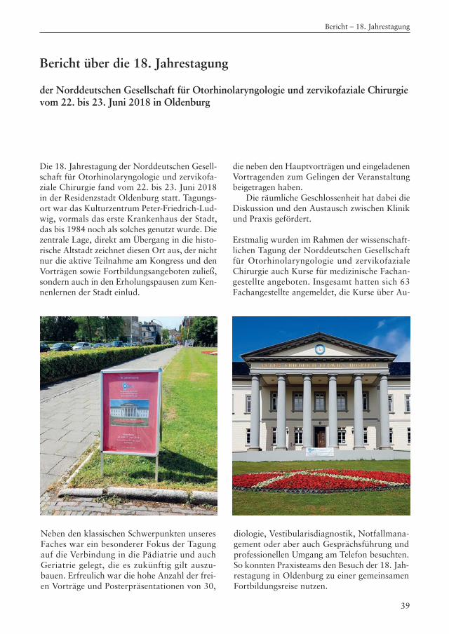
Die 18. Jahrestagung der Norddeutschen Gesell-schaft für Otorhinolaryngologie und zervikofa-ziale Chirurgie fand vom 22. bis 23. Juni 2018in der Residenzstadt Oldenburg statt. Tagungs-ort war das Kulturzentrum Peter-Friedrich-Lud-wig, vormals das erste Krankenhaus der Stadt,das bis 1984 noch als solches genutzt wurde. Diezentrale Lage, direkt am Übergang in die histo-rische Altstadt zeichnet diesen Ort aus, der nichtnur die aktive Teilnahme am Kongress und denVorträgen sowie Fortbildungsangeboten zuließ,sondern auch in den Erholungspausen zum Ken-nenlernen der Stadt einlud.
die neben den Hauptvorträgen und eingeladenenVortragenden zum Gelingen der Veranstaltungbeigetragen haben.
Die räumliche Geschlossenheit hat dabei dieDiskussion und den Austausch zwischen Klinikund Praxis gefördert.
Erstmalig wurden im Rahmen der wissenschaft-lichen Tagung der Norddeutschen Gesellschaftfür Otorhinolaryngologie und zervikofazialeChirurgie auch Kurse für medizinische Fachan-gestellte angeboten. Insgesamt hatten sich 63Fachangestellte angemeldet, die Kurse über Au-
Neben den klassischen Schwerpunkten unseresFaches war ein besonderer Fokus der Tagung auf die Verbindung in die Pädiatrie und auchGeriatrie gelegt, die es zukünftig gilt auszu -bauen. Erfreulich war die hohe Anzahl der frei-en Vorträge und Posterpräsentationen von 30,
diologie, Vestibularisdiagnostik, Notfallmana-gement oder aber auch Gesprächsführung undprofessionellen Umgang am Telefon besuchten.So konnten Praxisteams den Besuch der 18. Jah-restagung in Oldenburg zu einer gemeinsamenFortbildungsreise nutzen.
39
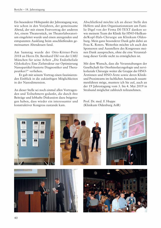
40
Ein besonderer Höhepunkt der Jahrestagung war,wie schon in den Vorjahren, der gemeinsameAbend, der mit einem Festvortrag der anderenArt, einem Theaterstück, im Theaterlaboratori-um eingeleitet wurde und einen anregenden undentspannten Ausklang beim anschließenden ge-meinsamen Abendessen fand.
Am Samstag wurde der Otto-Körner-Preis 2018 an Herrn Dr. Bernhard Uhl von der LMU München für seine Arbeit „Die Endotheliale Glykokalyx: Eine Zielstruktur zur OptimierungNanopartikel-basierte Diagnostiker und Thera-peutiker?“ verliehen.
Er gab mit seinem Vortrag einen faszinieren-den Einblick in die zukünftigen Möglichkeitenin der Nanodimension.
An dieser Stelle sei noch einmal allen Vortragen-den und Teilnehmern gedankt, die durch ihreBeiträge und lebhafte Diskussion dazu beigetra-gen haben, dass wieder ein interessanter undkonstruktiver Kongress zustande kam.
Abschließend möchte ich an dieser Stelle denHelfern und dem Organisationsteam um Fa mi-lie Digel von der Firma DI-TEXT danken so -wie meinem Team der Klinik für HNO-Heilkun-de/Kopf-Hals-Chirurgie am Klinikum Olden-burg. Mein ganz besonderer Dank geht dabei anFrau K. Roters. Weiterhin möchte ich auch denSponsoren und Ausstellern des Kongresses mei-nen Dank aussprechen, ohne die eine Veranstal-tung dieser Größe nicht zu ermöglichen ist.
Mit dem Wunsch, dass die Veranstaltungen derGesellschaft für Otorhinolaryngologie und zervi-kofaziale Chirurgie weiter die Gruppe der HNO-Ärztinnen und HNO-Ärzte sowie deren Klinik-und Praxisteams im fachlichen Austausch zusam-menführen möge, muntere ich Sie auf, auch ander 19 Jahrestagung vom 3. bis 4. Mai 2019 inStralsund möglichst zahlreich teilzunehmen.
Prof. Dr. med. F. Hoppe(Klinikum Oldenburg AöR)
Bericht – 18. Jahrestagung
ATMOS® S 61 CORIAN®HNO-Arbeitsplätze - Design und Funktionalität in Perfektion
Eine hohe Maxime bei der Gestaltung vonEin richtungen im Gesundheitswesen ist die Infektionsprävention. Deshalb vertrauen wir auf die ausgezeichneten Hygieneeigenschaften von CORIAN®.
Be anspruchung im Praxisalltag jederzeit gewachsen.
Die neuen ATMOS® S 61 CORIAN® Einheiten
ATMOS MedizinTechnik GmbH & Co. KG Ludwig-Kegel-Str. 16 79853 Lenzkirch/Germany Tel: +49 7653 689-642 [email protected] www.atmosmed.com
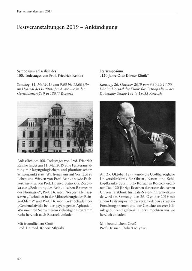
42
Festveranstaltungen 2019
Festveranstaltungen 2019 – Ankündigung
Symposium anlässlich des 100. Todestages von Prof. Friedrich Reinke
Samstag, 11. Mai 2019 von 9.00 bis 15.00 Uhrim Hörsaal des Instituts für Anatomie in derGertrudenstraße 9 in 18055 Rostock
Festsymposium „120 Jahre Otto-Körner-Klinik“
Samstag, 26. Oktober 2019 von 9.30 bis 15.00Uhr im Hörsaal der Klinik für Orthopädie in derDoberaner Straße 142 in 18055 Rostock
Anlässlich des 100. Todestages von Prof. FriedrichReinke findet am 11. Mai 2019 eine Festveranstal-tung mit laryngologischem und phoniatrischemSchwerpunkt statt. Wir freuen uns auf Vorträge zuLeben und Wirken von Prof. Reinke sowie Fach-vorträge, u.a. von Prof. Dr. med. Patrick G. Zorow -ka zur „Bedeutung des Reinke´schen Raumes inder Phoniatrie“, Prof. Dr. med. Norbert Kleinsas-ser zu „Techniken in der Mikrochirurgie des Rein-ke-Ödems“ und Prof. Dr. med. Götz Schade über„Gehirnaktivität bei der psychogenen Aphonie“.Wir möchten Sie zu diesem vielseitigen Programmrecht herzlich nach Rostock einladen.
Mit freundlichem Gruß Prof. Dr. med. Robert Mlynski
Am 25. Oktober 1899 wurde die GroßherzöglicheUniversitätsklinik für Ohren-, Nasen- und Kehl-kopfkranke durch Otto Körner in Rostock eröff-net. Das 120-jährige Bestehen der ersten deutschenUniversitätsklinik für Hals-Nasen-Ohrenheilkun-de wird am Samstag, den 26. Oktober 2019 miteinem Festsymposium zu verschiedenen aktuellenForschungsthemen und zur Gesichte unserer Kli-nik gebührend gefeiert. Hierzu möchten wir Sieherzlich einladen.
Mit freundlichem Gruß Prof. Dr. med. Robert Mlynski
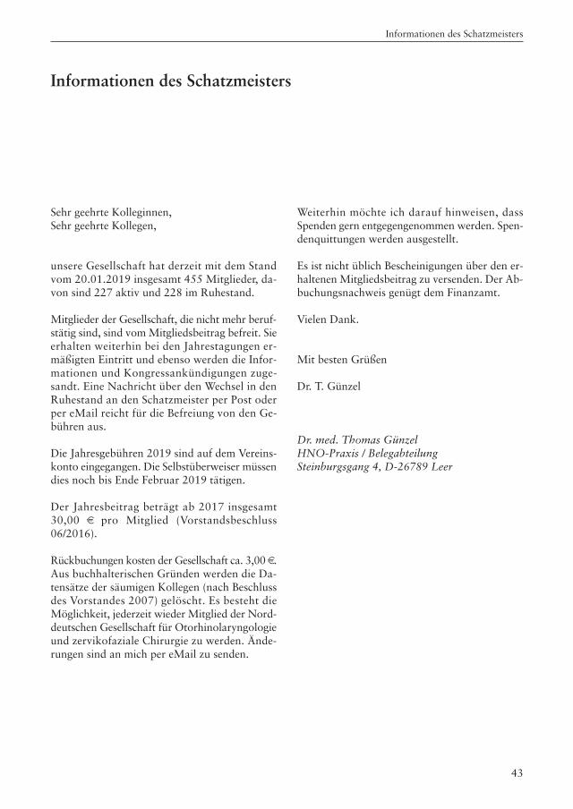
43
Informationen des Schatzmeisters
Informationen des Schatzmeisters
Sehr geehrte Kolleginnen,Sehr geehrte Kollegen,
unsere Gesellschaft hat derzeit mit dem Standvom 20.01.2019 insgesamt 455 Mitglieder, da-von sind 227 aktiv und 228 im Ruhestand.
Mitglieder der Gesellschaft, die nicht mehr beruf-stätig sind, sind vom Mitgliedsbeitrag befreit. Sieerhalten weiterhin bei den Jahrestagungen er-mäßigten Eintritt und ebenso werden die Infor-mationen und Kongressankündigungen zuge-sandt. Eine Nachricht über den Wechsel in denRuhestand an den Schatzmeister per Post oderper eMail reicht für die Befreiung von den Ge-bühren aus.
Die Jahresgebühren 2019 sind auf dem Vereins-konto eingegangen. Die Selbstüberweiser müssendies noch bis Ende Februar 2019 tätigen.
Der Jahresbeitrag beträgt ab 2017 insgesamt30,00 € pro Mitglied (Vorstandsbeschluss06/2016).
Rückbuchungen kosten der Gesellschaft ca. 3,00 €.Aus buchhalterischen Gründen werden die Da-tensätze der säumigen Kollegen (nach Beschlussdes Vorstandes 2007) gelöscht. Es besteht dieMöglichkeit, jederzeit wieder Mitglied der Nord-deutschen Gesellschaft für Otorhinolaryngologieund zervikofaziale Chirurgie zu werden. Ände-rungen sind an mich per eMail zu senden.
Weiterhin möchte ich darauf hinweisen, dassSpenden gern entgegengenommen werden. Spen-denquittungen werden ausgestellt.
Es ist nicht üblich Bescheinigungen über den er-haltenen Mitgliedsbeitrag zu versenden. Der Ab-buchungsnachweis genügt dem Finanzamt.
Vielen Dank.
Mit besten Grüßen
Dr. T. Günzel
Dr. med. Thomas GünzelHNO-Praxis / BelegabteilungSteinburgsgang 4, D-26789 Leer
44
Beitrittserklärung
q Ich erkläre meinen Beitritt zur Norddeutschen Gesellschaft für Otorhinolaryngologie und zervikofaziale Chirurgie.
q Adressänderung: Ich bin bereits Mitglied der Norddeutschen Gesellschaft für Otorhino -laryn gologie und zervikofaziale Chirurgie.
Adresse: Straße ________________________________________________________________
PLZ, Ort ______________________________________________________________
Fax___________________________________________________________________
eMail _________________________________________________________________
Name: _______________________________________________________________________
Titel: _______________________________________________________________________
IBAN: _______________________________________________________________________
BIC: _______________________________________________________________________
Geldinstitut: _______________________________________________________________________
q Ich bin mit der Abbuchung des Jahresbeitrages von 30,00 € bis auf Widerruf einverstanden.
q Ab dem _________________ bin ich im Ruhestand.
q Ich möchte aus der Gesellschaft austreten.
____________________ ____________________ ____________________Unterschrift Datum Stempel
Bitte senden an:
Dr. med. Thomas GünzelHNO-Praxis Leer Steinburgsgang 4, D-26789 LeerTel. (0491) 65840, Fax (0491) 65859, eMail: [email protected]
Beitrittserklärung / Aktualisierung der Adresse
Beitragsbescheinigung
45
Norddeutsche Gesellschaft für Otorhinolaryngologie und zervikofaziale Chirurgie
Hiermit wird bescheinigt, dass
Frau/Herr ______________________________________________________
wohnhaft ______________________________________________________
______________________________________________________
Mitglied der Norddeutschen Gesellschaft für Otorhinolaryngologie und zervikofaziale Chirurgie ist und den Jahresbeitrag für 2018 in Höhe von
30,00 €
an die Gesellschaft entrichtet hat.
gez.: Dr. med. Thomas Günzel(Schatzmeister der Gesellschaft)
Beitragskonto:Brandenburger BankKonto-Nr. 6077501, BLZ 16062073IBAN: DE18 1606 2073 0006 0775 01 BIC: GENODEF1BRB
Beitragsbescheinigung
46
Ehrenpräsidenten und Ehrenmitglieder
Ehrenpräsidenten:
Prof. Dr. med. habil. Bernd Freigang, Magdeburg
Prof. Dr. med. habil. Jan Helms, Würzburg
Ehrenmitglieder:
PD Dr. med. habil. Hans-Peter Jung, Brandenburg
Prof. Dr. med. habil. Rainer Laskawi, Göttingen
Frau Ingrid Kilian, Göttingen
Prof. Dr. med. Thomas Eichhorn, Cottbus
Ehrenpräsidenten und Ehrenmitglieder


