83
Medizinische Forschungsförderung Innsbruck (MFI) , Oktober 2011 Abschlussbericht 2006 – 2011

Medizinische Forschungsförderung Innsbruck (MFI) , Oktober 2011
Abschlussbericht
2006 – 2011

VORWORT Liebe Kolleginnen und Kollegen!
Die Förderung des wissenschaftlichen Nachwuchses ist eine der wichtigsten, wenn nicht die wichtigste Aufgabe einer Universität. Es gilt dabei junge Wissenschaftler/inne/n an die Anforderungen nationaler und internationaler Fördergeber heranzuführen, ihnen die Möglichkeit zu geben zu lernen, eigenständige Anträge zu verfassen und Vorarbeiten als Grundlage für solche Anträge durchzuführen.
2005 wurde vom Rektorat Sorg das Nachwuchsförderprogramm MFI „Medizinische Forschungsförderung Innsbruck“ eingerichtet, das speziell für junge Postdocs der MUI gewidmet war und die erwähnten Ziele verfolgte. Das Programm wurde in vier halbjährigen Tranchen ausgeschrieben. Vergeben wurden insgesamt € 3.077.785,95 an 13 Jungwissenschaftlerinnen und 24 Jungwissenschaftler für Personal-‐, Material-‐ und Reisekosten, die zur Durchführung der Projekte benötigt wurden.
Es wurde von Anfang an darauf Wert gelegt, dass die Projekte auswärtig begutachtet und unter strengen Kriterien von einer internen Kommission (MFI-‐Jury) vergeben wurden.
Der vorliegende Endbericht soll einen Rückblick auf fünf Jahre Nachwuchsförderung durch das MFI geben. Beeindruckend dokumentiert er den Erfolg des Programms anhand zahlreicher daraus entstandener Publikationen, Vorträgen auf Kongressen und bewilligten Anschlussprojekten beim FWF. Besonders zu erwähnen ist, dass ein Projektwerber (Univ.-‐Prof. Arthur Kaser) inzwischen einen Startpreis, ein ERC Starting-‐Grant und einen Ruf nach Cambridge erhalten hat.
Das Förderinstrument MFI ist nun 2011 ausgelaufen. Der Erfolg des Projekts hat das Rektorat bereits im Jahr 2010 dazu bewogen, ein neues Programm mit ähnlicher Zielsetzung, MUI Start, einzusetzen. Aus Budgetgründen sind die Mittel hierfür deutlich geringer, als diese für das MFI waren. Es wird meine Aufgabe sein, darauf zu dringen, dass diese Mittel erhalten bleiben bzw. wieder erhöht werden.
Mein besonderer Dank gilt aber nun dem Vorsitzenden der Jury o. Univ.-‐Prof. Wick und seinen Stellvertreter/innen o. Univ.-‐Prof. Dr. Monika Ritsch-‐Marte und Univ.-‐Prof. Dr. Günter Weiss, den weiteren Mitgliedern der internen MFI-‐Jury und den zahlreichen externen Gutachter/inne/n, die eine Auswahl der Projekte auf höchstem Niveau realisiert haben. Ohne deren persönlichen Einsatz wäre die Umsetzung des Programms nicht mit dieser Qualität möglich gewesen.
Besonderen Respekt und Bewunderung zolle ich unseren beteiligten Nachwuchswissenschaftler/inne/n für die zumeist sehr hohe Qualität der wissenschaftlichen Leistungen und wünsche ihnen für ihren weiteren Lebensweg alles Gute.
Zu guter Letzt möchte ich meiner Hoffnung Ausdruck verleihen, dass die Universität trotz der Sparzwänge die erfolgreiche Förderung von Postdoktoranden nicht nur beibehalten, sondern in Zukunft auch wieder ausbauen kann.
Univ.-‐Prof. Dr. Günther Sperk, Vizerektor für Forschung

INHALTSVERZEICHNIS EINLEITUNG ........................................................................................................................................... 5
MFI-‐JURY ................................................................................................................................................ 7
ANTRAGSPERIODEN ............................................................................................................................... 9
GENEHMIGTE PROJEKTE IN DER ÜBERSICHT ....................................................................................... 11
Antragsperiode 1 ............................................................................................................................. 11
Antragsperiode 2 ............................................................................................................................. 12
Antragsperiode 3 ............................................................................................................................. 13
Antragsperiode 4 ............................................................................................................................. 14
Abschlussberichte der MFI-‐Projekte .................................................................................................... 15
Abschlussberichte Antragsperiode 1 ............................................................................................... 15
Abschlussberichte Antragsperiode 2 ............................................................................................... 30
Abschlussberichte Antragsperiode 3 ............................................................................................... 42
Abschlussberichte Antragsperiode 4 ............................................................................................... 56
LITERATURVERZEICHNIS (Publikationen mit MFI-‐Erwähnung) ............................................................ 75
FINANZIELLE ANGABEN ZUM MFI ........................................................................................................ 81

Einleitung
5
EINLEITUNG Im Rahmen der Umsetzung der 2005 begonnenen Förderung von Forschungspools an der Medizinischen Universität Innsbruck wurde das Programm „Medizinische Forschungsförderung Innsbruck“ (MFI) etabliert. Das MFI sollte neben dem IFTZ („Integriertes Forschungs-‐ und Therapiezentrum“) das zweite wichtige Instrument zur eigenen intramuralen Forschungsförderung werden.
Das Forschungsinstrument diente vorrangig der individuellen Projektförderung mit Fokus auf den wissenschaftlichen Nachwuchs und stand allen promovierten Mitgliedern (außer berufenen Professor/innen) der Universität bis zu einem Alter von 40 Jahren unabhängig von der Zuordnung zu definierten Forschungsschwerpunkten zur Verfügung. Für jedes Projekt standen max. 65.000 Euro pro Jahr zur Verfügung, die für Personal, Sachmittel und Investitionen verwendet werden konnten (max. Fördersumme 130.000 Euro für 2 Jahre). Es war dabei auch möglich, die eigene Stelle im Rahmen eines Projektes zu finanzieren.
Eine Jury unter dem Vorsitz des früheren FWF-‐Präsidenten Univ.-‐Prof. Dr. Wick hat die eingereichten Projektanträge jeweils von 2 externen Gutachtern bewerten lassen und auf Basis dieser Gutachten dann über eine mögliche Förderung entschieden. Die MFI-‐Jury wurde durch den damaligen Rektor Univ.-‐Prof. Dr. Clemens Sorg bestimmt.
Die Projekte, die innerhalb des Programmes gefördert wurden, sind inzwischen beendet. Die Ergebnisse, die aus diesen Projekten hervor gegangen sind, sind in diesem Bericht zusammengefasst. Besonders hervorzuheben ist, dass zwei Kandidat/innen, die ein MFI-‐Grant bekommen haben, inzwischen einen Ruf an andere Universitäten erhalten haben:
• Univ.-‐Prof.Dr. Arthur Kaser, ehemals Universitätsklinik für Innere Medizin II, jetzt Professur Cambridge ERC-‐Starting Grant, Division of Gastroenterology and Hepatology, Department of Medicine, University of Cambridge
• Univ.-‐Prof. Dr. Martina Prelog, ehemals Universitätsklinik für Pädiatrie I, jetzt Professur in Würzburg, Universitätsklinikum Würzburg, Kinderklinik der Bayerischen Julius-‐Maximilians-‐Universität
Insgesamt wurden von 2006 – 2008 5 Antragsperioden durchlaufen. Die Projekte der 5. Antragsperiode wurden zwar begutachtet, jedoch sprach die MFI-‐Jury keinerlei Förderungsempfehlung mehr aus (Beschluss Universitätsrat 2008).
Innerhalb des Förderzeitraums und von den geförderten 35 Projekten wurden seither 63 Publikationen angefertigt. 11 Projekte wurden in eine externe Förderung überführt.

6

MFI-‐Jury
7
MFI-‐JURY
Vorsitzender
em.O.Univ.-‐Prof. Dr. Georg WICK Sektion für Experimentelle Pathophysiologie und Immunologie
Engerer Vorstand
O.Univ.-‐Prof. Dr. Monika RITSCH-‐MARTE Sektion für Biomedizinische Physik
Ao.Univ.-‐Prof. Dr. Günter WEISS Univ.-‐Klinik für Innere Medizin I
Weitere Mitglieder
Univ.-‐Prof. Dr. Christine BANDTLOW Sektion für Neurobiochemie
Ao.Univ.-‐Prof. Dr. Bernhard FLUCHER Sektion für Physiologie
Univ.-‐Prof. Dr. Beatrix GRUBECK-‐LOEBENSTEIN Institut für Biomedizinische Alternsforschung
Ao.Univ.-‐Prof. Dr. Christine HEUFLER-‐TIEFENTHALER Univ.-‐Klinik für Dermatologie
Univ.-‐Prof. Dr. Lukas HUBER Sektion für Zellbiologie
Ao.Univ.-‐Prof. Dr. Stefan KIECHL Univ.-‐Klinik für Neurologie
em. Univ.-‐Prof. Dr. Raimund MARGREITER Direktor der Univ.-‐Klinik für Visceral-‐, Transplantations-‐ und Thoraxchirurgie
Prim. Univ.-‐Doz. Dr. Herbert TILG Vorstand der Abteilung für Innere Medizin, Bezirkskrankenhaus Hall i. T.
em. O.Univ.-‐Prof. Dr. Gerd UTERMANN Direktor der Sektion für Humangenetik

8

Antragsperioden
9
ANTRAGSPERIODEN
Antragsperiode 1
Auswahlsitzung der MFI-‐Jury: 06.11.2006
47 (!) Projektanträge wurden an die MFI-‐Jury gesendet. Die MFI-‐Jury hat in einem ersten Schritt die Anträge streng auf formelle Richtigkeit geprüft: 17 der eingereichten Anträge wurden aus formellen Gründen abgelehnt. 30 Anträge wurden extern begutachtet. Von den 30 Anträgen wurden von der MFI-‐Jury 10 Anträge zur Förderung empfohlen.
Antragsperiode 2
Auswahlsitzung der MFI-‐Jury: 06.02.2007
26 Projektanträge wurden an die MFI-‐Jury gesendet. 6 Anträge sind aus formellen Gründen abgelehnt worden. Von den 20 extern begutachteten Anträgen wurden schließlich 7 zur Förderung empfohlen.
Antragsperiode 3
Auswahlsitzung der MFI-‐Jury: 19.09.2007
15 eingereichte Anträge. 9 davon wurden nach Begutachtung von der MFI-‐Jury zur Förderung empfohlen.
Antragsperiode 4
Auswahlsitzung der MFI-‐Jury: 30.04.2008
21 Projektanträge wurden eingereicht. Davon wurden 10 Anträge von der MFI-‐Jury zur Förderung empfohlen.
Antragsperiode 5
8 Projektanträge wurden eingereicht. 1 Antrag wurde aus formellen Gründen abgelehnt. 7 Anträge wurden bereits extern begutachtet. Eine Förderempfehlung der MFI-‐Jury liegt aufgrund der Entscheidung des Universitätsrates nicht vor.
Insgesamt wurden 117 Anträge an die MFI-‐Jury gestellt. Von diesen wurden 36 Projektanträge genehmigt (35 Projektanträge gefördert).

10
Genehmigte Projekte in der Übersicht
11
GENEHMIGTE PROJEKTE IN DER ÜBERSICHT
Antragsperiode 1 Projektleiter Institut/Klinik* Titel Seite
Ivan Tancevski Universitätsklinik für Innere Medizin I
shRNA fort the treatment of sepsis 15
Martin Offterdinger Sektion für Neurobiochemie
Nuclear receptor tyrosine kinases: Ligand-‐dependent or independent transport
16
Sandrine Dubrac Universitätsklinik für Dermatologie und Venerologie
The role of Langerhans cells in atopic dermatitis
17
Nikolaus Thuille Sektion für Zellgenetik Role of the PKCtheta/ITK complex in primary CD3+ T cell activation processes
18
David Bernhard Universitätsklinik für Herzchirurgie
Heat shock protein 60 -‐ A signalling molecule in atherogenesis?
20
Martin Eisendle Sektion für Molekularbiologie
The role of Aspergillus fumigatu CccA in iron storage and virulence
22
Heidelinde Fiegl Universitätsklinik für Gynäkologie und Geburtshilfe
TLR9 mediated effects on breast cancer cells by means of extracellular DNA
23
Walter Kaufmann Institut für Pharmakologie
Ion channel interactions within neuronal micro-‐ and nanodomains: A freeze-‐fracture immunogold approach
25
Claudia Sailer Sektion für Molekularbiologie
The composition and function of TRPV1 channel microdomains in sensory neurons
28
Rüdiger Schweigreiter Sektion für Neurobiochemie
The interactosome of the nerve growth inhibitor Nogo-‐A
29
* Zugehörigkeit während der Projektleitung
Genehmigte Projekte in der Übersicht
12
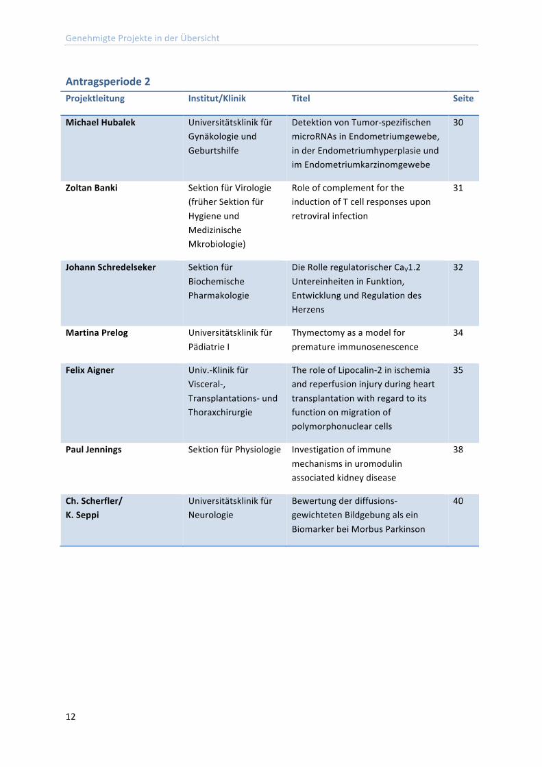
Antragsperiode 2 Projektleitung Institut/Klinik Titel Seite
Michael Hubalek Universitätsklinik für Gynäkologie und Geburtshilfe
Detektion von Tumor-‐spezifischen microRNAs in Endometriumgewebe, in der Endometriumhyperplasie und im Endometriumkarzinomgewebe
30
Zoltan Banki Sektion für Virologie (früher Sektion für Hygiene und Medizinische Mkrobiologie)
Role of complement for the induction of T cell responses upon retroviral infection
31
Johann Schredelseker Sektion für Biochemische Pharmakologie
Die Rolle regulatorischer CaV1.2 Untereinheiten in Funktion, Entwicklung und Regulation des Herzens
32
Martina Prelog Universitätsklinik für Pädiatrie I
Thymectomy as a model for premature immunosenescence
34
Felix Aigner Univ.-‐Klinik für Visceral-‐, Transplantations-‐ und Thoraxchirurgie
The role of Lipocalin-‐2 in ischemia and reperfusion injury during heart transplantation with regard to its function on migration of polymorphonuclear cells
35
Paul Jennings Sektion für Physiologie Investigation of immune mechanisms in uromodulin associated kidney disease
38
Ch. Scherfler/ K. Seppi
Universitätsklinik für Neurologie
Bewertung der diffusions-‐gewichteten Bildgebung als ein Biomarker bei Morbus Parkinson
40
Genehmigte Projekte in der Übersicht
13
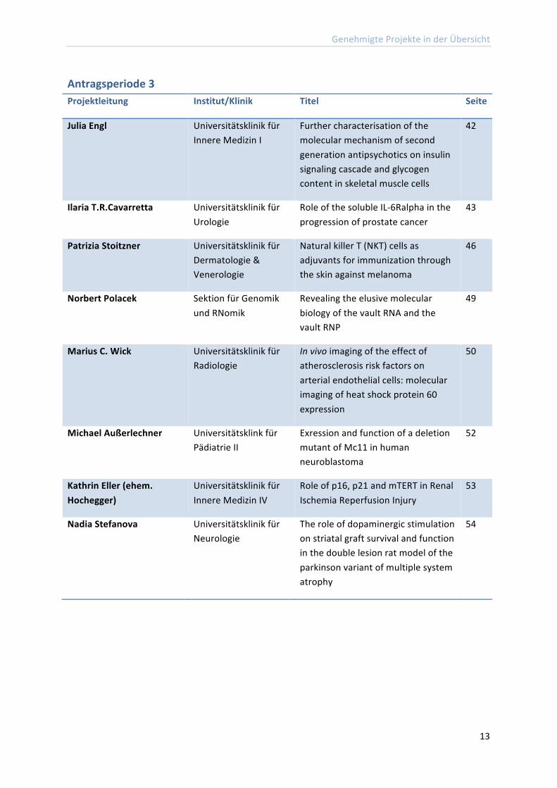
Antragsperiode 3 Projektleitung Institut/Klinik Titel Seite
Julia Engl Universitätsklinik für Innere Medizin I
Further characterisation of the molecular mechanism of second generation antipsychotics on insulin signaling cascade and glycogen content in skeletal muscle cells
42
Ilaria T.R.Cavarretta Universitätsklinik für Urologie
Role of the soluble IL-‐6Ralpha in the progression of prostate cancer
43
Patrizia Stoitzner Universitätsklinik für Dermatologie & Venerologie
Natural killer T (NKT) cells as adjuvants for immunization through the skin against melanoma
46
Norbert Polacek Sektion für Genomik und RNomik
Revealing the elusive molecular biology of the vault RNA and the vault RNP
49
Marius C. Wick Universitätsklinik für Radiologie
In vivo imaging of the effect of atherosclerosis risk factors on arterial endothelial cells: molecular imaging of heat shock protein 60 expression
50
Michael Außerlechner Universitätsklink für Pädiatrie II
Exression and function of a deletion mutant of Mc11 in human neuroblastoma
52
Kathrin Eller (ehem. Hochegger)
Universitätsklinik für Innere Medizin IV
Role of p16, p21 and mTERT in Renal Ischemia Reperfusion Injury
53
Nadia Stefanova Universitätsklinik für Neurologie
The role of dopaminergic stimulation on striatal graft survival and function in the double lesion rat model of the parkinson variant of multiple system atrophy
54
Genehmigte Projekte in der Übersicht
14
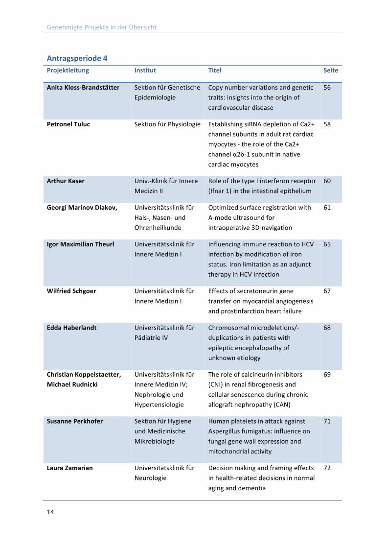
Antragsperiode 4 Projektleitung Institut Titel Seite
Anita Kloss-‐Brandstätter Sektion für Genetische Epidemiologie
Copy number variations and genetic traits: insights into the origin of cardiovascular disease
56
Petronel Tuluc Sektion für Physiologie Establishing siRNA depletion of Ca2+ channel subunits in adult rat cardiac myocytes -‐ the role of the Ca2+ channel α2δ-‐1 subunit in native cardiac myocytes
58
Arthur Kaser Univ.-‐Klinik für Innere Medizin II
Role of the type I interferon receptor (Ifnar 1) in the intestinal epithelium
60
Georgi Marinov Diakov, Universitätsklinik für Hals-‐, Nasen-‐ und Ohrenheilkunde
Optimized surface registration with A-‐mode ultrasound for intraoperative 3D-‐navigation
61
Igor Maximilian Theurl Universitätsklinik für Innere Medizin I
Influencing immune reaction to HCV infection by modification of iron status. Iron limitation as an adjunct therapy in HCV infection
65
Wilfried Schgoer Universitätsklinik für Innere Medizin I
Effects of secretoneurin gene transfer on myocardial angiogenesis and prostinfarction heart failure
67
Edda Haberlandt Universitätsklinik für Pädiatrie IV
Chromosomal microdeletions/-‐duplications in patients with epileptic encephalopathy of unknown etiology
68
Christian Koppelstaetter, Michael Rudnicki
Universitätsklinik für Innere Medizin IV; Nephrologie und Hypertensiologie
The role of calcineurin inhibitors (CNI) in renal fibrogenesis and cellular senescence during chronic allograft nephropathy (CAN)
69
Susanne Perkhofer Sektion für Hygiene und Medizinische Mikrobiologie
Human platelets in attack against Aspergillus fumigatus: influence on fungal gene wall expression and mitochondrial activity
71
Laura Zamarian Universitätsklinik für Neurologie
Decision making and framing effects in health-‐related decisions in normal aging and dementia
72
Abschlussberichte der MFI-‐Projekte
15
Abschlussberichte der MFI-‐Projekte Die hier abgedruckten Abschlussberichte sind durch die Projektleiter/innen der MFI Projekte angefertigt worden.
Abschlussberichte Antragsperiode 1
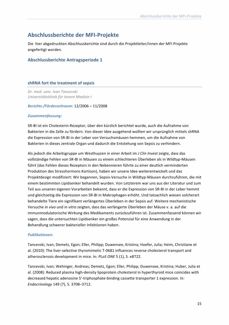
shRNA fort the treatment of sepsis
Dr. med. univ. Ivan Tancevski Universitätsklinik für Innere Medizin I
Berichts-‐/Förderzeitraum: 12/2006 – 11/2008
Zusammenfassung:
SR-‐BI ist ein Cholesterin-‐Rezeptor, über den kürzlich berichtet wurde, auch die Aufnahme von Bakterien in die Zelle zu fördern. Von dieser Idee ausgehend wollten wir ursprünglich mittels shRNA die Expression von SR-‐BI in der Leber von Versuchsmäusen hemmen, um die Aufnahme von Bakterien in dieses zentrale Organ und dadurch die Entstehung von Sepsis zu verhindern.
Als jedoch die Arbeitsgruppe um Westhuyzen in einer Arbeit im J Clin Invest zeigte, dass das vollständige Fehlen von SR-‐BI in Mäusen zu einem schlechteren Überleben als in Wildtyp-‐Mäusen führt (das Fehlen dieses Rezeptors in den Nebennieren führte zu einer deutlich verminderten Produktion des Stresshormons Kortison), haben wir unsere Idee weiterentwickelt und das Projektdesign modifiziert: Wir begannen, Sepsis-‐Versuche in Wildtyp-‐Mäusen durchzuführen, die mit einem bestimmten Lipidsenker behandelt wurden. Von Letzterem war uns aus der Literatur und zum Teil aus unseren eigenen Vorarbeiten bekannt, dass er die Expression von SR-‐BI in der Leber hemmt und gleichzeitig die Expression von SR-‐BI in Makrophagen erhöht. Und tatsachlich wiesen solcherart behandelte Tiere ein signifikant verlängertes Überleben in der Sepsis auf. Weitere mechanistische Versuche in vivo und in vitro zeigten, dass das verlängerte Überleben der Mäuse v. a. auf die immunmodulatorische Wirkung des Medikaments zurückzuführen ist. Zusammenfassend können wir sagen, dass die untersuchten Lipidsenker ein großes Potenzial für eine Anwendung in der Behandlung schwerer bakterieller Infektionen haben.
Publikationen:
Tancevski, Ivan; Demetz, Egon; Eller, Philipp; Duwensee, Kristina; Hoefer, Julia; Heim, Christiane et al. (2010): The liver-‐selective thyromimetic T-‐0681 influences reverse cholesterol transport and atherosclerosis development in mice. In: PLoS ONE 5 (1), S. e8722.
Tancevski, Ivan; Wehinger, Andreas; Demetz, Egon; Eller, Philipp; Duwensee, Kristina; Huber, Julia et al. (2008): Reduced plasma high-‐density lipoprotein cholesterol in hyperthyroid mice coincides with decreased hepatic adenosine 5'-‐triphosphate-‐binding cassette transporter 1 expression. In: Endocrinology 149 (7), S. 3708–3712.
Abschlussberichte der MFI-‐Projekte
16
Tancevski, Ivan; Wehinger, Andreas; Demetz, Egon; Hoefer, Julia; Eller, Philipp; Huber, Eva et al. (2009): The thyromimetic T-‐0681 protects from atherosclerosis. In: J. Lipid Res 50 (5), S. 938–944.
Ritsch, Andreas; Scharnagl, Hubert; Eller, Philipp; Tancevski, Ivan; Duwensee, Kristina; Demetz, Egon et al. (2010): Cholesteryl ester transfer protein and mortality in patients undergoing coronary angiography: the Ludwigshafen Risk and Cardiovascular Health study. In: Circulation 121 (3), S. 366–374.
Weitere Angaben zum Projekt:
Preise, aus den o.g. Publikationen, in denen das MFI genannt wurde:
1.Tancevski: ALSA -‐ Austrian Life Science Award 2009 2. Tancevski: ÖGES -‐ Hauptpreis Österr Ges f Endokrin. und Stoffwechsel 2010 3. Tancevski: AAS -‐ Hauptpreis der Österr Ges für Atherosklerose 2010
Nuclear Receptor Tyrosine Kinases: Ligand-‐dependent or independent transport
Priv. Doz. Dr. Martin Offterdinger Sektion für Neurobiochemie
Berichtszeitraum/Förderzeitraum: 05/2008 – 02/2009
Zusammenfassung:
Aufgrund der überaus stark verkürzten Laufzeit des Projektes konnten viele der Vorhaben, die im Projektantrag angeführt waren, noch nicht bearbeitet werden. Insgesamt kann aber aufgrund der vorliegenden Daten gesagt werden, dass nicht davon auszugehen ist, dass die jeweiligen Liganden der Rezeptortyrosinkinasen (RTK) eine wichtige Rolle beim Kernimport der RTKs spielen, da sowohl ruhende als auch Liganden stimulierte Zellen klar detektierbare nukleare RTKs aufwiesen. Konkret wurde EGFP markiertes FGFR1, erbB2 und erbB3 in Zellkernen lebender Zellen mit Hilfe von Konfokalmikroskopie nachgewiesen. Mit Hilfe von FRAP (,Fluorescence Recovery after Photobleaching') wurde darüber hinaus festgestellt, dass nukleare RTKs eine deutlich höhere Mobilität als membran-‐gebundene RTKs aufweisen und daher in einem ,Iöslichen' Zustand vorliegen müssen. Diese These wurde weiter erhärtet durch parallel durchgeführte Fluoreszenzkorrelationsspektroskopie (FCS) Studien.
Bei diesen Experimenten stellten wir fest, dass obwohl nukleare RTKs deutlich schneller diffundierten als ihre membrangebundenen Gegenstücke, sie dennoch langsamer waren als nur aufgrund des Molekulargewichts zu erwarten wäre. Da seit langem bekannt ist, dass RTKs in Gegenwart von Ligand miteinander assoziieren, untersuchten wir die Dimerisierung von RTKs im Zellkern und an der Zellmembran mit Hilfe von Fluoreszenz-‐Kreuz-‐Korrelationsspektroskopie (FCCS). Dabei erhielten wir den überraschenden Befund, dass erstens nukleare RTKs teilweise dimerisiert sind, und zwar in sehr ähnlichem Ausmaß wie auch an der Zellmembran.
Zweitens stellten wir fest, dass entgegen der allgemeinen Meinung in diesem Gebiet Behandlung ruhender, RTK exprimierender Zellen mit dem jeweiligen Liganden zu keinerlei Erhöhung des Anteils
Abschlussberichte der MFI-‐Projekte
17
an dimerisierten RTKs führte, und zwar weder an der Plasmamembran noch im Zellkern. Die quantitative Auswertung der FCCS Studien ermöglichte es, die Dissoziationskonstante (Kd) der RTKs zu messen, wobei Kds im niedrigen micromolaren Bereich gefunden wurden (i.e. 1 bis 3 μM).
The role of Langerhans cells in atopic dermatitis
Dr. Sandrine Dubrac Universitätsklinik für Dermatologie und Venerologie
Berichts-‐/Förderzeitraum: 01/2007 -‐ 12/2008
Zusammenfassung:
Thema des Projekts: To study the role of Langerhans cells (LC) in the development of atopic dermatitis (AD). LC belong to a subset of dendritic cells that reside in the epidermis. They are highly specialized in inducing immunity as well as in maintaining tolerance. A large body of evidence proves their extraordinary immunogenic capacities in vitro. In contrast, much less is known about their function in vivo. Regarding atopic dermatitis, high affinity IgE receptor-‐bearing LC have repeatedly been described, but there was no direct evidence so far confirming or excluding a pathogenetic role for LC.
Genetic or vitamin D3-‐induced overexpression of TSLP by keratinocytes results in an atopic dermatitis (AD)-‐like inflammatory phenotype in mice echoing the discovery of high TSLP expression in epidermis from AD patients. Although skin dendritic cells (DC) are suspected to be involved in AD, direct evidence of a pathogenetic role for skin DC in TSLP-‐induced skin inflammation has not yet been demonstrated. In a mouse model of AD i. e. mice treated with the low-‐calcemic vitamin D3 analogue, MC903, we show that epidermal Langerhans cells (LC)-‐depleted mice treated with MC903 do neither develop AD-‐like inflammation nor increased serum IgE as compared to vitamin D3 analogue-‐treated control mice.
Accordingly, we show that, in mice treated with MC903 or in K14-‐TSLP transgenic mice, expression of maturation markers by LC is increased whereas maturation of dermal DC is not altered. Moreover, only LC are responsible for the polarization of naive CD4+ T cells to a Th2 phenotype i. e., decrease in IFN-‐gamma and increase in IL-‐13 production by CD4+ T cells. This effect of LC on T-‐Iymphocytes does not require OX40-‐L/CD134 and is mediated by a concommitant downregulation of IL-‐12 and CD70.
While it was previously stated that TSLP up-‐regulates the production of TARC/CCL17 and MDC/CCL22 by human LC in vitro, our work shows that production of these Th2-‐ T cell attracting chemokines is increased only in keratinocytes in response to TSLP overexpression. These results demonstrate that LC are required for the development of AD in mouse models of AD involving epidermal TSLP overexpression. These data identify LC and the cytokine TSLP as potential and promising targets for anti-‐allergic therapies.
"Überraschungen" im Projektverlauf und bei den Ergebnissen: Surprise 1 about the change of our employement status: (i) the grant turned into a stipendium and (ii) we turned from Med Uni
Abschlussberichte der MFI-‐Projekte
18
employee into free-‐lancer researcher. Although this unexpected problem was eventually solved in a satisfactory manner, it initially absorbed much time, energy and motivation.
Surprise 2 about the research itself: Although we suspected LC to be involved in AD, we did not expect such a crucial implication of LC in the development of the disease. The results open new avenues of treatment for AD.
Major Results: This work provided the first evidence that LC are required to develop AD at least in animals. Moreover, LC are responsible of the Th-‐2 polarization of the immune system that occurs in the early development of the disease.
Publikationen:
Elentner, Andreas; Finke, Daniela; Schmuth, Matthias; Chappaz, Stéphane; Ebner, Susanne; Malissen, Bernard et al. (2009): Langerhans cells are critical in the development of atopic dermatitis-‐like inflammation and symptoms in mice. In: J. Cell. Mol. Med 13 (8B), S. 2658–2672.
Role of the PKCθ /ITK complex in primary CD3+ T cell activation processes
Dr. Mag. Nikolaus Thuille Sektion für Zellgenetik
Berichts-‐/Förderzeitraum: 12/2006 – 11/ 2008
Zusammenfassung:
The aim of this project was to investigate the functional role of an observed PKCθ/ITK signal transduction cross-‐talk in mouse CD3+ T lymphocytes, leading to proliferation, cytokine secretion, cytoskeletal remodelling, and apoptosis resistance of T cells. The parallel use of a gene ablation approach employing our established single and double Ko mice as well as a knockdown strategy with cell-‐permanent pharmacological inhibitors allowed me to directly compare the distinct enzymatic versus scaffold function of PKCθ and/or Itk in TCR-‐induced signalling complex formation.
PKCθ/Itk doubly deficient mice mainly resembled Itk-‐deficient mice in their T cell developmental phenotype. Also in the periphery, measured by Flow cytometric analyses (FACS) of cellularity of spleen and lymph nodes, PKCθ-‐/-‐/Itk-‐/-‐ mice revealed no additive differences in the distribution of CD3+, CD4+, CD8+, and CD19+ cells but resembled mostly the Itk-‐/-‐ phenotype.
The FACS analysis of the surface expression of CD25, CD44, and CD69 activation markers revealed a significant reduction of the mean fluorescence intensities in the PKCθ-‐/-‐/Itk-‐/-‐ and the Itk-‐/-‐ knockouts indicating a defect in the up-‐regulation of both the IL-‐2 receptor α-‐chain (CD25) and the activation marker CD69.
The combined loss of both PKCθ and Itk resulted in a reduction of proliferation and IL-‐2 secretion responses upon CD3/CD28 costimulation both by genetic and LMWI-‐mediated inhibition, however, this effect did not significantly exacerbate the defects observed in single deficient as well as single-‐inhibited T cells.
Abschlussberichte der MFI-‐Projekte
19
Further investigations of the transactivation status of important IL-‐2 gene transcription factors by EMSA assays revealed similarly NF-‐AT, NFκB and AP-‐1 activation defects in PKCθ and/or ITK single and double-‐deficient T cells. Consistently, the impaired activation status of PLCγ1 and ERK1/2 in the PKCθ-‐/-‐/Itk-‐/-‐ T cells as well as in the combined inhibitor treated cells, examined by phosphostatus analysis, were comparable to the defects in Itk-‐/-‐ T cells and Itk inhibitor treated T cells.
It has been published that in T cells, deficiency of Itk decreases antigen-‐induced cell death of mature cells, therefore I reinvestigated the apoptotic responses of PKCθ-‐/-‐/Itk-‐/-‐ T cell blasts in comparison to wild type and single knockout controls, however, in my hands, no significant differences in activation-‐induced cell death (AICD) susceptibility was revealed between the four genotypes. Similarly, panPKC and Itk inhibitor treated T cell blasts showed no significantly altered susceptibility to AICD in vitro.
Since a requirement of Itk and PKCθ for integrin-‐mediated adhesion of primary T cells has been demonstrated, we expected a stronger defect in the absence of both proteins, but surprisingly no additive adhesion defect of PKCθ-‐/-‐/Itk-‐/-‐ T cells was observed.
As conclusion from the present study, combined inhibitor treatment as well as the PKCθ-‐/-‐/Itk-‐/-‐ double deficiency failed to reveal new insights in the T cell activation functions of these two protein kinases, providing experimental evidence that Itk and PKCθ according to the classical concept of TCR signalling, are functioning in the same pathway during T cell activation.
The characterisation of the role of Thr-‐51 phosphorylation on PKCθ as possible structural requirement for the physical interaction of PKCθ and ITK upon T cell activation is still under investigation and could not been resolved during this funding period.
Publikationen:
Evenou, Jean-‐Pierre; Wagner, Jürgen; Zenke, Gerhard; Brinkmann, Volker; Wagner, Kathrin; Kovarik, Jiri et al. (2009): The potent protein kinase C-‐selective inhibitor AEB071 (sotrastaurin) represents a new class of immunosuppressive agents affecting early T-‐cell activation. In: J. Pharmacol. Exp. Ther 330 (3), S. 792–801.
Thuille, Nikolaus; Lutz-‐Nicoladoni, Christina; Letschka, Thomas; Hermann-‐Kleiter, Natascha; Heit, Isabelle; Baier, Gottfried (2009): PKCtheta and Itk functionally interact during primary mouse CD3+ T cell activation. In: Immunol. Lett 126 (1-‐2), S. 54–59.
Abschlussberichte der MFI-‐Projekte
20
Heat shock protein 60 -‐ A signalling molecule in atherogenesis?
Priv.-‐Doz. Dr. David Bernhard Universitätsklinik für Herzchirurgie (jetzt Wien)
Berichts-‐/Förderungszeitraum: 12/2006 – 11/2008
Zusammenfassung:
Im Rahmen des Projektes "Heat Shock Protein 60 -‐ A Signalling Molecule in Atherogenesis?" wurde in den vergangenen 2 Jahren zunächst die Bedeutung des Moleküls in der durch Rauchen verursachten Atherogenese untersucht. So konnte zunächst gezeigt werden, dass Rauchextrakte die Expression of HSP60 auf Transkriptionsebene erhöhen. Als Folge der Exposition von Endothelzellen wurde mittels Herstellung und Einsatzes eine HSP60-‐EYFP Fusionsproteins und Live Cell Imaging gezeigt, dass HSP60 aus den Mitochondrien als Folge von Stress freigesetzt wird (zyotsolische Verteilung) und konnte später an der Zelloberfläche nachgewiesen werden (Konfokale Laserscanning Mikroskopie).
Dies bildet den zentralen Punkt im Modell der Autoimmunhypothese der Atherosklerose und konnte diese weiter festigen. Zusätzlich wurde nachgewiesen, dass HSP60 auch freigesetzt wird (in vitro, Zellkulturfiberstande) und erhöhte Werte von löslichem HSP60 wurden in der Folge auch im Serum von Passivrauchern festgestellt, was die in vivo Bedeutung der Entdeckung unterstreicht. In Studien, in denen mittels FITC-‐markierten rekombinanten HSP60 die Affinität zu Endothelzellen untersucht wurde, konnte nur eine sehr schwache, jedoch spezifische (Kompetition) Affinität beobachtet werden.
In dieser Studie konnte also zunächst klar nachgewiesen werden, dass auch der Risikofaktor Rauchen im Sinne der Autoimmunhypothese der Atherosklerose aktiv ist und bei Vorhandensein von Autoantikörpern gegen körpereigenes HSP60 die immunologische Schädigung des Gefäßendothels fördern und/oder auslösen kann. Die Bedeutung von HSP60 als Signalmolekül, das beispielsweise über Bindung und Aktivierung von TOLL-‐like Rezeptoren zu Pro-‐Inflammatorischen Signalling im Rahmen der Atherogenese beiträgt, bleibt weiter unklar. Es konnte zwar nachgewiesen werden, dass HSP60 an Endothelzellen bindet, die niedrige Affinität stellt jedoch ein Problem für detaillierte Bindungsstudien dar.
Die Tatsache, dass lediglich eine schwache Affinität nachgewiesen werden konnte, bedeutet jedoch nicht, dass HSP60 keine Signalwege in Endothelzellen aktiviert, z.B. über TOLL-‐like Rezeptoren. Zukünftige Microarrayanalysen sollten darüber Aufschluss geben. Besonders wichtig in diesem Zusammenhang ist, dass hochreines rekombinantes Protein verwendet wird, da geringste Verunreinigungen mit Lipopolysacchariden ebenfalls zur Aktivierung von TOLL-‐like Rezeptor vermitteltem Signalling führen würden. Die Herstellung von hochreinem rekombinantem HSP60 stellt für die weiteren Analysen den zentralen Punkt dar.
Im Rahmen des vom MFI geförderten Projektes konnte eine naturwissenschaftliche Dissertation angefertigt (von Frau Dr. Barbara Messner) und zahlreiche Publikationen verfasst werden (siehe oben). Die zentrale Arbeit zum Thema (Publikation # 4) ist experimentell angeschlossen, ein Manuskript derzeit in Vorbereitung.
Abschlussberichte der MFI-‐Projekte
21
Ich möchte mich im Namen meiner Arbeitsgruppe sowie in meinem Namen herzlichst beim MFI für die Unterstützung bedanken!!
Publikationen:*
Bernhard D, Wang XL. (2007): Smoking, Oxidative Stress and Cardiovascular Diseases -‐ Do Anti-‐Oxidative Therapies Fail? In: Current Medicinal Chemistry 14, 1689-‐1699
Henderson, Blair; Csordas, Adam; Backovic, Aleksandar; Kind, Michaela; Bernhard, David; Wick, Georg (2008): Cigarette smoke is an endothelial stressor and leads to cell cycle arrest. In: Atherosclerosis 201 (2), S. 298–305.
Bernhard, D.; Laufer, G. (2008): The aging cardiomyocyte: a mini-‐review. In: Gerontology 54 (1), S. 24–31.
Kreutmayer, Simone Barbara; Messner, Barbara; Knoflach, Michael; Henderson, Blair; Niederegger, Harald; Böck, Günther et al. (2011): Dynamics of heat shock protein 60 in endothelial cells exposed to cigarette smoke extract. In: Journal of molecular and cellular cardiology.
Gangl, K.; Reininger, R.; Bernhard, D.; Campana, R.; Pree, I.; Reisinger, J. et al. (2009): Cigarette smoke facilitates allergen penetration across respiratory epithelium. In: Allergy 64 (3), S. 398–405.
Messner B, Knoflach M, Seubert A, Ritsch A, Pfaller K, Henderson B, Zeller I, Willeit J, Laufer G, Wick G, Kiechl S, Bernhard D.(2009): Cadmium a Novel and Independent Risk Factor for Early Atherosclerosis: Mechanisms and in vivo Relevance. Arterioscler Thromb Vasc Biol. (9):1392-‐8. Epub 2009 Jun 25.
Reisinger U, Zeller I, Schwaiger S, Messner B, Mayr T, Stigler R, Wiedemann D, Seger C, Schachner T, Dirsch V, Vollmar A, Bonatti J, Stuppner H, Laufer G, Bernhard D.(2009): Leoligin -‐ the Major Lignan from Edelweiss -‐ Inhibits Intimal Hyperplasia of Venous Bypass Grafts.In: Cardiovasc Res. 82(3):542-‐9. Epub 2009 Feb 19.
Csordas, Adam; Wick, Georg; Laufer, Günther; Bernhard, David (2008): An Evaluation of the Clinical Evidence on the Role of Inflammation and Oxidative Stress in Smoking-‐Mediated Cardiovascular Disease. In: Biomark Insights 3, S. 127–139.
*Vollständig kursiv angeführte Publikationen ohne MFI-‐Nennung
Abschlussberichte der MFI-‐Projekte
22
The role of Aspergillus fumigatu CccA in iron storage and virulence
Mag. Dr. Martin Eisendle Sektion für Molekularbiologie
Berichts-‐/Förderzeitraum: 1/2007 – 10/2008
Zusammenfassung:
The opportunistic fungal pathogen Aspergillus fumigatus synthesizes three structurally different siderophores (low molecular mass, ferric iron-‐specific chelators): extracellular triacetylfusarinine C for iron uptake, intracellular ferricrocin for hyphal iron storage and intracellular hydroxyferricrocin for conidial iron storage. On one hand, iron is essential for all living organisms, but on the other hand intracellular iron overload is toxic due to the generation of reactive oxygen species.
Therefore both, iron uptake and iron storage have to be tightly regulated and each organism has to develop mechanisms to detoxify excessive iron. Siderophore biosynthesis is repressed by iron via the GATA-‐transcription factor SreA and, consequently, inactivation of SreA results in excessive iron uptake during iron-‐replete conditions (Haas et al. 2008). Recently, iron metabolism of A. fumigatus attracted considerable interest as siderophore biosynthesis was identified to essential for this opportunistic fungal pathogen.
In the course of his project I was able to demonstrate that A. fumigatus employs in addition to ferricrocin a second mechanism for hyphal iron storage. Transcription of cccA, a homolog of the Saccharomyces cerevisiae vacuolar iron importer CCC1, was induced by iron, suggesting a role of CccA in iron homeostasis. A generated CccA-‐deficient A. fumigatus mutant displayed wild type-‐like growth during iron starvation but reduced growth during conditions of high iron availability. In particular the combination of CccA-‐deficiency and SreA-‐deficiency drastically decreased iron resistance.
A GFP (green fluorescence protein)-‐CccA fusion protein cured the defects caused by CccA-‐deficiency proving that the GFP-‐CccA fusion protein is functional. Epiluorescence microscopy revealed that GFP-‐CccA localizes to the vacuolar membrane similar to the yeast ortholog Ccc1. Importantly, inactivation of ferricrocin biosynthesis decreased growth during iron starvation but not iron-‐replete conditions.
Taken together, these results demonstrate that CccA is crucial for iron detoxification by transporting excessive cytosolic iron into the vacuole. In contrast, ferricrocin biosynthesis appears not to be involved in iron detoxification. Interestingly, various experimental approaches indicated that the CccA-‐mediated iron loading of the vacuole is not beneficial during iron starvation. The latter might indicate that the vacuole does not serve as nutritional iron store in A. fumigatus, which contrast the situation in S. cerevisiae. The results gained during this study are adequate for publication in a scientific journal. The manuscript is currently in preparation.
Publikationen:
Haas, Hubertus; Eisendle, Martin; Turgeon, B. Gillian (2008): Siderophores in fungal physiology and virulence. In: Annu Rev Phytopathol 46, S. 149–187.
Abschlussberichte der MFI-‐Projekte
23
Weitere Angaben zum Projekt:
Eisendle M, Abt B, Haas H (2008). CccA is involved in iron storage in Aspergillus fumigatus2008: Abstract at the 9th European Conference of Fungal Genetics (ECFG) 05.-‐08. 04. 2008, Edinburgh Scotland/UK.
TLR9 mediated effects on breast cancer cells by means of extracellular DNA
Priv.-‐Doz. Mag.Dr. Heidelinde Fiegl Universitätsklinik für Gynäkologie und Geburtshilfe
Berichts-‐/Förderzeitraum: 02/2007 – 01/2009
Zusammenfassung:
Although TLR9 has been previously considered to be expressed only in immune cells, there is now increasing evidence that TLR9 expression is present in non immune cells as well. We analyzed TLR9 mRNA expression in 124 human breast cancer samples, 10 benign breast tissues, 138 ovarian cancer samples and 30 non neoplastic ovarian tissues. We found that TLR9 mRNA expression increases with grade (p = 0.03) and is higher in estrogen receptor negative breast cancers samples (p= 0.005). An immunohistochemical analysis of paraffin embedded tissue slides of 116 breast cancer patients showed a stronger TLR9 protein expression in hormone receptor negative breast cancer specimens in comparison to hormone receptor positive breast cancer specimens (p<0.001).
In order to determine whether TLR9 expression is unique to breast cancer or is a feature in common with other solid tumors, we analyzed ovarian tissue samples from 138 patients with ovarian cancer 138 ovarian cancer and 30 patients with benign diseases. We observed a statistically significant augmentation of TLR9 mRNA expression from non-‐neoplastic ovarian tissues in comparison to serous ovarian cancers (p=0.02) or from serous to mucinous ovarian cancers respectively (p<0.001). Further we identified a correlation between tumor grade and TLR9 mRNA expression (p= 0.039).
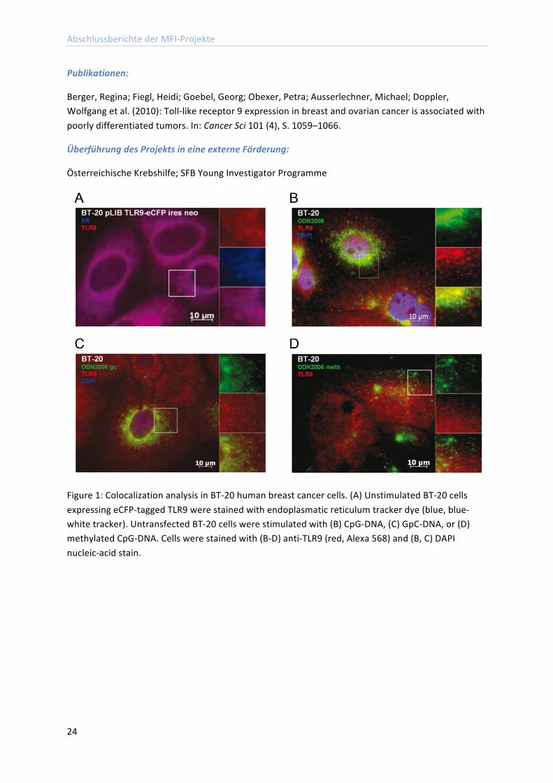
TLR9 is activated by nonmethylated CpG motifs. To visualize the entrance of nonmethylated CpG DNA structures in BT-‐20 and Hs578T breast cancer cells and the binding to TLR9, we performed immunofluorescence analysis. In untransfected human breast cancer cells, TLR9 was recruited to CpG-‐DNA within 90 min after incubation, and followed CpG-‐DNA to the intracellular compartments as expected. When these cells were incubated with fluorescence labeled control GpC-‐DNA or methylated CpG-‐DNA, translocation of TLR9 was greatly diminished. The results for BT-‐20 breast cancer cells are shown in Figure 1.
Summary: We demonstrated in this study that TLR9 expression is associated with poor differentiation in breast and ovarian tumors, and that TLR9 overexpression and stimulation with hypomethylated DNA augments the migratory capacity of cancer cell lines.
Abschlussberichte der MFI-‐Projekte
24
Publikationen:
Berger, Regina; Fiegl, Heidi; Goebel, Georg; Obexer, Petra; Ausserlechner, Michael; Doppler, Wolfgang et al. (2010): Toll-‐like receptor 9 expression in breast and ovarian cancer is associated with poorly differentiated tumors. In: Cancer Sci 101 (4), S. 1059–1066.
Überführung des Projekts in eine externe Förderung:
Österreichische Krebshilfe; SFB Young Investigator Programme
Figure 1: Colocalization analysis in BT-‐20 human breast cancer cells. (A) Unstimulated BT-‐20 cells expressing eCFP-‐tagged TLR9 were stained with endoplasmatic reticulum tracker dye (blue, blue-‐white tracker). Untransfected BT-‐20 cells were stimulated with (B) CpG-‐DNA, (C) GpC-‐DNA, or (D) methylated CpG-‐DNA. Cells were stained with (B-‐D) anti-‐TLR9 (red, Alexa 568) and (B, C) DAPI nucleic-‐acid stain.
Abschlussberichte der MFI-‐Projekte
25
Ion channel interactions within neuronal micro-‐ and nanodomains: A freeze-‐fracture immunogold approach
Dr. Walter Kaufmann Institut für Pharmakologie
Berichts-‐/Förderzeitraum: 02/2007 – 04/2009
Initially, the project was funded for the duration of one year, and it was started with the 1st of February 2007. It was then prolonged for another year after a meeting of the MFI jury in December 2007 (project duration from February 2008 to January 2009), and another 3 month (February 2009 to April 2009) within the budget of the project. Research activities during the whole period strictly followed the work plan and time table given in the project proposal.
Zusammenfassung:
During the first period (1st to 8th month, objective 1: subcellular expression profile of KCa 1.1 channels), the ultrastructural localization of KCa 1.1 (also called BKCa) channels was established in cerebellar Purkinje neurons and dentate gyrus granule cells of rat and mouse brain. Basically, pre-‐embedding electron microscopy (immunoperoxidase and immunometal techniques), post-‐embedding immunogold electron microscopy and SDS-‐digested freeze-‐fracture replica labeling (SDS-‐FRL) was applied. BKCa channels were localized to somato-‐dendritic as well as axonal sites in these types of neurons.
At distal dendritic compartments, BKCa channels were found scattered in the extrasynaptic plasma membrane of dendritic shafts and spines, sparing postsynaptic membrane specializations. At the level of the soma and proximal dendritic compartments, two pools of channels were detected. One pool was scattered in the extrasynaptic plasma membrane, and one pool was clustered in areas of the plasma membrane with underlying subsurface membrane cisterns. Quantitative analysis by means of SDS-‐FRL revealed that about 1/3 of BKCa channels belong to the clustered pool and about 2/3 to the scattered pool in principal cell somata. In contrast, the labeling density ratio of clustered to scattered channels was about 60:1, varying slightly with the cell type analyzed.
The two pools might represent independent channel pools that differ in their routes of Ca2+ activation. This was indicated in co-‐localization studies of BKCa channels with metabotropic glutamate receptors (mGlu) type-‐1, voltage-‐gated Ca2+ channels of the L-‐, P/Q-‐ and N-‐type, and inositol 1,4,5-‐triphosphate receptors (IP3-‐R). Whereas scattered BKCa channels may be activated via voltage-‐gated Ca2+ channels and/or metabotropic glutamate-‐receptors, clustered channels are likely to be activated via Ca2+ release from internal stores, specifically from junctional components of the endoplasmic reticulum (ER). Such ER formations, including subsurface membrane cisterns, are enriched in IP3-‐Rs that mediate the effects of several neurotransmitters, hormones, and growth factors by releasing Ca2+ into the cytosol, thus generating local Ca2+ sparks. These increases in cytosolic Ca2+ levels may be sufficient for BKCa channel activation in the plasma membrane. Clustered BKCa channels may thus participate in building a functional unit (a PLasmERosome) with the underlying ER calciosome contributing significantly to local signaling in principal neurons.
At axonal domains of principal neurons, BKCa channels were localized in the axon terminal of excitatory synapses, enriched in the pre-‐synaptic active zone. Moreover, some scattered channels
Abschlussberichte der MFI-‐Projekte
26
were detected at perisynaptic sites of axon terminals, and in the plasma membrane of axon initial segments, although at low densities.
During the second period (9th to 15th month, objective 2: Do BKCa channels and voltage-‐gated Ca2+ channels interact within specific subdomains and which Ca2+ channel subtypes are involved?), co-‐assembly of BKCa channels with voltage-‐gated Ca2+ channels was analyzed, focusing on P/Q-‐type channels. Ca2+ influx from the extracellular milieu through these channels induces a local increase in the cytosolic free Ca2+ concentration that is sufficient for rapid BKCa channel activation within nano-‐domains. P/Q-‐type channels were detected in the extrasynaptic plasma membrane of dendritic stems and spines at high densities, whereas immunolabeling in the somata was low. The somatic labeling of P/Q-‐type channels resulted mainly from the abundance of the proteins in the ER, as revealed by means of pre-‐embedding electron microscopy.
Efforts to immunolocalize P/Q-‐type channels using SDS-‐FRL were unsuccessful despite intensive testing of several commercial and noncommercial antibodies raised against different species. SDS-‐FRL reveals pseudothree-‐dimensional views of cell membranes and would allow qualitative as well as quantitative analysis of integral membrane proteins beyond the limitations of thin-‐section electron microscopy. It can therefore only be surmised, that P/Q-‐type channels are associated primarily with the scattered pool of BKCa channels at dendritic sites, insofar as P/Q-‐type channels are almost absent from the somata and proximal dendritic stems, where the clustered pool of BKCa channels is present.
It was analyzed next, whether BKCa channels are associated with postsynaptic membrane specializations of GABAergic synapses. GABAA receptors containing the α1 subunit are known being enriched in the postsynaptic specialization of inhibitory synapses. Hence double labeling for GABAA α1 and BKCa channels was performed using SDS-‐FRL. Specificity of immunolabeling was tested and confirmed on samples of GABAA α1 and BKCa channel null mice, respectively. Although the clustered pool of BKCa channels was always segregated from clusters of GABAA α1, scattered BKCa channels were found within GABAA α1 clusters every now and then. In conclusion, clusters of BKCa channels are always segregated from postsynaptic specializations of GABAergic synapses. Scattered BKCa channels reside mainly at extrasynaptic sites, and a few channels may be present in the postsynaptic membrane of GABAergic synapses.
In the third period (16th to 27th month, objective 3: association of BKCa channels with subsurface membrane cisterns), the expression of BKCa channels at sites of subsurface cisterns and the interaction of these channels with internal Ca2+ release channels as well as mGlu1 was studied more precisely in various types of central principal neurons. Specifically, the subcellular distribution profile of immunogold particles labeling BKCa channels was established for specific somatic domains, and compared to the distribution of IP3-‐Rs by means of post-‐embedding immunogold labeling techniques. IP3-‐Rs were localized to junctional and nonjunctional components of the rough and smooth ER, whereas plasma membranes were immunonegative for IP3-‐Rs. The peak of the subcellular IP3-‐R distribution was located in the cell interior at a distance of a few nanometers from the plasma membrane, whereas the peak of the BKCa channel distribution was lying very close to the inner aspect of the plasma membrane. This pointed to the presence of a large pool of BKCa channels in the plasma membrane, and a minor pool of intracellular channels in areas of subsurface membrane cisterns.
Abschlussberichte der MFI-‐Projekte
27
Co-‐immunolabeling for BKCa channels and mGlu1α in cerebellar Purkinje cells revealed
90% of dendritic spines immunopositive for both proteins, indicating that most if not all spines in Purkinje cells co-‐express mGlu1α and scattered BKCa channels. Co-‐localization of these channel types was observed not only at the level of the spines, but also at dendritic stems and neuronal somata. In contrast to the scattered pool of channels, clustered BKCa channels were not found co-‐localized with mGlu1α. Metabotropic glutamate receptors control cytosolic Ca2+ transients triggering Ca2+ influx via voltage-‐gated Ca2+ channels and Ca2+ release from internal stores upon activation of IP3-‐Rs. Activation of mGlu1α can thus induce activation of scattered BKCa channels at somato-‐dendritic domains of central principal neurons.
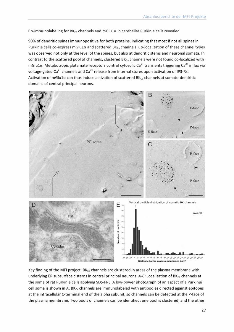
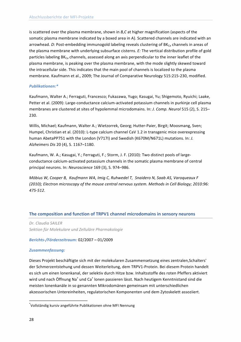
Key finding of the MFI project: BKCa channels are clustered in areas of the plasma membrane with underlying ER subsurface cisterns in central principal neurons. A-‐C: Localization of BKCa channels at the soma of rat Purkinje cells applying SDS-‐FRL. A low-‐power photograph of an aspect of a Purkinje cell soma is shown in A. BKCa channels are immunolabeled with antibodies directed against epitopes at the intracellular C-‐terminal end of the alpha subunit, so channels can be detected at the P-‐face of the plasma membrane. Two pools of channels can be identified; one pool is clustered, and the other
Abschlussberichte der MFI-‐Projekte
28
is scattered over the plasma membrane, shown in B,C at higher magnification (aspects of the somatic plasma membrane indicated by a boxed area in A). Scattered channels are indicated with an arrowhead. D: Post-‐embedding immunogold labeling reveals clustering of BKCa channels in areas of the plasma membrane with underlying subsurface cisterns. E: The vertical distribution profile of gold particles labeling BKCa channels, assessed along an axis perpendicular to the inner leaflet of the plasma membrane, is peaking over the plasma membrane, with the mode slightly skewed toward the intracellular side. This indicates that the main pool of channels is localized to the plasma membrane. Kaufmann et al., 2009; The Journal of Comparative Neurology 515:215-‐230, modified.
Publikationen:*
Kaufmann, Walter A.; Ferraguti, Francesco; Fukazawa, Yugo; Kasugai, Yu; Shigemoto, Ryuichi; Laake, Petter et al. (2009): Large-‐conductance calcium-‐activated potassium channels in purkinje cell plasma membranes are clustered at sites of hypolemmal microdomains. In: J. Comp. Neurol 515 (2), S. 215–230.
Willis, Michael; Kaufmann, Walter A.; Wietzorrek, Georg; Hutter-‐Paier, Birgit; Moosmang, Sven; Humpel, Christian et al. (2010): L-‐type calcium channel CaV 1.2 in transgenic mice overexpressing human AbetaPP751 with the London (V717I) and Swedish (K670M/N671L) mutations. In: J. Alzheimers Dis 20 (4), S. 1167–1180.
Kaufmann, W. A.; Kasugai, Y.; Ferraguti, F.; Storm, J. F. (2010): Two distinct pools of large-‐conductance calcium-‐activated potassium channels in the somatic plasma membrane of central principal neurons. In: Neuroscience 169 (3), S. 974–986.
Möbius W, Cooper B, Kaufmann WA, Imig C, Ruhwedel T, Snaidero N, Saab AS, Varoqueaux F (2010); Electron microscopy of the mouse central nervous system. Methods in Cell Biology; 2010:96: 475-‐512.
The composition and function of TRPV1 channel microdomains in sensory neurons
Dr. Claudia SAILER Sektion für Molekulare und Zelluläre Pharmakologie
Berichts-‐/Förderzeitraum: 02/2007 – 01/2009
Zusammenfassung:
Dieses Projekt beschäftigte sich mit der molekularen Zusammensetzung eines zentralen,Schalters' der Schmerzentstehung und dessen Weiterleitung, dem TRPV1-‐Protein. Bei diesem Protein handelt es sich um einen lonenkanal, der selektiv durch Hitze bzw. Inhaltsstoffe des roten Pfeffers aktiviert wird und nach Öffnung Na+ und Ca+ lonen passieren lässt. Nach heutigem Kenntnistand sind die meisten lonenkanäle in so genannten Mikrodomänen gemeinsam mit unterschiedlichen akzessorischen Untereinheiten, regulatorischen Komponenten und dem Zytoskelett assoziiert.
*Vollständig kursiv angeführte Publikationen ohne MFI Nennung
Abschlussberichte der MFI-‐Projekte
29
Die zentrale Frage dieses Projektes war die Aufklärung der ,TRPV1 Mikrodomäne'. Genaue Kenntnis dieses Proteasoms wurde langfristig zum besseren Verständnis der Schmerzentstehung und -‐weiterleitung beitragen sowie eine selektivere Entwicklung von entsprechenden Pharmaka ermöglichen.
„Überraschungen“ im Projektverlauf und bei den Ergebnissen: Das Projekt wurde zu Beginn durch das limitierte Vorhandensein von Untersuchungsmaterial (gereinigte Membranen aus Hinterwurzelganglien der Maus und Ratte) verzögert. Weiters stellte das ,Überführen' des TRPV1 Rezeptors in lösliche Form (,Solubilisation') eine große Herausforderung dar. Diese Probleme konnten gelöst werden. Zwischenzeitlich wird das Projekt von einem Doktoranden weiterbearbeitet. Kürzlich waren wir in der Lage, einige Komponenten der TRPV1-‐Mikrodomäne zu identifizieren. Eine funktionelle Anklärung ist derzeit im Gange. Wir erwarten erste publizierbare Ergebnisse innerhalb der nächsten 6 Monate.
Hinweise auf mögliche Erfolgsberichte in den Publikationsmedien: Das Projekt ist bisher wissenschaftlich nicht abgeschlossen.
The interactosome of the nerve growth inhibitor Nogo-‐A
Dr. Rüdiger Schweigreiter Sektion für Neurobiochemie
Berichts-‐/Förderzeitraum: 01/2007 – 08/2007
Zusammenfassung:
Projekt wurde in eine externe Förderung überführt und daher beendet.
Externe Förderung:
FWF-‐Projekt ZFP199080
Abschlussberichte der MFI-‐Projekte
30
Abschlussberichte Antragsperiode 2
Detektion von Tumor-‐spezifischen microRNAs in Endometriumgewebe, in der Endometriumhyperplasie und im Endometriumkarzinomgewebe
Dr. Michael Hubalek Klinische Abteilung für Gynäkologie und Geburtshilfe
Berichts-‐/Förderzeitraum: 05/2007 -‐ 06/2008
Zusammenfassung:
In diesem Projekt gelang es, Unterschiede in der microRNA-‐Expression zwischen dem Endometriumkarzinom und der Endometriumhyperplasie im Vergleich zu benignem Gewebe festzustellen. Es konnte für das Karzinom die Beteiligung von schon als onkogen bekannten microRNAs sichergestellt werden. Auch in der Regulation von hormonellem Ansprechen bzw. Sensibilität von Geweben spielen microRNAs eine Rolle. Die Detektion einiger dieser microRNAs, die schon mit hormoneller Regulation in Zusammenhang gebracht wurden, im Endometriumkarzinom und vor allem in der Endometrumhyperplasie scheint ein weiterer Beleg dafür. Mit dieser Arbeit kann ein weiteres Malignom ergänzt werden, in deren Karzinogenese microRNAs eine Rolle spielen.
Auch in der Exploration prädiktiver Zielgene konnten durch unsere Ergebnisse Fortschritte gemacht werden, da sie teilweise sehr gut mit schon veröffentlichen Ergebnissen übereinstimmen und diese dadurch bestätigen.
Von besonderer Wichtigkeit in der Erforschung von microRNAs ist es das große Netz von Signalwegen mit microRNA-‐Beteiligung zu verstehen. Eine Herausforderung dieses Themengebietes ist die große Zahl schon bekannter microRNAs.
Ein Charakteristikum von microRNAs ist die Gewebsspezifität und es gelang dadurch, allein anhand von deren Expressionsmuster Gewebe unbekannten Ursprungs zu identifizieren.
Die großen Hoffnungen in microRNA liegen gerade in deren vorhergesagter Gewebsspezifität. Gerade in der Krebstherapie besteht Notwendigkeit an hochspezifischen Medikamenten, die nur maligne Zellen bekämpfen und nicht wie bisher auch gesunde Körperzellen in hohem Maße schädigen. Diese Medikamente führen zu massiven Nebenwirkungen bei schon vorgeschwächten Patienten. Gerade die unerwünschte Toxizität auf gesundes Gewebe wie z. B. den Gastrointestinaltrakt das blutbildenden System limitiert oft die Radikalität der Krebstherapie.
Es gibt bereits einige viel versprechende Versuche an Mäusen, wo es gelang, durch direkte Manipulation von microRNAs, Tumorwachstum-‐ und Migration zu hemmen, ohne dass man systemische Auswirkungen auf den Körper feststellen konnte. Um diese Therapiestrategien auch erfolgreich für den Menschen anwendbar zu machen, bedarf es noch viel genauerer Kenntnisse über die Verbreitung von microRNAs in den unterschiedlichen Geweben und deren Regulationsvorgängen.
Abschlussberichte der MFI-‐Projekte
31
Ein weiterer wichtiger Aspekt im Rahmen dieses geförderten Projektes betrifft die Erlangung von labortechnischen Fähigkeiten. Die Detektion von microRNAs ist ein technisch anspruchsvoller Vorgang und kann nur durch umfangreiche Versuche standardisiert werden. Im Rahmen dieses Projektes wurde viel know-‐how diesbezüglich gewonnen und letztendlich gab dieses Wissen sicherlich einen Auschlag, dass ein weiteres Projekt zur Detektion von onkogenen microRNAs im Brustkrebsgewebe im Rahmen des OncoTyrol Projektes gefördert wird.
Weitere Angaben zum Projekt:
Dissertation Fr. Dr. Johanna Tichl
Role of complement for the induction of T cell responses upon retroviral infection
Dr. Zoltan Banki Sektion für Virologie
Berichts-‐/Förderzeitraum: 06/2007 -‐ 05/2010
Zusammenfassung:
The induction of a robust cytotoxic T cell response is crucial to control retroviral infection. For the induction of both primary and secondary immune responses, DCs are thought to be the most potent antigen presenting cells. As DCs express complement receptors (CRs) and retroviruses activate the complement system, the role of complement in the induction of virus-‐specific T cell responses was investigated. As a model, the Friend virus (FV) complex was chosen, as this murine retrovirus shares several features with HIV.
First, we confirmed that opsonization of Friend murine leukemia virus (F-‐MuLV) with complement enhanced infection of DCs as observed with HIV. For this, F-‐MuLV was incubated in vitro in normal mouse serum (NMS) to deposit C3-‐fragments on the viral surface. Next, we performed infection experiments of mouse bone marrow-‐derived DCs (bmDCs) with differentially opsonized F-‐MuLV. Non-‐ and C-‐opsonized F-‐MuLV productively infected bmDCs, but significantly higher virus levels were obtained from culture supernatants of DCs infected with F-‐MuLV-‐C. We next determined the capacity of bmDCs loaded with non-‐ or C-‐opsonized F-‐MuLV to activate transgenic CD8+ T cells expressing a T cell receptor specific for an FV gag peptide (TCRtg CD8+ T cells) in vitro.
Co-‐culture of TCRtg CD8+ T cells with bmDCs exposed to F-‐MuLV significantly induced expression of the early activation marker CD69 on CD8+ T cells, when compared to control DCs. CD8+ T cell activation was significantly enhanced when FV-‐specific CD8+ T cells were co-‐cultured with bmDCs loaded to C-‐opsonized F-‐MuLV. In addition to CD69, CD25 on F-‐MuLV-‐C/bmDCactivated CD8+ T cells was also expressed. To study proliferation of FV-‐specific CD8+ T cells induced by virus loaded bmDCs, isolated FV-‐specific TCRtg CD8+ T cells were stained with CFSE prior to co-‐culture with bmDCs. We found that after 4 days of co-‐culture both non-‐ and C-‐opsonized F-‐MuLV-‐loaded bmDCs induced proliferation of FV-‐specific CD8+ T cells.
However, the proliferation of CD8+ T cells induced by F-‐MuLV-‐C loaded bmDCs was more pronounced.
Abschlussberichte der MFI-‐Projekte
32
To determine if the activation and proliferation of virus-‐specific CD8+ T cells was impaired in C3-‐deficient mice, splenic CD8+ T cells were stained with tetramers. We observed a significantly reduced proportion of tetramer positive CD8+ T cells in the C3-‐deficient mice compared to wild-‐type (wt). Furthermore, there was significantly reduced expression of the activation-‐induced isoform of CD43 on CD8+ T cells. Since previous results indicated that CD8+ T cells are critical for recovery from FV infection, it was expected that impaired CD8+ T cell responses in C3-‐deficient mice would exacerbate FV infection. Indeed, there were significantly higher proportions of infected spleen cells in B6 C3-‐/-‐ mice at 7 dpi compared to B6 wt animals. These data indicated the importance of C3 for the control of retroviral infection in vivo, which correlates with the activation and proliferation of CD8+ T cells.
Publikationen:
Bánki, Zoltán; Posch, Wilfried; Ejaz, Asim; Oberhauser, Verena; Willey, Suzanne; Gassner, Christoph et al. (2010): Complement as an endogenous adjuvant for dendritic cell-‐mediated induction of retrovirus-‐specific CTLs. In: PLoS Pathog 6 (4), S. e1000891.
Bila, Custodio; Oberhauser, Verena; Ammann, Christoph G.; Ejaz, Asim; Huber, Georg; Schimmer, Simone et al. (2011): Complement opsonization enhances friend virus infection of B cells and thereby amplifies the virus-‐specific CD8+ T cell response. In: J. Virol 85 (2), S. 1151–1155.
Überführung des Projekts in eine externe Förderung:
ja, FWF-‐Projekt P21508-‐B13
Die Rolle regulatorischer CaV1.2 Untereinheiten in Funktion, Entwicklung und Regulation des Herzens
Dr. Johann Schredelseker Sektion für Biochemische Pharmakologie
Berichts-‐/Förderzeitraum: 07/2007 – 06/2009
Zusammenfassung:
Im Rahmen des MFI Projekts wurden erfolgreich einige Methoden am Modellsystem Zebrafisch an der Medizinischen Universität Innsbruck neu etabliert. Zunächst wurden Mikroinjektionen in Zebrafisch-‐Embryonen durchgeführt. Diese Methode wurde erfolgreich eingesetzt, um in der Zebrafisch-‐Mutante relaxed heterolog ein aufgrund einer Mutation fehlendes Protein zu expremieren. Die homozygoten Larven von relaxed sind absolut bewegungsunfähig, da eine Mutation in der beta-‐Untereinheit des muskulären Kalziumkanals dazu führt, dass der Kalziumkanal seine korrekte Position nicht mehr einnehmen kann und somit das Signal einer elektrischen Erregung nicht mehr an das Zell-‐Innere weitergeben kann.
Die Injektion von intakter, nicht mutierter mRNA, die die betroffene Untereinheit codiert, konnte die volle Bewegungsfähigkeit in relaxed Embryonen wiederherstellen. Wir konnten weiters zeigen, dass bestimmte Proteinsequenzen der beta Untereinheit für diesen „rescue“ notwendig sind, die nur in
Abschlussberichte der MFI-‐Projekte
33
einer Isoform der beta Untereinheit vorhanden sind. (Schredelseker et al., 2009, J Biol Chem) Weiters wurde im MFI-‐Projekt erfolgreich die Quantifizierung der Expression verschiedener Kalziumkanäle in Zebrafisch-‐Embryonen mittels real-‐time-‐PCR etabliert. Mit dieser Methode konnten wir zeigen, dass in Zebrafischen zwei Isoformen des muskulären Kalziumkanals in verschiedenen Muskeltypen expremiert werden (Schredelseker et al., 2010, Proc Natl Acad Sci USA).
Im Rahmen des Projekts wurden außerdem morpholinos gegen die regulatorischen Untereinheiten des kardialen Kalziumkanals CaV1.2 eingesetzt. Entgegen erster Annahmen stellte sich im Laufe des Projekts heraus, dass einige dieser Untereinheiten im Genom des Zebrafischs dupliziert sind, was den Knockdown (Ausschalten) dieser Gene weitaus schwieriger machte als zu Beginn angenommen. Die Ergebnisse dieser Untersuchungen konnten daher leider innerhalb der 2 Jahre Laufzeit von MFI-‐6180 nicht in einer Publikation zusammengefasst werden, stellen aber die Basis für ein Folgeprojekt dar, das der Antragsteller aufgrund unten genannter Gründe im Ausland weiter verfolgt.
In weiterer Folge sollen die im Ausland durchgeführten Studien nach einer Rückkehr nach Innsbruck dort weitergeführt werden. Die Entdeckung, dass Kalziumkanal-‐Gene dupliziert sind, stellte allerdings die Basis für eine erfolgreiche Publikation an muskulären Kalziumkanälen dar (Schredelseker et al., 2010, Proc Natl Acad Sci USA).
Publikationen:
Schredelseker, Johann; Dayal, Anamika; Schwerte, Thorsten; Franzini-‐Armstrong, Clara; Grabner, Manfred (2009): Proper restoration of excitation-‐contraction coupling in the dihydropyridine receptor beta1-‐null zebrafish relaxed is an exclusive function of the beta1a subunit. In: J. Biol. Chem 284 (2), S. 1242–1251.
Schredelseker, Johann; Shrivastav, Manisha; Dayal, Anamika; Grabner, Manfred (2010): Non-‐Ca2+-‐conducting Ca2+ channels in fish skeletal muscle excitation-‐contraction coupling. In: Proc. Natl. Acad. Sci. U.S.A 107 (12), S. 5658–5663.
Dayal, Anamika; Schredelseker, Johann; Franzini-‐Armstrong, Clara; Grabner, Manfred (2010): Skeletal muscle excitation-‐contraction coupling is independent of a conserved heptad repeat motif in the C-‐terminus of the DHPRbeta(1a) subunit. In: Cell Calcium 47 (6), S. 500–506.
Pirone, Antonella; Schredelseker, Johann; Tuluc, Petronel; Gravino, Elvira; Fortunato, Giuliana; Flucher, Bernhard E. et al. (2010): Identification and functional characterization of malignant hyperthermia mutation T1354S in the outer pore of the Cavalpha1S-‐subunit. In: Am. J. Physiol., Cell Physiol 299 (6), S. C1345-‐54.
Überführung in eine externe Förderung:
Da zu jenem Zeitpunkt, an dem das MFI Projekt auslief, eine Anstellung des Antragstellers an der Medizinischen Universität Innsbruck aufgrund der „Kettenvertragsregelung“ nicht erlaubt wurde und der FWF alle seine Fördermittel aufgrund fehlender Budgetierung vorübergehend einstellte, bestand leider keine Möglichkeit für den Verbleib des Antragstellers an der Medizinischen Universität Innsbruck.
Abschlussberichte der MFI-‐Projekte
34
Daher entschied sich der Antragsteller die aus dem Projekt gewonnenen Erkenntnisse und Fertigkeiten durch einen an das MFI Projekt anschließenden Auslandsaufenthalt zu verstärken. Ergebnisse, neu etablierte Methoden und Kontakte, die während der Arbeit (MFI) entstanden sind, wurden verwendet, um ein Schrödinger-‐Auslandsstipendium mit Rückkehrphase zu beantragen und einen Forschungsaufenthalt an der University of California in Los Angeles zu ermöglichen. (FWF: Projektnummer J 3065-‐B11).
Thymectomy as a model for premature immunosenescence
Priv.-‐Doz. Dr. Martina Prelog Universitätsklinik für Pädiatrie I
Berichts-‐/Förderzeitraum: 08/2007 – 12/2010
Zusammenfassung:
The thymus is the main source of recent thymic emigrants (RTE) and naive T cells. The aging of the immune system (immunosenescence) is characterized by loss of thymic function, decreased numbers of RTE, peripheral proliferation of mature T cells and oligoclonal expansions of specific T cell subpopulations. As shown in our studies, thymectomized patients demonstrate signs of premature immunosenescence reminiscent of aged people, such as decreased proportions of naive T cells and RTE, compensatory increase of mature T cell subpopulations with increased proliferation rates, restriction of the T cell receptor repertoire and a delayed response to new antigens and vaccinations.
The present review demonstrates that, despite some limitations, childhood thymectomy may serve as an useful model for premature immunosenescence mimicking changes expected after physiological thymus involution in the elderly. Thus it may prove an insightful tool to get a better understanding of human naive T cell development, thymic function and maintenance of the naive T cell pool.
Publikationen:*
Prelog, Martina; Keller, Michael; Geiger, Ralf; Brandstätter, Anita; Würzner, Reinhard; Schweigmann, Ulrich et al. (2009): Thymectomy in early childhood: significant alterations of the CD4(+)CD45RA(+)CD62L(+) T cell compartment in later life. In: Clin. Immunol 130 (2), S. 123–132.
Ziamy M, Würzner R, Holzmann H, Brandstätter A, Raschenberger J, Jeller V, Zimmerhackl LB, Prelog M (2010: Antibody dynamics after tick-‐borne encephalitis and measles-‐mumps-‐rubella vaccination in children post early thymectomy. In Vaccine 28: S. 8053-‐8060.
Appay V, Sauce D, Prelog M (2010): The role of the thymus in immunosenescence: lessons from the study ofthymectomized individuals. In: Aging 2(2): S. 78-‐81.
*Vollständig kursiv angeführte Publikationen ohne MFI-‐Nennung
Abschlussberichte der MFI-‐Projekte
35
Ziamy M, Prelog M.(2009): Thymectomy in early childhood: a model for premature T cell immunosenscence? Rejuvenation Research 12(4): S. 249-‐258.
The role of Lipocalin-‐2 in ischemia and reperfusion injury during heart transplantation with regard to its function on migration of polymorphonuclear cells
Dr. Felix Aigner Univ.-‐Klinik für Visceral-‐, Transplantations-‐ und Thoraxchirurgie
Berichts-‐/Förderzeitraum: 08/2007 – 10/2009
Zusammenfassung:
Ziel des beantragten Projektes war es, die molekularen Mechanismen der Regulation eines pro-‐inflammatorischen Proteins (LipocaIin-‐2, im Folgenden als Lcn-‐2 abgekürzt) während des Ischämie/Reperfusionsschadens im Rahmen von soliden Organtransplantationen am Beispiel der Herztransplantation im Mausmodell zu untersuchen. Die Rolle der Lcn-‐2-‐Expression auf Zielzellen und Transmittermoleküle unter Hypoxie-‐ und Reoxygenierungsbedingungen in einem in vivo und in vitro Modell herauszufinden und dabei Kontrollmechanismen auf molekularer Ebene zu analysieren waren die Hauptaugenmerke, auf die sich unsere Forschung im geforderten Zeitraum gerichtet hat.
1. Fragestellungen/Themenbereiche (Methoden und Ergebnisse)
1.1. Regulation der Expression von Lcn-‐2
1.1.1. Analyse der endogenen Lcn-‐2 Expression in verschiedenen Zellen/Zelllinien in vitro. Primare murine Granulozyten wurden aus der Peritoneal-‐Lavage nach Casein-‐Induktion mittels Percollgradientenzentrifugation isoliert. Die murine pro-‐myeloide Zelllinie 32-‐D, die murine Kardiomyocyten-‐ZellIinie HL-‐I sowie die Fibroblasten Zelllinie NIH-‐3T3 wurden unter den entsprechenden Standardbedingungen kultiviert.
Aus den Zellen wurde Gesamt-‐RNA isoliert und die mRNA mittels oIigo-‐(dT) Primem revers transkribiert. Die Lcn-‐2 mRNA Expression wurde anschließend mittels semiquantitativer PCR mit B-‐Actin als Referenzgen analysiert. Sowohl in primären Granulozyten als auch in 32-‐D-‐Zellen konnte eine signifikante Lcn-‐2-‐Expression nachgewiesen werden, wobei die Expression in primären Granulozyten wesentlich stärker war (mind. 3 unabhängige Experimente). Diese Ergebnisse konnten auf Proteinebene mittels Western-‐Blot-‐Analyse bestätigt werden. Im Gegensatz zu den primären Granulozyten und 32-‐D-‐Zellen wurde in NIH-‐3T3-‐Fibroblasten nur eine sehr schwache konstitutive Lcn-‐2-‐Expression beobachtet.
Unterzog man diese Zellen jedoch einer Stimulation durch Hypoxie (6 h) und Reoxygenierung (2 h, 24 h, 48 h), konnte eine signifikante Hochregulation der Lcn-‐2-‐Expression nach 48 h Reoxygenierung beobachtet werden. Dieses Ergebnis konnte in weiteren Experimenten mittels quantitativer RT-‐PCR bestätigt werden. In primären Granulozyten und 32-‐D Zellen wurde eine Drosselung der Lcn-‐2-‐Expression nach Hypoxie allein (18 h) beobachtet.
Abschlussberichte der MFI-‐Projekte
36
In HL-‐1 Zellen konnte weder vor noch nach Stimulation durch Hypoxiel Reoxygenierung eine Lcn-‐2 Expression nachgewiesen werden.
Diese Ergebnisse lassen vermuten, dass sowohl die konstitutive Lcn-‐2-‐Expression als auch die Expression nach Hypoxie/Reoxygenierung zelltyp-‐spezifisch reguliert ist.
1.1.2. Regulation des Lcn-‐2 Promotors (Lcn-‐2 Transkription) durch Hypoxie/HIF-‐1. Primäre Kardiomyozyten und Granulozyten sowie COS-‐7 Zellen wurden mittels Reporterplasmiden und Expressionsplasmiden transfiziert und einer Hypoxie ausgesetzt. Reporter gene assays zeigten, dass weder Hypoxie noch in Kombination mit HIF-‐1α oder β Überexpression den Lcn-‐2-‐Promoter induzierten. Transfektion von HIF-‐1b, jedoch nicht HIF-‐1a erhöhten die Reporter Genaktivität (2.29-‐fold +/-‐ 0.68).
1.1.3. Regulation des Lcn-‐2-‐Promotors durch Hypoxie/Reoxygenierung (Reportergen-‐Assays in vitro). Die Frage, ob die Lcn-‐2-‐Transkription durch regulatorische Sequenzelemente im Promotor durch Hypoxie/Reoxygenierung reguliert wird, wurde durch transiente Transfektion des Reporterplasmides pGL3-‐24p3-‐luc, das ca. 1400 bp der Lcn-‐2 "Upstream"-‐Region enthält (s. o.), in COS-‐7-‐Zellen und anschließenden Reportergen-‐Assays (Luciferase) untersucht. Die transfizierten Zellen wurden in Medium, das 0.3 % FCS enthielt, für 6 h bzw. 18 h einer Hypoxieperiode (1 % O2) unterzogen und anschließend mit Medium mit 10 % FCS in Anwesenheit von 21 % 02 (Normoxie) für 24 h stimuliert.
In 2 unabhängigen Transfektionsexperimenten wurde nach 6 h bzw. 18 h Hypoxie eine Drosselung der durch den Lcn-‐2-‐Promotor getriebenen Reportergenexpression um den Faktor 1.25 bzw. 1.54 im Vergleich zu der Kontrolle unter Normoxie beobachtet. In beiden Fällen konnte jedoch nach anschließender 24-‐stündiger Stimulation der Zellen mit 10 % FCSI2I % 02 eine Steigerung der Reportergenaktivität auf das 1.14-‐ bzw. 1.42-‐fache der unstimulierten Kontrolle verzeichnet werden. Diese Ergebnisse lassen vermuten, dass die Lcn-‐2-‐Transkription unter Hypoxiebedingungen allein herunterreguliert wird, durch Reoxygenierung und Serum jedoch stimuliert wird und bestätigen die Ergebnisse bezüglich der Regulation der endogenen Lcn-‐2-‐Expression in verschiedenen Zellen (s. 1.1.1.).
1.2. Expression des Lcn-‐2-‐Rezeptors (24p3R) in verschiedenen Zellen in vitro. Die Expression des Lcn-‐2-‐Rezeptors (24p3R, Devireddy et aI., 2005) wurde mittels semiquantitativer PCR und Western-‐Blot Analyse mit 24p3R Antiserum analysiert. In 3 unabhängigen Experimenten konnte keine 24p3R mRNA Expression in 32-‐D-‐Zellen nachgewiesen werden, in primären Granulozyten wurde nur eine sehr schwache Expression beobachtet. In HL-‐1-‐Kardiomyocyten hingegen konnten nach RT-‐PCR mehrere Banden beobachtet werden, was auf die Existenz von 24p3R Spleißvarianten hindeutet. Die Western-‐Blot-‐Analyse zeigte nur in Proteinextrakten aus primären Granulozyten eine schwache Bande.
1.3. Analyse der Expression von Chemokinen, Chemokin-‐Rezeptoren und Adhäsionsmolekülen in Mausherzen während Ischämie/Reperfusion nach syngener Herztransplantation (Mausmodell). Anhand eines syngenen Herztransplantationsmodell in der Maus mit 6 Stunden kalter Ischämiezeit und 2 h, 12 h, 24 h und 48 h Reperfusion wurde jeweils im Lcn-‐2 wt und Lcn-‐2 knockout Setting die Expression unterschiedlicher Chemokine (MIP-‐2, LIX, KC, MCP-‐1, IL-‐6 und CCL-‐6) sowie Chemokin-‐Rezeptoren (CXCR2 und CCR2) und Adhäsionsmoleküle (ICAM-‐1) auf mRNA-‐Ebene untersucht wurden (für jedes Gen wurde ein qRT-‐PCR Assay etabliert). Die Granulozyteninfiltration und die
Abschlussberichte der MFI-‐Projekte
37
Serum Creatininkinase Spiegel nahmen im Lcn-‐2 knockout Setting ab korrelierend zu einer stabilen ICAM-‐1-‐Expression verglichen mit der im wt-‐Setting (> 5fache Expression nach 2 h Reperfusion). In der Frühphase nach Reperfusion (2 h) waren MCP-‐1, KC, LIX und MIP-‐2 im Lcn-‐2 knockout Setting niedriger exprimiert mit einer verzögerten Hochregulation nach 12 h (LIX, MIP-‐2). Unsere Ergebnisse deuten auf einen direkten Zusammenhang zwischen Lcn-‐2-‐Expression und Chemotaxisregulation in Abhängigkeit vom Zeitpunkt der Reperfusion.
1.4. Simultan zu unserem in vivo-‐Modell wurde die Expression von Chemokinen und Chemokin-‐Rezeptoren in primären Granulozyten und Kardiomyozyten aus Lcn-‐2 wt und Lcn-‐2 ko Mäusen in vivo analysiert (qRT-‐PCR) und unterschiedliche Expressionsmuster zu den verschiedenen Zeitpunkten nach Reperfusion festgestellt.
1.5. Etablierung eines Protokolls zur Applikation von rekombinantem, an einen Eisen-‐Siderophore-‐Komplex gebundenen Lcn-‐2 in einem murinen heterotopen Mäuseherztransplantationsmodell mit 6 h kalter Ischämiezeit und 24 h Reperfusion (Diplomarbeit Markus Kofler) unter Verwendung der Lcn-‐2-‐Knockout und wildtype Mäuse (Daten noch ausständig).
1.6. Auswertung von Teilergebnissen (Leber) der Studie "Bestimmung von Neutrophil Gelatinase-‐Associated Lipocalin (NGAL) im Serum und Urin nach Nieren-‐ und Lebertransplantation im Zusammenhang mit der akuten Organabstoßung" (AN3710) 279/4.17 -‐ positives Ethikkommissionsvotum. Ziel der vorliegenden klinischen Studie ist die Bestimmung von Serum und Urin NGAL vor und nach Nieren-‐ und Lebertransplantation mittels kommerziell erworbener humanNGAL ELISA kits (Quantikine® Human Lipocalin-‐2/NGAL Immunoassay R&D, Minneapolis, MN, USA) und Korrelation der NGAL-‐Expression mit Routineparametern der Transplantatfunktion Niere: Kreatinin, Harnstoff, Harnausscheidungsmenge; Leber: Gerinnung, Transaminasen, Cholestaseparameter).
Dadurch sollen die Rolle von NGAL als prädiktiver Faktor der DGF bzw. einer akuten Transplantatabstoßungsreaktion ermittelt und eventuell Schlüsse auf das Langzeitüberleben der Transplantate gezogen werden. Erste Ergebnisse aus der Auswertung der Leberseren von knapp 70 Patienten nach Lebertransplantation zeigten eine enge Korrelation der Lcn-‐2-‐Expression im Serum mit akuter Abstoßungsreaktion bzw. einen Zusammenhang zwischen verschiedenen patientenspezifischen Parameter (Grunderkrankung, postoperative Nierenfunktionseinschränkung und Immunsuppression) und der Hochregulation von Lcn-‐2 (Manuskript in Vorbereitung).
Weitere Angaben zum Projekt:
Abstracts:
Aigner F, Sickinger S, Maier H, Schwelberger H, Valiant N, Kofler M, König S, Öllinger S, Schneeberger S, Troppmair J and Margreiter R: Role of Lipocalin-‐2 in chemotaxis during ischemia and reperfusion injury. Eur Surg, 41, Suppl. Nr. 231,23,2009.
Gehwolf P, Bösch F, Kostron A, Stromberger K, Struller FM, Aigner F, Sickinger S, Sucher R, Margreiter R, Öllinger R: Resveratrol und Ginseng-‐Extrakt verbessern die Organfunktion in einem Rattennieren-‐Ischämie-‐Reperfusionsmodell. Eur Surg, 41, Suppl. Nr. 231,34,2009.
Abschlussberichte der MFI-‐Projekte
38
Aigner F, Maier H, Schwelberger H, Sickinger S, Kofler M, König S, Redl B, Schneeberger S, Troppmair J and Margreiter R: Chemotactic function of Lipocalin-‐2 in ischemia and reperfusion injury. American Transplant Congress, May 30 -‐ June 3, 2009, Boston, MA, USA. Am J Transpl., 9, Suppl. 2, 490.
Sickinger S, Maier H, Schwelberger H, Hermann M, Fluckinger M, Redl B, Muigg A, Wallnöfer E, Margreiter R, Troppmair J, Aigner F: Lipocalin-‐2: a new regulator of the inflammatory response during ischemia/reperfusion. Eur Surg, 40, Suppl. Nr. 227, 9, 2008.
Aigner F, Maier H, Obrist P, Schwelberger H, Sickinger S, Troppmair J, Margreiter R: The role of Lipocalin-‐2 as an inflammatory marker in acute allograft rejection. Eur Surg, 40, Suppl. Nr. 226, 1,2008.
Sickinger S, Maier H, Schwelberger H, Redl B, Margreiter R, Troppmair J, Aigner F: Lipocalin-‐2: a new regulator of the inflammatory response during ischemia/reperfusion. Eur Surg, 40, Suppl. Nr. 226, 34, 2008.
Sickinger S, Maier H, Schwelberger H, Hermann M, Muigg A, Wallnöfer E, Margreiter R, Troppmair J, Aigner F (2008): Lipocalin-‐2: a new regulator of the inflammatory response during ischemia/reperfusion. FEBS Journal, 275.
Posterpräsentationen:
Gehwolf P, Bösch F, Kostron A, Stromberger K, Struller FM, Aigner F, Sickinger S, Sucher R, Margreiter R, Öllinger R: Resveratrol und Ginseng-‐Extrakt verbessern die Organfunktion in einem Rattennieren-‐Ischämie-‐Reperfusionsmodell. 23rd Meeting of the Austrian Society of Transplantation, Transfusion and Genetics, Seefeld (Austria), October 21-‐23,2009.
Aigner F, Maier H, Schwelberger H, Sickinger S, Kofler M, König S, Redl B, Schneeberger S, Troppmair J and Margreiter R: Chemotactic function of Lipocalin-‐2 in ischemia and reperfusion injury. American Transplant Congress, May 30 -‐ June 3, 2009, Boston, MA, USA. Awarded as "Poster of distinction".
Sickinger S, Maier H, Schwelberger H, Hermann M, Muigg A, Wallnöfer E, Margreiter R, Troppmair J, Aigner F (2008): Lipocalin-‐2: a new regulator of the inflammatory response during ischemialreperfusion. 33rd FEBS Congress & 11 th IUBMB Conference, Athen, Greece, 28.6.-‐3.7.2008.
Investigation of immune mechanisms in uromodulin associated kidney disease
Priv.-‐Doz. Dr. Paul Jennings Sektion für Physiologie
Berichts-‐/Förderzeitraum: 09/2007 -‐ 08/2009
Zusammenfassung:
The urinary protein, uromodulin, possesses diverse functions such as protection of the kidney against stone formation and protection against ascending urinary tract infection. Uromodulin is
Abschlussberichte der MFI-‐Projekte
39
synthesised in the cells of the thick ascending limb (TAL), apically sorted and secreted into the lumen and is found in high amounts in the urine. However, uromodulin is also found in low quantities in the blood, which can increase upon renal tubular damage. The aim of this project was to investigate the role of uromodulin in the progression of chronic renal disease and to further investigate immunoproperties of this glycoprotein.
In clinical samples of patients with chronic renal disease, we could demonstrate that low urinary uromodulin and high blood uromodulin correlated to renal disease progression. Additionally, patients with high serum uromodulin also had elevated levels of inflammatory markers. These data suggest that when the kidney cells that produce uromodulin are damaged (thick ascending limb) there is an elevation in interstitial uromodulin which causes renal inflammation. Persistent chronic thick ascending limb (TAL) damage reduces TAL cell numbers and consequently lowers both urinary and serum uromodulin concentrations. The combined analysis of serum and urinary uromodulin provides new insights into the role of uromodulin in CKD and suggest that uromodulin may be an active player in CKD progression.
In order to better understand the interaction of uromodulin with the immune system we investigated the potential of uromodulin to induce pro-‐inflammatory cytokine release from human whole blood cells. Additionally we investigated the effects of uromodulin on neutrophil adherence and migration across renal epithelial cell cultures. Neutrophils are a subclass of white blood cells that (leukocytes) that are the first immune cells to arrive at a site of infection. Uromodulin induced a dose dependent release of TNF alpha, IL-‐1 beta, IL-‐6 and IL-‐8 in human whole blood cells. Both neutrophil adherence and transepithelial migration was significantly higher in uromodulin expressing cells than in non-‐uromodulin expressing cells. These experiments demonstrate that uromodulin can interact with and activate circulating immune cells and that uromodulin facilitates neutrophil transepithelial migration.
The results of this project have demonstrated that uromodulin is an inflammatory protein and can facilitate migration of neutrophils across the epithelial cells of the nephron. Additionally, we have demonstrated that uromodulin is likely to be a factor in the rate of chronic renal disease progression. This later finding may present an opportunity for therapeutic intervention.
Publikationen:
Prajczer, Sinikka; Heidenreich, Ursula; Pfaller, Walter; Kotanko, Peter; Lhotta, Karl; Jennings, Paul (2010): Evidence for a role of uromodulin in chronic kidney disease progression. In: Nephrol. Dial. Transplant 25 (6), S. 1896–1903.
Schmid, Marianne; Prajczer, Sinikka; Gruber, Leonhard N.; Bertocchi, Cristina; Gandini, Rosaria; Pfaller, Walter et al. (2010): Uromodulin facilitates neutrophil migration across renal epithelial monolayers. In: Cell. Physiol. Biochem 26 (3), S. 311–318.
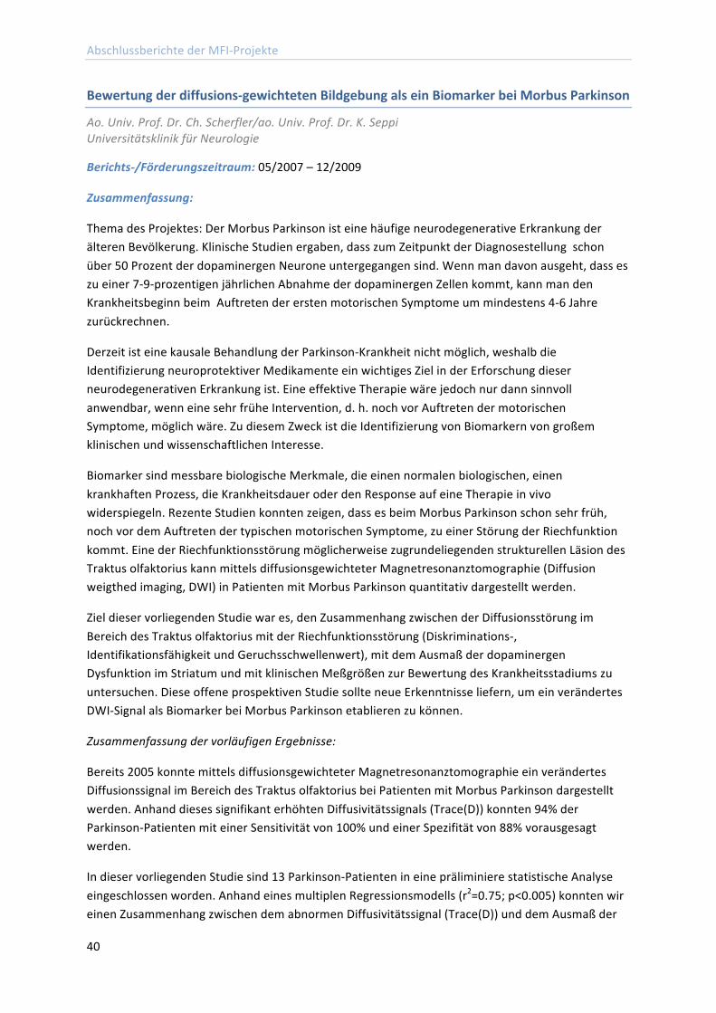
Abschlussberichte der MFI-‐Projekte
40
Bewertung der diffusions-‐gewichteten Bildgebung als ein Biomarker bei Morbus Parkinson
Ao. Univ. Prof. Dr. Ch. Scherfler/ao. Univ. Prof. Dr. K. Seppi Universitätsklinik für Neurologie
Berichts-‐/Förderungszeitraum: 05/2007 – 12/2009
Zusammenfassung:
Thema des Projektes: Der Morbus Parkinson ist eine häufige neurodegenerative Erkrankung der älteren Bevölkerung. Klinische Studien ergaben, dass zum Zeitpunkt der Diagnosestellung schon über 50 Prozent der dopaminergen Neurone untergegangen sind. Wenn man davon ausgeht, dass es zu einer 7-‐9-‐prozentigen jährlichen Abnahme der dopaminergen Zellen kommt, kann man den Krankheitsbeginn beim Auftreten der ersten motorischen Symptome um mindestens 4-‐6 Jahre zurückrechnen.
Derzeit ist eine kausale Behandlung der Parkinson-‐Krankheit nicht möglich, weshalb die Identifizierung neuroprotektiver Medikamente ein wichtiges Ziel in der Erforschung dieser neurodegenerativen Erkrankung ist. Eine effektive Therapie wäre jedoch nur dann sinnvoll anwendbar, wenn eine sehr frühe Intervention, d. h. noch vor Auftreten der motorischen Symptome, möglich wäre. Zu diesem Zweck ist die Identifizierung von Biomarkern von großem klinischen und wissenschaftlichen Interesse.
Biomarker sind messbare biologische Merkmale, die einen normalen biologischen, einen krankhaften Prozess, die Krankheitsdauer oder den Response auf eine Therapie in vivo widerspiegeln. Rezente Studien konnten zeigen, dass es beim Morbus Parkinson schon sehr früh, noch vor dem Auftreten der typischen motorischen Symptome, zu einer Störung der Riechfunktion kommt. Eine der Riechfunktionsstörung möglicherweise zugrundeliegenden strukturellen Läsion des Traktus olfaktorius kann mittels diffusionsgewichteter Magnetresonanztomographie (Diffusion weigthed imaging, DWI) in Patienten mit Morbus Parkinson quantitativ dargestellt werden.
Ziel dieser vorliegenden Studie war es, den Zusammenhang zwischen der Diffusionsstörung im Bereich des Traktus olfaktorius mit der Riechfunktionsstörung (Diskriminations-‐, Identifikationsfähigkeit und Geruchsschwellenwert), mit dem Ausmaß der dopaminergen Dysfunktion im Striatum und mit klinischen Meßgrößen zur Bewertung des Krankheitsstadiums zu untersuchen. Diese offene prospektiven Studie sollte neue Erkenntnisse liefern, um ein verändertes DWI-‐Signal als Biomarker bei Morbus Parkinson etablieren zu können.
Zusammenfassung der vorläufigen Ergebnisse:
Bereits 2005 konnte mittels diffusionsgewichteter Magnetresonanztomographie ein verändertes Diffusionssignal im Bereich des Traktus olfaktorius bei Patienten mit Morbus Parkinson dargestellt werden. Anhand dieses signifikant erhöhten Diffusivitätssignals (Trace(D)) konnten 94% der Parkinson-‐Patienten mit einer Sensitivität von 100% und einer Spezifität von 88% vorausgesagt werden.
In dieser vorliegenden Studie sind 13 Parkinson-‐Patienten in eine präliminiere statistische Analyse eingeschlossen worden. Anhand eines multiplen Regressionsmodells (r2=0.75; p<0.005) konnten wir einen Zusammenhang zwischen dem abnormen Diffusivitätssignal (Trace(D)) und dem Ausmaß der
Abschlussberichte der MFI-‐Projekte
41
Riechfunktionsstörung, im Speziellen der Störung der Diskriminationsfähigkeit (p=0.05) und der Krankheitsdauer (p=0.005) in Patienten mit Morbus Parkinson nachweisen. (Abb.1)
Die Korrelation zwischen der erhöhten Diffusivität (Trace(D)) im Bereich des Traktus olfaktorius und der Riechfunktionsstörung erhärtet die Annahme, dass es sich bei den gezeigten Veränderungen um eine strukturelle Läsion des Riechnervs handelt. Zusätzlich deuten die vorläufigen Studienresultate darauf hin, dass die diffusionsgewichtete Magnetresonanztomographie als objektivierbarer Marker für eine progrediente Pathologie im Traktus olfaktorius bei Patienten mit Morbus Parkinson dienen kann.
Abb.1):
A) B)
A) Signifikant Zunahme des Trace(D) Signals im Traktus olfaktorius mit der Krankheitsdauer bei Patienten mit der Parkinson Krankheit.
B) Signifikante Korrelation des Trace(D) Signals im Traktus olfaktorius mit der Diskriminationsfähigkeit von Gerüchen bei Patienten mit der Parkinson-‐Krankheit.
Weitere Angaben zum Projekt:
Diffusion weighted imaging of the olfactory tract and its association with hyposmia in PD. Movement Disorders Society Congress 2009, Paris
Hyposmie und diffusions-‐gewichtete Bildgebung des Traktus olfaktorius bei Patienten mit Morbus Parkinson. Kongress der Österreichischen Parkinson-‐Gesellschaft 2009, Graz
p=0.05 p=0.005
Abschlussberichte der MFI-‐Projekte
42
Abschlussberichte Antragsperiode 3
Further characterisation of the molecular mechanism of second generation antipsychotics on insulin signaling cascade and glycogen content in skeletal muscle cells
Dr.med.univ. Julia Engl Universitätsklinik für Innere Medizin I
Berichts-‐/Förderzeitraum: 02/2008 -‐ 03/2010
Zusammenfassung:
Das Ziel des Projektes war es, folgende Punkte zu untersuchen:
>-‐ Wirkung von Ketanserin auf die Insulinsignaltransduktionskaskade >-‐ Antagonisierung dieses Effekts mittels Methylserotonin >-‐ Wichtige Interaktionspunkte der Serotonin-‐ und Insulintransduktionskaskade Unsere Ergebnisse zeigten, dass Ketanserin, genauso wie Olanzapin, den Glykogengehalt in L6-‐Myozyten signifikant verringert. Bei näherer Untersuchung des klassischen Insulinsignaltransduktionsweges zeigte sich jedoch, dass Ketanserin nur pAkt herabreguliert, aber andere Signaltransduktionsproteine nicht beeinflusst. Olanzapin hingegen wirkte inhibierend auf sämtliche Proteine der Insulinsignaltransduktionskaskade. Des Weiteren zeigten Inkubationen der Zellen mit Methyserotonin einen signifikanten Anstieg des Glykogengehaltes. Allerdings konnte durch eine Koinkubation von Ketanserin beziehungsweise Olanzapin mit Methylserotonin nicht die erwarterte Antagonisierung erreicht werden.
Daraus lässt sich schließen, dass Ketanserin und möglicherweise auch Antipsychotika der zweiten Generation entweder über ein AKT-‐Adapterprotein oder einen anderen, noch unbekannten, Signaltransduktionsweg den Glykogengehalt in L6-‐Myozyten verringern und dadurch zu einer Insulinresistenz führen.
Aufgrund dieser Ergebnisse fokussieren wir unsere Versuche nun ausschließlich auf die Serotoninsignaltransduktion und dessen Wirkung auf den Glukosestoffwechsel in Skelettmuskelzellen, da in der Literatur zu diesem Thema nur dürftige Ergebnisse vorliegen und der genaue Wirkmechanismus noch nicht geklärt ist.
Um den genauen Mechanismus nachzuweisen konzentrieren wir uns nun auf die Glukoseaufnahme. Dabei versuchen wir durch Inhibition der in Frage kommenden Signaltransduktionswege mittels Proteinkinaseinhibitioren, die Signaltransduktionskaskade darzustellen, über welche Methylserotonin den Glukosestoffwechsel in Skelettmuskelzellen verbessert.
Weitere Angaben zum Projekt:
Vorträge: Jahrestagung der Österreichischen Diabetes-‐Gesellschaft, Salzburg 2010
Posterpräsentationen: Kongress Prediabetes and Metabolic Syndrome, Madrid 2011
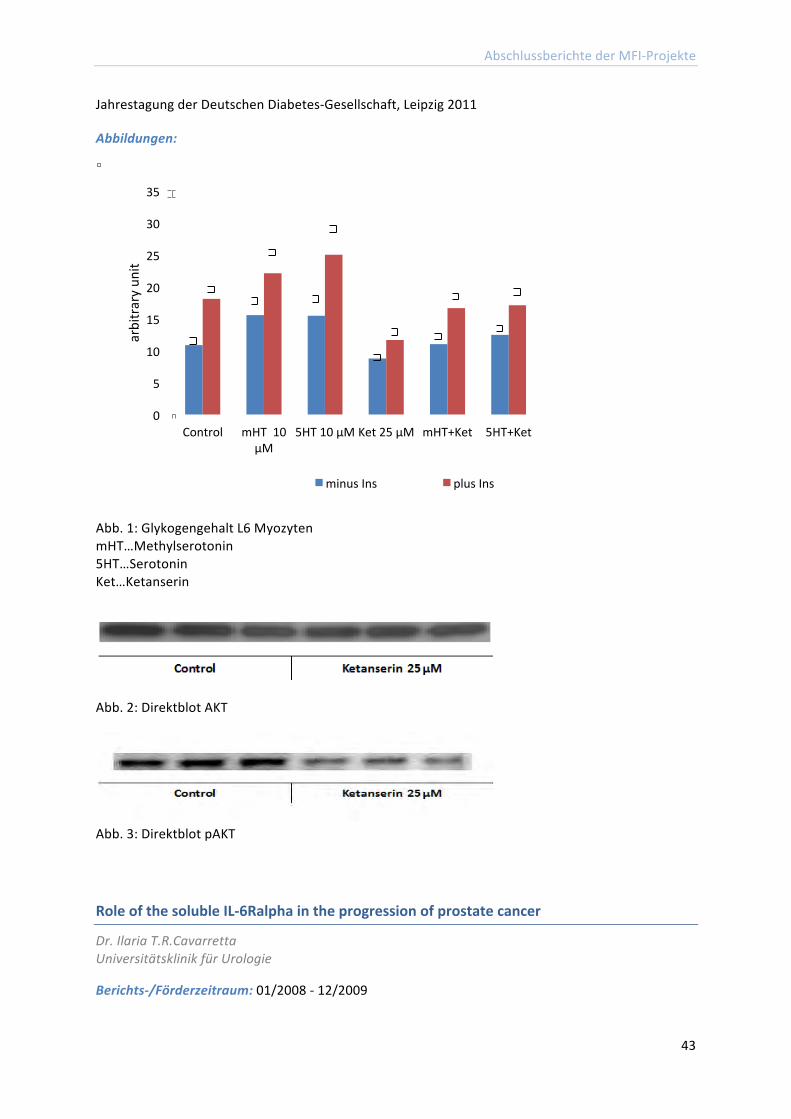
Abschlussberichte der MFI-‐Projekte
43
Jahrestagung der Deutschen Diabetes-‐Gesellschaft, Leipzig 2011
Abbildungen:
Abb. 1: Glykogengehalt L6 Myozyten mHT…Methylserotonin 5HT…Serotonin Ket…Ketanserin
Abb. 2: Direktblot AKT
Abb. 3: Direktblot pAKT
Role of the soluble IL-‐6Ralpha in the progression of prostate cancer
Dr. Ilaria T.R.Cavarretta Universitätsklinik für Urologie
Berichts-‐/Förderzeitraum: 01/2008 -‐ 12/2009
0
5
10
15
20
25
30
35
Control mHT 10 µM
5HT 10 µM Ket 25 µM mHT+Ket 5HT+Ket
arbitrary un
it
minus Ins plus Ins
Abschlussberichte der MFI-‐Projekte
44
Zusammenfassung:
A pathophysiological role of IL-‐6 in the progression of prostate cancer (PC) has been suggested and supported by several in vitro and in vivo investigations leading to the idea that anti-‐cytokine therapies could represent a promising attempt to slow down the evolution of this disease. However, in vitro and preclinical studies have yielded contrasting responses to IL-‐6 or anti-‐IL-‐6 antibodies. Thus, the outcome of IL-‐6-‐targeting therapies may be unpredictable also in PC patients, ranging from a lack of response to beneficial or detrimental effects. In order to better understand the mechanisms underlying the different responses to the cytokine, we focused our attention on IL-‐6 receptors that represent the first element in the cascade of signalling activated by IL-‐6. IL-‐6 signal transduction may indeed occur through the membrane IL-‐6R (classical signalling) and/or through the less studied soluble IL-‐6R (sIL-‐6R; IL-‐6 transsignalling (IL-‐6TS)). Additionally, the soluble form of the gp130 receptor, sgp130, selectively inhibits the IL-‐6TS. We chose for our investigations the LNCaP cells that are growth-‐inhibited by IL-‐6 (Hobisch et al. 2001) and the Du145 cells that are growth-‐stimulated by the cytokine (Onuma et al. 2003). These cells express both membrane and soluble IL6 receptors. The androgen dependent prostate cancer cell line LAPC4 was utilized as a natural sIL-‐6R-‐negative model since they possess virtually only the mIL-‐6R. By exploiting different approaches, we demonstrated for the first time that IL-‐6TS specifically accounts for an anti-‐proliferative effect of the cytokine in all PC cell lines considered. One of the approaches we utilized is illustrated in Figure 1. Although high levels of IL-‐6 are considered indicators of bad prognosis and consistently found associated with metastases, very little data showing a direct link between IL-‐6 and metastatogenesis in different malignancies are known (Kanazawa et al. 2007, Studebaker et al. 2008). Based on the higher incidence of PC relapse in patients with increased preoperative levels of sIL-‐6R and IL-‐6, we turned our attention to the possible involvement of IL-‐6/sIL-‐6R in mechanisms responsible for cancer dissemination to secondary organs. In particular, we found that IL-‐6TS increases motility and migration of prostate cells. One of the approaches we utilized is illustrated in Figure 2 facilitating thereby processes that determine metastasis initiation and spread. We measured also an IL-‐6/sIL-‐6R-‐dependent decrease of attachment of Du145 cells to one of the major components of the prostate extracellular matrix. This is the first evidence that IL-‐6TS activation may promote PC dissemination. In order to investigate the mechanism by which IL-‐6TS may promote the spread of PC cells, we looked at the expression of maspin, a tumour suppressor recently shown to be involved in regulation of cell adhesion and motility in PC (Sheng et al. 1996, Abraham et al. 2003, Ngamkitidechakul et al. 2003). We measured dramatically higher basal levels of Maspin in the non cancerous prostate epithelial cells (BPH.1 and EP156T) compared to PC cells. Interestingly, we found that IL-‐6 decreased the expression of Maspin in Du145, LNCaP, and LAPC4, but not in EP156T cells. Inhibition of IL-‐6TS prior to exposure to IL-‐6 antagonised the decrease of Maspin in LNCaP and abrogated it in Du145 cells. Finally, a further decrease of Maspin was observed in LAPC4 cells after exposure to sIL-‐6R and to IL-‐6. These studies published in a peer review Journal (Santer et al. 2010) were further developed in the following years and resulted in a second publication (Shariat et al. 2011). The relevance of such findings resides in the possibility to refine the currently available experimental anti-‐IL-‐6 therapies.
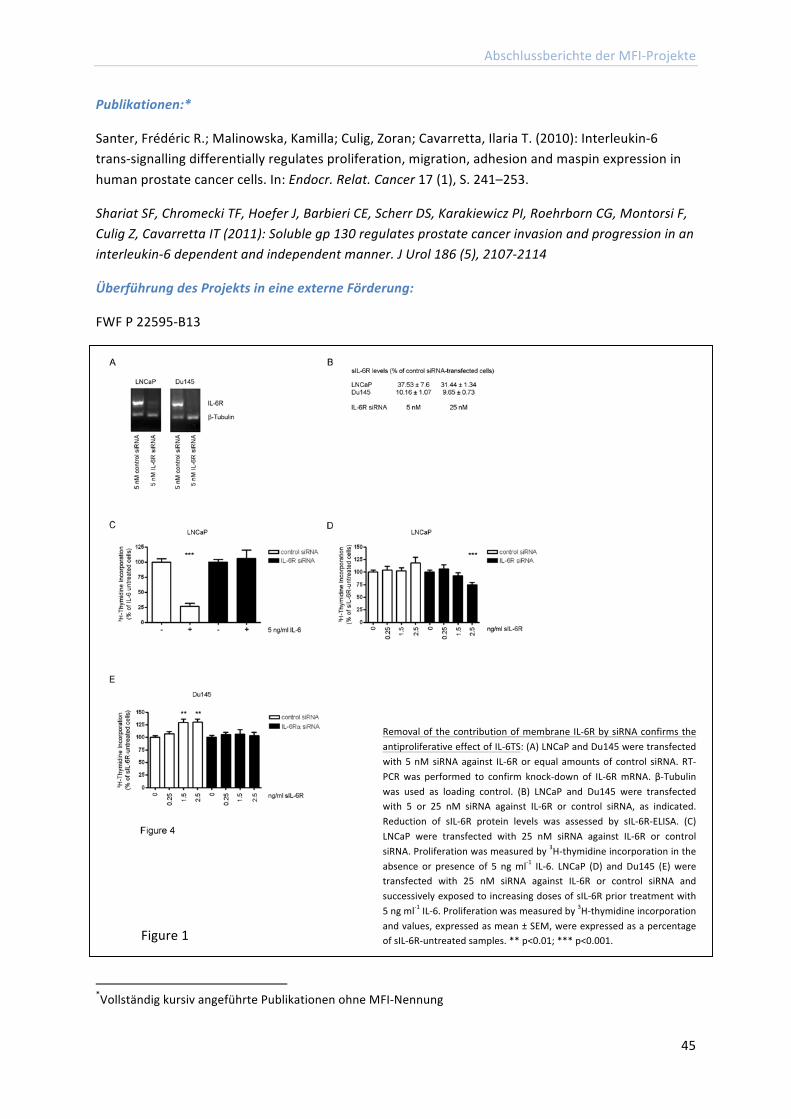
Abschlussberichte der MFI-‐Projekte
45
Publikationen:*
Santer, Frédéric R.; Malinowska, Kamilla; Culig, Zoran; Cavarretta, Ilaria T. (2010): Interleukin-‐6 trans-‐signalling differentially regulates proliferation, migration, adhesion and maspin expression in human prostate cancer cells. In: Endocr. Relat. Cancer 17 (1), S. 241–253.
Shariat SF, Chromecki TF, Hoefer J, Barbieri CE, Scherr DS, Karakiewicz PI, Roehrborn CG, Montorsi F, Culig Z, Cavarretta IT (2011): Soluble gp 130 regulates prostate cancer invasion and progression in an interleukin-‐6 dependent and independent manner. J Urol 186 (5), 2107-‐2114
Überführung des Projekts in eine externe Förderung:
FWF P 22595-‐B13
*Vollständig kursiv angeführte Publikationen ohne MFI-‐Nennung
Removal of the contribution of membrane IL-‐6R by siRNA confirms the antiproliferative effect of IL-‐6TS: (A) LNCaP and Du145 were transfected with 5 nM siRNA against IL-‐6R or equal amounts of control siRNA. RT-‐PCR was performed to confirm knock-‐down of IL-‐6R mRNA. β-‐Tubulin was used as loading control. (B) LNCaP and Du145 were transfected with 5 or 25 nM siRNA against IL-‐6R or control siRNA, as indicated. Reduction of sIL-‐6R protein levels was assessed by sIL-‐6R-‐ELISA. (C) LNCaP were transfected with 25 nM siRNA against IL-‐6R or control siRNA. Proliferation was measured by 3H-‐thymidine incorporation in the absence or presence of 5 ng ml-‐1 IL-‐6. LNCaP (D) and Du145 (E) were transfected with 25 nM siRNA against IL-‐6R or control siRNA and successively exposed to increasing doses of sIL-‐6R prior treatment with 5 ng ml-‐1 IL-‐6. Proliferation was measured by 3H-‐thymidine incorporation and values, expressed as mean ± SEM, were expressed as a percentage of sIL-‐6R-‐untreated samples. ** p<0.01; *** p<0.001. Figure 1
Abschlussberichte der MFI-‐Projekte
46
References:
Abraham S, Zhang W, et al. 2003 Journal of Urology 169 1157-‐1161. Hobisch A, Ramoner R, et al. 2001 Clinical Cancer Research 7 2941-‐2948. Kanazawa T, Nishino H, et al. 2007 European Archives of Otorhinolaryngology 264 815-‐821. Ngamkitidechakul C, Warejcka DJ, et al. 2003 Journal of Biological Chemistry 278 31796-‐31806. Onuma M, Bub JD, et al. 2003 Journal of Biological Chemistry 278 42660-‐42667. Sheng S, Carey J, et al. 1996 Proceedings of National Academy of Science USA 93 11669-‐11674. Studebaker AW, Storci G, et al. 2008 Cancer Research 68 9087-‐9095.
Natural killer T (NKT) cells as adjuvants for immunization through the skin against melanoma
Priv.Doz.Dr.Mag. Patrizia Stoitzner Universitätsklinik für Dermatologie und Venerologie
Berichts-‐/Förderzeitraum: 02/ 2008 – 01/2010
Zusammenfassung:
The aim of the study was to assess whether skin DC are able to interact with NKT cells in the skin-‐draining lymph node, and whether this interaction can be exploited to enhance vaccination through the skin to treat melanoma. We successfully published in the "Journal of Immunology" that skin dendritic cells are able to present the synthetic gylcolipid antigen α-‐Galactosylceramide to NKT cells in vitro. Furthermore, the addition of α-‐Galactosylceramide to an intradermal immunization with a tumor model antigen improved cytotoxic CD8+ T cell responses that could inhibit tumor growth. Interestingly, these improved T cell responses were induced independently of skin dendritic cells, most probably by lymph node resident dendritic cells.
This project is now continued in a FWF-‐Einzelprojekt that was granted to me in June 2009 in which we investigate the role of skin dendritic cells in skin cancer and the possibilities of using these cells for immunotherapy. Besides this successful publication of our data I was invited to write a book
Blocking of IL-‐6TS decreases Du145 cell migration: Du145 cells were treated with 5 ng ml-‐1 IL-‐6 in the presence of 0.25 µg ml-‐1 sgp130 or vehicle. At cell confluence, a scratch was performed using a 10 µl tip. After 24 h remaining free area was measured. Values are expressed as mean ± SEM. ** p<0.01 Figure 2
Abschlussberichte der MFI-‐Projekte
47
chapter about the isolation of skin dendritic cells for "Methods in Molecular Biology" and a review about Langerhans cells as targets for immunotherapy for the journal "Immunology and Cell Biology". The MFI-‐project is acknowledged in both reviews and the original paper.
Publikationen:
Tripp, Christoph H.; Sparber, Florian; Hermans, Ian F.; Romani, Nikolaus; Stoitzner, Patrizia (2009): Glycolipids injected into the skin are presented to NKT cells in the draining lymph node independently of migratory skin dendritic cells. In: J. Immunol 182 (12), S. 7644–7654.
Stoitzner, Patrizia; Romani, Nikolaus; McLellan, Alexander D.; Tripp, Christoph H.; Ebner, Susanne (2010): Isolation of skin dendritic cells from mouse and man. In: Methods Mol. Biol 595, S. 235–248.
Stoitzner, Patrizia; Sparber, Florian; Tripp, Christoph H.: Langerhans cells as targets for immunotherapy against skin cancer. In: Immunol. Cell Biol 88 (4), S. 431–437.
Überführung des Projekts in externe Förderung:
FWF P21487-‐B13
Weitere Angaben zum Projekt:
Präsentationen an nationalen und internationalen Kongressen und Seminaren:
1. Stoitzner P, Langerhans cells and skin cancer: potential targets for immunotherapy? Invited Speaker at the"Spring Meeting of the Austrian Society for Dermatology & Venereology, Vienna, Austria, February 2010.
2. Tripp CH, Sparber F, Hermans IF, Idoyaga J, Steiman RM, Romani N, Stoitzner P. Improvement of intradermal immunization against melanoma. Karl Landseteiner Meeting, Salzburg, Austria, November 2009.
3. Stoitzner P, Langerhans cells in skin cancer: potential targets for immunotherapy? Invited Speaker at the 11thInternational Workshop on Langerhans Cells, Funchal, Madeira, September 2009.
4.Stoitzner P. Novel immunization strategies through the skin mediated by dendritic cells. Seminar given at the Queens University Belfast, UK, May 2009.
5.Stoitzner P. Skin dendritic cells and their potential for immunotherapy. Invited speaker at the Minisymposium on "Dendritic cells" at the Erasmus MC, Rotterdam, October, 2008.
6.Stoitzner P, Hermans IF, Romani N. Skin dendritic cells stimulate cytokine production by natural killer T cells. Joint Annual Meeting of Immunology of the Austrian and German Societies (OGAI, DGfI), Vienna, Austria, September 2008.
7.Stoitzner P. Are Langerhans cells major player in the skin immune system? Seminar given at the Department of Immunology, Ludwig-‐Maximilians University In Munich, Germany, July 2008.
8.Stoitzner P. Epicutaneous immunization as a new cancer immunotherapy? Seminar given at the Biozentrum in Innsbruck, Austria, April 2008.
Abschlussberichte der MFI-‐Projekte
48
Posterpräsentationen:
1. Tripp CH, Sparber F, Hermans IF, Idoyaga J, Steiman RM, Romani N, Stoitzner P. Improvement of intradermal immunization against melanoma.11th International Symposium on Dendritic Cells in Fundamental and Clinical Immunology, Lugano, Switzerland, September 2010.
2. Tripp CH, Sparber F, Hermans IF, Idoyaga J, Steiman RM, Romani N, Stoitzner P. Improvement of intradermal immunization against melanoma. The Annual Meeting of the Arbeitsgemeinschaft Dermatologische Forschung (ADF) , Luebeck, Germany, February 2010.
3. Tripp CH, Sparber F, Hermans IF, Idoyaga J, Steiman RM, Romani N, Stoitzner P. Improvement of intradermal immunization against melanoma. Annual Meeting of the Austrian Society of Dermatology& Venereology, Salzburg, Austria, Novermber 2009.
4. Tripp CH, Sparber F, Hermans IF, Romani N, Stoitzner P. Glycolipid antigen as adjuvant for skin immunization against melanoma. 2nd European Congress of Immunology, Berlin, Germany, September 2009.
5. Tripp CH, Sparber F, Hermans IF, Romani N, Stoitzner P. Glycolipid antigen as adjuvant for skin immunization against melanoma. 11th International Workshop on Langerhans cells, Funchal, Madeira, September 2009.
6. Tripp CH, Sparber F, Hermans IF, Romani N, Stoitzner P. Glycolipid antigen as adjuvant for skin immunization against melanoma. Joint Meeting of th World Congress on Melanoma and Congress of the European Association of Dermato-‐Oncology (EADO), Vienna, Austria, May 2009.
7. Tripp CH, Hermans IF, Romani N, Stoitzner P. Intradermal administration of synthetic glycolipid antigen together with a tumor model antigen induces efficient T cell responses against melanoma. The Annual Meeting of the Arbeitsgemeinschaft Dermatologische Forschung (ADF), Heidelberg, Germany, March 2009.
8. Stoitzner P, Hermans IF, Romani N. Skin dendritic cells stimulate cytokine production by natural killer T cells. The 10th International Symposium on Dendritic Cells, Kobe, Japan, October 2008.
9. Stoitzner P, Ronchese F, Romani N, Hermans IF. Skin dendritic cells upregulate CD1d expression upon migration to the lymph nodes. NK and NKT cell Biology, Keystone Symposia, Keystone, Colorado, US. February 2008.
Abschlussberichte der MFI-‐Projekte
49
Revealing the elusive molecular biology of the vault RNA and the vault RNP
Priv. Doz. Dr. Norbert Polacek Sektion für Genomik und RNomik
Berichts-‐/Förderzeitraum: 9/2007 – 02/2010
Zusammenfassung:
Non-‐protein coding RNAs (ncRNAs) have recently emerged on the scene of genomic research as prominent players in the regulation of gene expression. Many functionally characterized ncRNAs have been shown to be differentially expressed in various organisms during specific environmental or developmental conditions thus establishing regulatory networks crucial for shaping cellular life.
In a previous study (Mrazek et aI., 2007) we could demonstrate that certain human ncRNAs were markedly up-‐regulated in human B cells upon Epstein-‐Barr virus (EBV) infection. The most significant extent of ncRNA up-‐regulation was observed for the so called vault RNAs (vtRNAs). vtRNAs are ncRNAs that are integral to the vault complex, a gigantic hollow ribonucleoprotein particle (RNP) of 13 MDa with thus far elusive biological role.
vtRNA expression is specifically stimulated by members of the γ-‐herpersviridae
During the course of this MFI project, we were able to show that the expression of the threeso far known human vtRNAs is specifically up-‐regulated in human lymphocytes upon infection by viruses of the γ-‐herpesviridae family, such as EBV or the Kaposi's Sarcoma virus, but not by other DNA or RNA viruses (Nandy et aI., 2009). Stimulation of vtRNA expression by EBV was evident for all three canonical vtRNAs (hvg1-‐3) and also for a novel ncRNA candidate, initially termed CBL-‐3.
The ncRNA CBL-‐3 is a novel fourth human vault RNA
The CBL-‐3 ncRNA shares clear primary and secondary structure similarities with the three known vtRNAs. The most important findings during this MFI research project was our observation that CBL-‐3 co-‐sediments with intact vault particles in density gradients of various human cell lines (Nandy et al., 2009). This strongly indicates that CBL-‐3 is a novel vault particle-‐associated ncRNA and we have thus classified it as a novel, fourth human vault vtRNA (Nandy et aI., 2009).
Independent in silico studies by the bioinformatician Dr. Peter Stadler (University of Leipzig) and his team fully confirmed our experimental findings and our interpretation (Stadler et aI., 2009, Mol. BioI. Evol. 26:1975-‐1991).
The role of vtRNAs in virus infection/propagation
Based on these promising results that we could obtain during the course of this MFI project, ongoing work in my group aims at elucidating the biological role of the vtRNAs during virus infection in more detail. We have already established a stably transfected human B-‐cell line that constantly over-‐expresses vtRNA 1. This cell line now paves the way to study the role of the vtRNA (vtRNP) during EBV infection and propagation in detail.
Abschlussberichte der MFI-‐Projekte
50
Publikationen:
Nandy, Constanze; Mrázek, Jan; Stoiber, Heribert; Grässer, Friedrich A.; Hüttenhofer, Alexander; Polacek, Norbert (2009): Epstein-‐barr virus-‐induced expression of a novel human vault RNA. In: J. Mol. Biol 388 (4), S. 776–784.
Trappl, K., and Polacek, N. (2011): The ribosome: A molecular machine powered by RNA In: Metal Ions Life Sci. 9:253-‐275.
Überführung des Projekts in eine externe Förderung:
DOC fFORTE stipend from the Austrian Academy of Sciences to Mag. Melanie Amort
Weitere Angaben zum Projekt:
Sigel, Astrid; Sigel, Helmut (Hg.) (2011): Metal Ions in Life Sciences. Cambridge: Royal Society of Chemistry. (Buch Sammelwerk)
In vivo imaging of the effect of atherosclerosis risk factors on arterial endothelial cells: molecular imaging of heat shock protein 60 expression"
Priv.-‐Doz. Dr. med. univ. Marius C. Wick Department Radiologie
Berichts-‐/Förderzeitraum: 06/2008 – 06/2011
Zusammenfassung:
Die "Autoimmunologische Hypothese der Entstehung der Atherosklerose" beruht auf der, mittlerweile durch zahlreiche experimentelle und klinische Studien nachgewiesenen Annahme, dass zellulärer Stress in Form verschiedenster klassischer Atherosklerose-‐Risikofaktoren wie Hochdruck, oxidiertes LDL, Rauchen, Diabetes, Clamydieninfektionen etc. zur Expression von Hitzeschockprotein 60 (HSP60) an der Oberfläche von arteriellen Endothelzellen führt. Diese HSP60-‐Expression stellt ein "Gefahrensignal" für die angeborene und adaptive Immunabwehr des Organismus dar, was als Folge kreuzreaktiver autoimmunologischer Prozesse zu einer Entzündungsreaktion in der Intima der Gefäßwand führt und somit der erste Schritt der Entstehung der Atherosklerose ist. Bakterielles Toxin kann als Surrogat für infektionsbedingten Endothelzellstress angesehen werden.
Das Ziel unserer Studie "In vivo molecular imaging of HSP60" war die Entwicklung einer neuen radiologischen Methode, welche die nicht-‐invasive in vivo molekulare Bildgebung des Effekts von Stress auf die Expression von HSP60 auf arteriellen Endothelzellen im normocholesterolämischen Kaninchenmodell ermöglicht.
Die Tierversuche waren vom Bundesministerium für Wissenschaft und Forschung genehmigt (BMBWK-‐66.01110011-‐BrGT/2005; BMBWK-‐66.01110017-‐BrGT/2006).
Endothelialer Stress wurde in 20 New-‐Zealand-‐White-‐Kaninchen mittels intravenöser Injektion von Lipopolysaccharid (LPS; 10)µg/kg) induziert. Acht Kaninchen bekamen eine "Scheinbehandlung"

Abschlussberichte der MFI-‐Projekte
51
mittels Puffer oder dienten als unbehandelte Kontrollen. Die in vivo molekulare Bildgebung der endothelialen Expression von HSP60 wurde mittels Fusion von Computertomografien (CT) und Positronen-‐Emmissions-‐Tomografien (PET) der Kaninchen 24 Stunden nach der intravenösen Injektion von 124I-‐markierten monoklonalen anti-‐HSP60 Antikörpem oder 124I-‐markierten Isotypen Kontrollantikörpem durchgeführt.
Verglichen mit den unbehandelten und scheinbehandelten Kontrollen zeigten die in vivo PET -‐CT Bilder der LPS-‐behandelten Kaninchen, eine signifikant erhöhte endotheliale Bindung von 124I-‐markierten monoklonalen anti-‐HSP60 Antikörpem, besonders an den bekannten Pradilektionsstellen der Atherosklerose in der Umgebung der Verzweigungen von Arterien. Die Resultate der nicht-‐invasiven in vivo Bildgebung wurden mittels Autoradiografien und Immunhistochemie der entnommenen Arterien bestätigt.
Somit haben die Ergebnisse unserer Studie gezeigt, dass die HSP60-‐Expression in normocholesterolämischen Kaninchen nach Induktion von endothelialem Stress signifikant erhöht ist und – als "Proof-‐of-‐principle" -‐ eine nicht-‐invasive in vivo molekulare Bildgebung der frühen endothelialen HSP60-‐Expression mittels PET -‐CT und 124I-‐markierten monoklonalen anti-‐HSP60-‐ Antikörpern zur Früherkennung der Atherosklerose möglich ist.
Publikationen:
Wick, Marius C.; Mayerl, Christina; Backovic, Aleksandar; van der Zee, Ruurd; Jaschke, Werner; Dietrich, Hermann; Wick, Georg (2008b): In vivo imaging of the effect of LPS on arterial endothelial cells: molecular imaging of heat shock protein 60 expression. In: Cell Stress Chaperones 13 (3), S. 275–285.
Machtoub L, Pfeiffer R, Backovic A, Frischauf S, Wick MC (2010):Molecular imaging cellular SPIO uptake with nonlinear optical microscopy. In: Journal of Medical Imaging and Radiation Sciences. 41(3), S. 159-‐164.
Übersichtsartikel:
Wick, Marius C.; Kremser, Christian; Frischauf, Stefan; Wick, Georg (2008a): In vivo molecular imaging of vascular stress. In: Cell Stress Chaperones 13 (3), S. 263–273.
Grundtman, Cecilia; Kreutmayer, Simone B.; Almanzar, Giovanni; Wick, Marius C.; Wick, Georg (2011): Heat shock protein 60 and immune inflammatory responses in atherosclerosis. In: Arterioscler. Thromb. Vasc. Biol 31 (5), S. 960–968.
Weitere Angaben zum Projekt:
Buchkapitel:
Henderson B, Rossmann A, Mayerl C, Wick M, Wick G. Kapitel: Atherosclerosis -‐ An Age-‐dependent Autoimmune Disease. In: Fulop T, Franceschi C, Hirokawa K (Herausg.). Handbook on lmmunosenescence Basic Understanding and Clinical Applications. Springer Science + Buisness Media B.V. 2009; DOl 10.1007/978-‐1-‐4020-‐9062-‐2_53.
Abschlussberichte der MFI-‐Projekte
52
Wick MC, Chemelli-‐Steingruber IE, and Kremser C.Chapter 16: Imaging of atherosclerosis. In: "Inflammation and Atherosclerosis". Wick G and Grundtman C (Herausg.). (ISBN 978-‐3-‐7091-‐0337-‐1) Springer-‐Verlag Wien New York 2011;in print.
Vorträge:
Wick MC, Mayerl C, Haubner R, Dobrozemsky G, Jaschke W, Dietrich H, Wick G. In vivo imaging of arterial heat shock protein 60 expression after application of toxic stress. 2nd World Conference of Stress; 22-‐26.08.2007, Budapest, Hungary. Cell Stress & Chaperones 2007:12, No. 2pp., el, 6F_05_S.
Wick MC, Mayerl C, Backovic A, van der Zee R, Jaschke W, Dietrich H, Wick G. In vivo imaging of the effect of LPS on arterial endothelial cells: molecular imaging of heat shock protein 60 expression. Annual meeting of the Austrian Atherosclerosis Society, 18.-‐ 19.04.2008, St. Gilgen, Austria.
Wick MC, Mayerl C, Backovic A, van der Zee R, Jaschke W, Dietrich H, Wick G. In vivo molecular imaging of heat shock protein 60 expression after arterial endothelial cellsstress using LPS. 2008 World Molecular Imaging Congress (WMIC 2008), September 10-‐13, 2008, Nice, France.
Wick MC and Backovic A. "Molecular imaging of the effect of stress on arterial endothelial cells". Biocentre Seminar, 20.03.2009, Biocentre, Medizinische Universität Innsbruck
Expression and function of a deletion mutant of Mcl1 in human neuroblastoma
Priv.Doz.Mag.Dr. Michael Außerlechner Universitätsklinik für Pädiatrie II
Berichts-‐/Förderzeitraum: 09/2008 – 08/2010
Zusammenfassung:
Das MFIProjekt beschäftigte sich mit der Frage, welche Rolle eine in unserer Arbeitsgruppe identifizierte Variante des Proteins MCL1 in Neuroblastomzellen spielt. MCL1 ist ein anti-‐apoptotisches Protein der BCL2-‐Familie, das in Leukämiezellen überexprimiert vorliegt und Resistenz gegenüber Zelltod und Chemotherapie mediiert. Wir konnten sowohl in Leukämie-‐ als auch in Neuroblastomzellen eine neue mRNA Variante von MCL1 nachweisen, der eine 45 bp lange Sequenz fehlt, was zum Verlust von 15 Aminosäuren (G158-‐D173), jedoch zu keiner Leserasterverschiebung führt. Dadurch entsteht ein bislang noch nicht beschriebenes Protein, dem zwei wichtige Aminosäuren für die Regulation der Stabilität fehlen.
Der N-‐terminale Bereich dieser Variante hat entsprechend der in silico Strukturvorhersage eine völlig unterschiedliche Sekundärstruktur im Vergleich zu dem Wildtyp MCL1. Durch die Markierung von MCL1del mit grün bzw. blau fluoreszierenden EGFP bzw ECFP konnten wir mittels „live cell imaging“ erstmals zeigen, dass diese MCL1 Variante sowohl zytoplasmatisch als auch im Kern von Neuroblastomzellen vorkommt, während Wildtyp-‐MCL1 exlusiv mitochondrial lokalisiert ist. Um direkt den Effekt von MCL1del auf die Apoptose-‐Sensitivität zu messen, wurde mittels retroviralen Gentransfer sowohl MCL1 als auch MCL1del in Neuroblastomzellen eingebracht.
Abschlussberichte der MFI-‐Projekte
53
Mit diesen neuronalen Tumorzelllinien konnten wir erstmals zeigen, dass die Expression der Variante MCL1del signifikant den TRAIL-‐induzierten Zelltod hemmt und somit Tumorzellen vor dem Angriff durch das Immunsystem schützt. Diese Ergebnisse weisen darauf hin, dass die identifizierte Variante MCL1del eindeutig einen Zelltod-‐hemmenden Einfluss hat und Neuroblastomtumorzellen effizient gegen Apoptose schützt.
Role of p16, p21 and mTERT in renal ischemia reperfusion injury
Dr. Kathrin Eller (ehem. Hochegger) Universitätsklinik für Innere Medizin IV
Berichts-‐/Förderzeitraum: 02/2008 – 01/2010
Zusammenfassung:
Im durch den Medizinischen Forschungsförderungsfonds Innsbruck geförderten Projekt „Role of p16, p21 und mTERT in Renal Ischemia Reperfusion Injury“ wurde postuliert, die Rolle von p16, p21 und mTERT durch eine in vivo Expressionsstrategie weiter zu untersuchen. Es gelang uns, erfolgreich einen Vektor für p16 und p21 herzustellen. Die Klonierung von mTERT war aufgrund der Größe technisch nicht möglich. In Folge injizierten wir den jeweiligen Vektor, gekoppelt an polyethylenimine (PEI), in unsere Mäuse und indizierten das renale Ischämie und Reperfusions-‐Modell (IRI).
Es gelang uns erfolgreich, das jeweilige Protein in der Niere hochzuregulieren. Die Mäuse, die den p21-‐Vektor erhielten, zeigten einen erstaunlichen Phänotyp. Sie präsentierten sich, im Unterschied zu den Kontrollen (die einen leeren Vektor erhielten), mit deutlich geringeren Erkrankungsindizes. Pathomechanistisch wurde die Apoptose in diesen Tieren durch die p21-‐Hochregulation signifikant vermindert. Dies ist der erste in vivo Beweis, ohne Verwendung von knock-‐out Tieren, dass p21 in der renalen IRI eine protektive Funktion hat. Das entsprechende Manuskript ist gerade in Bearbeitung.
Fr. Mag. Huber, die über das MFI-‐Projekt finanziert wurde, war maßgeblich an obigen Daten beteiligt. Weiters war sie maßgeblich an weiteren Projekten beteiligt. Sie unterstützte das Forschungsprojekt „Tim-‐3 in nephrotoxic serum nephritis“ erfolgreich. Die von ihr in unserem Labor etablierten Methoden der TUNEL-‐Färbung wurden in der Arbeit „Tim-‐3 is upregulated and protective in nephrotoxic serum nephritis“, die rezent im American Journal of Pathology publiziert wurden, von ihr durchgeführt und evaluiert. Weiters ist sie maßgeblich an dem Projekt „Bortezomib in nephrotoxic serum nephritis“ beteiligt.
Hier wird der Einfluß von Bortezomib, das p21 effektiv beeinflusst, in der nephrotoxischen Serum Nephritis untersucht. Wir konnten nachweisen, dass Bortezomib in der verwendeten Dosierung in der frühen Phase zu einem Tubulusschaden führt und in der protrahierten Phase die Krankheitsindizes signifikant durch eine Inhibition der B-‐ und T-‐Zellantwort supprimiert. Auch hier ist ein Manuskript derzeit in Bearbeitung.
Abschlussberichte der MFI-‐Projekte
54
Publikationen:
Schroll, Andrea; Eller, Kathrin; Huber, Julia M.; Theurl, Igor M.; Wolf, Anna M.; Weiss, Günter; Rosenkranz, Alexander R. (2010): Tim3 is upregulated and protective in nephrotoxic serum nephritis. In: Am. J. Pathol 176 (4), S. 1716–1724.
The role of dopaminergic stimulation on striatal graft survival and function in the double lesion rat model of the parkinson variant of multiple system atrophy
Priv.-‐Doz. Dr. med.univ. Nadia Stefanova, PhD Universitätsklinik für Neurologie
Berichts-‐/Förderzeitraum: 02/2008 – 01/2009
Zusammenfassung:
Multiple system atrophy (MSA) is a progressive, neurodegenerative disorder of unknown aetiology (Wenning, 2001). Treatment of parkinsonism in MSA (MSA-‐P) is still a challenge. Although initial levodopa efficacy is documented in up to 40% of MSA patients (Parati, 1993b, Wenning, 1994, Albanese, 1995), the response typically declines after a few years of treatment, leaving 90% of the MSA-‐P patients levodopa-‐unresponsive in the long term (Wenning, 2004). The double lesion rat model of MSA-‐P has been established by sequential unilateral injection of 6-‐hydroxidopamine (6-‐OHDA) into the medial forebrain bundle and of quinolinic acid (QA) into the ipsilateral striatum (Wenning, 1996).
Studies of our group have shown that embryonic striatal allografts survive in denervated and lesioned host striatum and restore responsiveness to dopaminergic stimulation in the MSA-‐P rat model (Wenning, 1996, Puschban, 2000a). This MFI project has been a natural continuation of the studies of our research group focused on developing of a pre-‐clinical rational for neurotransplantation in MSA-‐P aiming towards future translation of these results into a clinical trial. We have now investigated the effects of E14 embryonic striatal allografts on dopaminergic responsiveness in the unilateral double-‐lesion rat model of MSA-‐P by using tests of complex motor behavior.
Both sham and graft animals showed an increase in apomorphine-‐induced rotations as well as an improvement in cylinder test performance following surgical intervention. In contrast, L-‐Dopa responsiveness of stepping behavior was improved only in grafted animals. The restoration of apomorphine-‐induced rotation correlated with the P-‐zone volume of grafts. Our findings indicated that transplantation of embryonic striatal grafts might, at least to some extent, restore responsiveness to L-‐Dopa in tasks of complex motor behavior. In the follow-‐up experiment we are now analysing the effects of a more complicated co-‐graft approach (striatal and mesencephalic co-‐grafts) on survival and function of striatal grafts in the double lesion MSA-‐P rat model.
Wistar rats received 6-‐OHDA and QA lesions to model MSA-‐P. Animals were randomised into groups based on there rotation behaviour and then grafted with striatal grafts, striatal and mesencephalic co-‐grafts or received sham grafts. The interim behavioural anaysis did not show significant group
Abschlussberichte der MFI-‐Projekte
55
differences in the performance of rotometer test, cylinder test or stepping test at selected time points after grafting. There were no differences among the groups in respect to pulsatile exogenous L-‐dopa-‐induced dyskinesias either. At present the histological stainings have been completed and image analysis with Stereo Investigator 7.0 is undergoing. The outcomes of the morphological studies will be of crucial importance to evaluate the success of the graft procedure as well as the survival and functional activity of the grafts. The first phase of our studies on striatal grafts in the MSA-‐P double lesion rat model has been just published at the end of December 2008 in J. Neurosci. Res (on-‐line publication : http://www3.interscience.wiley.com/cgi-‐bin/fulltext/121589677/HTMLSTART).
Publikationen:
Köllensperger, Martin; Stefanova, Nadia; Pallua, Anton; Puschban, Zoe; Dechant, Georg; Hainzer, Monika et al. (2009): Striatal transplantation in a rodent model of multiple system atrophy: effects on L-‐Dopa response. In: J. Neurosci. Res 87 (7), S. 1679–1685.
Abschlussberichte der MFI-‐Projekte
56
Abschlussberichte Antragsperiode 4
Copy Number Variations and genetic traits: insights into the origin of cardiovascular disease
Priv.-‐Doz. Dipl.-‐Ing. Mag. Dr. Anita Kloss-‐Brandstätter Sektion für Genetische Epidemiologie
Berichts-‐/Förderzeitraum: 07/2008 – 06/2010
Zusammenfassung:
Hintergrund: Das Risiko für Herz-‐Kreislauf-‐Erkrankungen steht in starkem Zusammenhang mit Plasmalipidkonzentrationen. Die Variabilität von Einzelbasen-‐Polymorphismen (SNPs) in Genen, die bekannterweise in die Regulation von Lipiden involviert sind, reicht nicht aus, um die beobachtbaren phänotypischen Unterschiede zu erklären. Die kürzlich beschriebenen "Copy Number Variations" (CNVs) beeinflussen massiv die Gen-‐Dosierung und könnten zusammen mit SNPs und Umweltfaktoren die Lipidkonzentrationen regulieren.
Erreichte Meilensteine
1) Entwicklung von bioinformatischen Lösungen für die Suche nach CNVs
Die Suche nach genetischen Ursachen von komplexen Erkrankungen wie Herz-‐Kreislauferkrankungen gestaltet sich häufig wie die sprichwörtliche Suche nach der Stecknadel im Heuhaufen. Dabei fallen sowohl auf Seite der genetischen Varianten wie auch auf Seite der beobachtbaren oder messbaren Merkmale riesige Datenmengen an, die es gilt, effizient zu verwalten. Dazu entwickelten wir in Zusammenarbeit mit dem Institut für Informatik der Universität Innsbruck eine Datenbankapplikation, die ein gemeinsames Speichern, Verwalten und Abfragen von Genotypen und Phänotypen ermöglicht (eCOMPAGT; [1,2]).
Um Daten aus genetischen Assoziationsstudien sinnvoll interpretieren zu können, erstellten wir einen Leitfaden zur sinnvollen bioinformatischen Auswertung und Interpretation von genetischen Varianten („GenEpi Toolbox“) [3].
CNV-‐Daten, die aus SNP-‐array-‐Intensitätsdaten extrahiert werden können, können in einer genomweiten Assoziationsstudie mit unserer Software CONAN nach Assoziationen mit Phänotypen wie z. B. Blutfettwerten gescreent werden [4].
2) Etablierung von molekularbiologischen Verfahren
Die Typisierung von SNPs (Single Nucleotide Polymorphisms) ist an unserem Institut eine etablierte Routineapplikation, die mit TaqMan-‐Assays robuste und schnelle Ergebnisse liefert. Genetische Varianten, die sich als Wiederholung von genomischen Bereichen darstellen, wie Telomere oder CNVs, gestalten sich schwieriger in der Typisierung. Ein möglicher Ansatz dafür ist quantitative real-‐time PCR (qPCR). Mit dieser Technologie haben wir ein Verfahren zur Hochdurchsatz-‐Typisierung von relativen Telomerlängen entwickelt, mit dem hoch-‐qualitative und damit äußerst verlässliche Daten
Abschlussberichte der MFI-‐Projekte
57
für genetische Assoziationsstudien, wie z. B. der Einfluss der Telomerlänge auf das Eintreten Herz-‐Kreislauferkrankungen generiert werden können [5]. Ein vom Prinzip her ähnliches Verfahren kann zur Bestimmung der CNVs verwendet werden; da sind wir gerade dabei, dieses Verfahren für die Typisierung von CNVs mit der SEQUENOM-‐Plattform zu optimieren.
Außerdem konnten wir im Rahmen des Projektes ein Verfahren entwickeln, mit dem in großen Studienpopulationen nach seltenen Varianten gesucht werden kann [6].
3) Evaluation des potenziellen Einflusses von CNVs auf Herz-‐Kreislauferkrankungen
Erhöhte Lipoprotein(a) [Lp(a)] Konzentrationen stehen in direktem Zusammenhang mit einem erhöhten Risiko für Herz-‐Kreislauferkrankungen [7]. Die Höhe des Lp(a) Spiegels im Blut wird größtenteils durch die Anzahl der Kopien einer CNVs im Apolipoprotein(a) Genlocus bestimmt [Kraft, 1992]. In einer Studie an übergewichtigen Kindern und Jugendlichen wollten wir überprüfen, ob Gewichtsreduktion zu einer Senkung des Lp(a) Spiegels führt und wie weit die CNV einen Einfluss auf die Lp(a) Senkung hat. Tatsächlich konnten wir feststellen, dass die Anzahl an Kopien dieses speziellen CNVs invers mit der gewichtsbedingten Lp(a) Reduktion korrelierte: je mehr Kopien ein Kind aufwies, umso geringer war die relative Abnahme im Lp(a)-‐Blutwert nach Beendigung der Diät [8].
In einer kürzlich in Atherosclerosis publizierten Arbeit [9] konnten wir außerdem beobachten, dass genau jene SNPs, die in copy-‐number variablen genomischen Bereichen liegen, einen sehr starken Einfluss auf den Harnsäure-‐Spiegel im Blut haben. Erhöhte Harnsäure-‐Spiegel stehen in starkem Zusammenhang mit Gicht, Arteriosklerose und damit in weiterer Folge mit Herz-‐Kreislauferkrankungen. SNPs, die außerhalb von CNVs liegen, zeigten einen viel schwächeren Einfluss auf den Harnsäure-‐Spiegel im Blut [9].
Publikationen:*
Schönherr, Sebastian; Weissensteiner, Hansi; Coassin, Stefan; Specht, Günther; Kronenberg, Florian; Brandstätter, Anita (2009): eCOMPAGT -‐-‐ efficient combination and management of phenotypes and genotypes for genetic epidemiology. In: BMC Bioinformatics 10, S. 139.
Weissensteiner, Hansi; Schönherr, Sebastian; Specht, Günther; Kronenberg, Florian; Brandstätter, Anita (2010): eCOMPAGT integrates mtDNA: import, validation and export of mitochondrial DNA profiles for population genetics, tumour dynamics and genotype-‐phenotype association studies. In: BMC Bioinformatics 11, S. 122.
Coassin, Stefan; Brandstätter, Anita; Kronenberg, Florian (2010): Lost in the space of bioinformatic tools: a constantly updated survival guide for genetic epidemiology. The GenEpi Toolbox. In: Atherosclerosis 209 (2), S. 321–335.
Forer, Lukas; Schönherr, Sebastian; Weissensteiner, Hansi; Haider, Florian; Kluckner, Thomas; Gieger, Christian et al. (2010): CONAN: copy number variation analysis software for genome-‐wide association studies. In: BMC Bioinformatics 11, S. 318.
*Vollständig kursiv angeführte Publikationen ohne MFI Nennung
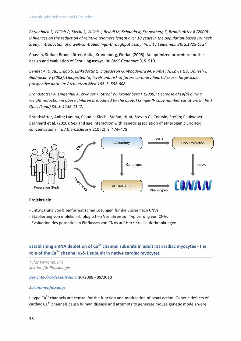
Abschlussberichte der MFI-‐Projekte
58
Ehrlenbach S, Willeit P, Kiechl S, Willeit J, Reindl M, Schanda K, Kronenberg F, Brandstätter A (2009): Influences on the reduction of relative telomere length over 10 years in the population-‐based Bruneck Study: introduction of a well-‐controlled high-‐throughput assay; In: Int J Epidemiol, 38, S.1725-‐1734.
Coassin, Stefan; Brandstätter, Anita; Kronenberg, Florian (2008): An optimized procedure for the design and evaluation of Ecotilling assays. In: BMC Genomics 9, S. 510.
Bennet A, Di AE, Erqou S, Eiriksdottir G, Sigurdsson G, Woodward M, Rumley A, Lowe GD, Danesh J, Gudnason V (2008): Lipoprotein(a) levels and risk of future coronary heart disease: large-‐scale prospective data. In: Arch Intern Med 168: S. 598-‐608.
Brandstätter A, Lingenhel A, Zwiauer K, Strobl W, Kronenberg F (2009): Decrease of Lp(a) during weight reduction in obese children is modified by the apo(a) kringle-‐IV copy number variation. In: Int J Obes (Lond) 33, S. 1136-‐1142.
Brandstätter, Anita; Lamina, Claudia; Kiechl, Stefan; Hunt, Steven C.; Coassin, Stefan; Paulweber, Bernhard et al. (2010): Sex and age interaction with genetic association of atherogenic uric acid concentrations. In: Atherosclerosis 210 (2), S. 474–478.
Projektziele
-‐ Entwicklung von bioinformatischen Lösungen für die Suche nach CNVs -‐ Etablierung von molekularbiologischen Verfahren zur Typisierung von CNVs -‐ Evaluation des potentiellen Einflusses von CNVs auf Herz-‐Kreislauferkrankungen
Establishing siRNA depletion of Ca2+ channel subunits in adult rat cardiac myocytes -‐ the role of the Ca2+ channel α2δ-‐1 subunit in native cardiac myocytes
Tuluc Petronel, PhD Sektion für Physiologie
Berichts-‐/Förderzeitraum: 10/2008 -‐ 09/2010
Zusammenfassung:
L-‐type Ca2+ channels are central for the function and modulation of heart action. Genetic defects of cardiac Ca2+ channels cause human disease and attempts to generate mouse genetic models were
Abschlussberichte der MFI-‐Projekte
59
hampered because knock-‐out animals of essential heart proteins are not vital. Most of the research done previously on the role of Ca2+ channel subunits in heart function was performed in heterologous expression systems. The goal of my project was to knockdown α2δ-‐1 subunit in adult rat cardiac myocytes using adenovirus delivered shRNAs.
The successful completion of this project is of significant interest for heart physiology because it will demonstrate the role of the α2δ-‐1 subunit in native cardiac muscle.
Additionally, it demonstrates that shRNA knock-‐down of essential proteins in cardiac function is possible thus, opening a multitude of experimental opportunities to study the role of cardiac proteins in their native environment. The success of the project was hampered by the fact that cardiac myocytes in vitro do not preserve their phenotype for more than 4 days.
While the successful mRNA knockdown of the auxiliary α2δ-‐1 subunit was achieved within 48 hours after cardiac myocytes dissociation, the slow turnover rate of protein allowed only a modest reduction in protein levels. Thus, the shRNA knockdown of cardiac proteins is feasible but, only for proteins with fast turnover rate. However, in the last 6 month we acquired a new mouse strain knockout for the α2δ-‐1 subunit of the Ca2+ channel (Fuller-‐Bicer et al. (2009). "Targeted Disruption of the Voltage-‐Dependent Ca2+ Channel α2δ-‐1 Subunit.", Am J Physiol Heart Circ Physiol). Currently I am preparing a grant proposal that will focus on the role of α2δ-‐1 subunit in cardiovascular and smooth muscle systems using these knockout mice.
The second project developed with the funding from the MFI (2007-‐417) was focused on the new splice variant of the skeletal muscle Cav1.1 Ca2+ channel that we have recently discovered and described. The Cav1.1 voltage-‐gated calcium (Ca2+) channel is the voltagesensor in skeletal muscle excitation-‐contraction (EC) coupling. Its primary role is to activate gating of the Ca2+ release channel (type 1 ryanodine receptor, RyR1) in the sarcoplasmic reticulum. Cav1.1 Ca2+ currents were described as slowly activating and with poor voltagesensitivity and they are not essential for EC-‐coupling in mature muscle fibers.
The new splice variant lacking exon 29 (Cav1.1-‐ΔE29) has an 8-‐fold higher current amplitude, fast activationkinetics, and a 30mV left-‐shifted voltage-‐dependence of activation while the extra Ca2+ influx substantially contributes to EC-‐coupling. Furthermore, Cav1.1-‐ΔE29 is the dominant Ca2+ channel in developing muscle thus, indicating a possibly new mechanism of EC-‐coupling.
The molecular determinant of its characteristic gating properties is the extracellular loop in repeat IV (IVS3-‐IVS4) mainly coded by exon 29. Currently I am designing and testing intramolecular chimeras between repeats I and IV of Cav1.1 to characterize the structural basis of the gating properties of Cav1.1. In order to elucidate the physiological relevance of Cav1.1-‐ΔE29, an application for further funding was submitted by Ao. Univ.-‐Prof. Dr. Bernhard E. Flucher to FWF (decision pending).
Abschlussberichte der MFI-‐Projekte
60
Publikationen:*
Pirone, Antonella; Schredelseker, Johann; Tuluc, Petronel; Gravino, Elvira; Fortunato, Giuliana; Flucher, Bernhard E. et al. (2010): Identification and functional characterization of malignant hyperthermia mutation T1354S in the outer pore of the Cavalpha1S-‐subunit. In: Am. J. Physiol., Cell Physiol 299 (6), S. C1345-‐54.
Tuluc P, Molenda N, Schlick B, Obermair GJ, Flucher BE, Jurkat-‐Rott K. (2009): A Ca(V)1.1 Ca(2+) Channel Splice Variant with High Conductance and Voltage Sensitivity Alters EC Coupling in Developing Skeletal Muscle. In: Biophysical journal:96:35-‐44
Überführung des Projekts in eine externe Förderung:
FWF-‐Project together with Ao. Univ.-‐Prof. Dr. Bernhard E. Flucher, (Expression and function of the skeletal muscle calcium channel splice variant Cav1.1ΔE29). Request funding € 399,000.
Role of the type I interferon receptor (Ifnar 1) in the intestinal epithelium
ao.Univ.-‐Prof. Dr. Arthur Kaser Universitätsklinik für Innere Medizin II
Berichts-‐/Förderzeitraum: 07/2008 – 06/2010
Zusammenfassung:
Im vorliegenden MFI Projekt, das zwischenzeitlich weiter yom FWF im Rahmen eines Einzelprojekts gefördert wird, untersuchen wir die Rolle des Typ I Interferon Rezeptors im intestinalen Epithel. Zu diesem Zweck haben wir eine konditionelle knock-‐out-‐Maus generiert, in der per Villin-‐Promoter spezifisch im intestinalen Epithel eine Cre-‐Recombinase aktiviert wird und diese spezifisch im Epithel des Dünn-‐ und Dickdarms ein gefloxtes Ifnar 1 Allel deletiert. Wie wir zeigen konnten, entwickelt dieser Mausstamm deutlich vermehrte Entzündung nach oraler Administration der Barriere-‐störenden Substanz Dextran Natrium Salz (DSS).
In weiteren Experimenten untersuchten wir einen möglichen Zusammenhang zwischen IFNAR1 im intestinalen Epithel und der Entstehung von entzündungsassoziierten Dickdarmkarzinomen. Zu diesem Zweck haben wir Azoxymethan und in weiterer Folge mehrere Zyklen DSS administriert. Dabei zeigte sich eine eindrückliche Vermehrung in der Anzahl und Größe von Tumoren im Dickdarm. Diese phänotypischen Daten werden derzeit noch erweitert im Sinne von Konfirmationsexperimenten. Desweiteren sind wir derzeit intensiv mit der deskriptiven, als auch insbesondere der funktionellen weiteren Aufarbeitung dieser eindrucksvollen phänotypischen Daten beschäftigt.
Ein sich aus diesen Daten ergeben habender zusätzlicher hoch interessanter Aspekt ergibt sich aus einer vor wenigen Monaten von Cadwell et al in Cell publizierten Arbeit. Dabei konnte gezeigt
*Vollständig kursiv angeführte Publikationen ohne MFI Nennung
Abschlussberichte der MFI-‐Projekte
61
werden, dass hypomorphes ATG16L1 (ein wichtiger genetischer Risikofaktor für Mb. Crohn, welcher mechanistisch mit Autophagie assoziiert ist) in Paneth -‐Zellen im Kontext einer spezifischen viralen Infektion mit einem murinen Norovirus einerseits zu strukturellen Alteration in Paneth-‐Zellen führt, andererseits im Kontext einer mikrobiellen Darmflora im DSS-‐Modell zu vermehrter Entzündung führt. Interessanterweise findet sich das genannte Norovirus nicht in Paneth-‐Zellen, sondern kann nur in der mononukleären der Lamina propria nachgewiesen werden.
Um die Hypothese zu testen, dass Typ I IFN, welches aufgrund der viralen Infektion aus mononukleären Zellen stammt, über den Typ I IFN Rezeptor auf Paneth-‐Zellen wirkt und auf diesem Weg den oben genannten Phänotyp induziert, haben wir in Zusammenarbeit mit Profs. Schreiber und Rosenstiel in Kiel eine konditionelle Doppel-‐knock-‐out-‐Maus für Atg16l1 und Ifnar 1 generiert, welche uns in Kürze zur weiteren Analyse zur Verfügung stehen wird.
Publikationen:
Kaser, Arthur; Blumberg, Richard S. (2008): Paneth cells and inflammation dance together in Crohn's disease. In: Cell Res 18 (12), S. 1160–1162.
Kaser, Arthur; Zeissig, Sebastian; Blumberg, Richard S. (2010): Inflammatory bowel disease. In: Annu. Rev. Immunol 28, S. 573–621.
Fritz, Teresa; Niederreiter, Lukas; Adolph, Timon; Blumberg, Richard S.; Kaser, Arthur (2011): Crohn's disease: NOD2, autophagy and ER stress converge. In: Gut.
Kaser, Arthur; Tomczak, Michal; Blumberg, Richard S. (2011): "ER stress(ed out)!": Paneth cells and ischemia-‐reperfusion injury of the small intestine. In: Gastroenterology 140 (2), S. 393–396.
Überführung des Projekts in eine externe Förderung:
FWF-‐Projekt „Epithelialer Typ I IFN Rezeptor in der Darmschleimhaut“
Optimized surface registration with A-‐mode ultrasound for intraoperative 3D-‐navigation
Georgi Marinov Diakov, PhD Universitätsklinik für Hals-‐, Nasen-‐ und Ohrenheilkunde
Berichts-‐/Förderzeitraum: 07/2008 -‐ 06/2010
Zusammenfassung:
Introduction
The manual selection of paired-‐points for registration of the patient to the preoperative CT dataset is the main source of errors in 3D-‐navigated surgery. In this research project the registration process was automated with an ultrasound acquisition of surface points on the occipital bone, which were used for surface registration with the iterative closest point algorithm (ICP). By measuring the transit time of the reflected signal, the spatial coordinates of the points of reflection on the bone surface
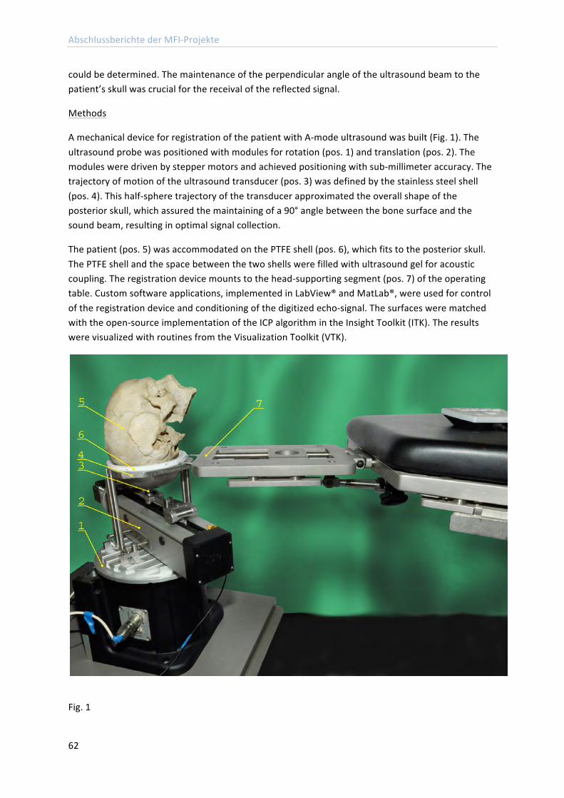
Abschlussberichte der MFI-‐Projekte
62
could be determined. The maintenance of the perpendicular angle of the ultrasound beam to the patient’s skull was crucial for the receival of the reflected signal.
Methods
A mechanical device for registration of the patient with A-‐mode ultrasound was built (Fig. 1). The ultrasound probe was positioned with modules for rotation (pos. 1) and translation (pos. 2). The modules were driven by stepper motors and achieved positioning with sub-‐millimeter accuracy. The trajectory of motion of the ultrasound transducer (pos. 3) was defined by the stainless steel shell (pos. 4). This half-‐sphere trajectory of the transducer approximated the overall shape of the posterior skull, which assured the maintaining of a 90° angle between the bone surface and the sound beam, resulting in optimal signal collection.
The patient (pos. 5) was accommodated on the PTFE shell (pos. 6), which fits to the posterior skull. The PTFE shell and the space between the two shells were filled with ultrasound gel for acoustic coupling. The registration device mounts to the head-‐supporting segment (pos. 7) of the operating table. Custom software applications, implemented in LabView® and MatLab®, were used for control of the registration device and conditioning of the digitized echo-‐signal. The surfaces were matched with the open-‐source implementation of the ICP algorithm in the Insight Toolkit (ITK). The results were visualized with routines from the Visualization Toolkit (VTK).
Fig. 1
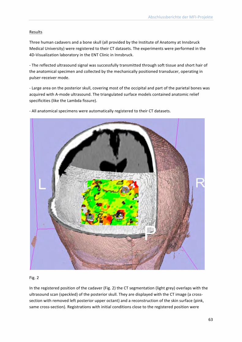
Abschlussberichte der MFI-‐Projekte
63
Results
Three human cadavers and a bone skull (all provided by the Institute of Anatomy at Innsbruck Medical University) were registered to their CT datasets. The experiments were performed in the 4D-‐Visualization laboratory in the ENT Clinic in Innsbruck.
-‐ The reflected ultrasound signal was successfully transmitted through soft tissue and short hair of the anatomical specimen and collected by the mechanically positioned transducer, operating in pulser-‐receiver mode.
-‐ Large area on the posterior skull, covering most of the occipital and part of the parietal bones was acquired with A-‐mode ultrasound. The triangulated surface models contained anatomic relief specificities (like the Lambda fissure).
-‐ All anatomical specimens were automatically registered to their CT datasets.
Fig. 2
In the registered position of the cadaver (Fig. 2) the CT segmentation (light grey) overlaps with the ultrasound scan (speckled) of the posterior skull. They are displayed with the CT image (a cross-‐section with removed left posterior upper octant) and a reconstruction of the skin surface (pink, same cross-‐section). Registrations with initial conditions close to the registered position were
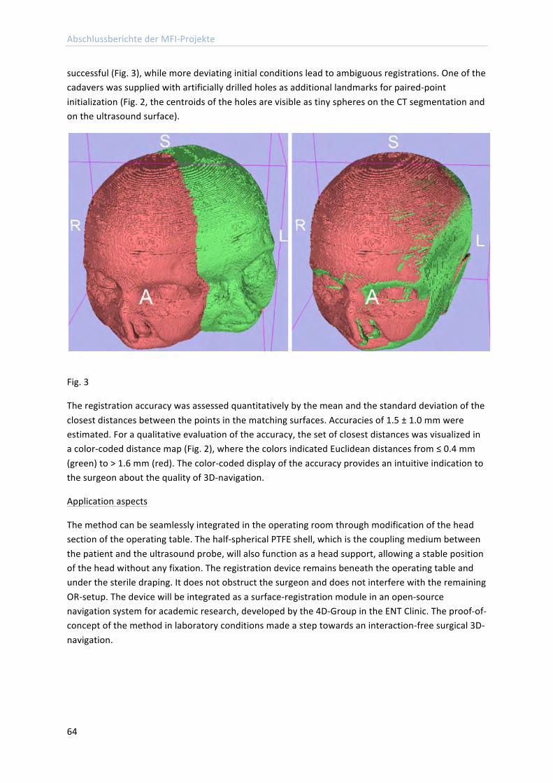
Abschlussberichte der MFI-‐Projekte
64
successful (Fig. 3), while more deviating initial conditions lead to ambiguous registrations. One of the cadavers was supplied with artificially drilled holes as additional landmarks for paired-‐point initialization (Fig. 2, the centroids of the holes are visible as tiny spheres on the CT segmentation and on the ultrasound surface).
Fig. 3
The registration accuracy was assessed quantitatively by the mean and the standard deviation of the closest distances between the points in the matching surfaces. Accuracies of 1.5 ± 1.0 mm were estimated. For a qualitative evaluation of the accuracy, the set of closest distances was visualized in a color-‐coded distance map (Fig. 2), where the colors indicated Euclidean distances from ≤ 0.4 mm (green) to > 1.6 mm (red). The color-‐coded display of the accuracy provides an intuitive indication to the surgeon about the quality of 3D-‐navigation.
Application aspects
The method can be seamlessly integrated in the operating room through modification of the head section of the operating table. The half-‐spherical PTFE shell, which is the coupling medium between the patient and the ultrasound probe, will also function as a head support, allowing a stable position of the head without any fixation. The registration device remains beneath the operating table and under the sterile draping. It does not obstruct the surgeon and does not interfere with the remaining OR-‐setup. The device will be integrated as a surface-‐registration module in an open-‐source navigation system for academic research, developed by the 4D-‐Group in the ENT Clinic. The proof-‐of-‐concept of the method in laboratory conditions made a step towards an interaction-‐free surgical 3D-‐navigation.
Abschlussberichte der MFI-‐Projekte
65
Publikationen:
Diakov, G.; Kral, F.; Güler, O.; Freysinger, W. (2010): Automatische Registration des Patienten mit A-‐Mode-‐Ultraschall für computerunterstützte Chirurgie. Funktionsnachweis im Labor. In: HNO 58 (11), S. 1067–1073.
Influencing immune reaction to HCV infection by modification of iron status. Iron limitation as an adjunct therapy in HCV infection
Priv.-‐Doz. Dr. med. univ. Igor Maximilian Theurl Universitätsklinik für Innere Medizin I
Berichts-‐/Förderzeitraum: 09/2008 – 08/2010
Zusammenfassung:
Das Ziel dieses Projektes war es, den schon lange postulierten Zusammenhang zwischen Eisenhomeostase und einer Infektion mit dem Hepatitis-‐C-‐Virus genauer zu untersuchen.
Hierfür ist es uns gelungen, in unserem Labor ein neues von Wakita und Kollegen beschriebenes Zellkultursystem zu etablieren und für die von uns geplanten Versuch zu modifizieren. Wir konnten in einem ersten Schritt große Mengen von infektiösen Partikeln herstellen, was gegenüber alten Systemen den Vorteil ergab, dass Infektions-‐ und Replikationsraten je nach vorliegendem zellularem Eisenstatus untersuchbar werden.
Zunächst untersuchten wir aber die Wirkung einer HCV-‐Infektion in vitro auf den Eisenstoffwechsel der Zelle, wobei sich in vitro keine großen Unterschiede im Eisenmetabolismus und seiner Zielgene zeigte. Dies kann auch daran hängen, dass der momentan für dieses System vorhandene Virussubty in vivo auch mit geringeren Veränderungen im Eisenstoffwechsel einhergeht als andere Virussubtypen. Auch könnten andere Zellen als Hepatozyten in vivo für die Veränderungen zuständig sein.
Tatsächlich zeigte sich auch in einem Maussystem das transgen HCV-‐Polyprotein exprimiert, ein deutlicher Unterschied im Eisenmetabolismus und vor allem in der Hepcidinexpression im Vergleich zu nicht transgenen Kontrollmäusen. Derzeit testen wir mit Partnern andere und neue Hepatitis-‐C-‐Virus Mausmodelle bezüglich Veränderungen im Eisenstoffwechsel. Modifizierte, sich replizierende Viren, könnten helfen zu zeigen, ob in vivo eine Hepatisvirusinfektion den Eisenhaushalt der infizierten Zelle wirklich modifiziert und ob eventuelle Virustatische Therapien den Eisenhaushalt mit beeinflussen, wie es für Ribavirin schon diskutiert wurde.
Der zweite wichtige Punkt ist die Frage, ob der Eisenstatus einer Zelle zum Zeitpunkt der Infektion einen Einfluss auf Infektions-‐ bzw. Replikationsrate hat. Hier konnten wir im Zellkultur-‐System zeigen, dass dies der Fall ist. Nunmehr geht es darum ,ob in den bereits erwähnten Maussystemen eine orale Eisenchelation zusätzlich zur Virustatischen Therapie einen additiven Behandlungserfolg bringt.
Abschlussberichte der MFI-‐Projekte
66
Somit konnten wir unser Projekt in vitro gut voranbringen, mussten aber, um bestimmte Fragen weiter verfolgen zu können auf in vivo Modelle umstellen, welche in Kooperation mit anderen Gruppen momentan hinsichtlich unserer Fragestellung evaluiert werden.
Ferritin stellte sich als wichtiger prädikativer als auch die Infektion im Allgemeinen beeinflussender Faktor dar. Da Ferritin sowohl über Eisen als auch Inflammationsstatus reguliert wird, war es wichtig eine Methode zu entwickeln, diese Unterscheidung sicher durchführen zu kennen. Hierfür wurde ein Projektvorschlag bei der OeNB eingereicht und angenommen bzw. gefördert.
Auch sind durch Erkenntnisse in diesem Projekt andere, mit diesem Projekt verbundene Projekte weiter vorangetrieben worden, was zu 3 Publikationen in hochrangigen peer review Journalen führte. 1 weitere Arbeiten ist derzeit in Revision.
Major Results:
• Etablierung eines Zellkultursystems zur Produktion infektiöser Hepatitis C Partikel • In vitro HCV Infektion keine große Auswirkung auf „Eisengene“ • Eisenmetabolismus der Zelle beeinflusst die Infektionsrat mit Hepatitis-‐C-‐Partikel in vitro.
Publikationen:
Theurl, Igor; Finkenstedt, Armin; Schroll, Andrea; Nairz, Manfred; Sonnweber, Thomas; Bellmann-‐Weiler, Rosa et al. (2010): Growth differentiation factor 15 in anaemia of chronic disease, iron deficiency anaemia and mixed type anaemia. In: Br. J. Haematol 148 (3), S. 449–455.
Theurl, Igor; Aigner, Elmar; Theurl, Milan; Nairz, Manfred; Seifert, Markus; Schroll, Andrea et al. (2009): Regulation of iron homeostasis in anemia of chronic disease and iron deficiency anemia: diagnostic and therapeutic implications. In: Blood 113 (21), S. 5277–5286.
Theurl, Igor; Schroll, Andrea; Sonnweber, Thomas; Nairz, Manfred; Theurl, Milan; Willenbacher, Wolfgang et al. (2011): Pharmacologic inhibition of hepcidin expression reverses anemia of chronic disease in rats. In: Blood.
Überführung des Projekts in eine externe Förderung:
OeNB: „New diagnostic strategies improving therapy of anemia of chronic disease”
Abschlussberichte der MFI-‐Projekte
67
Effects of Secretoneurin Gene Transfer on Myocardial Angiogenesis and Prostinfarction Heart Failure
Dr. Wilfried Schgoer Universitätsklinik für Innere Medizin I
Berichts-‐/Förderzeitraum: 08/2008 – 07/2010
Zusammenfassung:
Recently it has been shown that gene therapy with the angiogenic neuropeptide Secretoneurin (SN) restores tissue integrity, function and perfusion in a mouse hind-‐limb ischemia model by induction of angiogenesis, arteriogenesis and vasculogenesis. Therefore we hypothesized that SN might also affect function of cardiac endothelial cells and exert beneficial effects in a rat model of myocardial infarction.
In vivo, SN gene therapy was performed by intramyocardial injection of 100 µg SN-‐plasmid (or empty vector as control), shortly after ligation of the left anterior descending artery in Sprague-‐Dawley rats. SN prevented the anterior wall thinning (LVEDD 9.1 vs. 10.5 mm; p=0.04; LVESD 6.5 vs. 9.1 mm; p=0.002; n=11) and significantly improved cardiac function as revealed by histological analysis (capillaries/HPF 63.5±4 vs. 44.9±3.7; arterioles/HPF 8.5±0.5 vs. 5.8±0.6; fibrosis 21.65±1.6% vs. 35.37±2.8%; all p<0.001 and n=11); and echocardiographic measurement (ejection fraction 64.4±3.1% vs. 40.3±2.7%; p<0.001;; n=11).
To investigate the effect of SN in cell culture, in vitro angiogenesis assays using human coronary artery endothelial cells (HCAEC) in the absence or presence of different concentrations of SN were performed. SN dose dependently induced angiogenesis. A specific SN-‐ antibody and interestingly, a VEGF-‐ antibody too, abolished the observed effect. Western blot analysis of HCAEC extracts revealed stimulation of MAPK by SN 100 ng/ml after 20 minutes and of Akt after 40 to 240 minutes, indicating involvement of the ERK1/2 and PI3-‐kinase/Akt signal transduction pathways.
The SN induced MAPK activation could be blocked with a VEGF antibody as well as with the specific VEGFR2 inhibitor, SU1498. Receptor tyrosine kinase profiler assays and immunoprecipitation confirmed the SN mediated VEGFR2 activation.
In conclusion, SN showed angiogenic effects in a rat model of myocardial infarction, clinical parameters such as LVEDD and EF were significantly improved with SN-‐plasmid gene therapy, probably by stimulation of VEGFR2.
References:
Kirchmair et al.: The neuropeptide Secretoneurin acts as a direct anglogenic cytokine In vitro and In vivo.Circulation 2004; 109(6):777-‐83.
Egger et al.: Hypoxia up.regulates the angiogenic cytokine secretoneurin via an HIF-‐1(alpha)-‐ and basic: FGF-‐dependent pathway In muscle cells. FASEB J. 2007; 21(11):2906-‐17.
Schgoer et al.: Gene Therapy With The Anglogenic Cytokine Secretoneurin Induces Therapeutic Angiogenesis by a Nitric Oxide-‐Dependant Mechanism. Circ. Res. 2009; 105(10):994-‐1002.
Abschlussberichte der MFI-‐Projekte
68
Chromosomal Microdeletions/-‐duplications in Patients with Epileptic Encephalopathy of Unknown Etiology
OÄ Dr. Edda Haberlandt Universitätsklink für Pädiatrie IV
Berichts-‐/Förderzeitraum: 09/2008 -‐ 08/2010
Zusammenfassung:
Epilepsy and epileptic syndromes ranging from age-‐related and benign forms to severe epileptic encephalopathy are the most frequent neurological disorder in children with a frequency of 0.7-‐0.8 % up to the age of 15 years. A substantial subgroup has an atypical course and/or additional symptoms such as mental retardation, facial dysmorphisms, and other anomalies. Recently, submicroscopic chromosomal deletions/duplications have been recognised as frequent causes of genetic diseases. Such aberrations of only a few Mb or clearly less in size can be easily detected by a recently developed technique called SNP-‐array analysis.
In this project funded by the MFI we investigated 44 patients with epilepsy/epileptic encephalopathy not classifiable according to the classification of the International League Against Epilepsy because of an atypical course or syndromal aspects with the Illumina® lnfinium Human1M-‐Duo V3 array, which contains almost 1.200.000 SNPs on a single array. The major aims of the study were: (a) finding microdeletions/-‐duplications in patients with epileptic encephalopathy, (b) mapping new epilepsy or epilepsy related genes and genomic regions (CNVs), (c) understanding the etiology and pathogenesis of epilepsies more exactly, and (d) improving the genetic counselling regarding prognosis and recurrence risk.
Of the 44 patients investigated, 23 have been finally evaluated so far, the remaining 21 are in the works. Among the 23 we found in 8 cases genetic aberrations which may significantly contribute to the patients' conditions. These are: a microdeletion of 3.3 Mb on 1q41q42, a microdeletion of 826 kb on 19p13.2, an almost equal microdeletion in two different patients of 640 kb and 635 kb on 17q21.31, a complex mosaicism microduplication of approximately 1 Mb on 17 p13.2p13.1, a microdeletion of 30 kb on 14q23.3, a microdeletion of 3.3 kb on 1q32.1, and a microdeletion of 1.3 Mb on 5q13.2.
The first 7 aberrations are all heterozygous and de novo. Of special interest is the case with the 1q41q42 deletion as the dysmorphic features of the patient resemble those characteristic for Angelman syndrome and as it contains a gene which is a very good candidate gene for epilepsy due to its expression in neuronal cells and homology with a well-‐known epilepsy gene.
Also, a comparable deletion on 19p13.2 was to the best of our knowledge not described before. The chromosome 19 is very gene-‐rich, and deletions on it are rarely found which supports the assumption that they are clinically relevant. The region deleted in our patient contains four genes which may contribute to his condition: One of these genes has already been related to mental disorders like mild cognitive impairment and schizophrenia, the other three have been linked to the development of the nervous system. Therefore, our finding is of utmost significance.
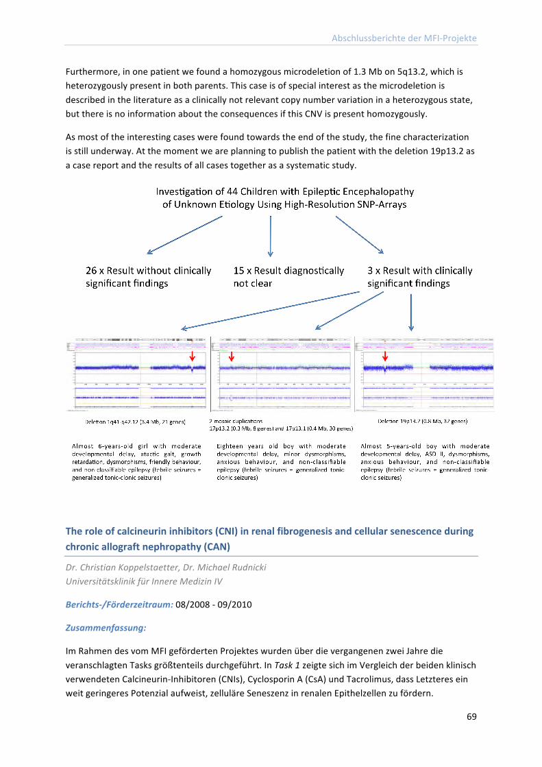
Abschlussberichte der MFI-‐Projekte
69
Furthermore, in one patient we found a homozygous microdeletion of 1.3 Mb on 5q13.2, which is heterozygously present in both parents. This case is of special interest as the microdeletion is described in the literature as a clinically not relevant copy number variation in a heterozygous state, but there is no information about the consequences if this CNV is present homozygously.
As most of the interesting cases were found towards the end of the study, the fine characterization is still underway. At the moment we are planning to publish the patient with the deletion 19p13.2 as a case report and the results of all cases together as a systematic study.
The role of calcineurin inhibitors (CNI) in renal fibrogenesis and cellular senescence during chronic allograft nephropathy (CAN)
Dr. Christian Koppelstaetter, Dr. Michael Rudnicki Universitätsklinik für Innere Medizin IV
Berichts-‐/Förderzeitraum: 08/2008 -‐ 09/2010
Zusammenfassung:
Im Rahmen des vom MFI geförderten Projektes wurden über die vergangenen zwei Jahre die veranschlagten Tasks größtenteils durchgeführt. In Task 1 zeigte sich im Vergleich der beiden klinisch verwendeten Calcineurin-‐Inhibitoren (CNIs), Cyclosporin A (CsA) und Tacrolimus, dass Letzteres ein weit geringeres Potenzial aufweist, zelluläre Seneszenz in renalen Epithelzellen zu fördern.
Abschlussberichte der MFI-‐Projekte
70
Die neu in die Versuchsreihe eingeführte epitheliale Zellinie RPTEC-‐TERT1 erbrachte dabei weitgehend vergleichbare Ergebnisse wie die etablierten HK-‐2 Zellen (das „klassische“ Modell), und sie erwiesen sich in weiterer Folge als erfolgversprechendes Zellkulturmodell und als guter Ersatz für (nur limitiert verfügbare) humane Primärzellen. Zusätzliche Versuche mit Rapamycin, einem erst vor wenigen Jahren eingeführten Immunsuppressivum mit abweichendem Wirkmechanismus, zeigten hier ein nochmals geringeres toxisches Potenzial und geringere Propagierung der Zellalterung.
Im Task 2 wurden die Versuche auf renale Bindegewebszellen ausgeweitet, wobei sich überraschenderweise eine Wachstumshemmung der Zellen unter Tacroilimus, nicht aber unter CsA zeigte, also eine Umkehrung CNI-‐Effekte verglichen mit Epithelzellen.
Die innerhalb von Task 3 durchgeführten cDNA-‐Microarrays (Vergleich von Fibroblasten und Epithelzellen bzw. CsA und Tacrolimus) ergaben insbesondere in Bezug auf Tacrolimus ein interessantes Bild. Während Tacrolimus in epithelialen Zellen nur eine geringe Verschiebung des genetischen Expressionsprofiles bewirkte, hatte es in den Fibroblasten weit umfassendere Wirkungen, die auf einen Beitrag zur langfristigen Fibrosierung des Organs hindeuten. Das ist auch insoferne bemerkenswert, als Tacrolimus (im Gegensatz zu Cyclosporin) selbst bei hohen Konzentrationen keine offensichtliche toxische Wirkung auf die Zellen zeigte.
Im Augenblick laufen Experimente zur Validierung und zum Ausbau der Microarray-‐Daten, mit besonderer Betonung auf fibrotische Effekte bei klinisch relevanten Konzentrationen. Besonders vielversprechend ist die beobachtete Induktion der NAD(P)H-‐Oxidase Typ 4 durch Tacrolimus, weil dieses Enzym neuerdings als wichtiger Modulator von fibrotischen Prozessen angesehen wird und längerfristig therapeutische Ansätze bieten könnte.
Zusammenfassend ergab der direkte Vergleich der drei in der Transplantationsmedizin am häufigsten verwendeten Immunsuppressiva deutliche Unterschiede in Bezug auf Zellalterung in humanen renalen Zellkulturmodellen. Hier zeigen sich möglicherweise neue mechanistische Zusammenhänge. Aktuell sind zwei Originalarbeiten in Ausarbeitung.
Überführung des Projekts in eine externe Förderung:
Industrial Grant Wyeth über € 30.000 für 3 Jahre
Abschlussberichte der MFI-‐Projekte
71
Human platelets in attack against Aspergillus fumigatus: influence on fungal gene wall expression and mitochondrial activity
Priv.-‐Doz. Mag. Dr. Susanne Perkhofer Sektion für Hygiene und Medizinische Mikrobiologie
Berichts-‐/Förderzeitraum: 10/2008 – 09/2010
Zusammenfassung:
Wichtiger Hinweis:
Der von mir bewilligte MFI mit der Projektnummer: 2007412 wurde von Seiten der MUI um die Personalkosten gekürzt. Durch diese Situation ergab sich nicht die Möglichkeit, die Untersuchungen im vollen Ausmaß durchzuführen. Daher fokussierte ich mich ausschließlich auf die mitochondriale Aktivität von Aspergillus fumigatus unter Thrombozyten Einfluss.
Bericht:
In Rahmen dieses Forschungsprojektes wurde der Einfluss humaner Thrombozyten auf die pilzliche mitochondriale Aktivität untersucht. Hierfür wurden die pilzlichen Mitochondrien von A. fumigatus nach Behandlung mit Thrombozyten nach einem Protokoll von Vödisch et al. isoliert und im Anschluss einer Proteomics Analyse unterzogen. Hierbei zeigte sich, dass die pilzlichen Superoxiddismutasen (SODs), welche unter Streßeinfluß und erhöhtem Vorhandensein von freien Radikalen hochreguliert werden, bei den pilzlichen, Thrombozyten-‐behandelten Mitochondrien einen starken Anstieg auswiesen.
Es wird auch bei antimykotischen Substanzen vermutet, dass diese durch Stress die pilzichen SODs hochregulieren. Die Ergebnisse dieser Untersuchungen zeigten einen kombinatorischen Effekt und die Ergebnisse wurden in den Journalen Journal of Anitmicrobial Chemotherapy (1) und Medical Mycology (2) 2010 publiziert. Weiters wurden die pilzlichen und Thrombozyten-‐behandelten Mitochondrien in die Oxygraphie zur Messung der mitochondrialen Atmung eingesetzt. Hierbei zeigte sich als Überraschung, dass die pilzlichen Mitochondrien nicht imstande sind, einen Cytochrom C Effekt aufzuweisen. Nach längeren Untersuchungen waren wir imstande, dieses Problem zu Iösen, da der Pilz uni sono unter den Eukaryonten über eine alternative Oxidase verfügt, welche wir nun durch Salicylhydroxamsäure inhibieren können.
Weitere Untersuchungen werden folgen, um dieses wichtige Thema weiterhin zu beforschen, da die Generierung dieser Daten einen weiteren Hinweis auf die Achillessehne des humanpathogenen Pilzes gibt, um zukünftig einerseits bessere Therapiemöglichkeiten anzubieten und andererseits durch diese Mitochondrienanalysen einen Hinweis auf spezifische Angriffspunkte zu identifizieren, die in weiterer Folge eine Entwicklung effektiver antimykotischer Substanzen zur Folge haben könnte.
Publikationen:
Perkhofer, Susanne; Trappl, Krista; Nussbaumer, Walter; Dierich, Manfred P.; Lass-‐Flörl, Cornelia (2010): Potential synergistic activity of antimycotic substances in combination with human platelets against Aspergillus fumigatus. In: J. Antimicrob. Chemother 65 (6), S. 1309–1311.
Abschlussberichte der MFI-‐Projekte
72
Perkhofer, Susanne; Trappl, Krista; Striessnig, Barbara; Nussbaumer, Walter; Lass-‐Flörl, Cornelia (2011): Platelets enhance activity of antimycotic substances against non-‐Aspergillus fumigatus Aspergillus species in vitro. In: Med. Mycol 49 (2), S. 157–166.
Decision making and framing effects in health-‐related decisions in normal aging and dementia
Dr. Laura Zamarian Universitätsklinik für Neurologie
Berichts-‐/Förderzeitraum: 10/2008 – 09/2010
Zusammenfassung:
In this MFI project, specific aspects of cognition – decision making under uncertainty, information sampling, judgment of health-‐related information, numerical processing, and executive functioning – were investigated in healthy older persons, in older persons with mild cognitive impairment (MCI) without dementia, and in older persons with dementia of Alzheimer’s type (AD), Parkinson’s disease (PD), or Parkinson’s disease dementia (PDD).
Results help to improve our insight into the cognitive changes related to normal aging and to pathological aging as well as to advance our understanding of the neural network supporting decision making, numerical processing, and executive functioning. Furthermore, results provide invaluable clues into the way caregivers and clinical practitioners can assist older persons with slight cognitive impairment to make appropriate and informed decisions.
In summary, results indicate that:
1) Healthy older adults like younger adults judge the medications that are described in positive terms (e.g., successful in 84% of cases) more favourably than the medications that are described in negative terms (e.g., unsuccessful in 16% of cases). However, these framing effects are reduced when natural frequencies (e.g., 84 out of 100) are used in alternative to percentages (e.g., 84%) to convey numerical information. Natural frequencies are easier to understand than percentages and may facilitate the estimation and the appreciation of the outcome of medical treatments. The use of natural frequencies in the communication of health-‐related information may thus reduce the bias of positive and negative descriptions, and help people to make informed decisions.
2) MCI patients and mild AD patients are considerably influenced by the way information about medications is presented and show pronounced framing effects relative to healthy aging peers. A correlation analysis suggests that the impact of positive and negative descriptions on the judgment of health-‐related information is stronger in the persons with the poorer episodic memory, numerical processing, and executive function profile. Greater attention to how information is presented should be paid when we communicate to patients with slight cognitive impairment. The use of standardized, easily understandable means of patient information is recommended.
Abschlussberichte der MFI-‐Projekte
73
3) MCI patients make poorer decisions than healthy aging peers when the decision situation is not well defined, information about risk is missing or conflicting, and learning from contingencies is needed. They also have problems when full information about a complex and changing decision situation is given, and learning from experience is not required. To some extent, decision-‐making impairments of MCI patients resemble those of patients with mild dementia (AD or PDD).
4) In general, our data suggest that, in patients with MCI, mild AD, PD, or mild PDD, the quality of life may be reduced not only as a result of memory or executive function deficits, depression, or motor disability, but also as a result of impaired decision making and judgment abilities. Reducing uncertainty in decision situations, providing complete information about risks and consequences of decisions, and making available easily understandable information material may facilitate older persons with slight cognitive impairment in making decisions in various domains of daily life including medical care, finances, or housing situations.
5) The methods implemented by this project have proven to be successful in the detection and characterization of deficits in decision making, numerical processing, and executive functioning of older adults with slight cognitive impairment. Noteworthy, they have also proven to be successful in the assessment of younger adults with temporal lobe epilepsy, of younger adults with selective disorders to reward-‐related neurotransmitter systems (as the case of narcolepsy patients), and of a younger adult with a rare form of neurodegenerative disease.
The main results of this project have been published in international peer-‐reviewed journal and have been presented at national and international congresses. General dissemination media (oral communication at the Brain Awareness Week 2009, Infoletter of the Gesellschaft für Neuropsychologie Österreich, Mypoint Newsletter of the Medical University Innsbruck, university seminars) have also been used to promote consciousness among the general public. Further publications are in preparation.
Publikationen:*
Zamarian L, Bodner T, Revkin SK, Benke T, Bösch S, Donnemiller E, Delazer M (2009): Numerical deficits in basal ganglia dysfunction. In: Neurocase, 15(5), S. 390-‐404.
Bonatti E, Kuchukhidze G, Zamarian L, Trinka E, Bodner T, Benke T, Delazer M (2009): Decision making in ambiguous and risky situations after unilateral temporal lobe epilepsy surgery. In: Epilepsy & Behaviour 14(4), S. 665-‐73.
Delazer, M.; Sinz, H.; Zamarian, L.; Stockner, H.; Seppi, K.; Wenning, G. K. et al. (2009): Decision making under risk and under ambiguity in Parkinson's disease. In: Neuropsychologia 47 (8-‐9), S. 1901–1908.
Delazer, Margarete; Zamarian, Laura; Bonatti, Elisabeth; Kuchukhidze, Giorgi; Koppelstätter, Florian; Bodner, Thomas et al. (2010): Decision making under ambiguity and under risk in mesial temporal lobe epilepsy. In: Neuropsychologia 48 (1), S. 194–200.
* Vollständig kursiv angeführte Publikationen ohne MFI Nennung
Abschlussberichte der MFI-‐Projekte
74
Zamarian, Laura; Benke, Thomas; Buchler, Max; Wenter, Johanna; Delazer, Margarete (2010): Information about medications may cause misunderstanding in older adults with cognitive impairment. In: J. Neurol. Sci 298 (1-‐2), S. 46–51.
Zamarian, Laura; Weiss, Elisabeth M.; Delazer, Margarete (2011): The impact of mild cognitive impairment on decision making in two gambling tasks. In: J Gerontol B Psychol Sci Soc Sci 66 (1), S. 23–31.
Delazer, Margarete; Zamarian, Laura; Bonatti, Elisabeth; Walser, Nicola; Kuchukhidze, Giorgi; Bodner, Thomas et al. (2011): Decision making under ambiguity in temporal lobe epilepsy: does the location of the underlying structural abnormality matter? In: Epilepsy Behav 20 (1), S. 34–37.
Delazer M, Högl B, Zamarian L, Wenter J, Gschliesser V, Ehrmann L, Brandauer E, Cevikkol Z, Frauscher B (2011): Executive functions, information sampling and decision making in narcolepsy with cataplexy. In Neuropsychology 25 (4), S. 477-‐487.
Zamarian, L.; Ischebeck, A.; Delazer, M. (2009): Neuroscience of learning arithmetic-‐-‐evidence from brain imaging studies. In: Neurosci Biobehav Rev 33 (6), S. 909–925.
Kuchukhidze, G.; Koppelstaetter, F.; Unterberger, I.; Dobesberger, J.; Walser, G.; Zamarian, L. et al. (2010): Hippocampal abnormalities in malformations of cortical development: MRI study. In: Neurology 74 (20), S. 1575–1582.
Literaturverzeichnis
75
LITERATURVERZEICHNIS (Publikationen mit MFI-‐Erwähnung)
Bánki, Zoltán; Posch, Wilfried; Ejaz, Asim; Oberhauser, Verena; Willey, Suzanne; Gassner, Christoph et al. (2010): Complement as an endogenous adjuvant for dendritic cell-‐mediated induction of retrovirus-‐specific CTLs. In: PLoS Pathog 6 (4), S. e1000891.
Berger, Regina; Fiegl, Heidi; Goebel, Georg; Obexer, Petra; Ausserlechner, Michael; Doppler, Wolfgang et al. (2010): Toll-‐like receptor 9 expression in breast and ovarian cancer is associated with poorly differentiated tumors. In: Cancer Sci 101 (4), S. 1059–1066.
Bernhard D, Wang XL. (2007): Smoking, Oxidative Stress and Cardiovascular Diseases -‐ Do Anti-‐Oxidative Therapies Fail? In: Current Medicinal Chemistry; 2007: 14, 1689-‐1699
Bernhard, D.; Laufer, G. (2008): The aging cardiomyocyte: a mini-‐review. In: Gerontology 54 (1), S. 24–31.
Bila, Custodio; Oberhauser, Verena; Ammann, Christoph G.; Ejaz, Asim; Huber, Georg; Schimmer, Simone et al. (2011): Complement opsonization enhances friend virus infection of B cells and thereby amplifies the virus-‐specific CD8+ T cell response. In: J. Virol 85 (2), S. 1151–1155.
Brandstätter, Anita; Lamina, Claudia; Kiechl, Stefan; Hunt, Steven C.; Coassin, Stefan; Paulweber, Bernhard et al. (2010): Sex and age interaction with genetic association of atherogenic uric acid concentrations. In: Atherosclerosis 210 (2), S. 474–478.
Coassin, Stefan; Brandstätter, Anita; Kronenberg, Florian (2008): An optimized procedure for the design and evaluation of Ecotilling assays. In: BMC Genomics 9, S. 510.
Coassin, Stefan; Brandstätter, Anita; Kronenberg, Florian (2010): Lost in the space of bioinformatic tools: a constantly updated survival guide for genetic epidemiology. The GenEpi Toolbox. In: Atherosclerosis 209 (2), S. 321–335.
Csordas, Adam; Wick, Georg; Laufer, Günther; Bernhard, David (2008): An Evaluation of the Clinical Evidence on the Role of Inflammation and Oxidative Stress in Smoking-‐Mediated Cardiovascular Disease. In: Biomark Insights 3, S. 127–139.
Dayal, Anamika; Schredelseker, Johann; Franzini-‐Armstrong, Clara; Grabner, Manfred (2010): Skeletal muscle excitation-‐contraction coupling is independent of a conserved heptad repeat motif in the C-‐terminus of the DHPRbeta(1a) subunit. In: Cell Calcium 47 (6), S. 500–506.
Delazer M, Högl B, Zamarian L, Wenter J, Gschliesser V, Ehrmann L, Brandauer E, Cevikkol Z, Frauscher B (2011): Executive functions, information sampling and decision making in narcolepsy with cataplexy. In Neuropsychology 25 (4), S. 477-‐487.
Delazer, M.; Sinz, H.; Zamarian, L.; Stockner, H.; Seppi, K.; Wenning, G. K. et al. (2009): Decision making under risk and under ambiguity in Parkinson's disease. In: Neuropsychologia 47 (8-‐9), S. 1901–1908.
Literaturverzeichnis
76
Delazer, Margarete; Zamarian, Laura; Bonatti, Elisabeth; Kuchukhidze, Giorgi; Koppelstätter, Florian; Bodner, Thomas et al. (2010): Decision making under ambiguity and under risk in mesial temporal lobe epilepsy. In: Neuropsychologia 48 (1), S. 194–200.
Delazer, Margarete; Zamarian, Laura; Bonatti, Elisabeth; Walser, Nicola; Kuchukhidze, Giorgi; Bodner, Thomas et al. (2011): Decision making under ambiguity in temporal lobe epilepsy: does the location of the underlying structural abnormality matter? In: Epilepsy Behav 20 (1), S. 34–37.
Diakov, G.; Kral, F.; Güler, O.; Freysinger, W. (2010): Automatische Registration des Patienten mit A-‐Mode-‐Ultraschall für computerunterstützte Chirurgie. Funktionsnachweis im Labor. In: HNO 58 (11), S. 1067–1073.
Elentner, Andreas; Finke, Daniela; Schmuth, Matthias; Chappaz, Stéphane; Ebner, Susanne; Malissen, Bernard et al. (2009): Langerhans cells are critical in the development of atopic dermatitis-‐like inflammation and symptoms in mice. In: J. Cell. Mol. Med 13 (8B), S. 2658–2672.
Evenou, Jean-‐Pierre; Wagner, Jürgen; Zenke, Gerhard; Brinkmann, Volker; Wagner, Kathrin; Kovarik, Jiri et al. (2009): The potent protein kinase C-‐selective inhibitor AEB071 (sotrastaurin) represents a new class of immunosuppressive agents affecting early T-‐cell activation. In: J. Pharmacol. Exp. Ther 330 (3), S. 792–801.
Forer, Lukas; Schönherr, Sebastian; Weissensteiner, Hansi; Haider, Florian; Kluckner, Thomas; Gieger, Christian et al. (2010): CONAN: copy number variation analysis software for genome-‐wide association studies. In: BMC Bioinformatics 11, S. 318.
Fritz, Teresa; Niederreiter, Lukas; Adolph, Timon; Blumberg, Richard S.; Kaser, Arthur (2011): Crohn's disease: NOD2, autophagy and ER stress converge. In: Gut.
Gangl, K.; Reininger, R.; Bernhard, D.; Campana, R.; Pree, I.; Reisinger, J. et al. (2009): Cigarette smoke facilitates allergen penetration across respiratory epithelium. In: Allergy 64 (3), S. 398–405.
Grundtman, Cecilia; Kreutmayer, Simone B.; Almanzar, Giovanni; Wick, Marius C.; Wick, Georg (2011): Heat shock protein 60 and immune inflammatory responses in atherosclerosis. In: Arterioscler. Thromb. Vasc. Biol 31 (5), S. 960–968.
Haas, Hubertus; Eisendle, Martin; Turgeon, B. Gillian (2008): Siderophores in fungal physiology and virulence. In: Annu Rev Phytopathol 46, S. 149–187.
Henderson, Blair; Csordas, Adam; Backovic, Aleksandar; Kind, Michaela; Bernhard, David; Wick, Georg (2008): Cigarette smoke is an endothelial stressor and leads to cell cycle arrest. In: Atherosclerosis 201 (2), S. 298–305.
Kaser, Arthur; Blumberg, Richard S. (2008): Paneth cells and inflammation dance together in Crohn's disease. In: Cell Res 18 (12), S. 1160–1162.
Kaser, Arthur; Tomczak, Michal; Blumberg, Richard S. (2011): "ER stress(ed out)!": Paneth cells and ischemia-‐reperfusion injury of the small intestine. In: Gastroenterology 140 (2), S. 393–396.
Literaturverzeichnis
77
Kaser, Arthur; Zeissig, Sebastian; Blumberg, Richard S. (2010): Inflammatory bowel disease. In: Annu. Rev. Immunol 28, S. 573–621.
Kaufmann, W. A.; Kasugai, Y.; Ferraguti, F.; Storm, J. F. (2010): Two distinct pools of large-‐conductance calcium-‐activated potassium channels in the somatic plasma membrane of central principal neurons. In: Neuroscience 169 (3), S. 974–986.
Kaufmann, Walter A.; Ferraguti, Francesco; Fukazawa, Yugo; Kasugai, Yu; Shigemoto, Ryuichi; Laake, Petter et al. (2009): Large-‐conductance calcium-‐activated potassium channels in purkinje cell plasma membranes are clustered at sites of hypolemmal microdomains. In: J. Comp. Neurol 515 (2), S. 215–230.
Köllensperger, Martin; Stefanova, Nadia; Pallua, Anton; Puschban, Zoe; Dechant, Georg; Hainzer, Monika et al. (2009): Striatal transplantation in a rodent model of multiple system atrophy: effects on L-‐Dopa response. In: J. Neurosci. Res 87 (7), S. 1679–1685.
Kreutmayer, Simone Barbara; Messner, Barbara; Knoflach, Michael; Henderson, Blair; Niederegger, Harald; Böck, Günther et al. (2011): Dynamics of heat shock protein 60 in endothelial cells exposed to cigarette smoke extract. In: Journal of molecular and cellular cardiology.
Kuchukhidze, G.; Koppelstaetter, F.; Unterberger, I.; Dobesberger, J.; Walser, G.; Zamarian, L. et al. (2010): Hippocampal abnormalities in malformations of cortical development: MRI study. In: Neurology 74 (20), S. 1575–1582.
Machtoub L, Pfeiffer R, Backovic A, Frischauf S, Wick MC (2010): Molecular Imaging Cellular SPIO Uptake with Nonlinear Optical Microscopy. In: Journal of Medical Imaging and Radiation Sciences;41(3), S. 159-‐164.
Nandy, Constanze; Mrázek, Jan; Stoiber, Heribert; Grässer, Friedrich A.; Hüttenhofer, Alexander; Polacek, Norbert (2009): Epstein-‐barr virus-‐induced expression of a novel human vault RNA. In: J. Mol. Biol 388 (4), S. 776–784.
Perkhofer, Susanne; Trappl, Krista; Nussbaumer, Walter; Dierich, Manfred P.; Lass-‐Flörl, Cornelia (2010): Potential synergistic activity of antimycotic substances in combination with human platelets against Aspergillus fumigatus. In: J. Antimicrob. Chemother 65 (6), S. 1309–1311.
Perkhofer, Susanne; Trappl, Krista; Striessnig, Barbara; Nussbaumer, Walter; Lass-‐Flörl, Cornelia (2011): Platelets enhance activity of antimycotic substances against non-‐Aspergillus fumigatus Aspergillus species in vitro. In: Med. Mycol 49 (2), S. 157–166.
Pirone, Antonella; Schredelseker, Johann; Tuluc, Petronel; Gravino, Elvira; Fortunato, Giuliana; Flucher, Bernhard E. et al. (2010): Identification and functional characterization of malignant hyperthermia mutation T1354S in the outer pore of the Cavalpha1S-‐subunit. In: Am. J. Physiol., Cell Physiol 299 (6), S. C1345-‐54.
Prajczer, Sinikka; Heidenreich, Ursula; Pfaller, Walter; Kotanko, Peter; Lhotta, Karl; Jennings, Paul (2010): Evidence for a role of uromodulin in chronic kidney disease progression. In: Nephrol. Dial. Transplant 25 (6), S. 1896–1903.
Literaturverzeichnis
78
Prelog, Martina; Keller, Michael; Geiger, Ralf; Brandstätter, Anita; Würzner, Reinhard; Schweigmann, Ulrich et al. (2009): Thymectomy in early childhood: significant alterations of the CD4(+)CD45RA(+)CD62L(+) T cell compartment in later life. In: Clin. Immunol 130 (2), S. 123–132.
Ritsch, Andreas; Scharnagl, Hubert; Eller, Philipp; Tancevski, Ivan; Duwensee, Kristina; Demetz, Egon et al. (2010): Cholesteryl ester transfer protein and mortality in patients undergoing coronary angiography: the Ludwigshafen Risk and Cardiovascular Health study. In: Circulation 121 (3), S. 366–374.
Santer, Frédéric R.; Malinowska, Kamilla; Culig, Zoran; Cavarretta, Ilaria T. (2010): Interleukin-‐6 trans-‐signalling differentially regulates proliferation, migration, adhesion and maspin expression in human prostate cancer cells. In: Endocr. Relat. Cancer 17 (1), S. 241–253.
Schmid, Marianne; Prajczer, Sinikka; Gruber, Leonhard N.; Bertocchi, Cristina; Gandini, Rosaria; Pfaller, Walter et al. (2010): Uromodulin facilitates neutrophil migration across renal epithelial monolayers. In: Cell. Physiol. Biochem 26 (3), S. 311–318.
Schönherr, Sebastian; Weissensteiner, Hansi; Coassin, Stefan; Specht, Günther; Kronenberg, Florian; Brandstätter, Anita (2009): eCOMPAGT -‐-‐ efficient combination and management of phenotypes and genotypes for genetic epidemiology. In: BMC Bioinformatics 10, S. 139.
Schredelseker, Johann; Dayal, Anamika; Schwerte, Thorsten; Franzini-‐Armstrong, Clara; Grabner, Manfred (2009): Proper restoration of excitation-‐contraction coupling in the dihydropyridine receptor beta1-‐null zebrafish relaxed is an exclusive function of the beta1a subunit. In: J. Biol. Chem 284 (2), S. 1242–1251.
Schredelseker, Johann; Shrivastav, Manisha; Dayal, Anamika; Grabner, Manfred (2010): Non-‐Ca2+-‐conducting Ca2+ channels in fish skeletal muscle excitation-‐contraction coupling. In: Proc. Natl. Acad. Sci. U.S.A 107 (12), S. 5658–5663.
Schroll, Andrea; Eller, Kathrin; Huber, Julia M.; Theurl, Igor M.; Wolf, Anna M.; Weiss, Günter; Rosenkranz, Alexander R. (2010): Tim3 is upregulated and protective in nephrotoxic serum nephritis. In: Am. J. Pathol 176 (4), S. 1716–1724.
Stoitzner, Patrizia; Romani, Nikolaus; McLellan, Alexander D.; Tripp, Christoph H.; Ebner, Susanne (2010): Isolation of skin dendritic cells from mouse and man. In: Methods Mol. Biol 595, S. 235–248.
Stoitzner, Patrizia; Sparber, Florian; Tripp, Christoph H.: Langerhans cells as targets for immunotherapy against skin cancer. In: Immunol. Cell Biol 88 (4), S. 431–437.
Tancevski, Ivan; Demetz, Egon; Eller, Philipp; Duwensee, Kristina; Hoefer, Julia; Heim, Christiane et al. (2010): The liver-‐selective thyromimetic T-‐0681 influences reverse cholesterol transport and atherosclerosis development in mice. In: PLoS ONE 5 (1), S. e8722.
Tancevski, Ivan; Wehinger, Andreas; Demetz, Egon; Eller, Philipp; Duwensee, Kristina; Huber, Julia et al. (2008): Reduced plasma high-‐density lipoprotein cholesterol in hyperthyroid mice coincides with decreased hepatic adenosine 5'-‐triphosphate-‐binding cassette transporter 1 expression. In: Endocrinology 149 (7), S. 3708–3712.
Literaturverzeichnis
79
Tancevski, Ivan; Wehinger, Andreas; Demetz, Egon; Hoefer, Julia; Eller, Philipp; Huber, Eva et al. (2009): The thyromimetic T-‐0681 protects from atherosclerosis. In: J. Lipid Res 50 (5), S. 938–944.
Theurl, Igor; Aigner, Elmar; Theurl, Milan; Nairz, Manfred; Seifert, Markus; Schroll, Andrea et al. (2009): Regulation of iron homeostasis in anemia of chronic disease and iron deficiency anemia: diagnostic and therapeutic implications. In: Blood 113 (21), S. 5277–5286.
Theurl, Igor; Finkenstedt, Armin; Schroll, Andrea; Nairz, Manfred; Sonnweber, Thomas; Bellmann-‐Weiler, Rosa et al. (2010): Growth differentiation factor 15 in anaemia of chronic disease, iron deficiency anaemia and mixed type anaemia. In: Br. J. Haematol 148 (3), S. 449–455.
Theurl, Igor; Schroll, Andrea; Sonnweber, Thomas; Nairz, Manfred; Theurl, Milan; Willenbacher, Wolfgang et al. (2011): Pharmacologic inhibition of hepcidin expression reverses anemia of chronic disease in rats. In: Blood.
Thuille, Nikolaus; Lutz-‐Nicoladoni, Christina; Letschka, Thomas; Hermann-‐Kleiter, Natascha; Heit, Isabelle; Baier, Gottfried (2009): PKCtheta and Itk functionally interact during primary mouse CD3+ T cell activation. In: Immunol. Lett 126 (1-‐2), S. 54–59.
Trappl, K., and Polacek, N. (2011): The ribosome: A molecular machine powered by RNA In: Metal Ions Life Sci. 9:253-‐275.
Tripp, Christoph H.; Sparber, Florian; Hermans, Ian F.; Romani, Nikolaus; Stoitzner, Patrizia (2009): Glycolipids injected into the skin are presented to NKT cells in the draining lymph node independently of migratory skin dendritic cells. In: J. Immunol 182 (12), S. 7644–7654.
Weissensteiner, Hansi; Schönherr, Sebastian; Specht, Günther; Kronenberg, Florian; Brandstätter, Anita (2010): eCOMPAGT integrates mtDNA: import, validation and export of mitochondrial DNA profiles for population genetics, tumour dynamics and genotype-‐phenotype association studies. In: BMC Bioinformatics 11, S. 122.
Wick, Marius C.; Kremser, Christian; Frischauf, Stefan; Wick, Georg (2008a): In vivo molecular imaging of vascular stress. In: Cell Stress Chaperones 13 (3), S. 263–273.
Wick, Marius C.; Mayerl, Christina; Backovic, Aleksandar; van der Zee, Ruurd; Jaschke, Werner; Dietrich, Hermann; Wick, Georg (2008b): In vivo imaging of the effect of LPS on arterial endothelial cells: molecular imaging of heat shock protein 60 expression. In: Cell Stress Chaperones 13 (3), S. 275–285.
Willis, Michael; Kaufmann, Walter A.; Wietzorrek, Georg; Hutter-‐Paier, Birgit; Moosmang, Sven; Humpel, Christian et al. (2010): L-‐type calcium channel CaV 1.2 in transgenic mice overexpressing human AbetaPP751 with the London (V717I) and Swedish (K670M/N671L) mutations. In: J. Alzheimers Dis 20 (4), S. 1167–1180.
Zamarian, L.; Ischebeck, A.; Delazer, M. (2009): Neuroscience of learning arithmetic-‐-‐evidence from brain imaging studies. In: Neurosci Biobehav Rev 33 (6), S. 909–925.

Literaturverzeichnis
80
Zamarian, Laura; Benke, Thomas; Buchler, Max; Wenter, Johanna; Delazer, Margarete (2010): Information about medications may cause misunderstanding in older adults with cognitive impairment. In: J. Neurol. Sci 298 (1-‐2), S. 46–51.
Zamarian, Laura; Weiss, Elisabeth M.; Delazer, Margarete (2011): The impact of mild cognitive impairment on decision making in two gambling tasks. In: J Gerontol B Psychol Sci Soc Sci 66 (1), S. 23–31.
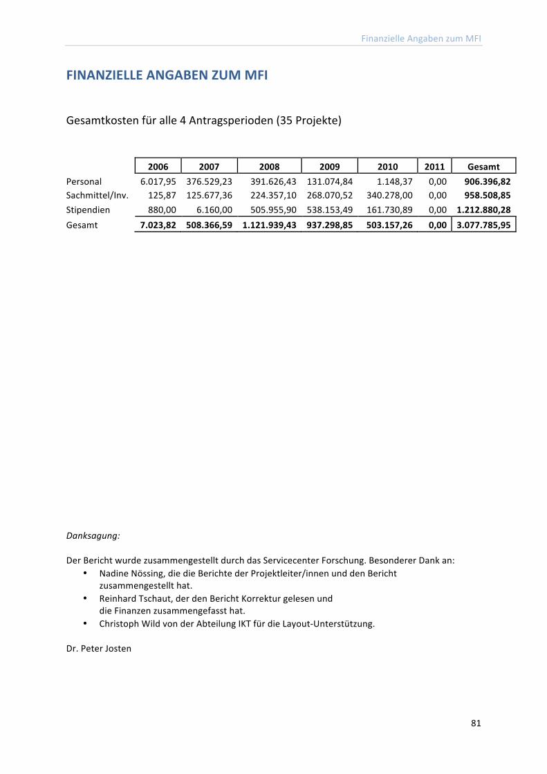
Finanzielle Angaben zum MFI
81
FINANZIELLE ANGABEN ZUM MFI
Gesamtkosten für alle 4 Antragsperioden (35 Projekte)
2006 2007 2008 2009 2010 2011 Gesamt Personal 6.017,95 376.529,23 391.626,43 131.074,84 1.148,37 0,00 906.396,82 Sachmittel/Inv. 125,87 125.677,36 224.357,10 268.070,52 340.278,00 0,00 958.508,85 Stipendien 880,00 6.160,00 505.955,90 538.153,49 161.730,89 0,00 1.212.880,28 Gesamt 7.023,82 508.366,59 1.121.939,43 937.298,85 503.157,26 0,00 3.077.785,95
Danksagung: Der Bericht wurde zusammengestellt durch das Servicecenter Forschung. Besonderer Dank an:
• Nadine Nössing, die die Berichte der Projektleiter/innen und den Bericht zusammengestellt hat.
• Reinhard Tschaut, der den Bericht Korrektur gelesen und die Finanzen zusammengefasst hat.
• Christoph Wild von der Abteilung IKT für die Layout-‐Unterstützung. Dr. Peter Josten


