This work has been digitalized and published in 2013 by Verlag Zeitschrift für Naturforschung in cooperation with the Max Planck Society for the Advancement of Science under a Creative Commons Attribution 4.0 International License. Dieses Werk wurde im Jahr 2013 vom Verlag Zeitschrift für Naturforschung in Zusammenarbeit mit der Max-Planck-Gesellschaft zur Förderung der Wissenschaften e.V. digitalisiert und unter folgender Lizenz veröffentlicht: Creative Commons Namensnennung 4.0 Lizenz. Lentinellic Acid, a Biologically Active Protoilludane Derivative from Lentinellus Species (Basidiomycetes) [1] A. Stärk, T. Anke, LB Biotechnologie der Universität, Paul-Ehrlich-Straße 23, D-6750 Kaiserslautern, Bundesrepublik Deutschland U. Mocek, W. Steglich, Institut für Organische Chemie und Biochemie der Universität, Gerhard-Domagk-Straße 1, D-5300 Bonn, Bundesrepublik Deutschland A. Kirfel and G. Will Mineralogisches Institut der Universität, Poppelsdorfer Schloß, D-5300 Bonn, Bundesrepublik Deutschland Z. Naturforsch. 43c, 177-183 (1988); received October 16, 1987 Dedicated to Professor Helmut Doerfel on the occasion of his 60th birthday Lentinellus, Basidiomycetes, Sesquiterpenoids, Protoilludanes, Lentinellic Acid, Antibiotics A new antimicrobial and cytotoxic sesquiterpenoid, lentinellic acid (1), has been isolated from submerged cultures of Lentinellus ursinus and L. omphalodes. The structure of the antibiotic was elucidated by spectroscopic methods and a single crystal X-ray analysis. 1 may be formed biogenetically by condensation of a protoilludane aldehyde 4 with a malonate unit. Introduction Species of the genus Lentinellus are of widespread occurrence, their usual habitat being decaying wood. They are characterized by tough carpophores with toothed lamellae and spores with amyloid spines. The hymenium contains gloeocystidia staining blue with sulfovanillic acid. The component responsible for this color reaction has been identified as the ses- quiterpenoid stearoylvelutinal [2], The taxonomy of the genus Lentinellus and its relationship to the Aphyllophorales has been discussed by Singer [3]. During our investigation of the secondary metabo- lism of mycelial cultures of Lentinellus species sever- al antibiotic and cytotoxic compounds have been iso- lated [4, 5]. In the following we wish to describe a new antibiotic, lentinellic acid (1), derived from fermentations of Lentinellus omphalodes and L. ursinus. Reprint requests to Prof. Dr. T. Anke or Prof. Dr. W. Steglich. Verlag der Zeitschrift für Naturforschung. D-7400 Tübingen 0341 - 0382/88/0003 - 0194 $ 01.30/0 Experimental Lentinellus omphalodes (Fr.) P. Karst, and L. ursinus (Fr.) Kühn. Mycelial cultures were obtained from spore prints or tissue plugs from specimens collected in Germany (L. ursinus 80163, L. omphalodes 8075, 80272), Canada (L. omphalodes 80116, 80131), or USA (L. omphalodes 8349). Fermentation and isolation For the maintenance on agar slants the strains were grown in a yeast extract-malt extract-glucose medium composed of (g/liter): yeast extract 4, malt extract 10, glucose 4, and agar 20. For the production of lentinellic acid the medium contained (g/liter): glu- cose 50, peptone 2, yeast extract 5, KH 2 P0 4 1, and MgS0 4 x7H 2 0 1. The fermentations were carried out in a Biolafitte fermentation apparatus (1 1 air/ min, 200 rpm, 22 °C). 200 ml of a well grown seed culture were used as inoculum for 20 1 of medium. Antibiotic production was followed by paper-disc/ agar-diffusion assay using Bacillus brevis as test or- ganism. After fermentation the mycelia were sepa- rated from the culture fluid by filtration. Lentinellic acid was extracted from the culture fluid (19 1) with 5 1 of ethyl acetate. After evaporation of the solvent the crude product (4.4 g) was applied to a column of
Transcript
This work has been digitalized and published in 2013 by Verlag Zeitschrift für Naturforschung in cooperation with the Max Planck Society for the Advancement of Science under a Creative Commons Attribution4.0 International License.
Dieses Werk wurde im Jahr 2013 vom Verlag Zeitschrift für Naturforschungin Zusammenarbeit mit der Max-Planck-Gesellschaft zur Förderung derWissenschaften e.V. digitalisiert und unter folgender Lizenz veröffentlicht:Creative Commons Namensnennung 4.0 Lizenz.
Lentinellic Acid, a Biologically Active Protoilludane Derivative from Lentinellus Species (Basidiomycetes) [1] A. Stärk, T. Anke , LB Biotechnologie der Universität, Paul-Ehrlich-Straße 23, D-6750 Kaiserslautern, Bundesrepublik Deutschland U. Mocek, W. Steglich, Institut für Organische Chemie und Biochemie der Universität, Gerhard-Domagk-Straße 1, D-5300 Bonn, Bundesrepublik Deutschland A. Kirfel and G. Will Mineralogisches Institut der Universität, Poppelsdorfer Schloß, D-5300 Bonn, Bundesrepublik Deutschland Z. Naturforsch. 43c, 177-183 (1988); received October 16, 1987 Dedicated to Professor Helmut Doerfel on the occasion of his 60th birthday Lentinellus, Basidiomycetes, Sesquiterpenoids, Protoilludanes, Lentinellic Acid, Antibiotics
A new antimicrobial and cytotoxic sesquiterpenoid, lentinellic acid (1), has been isolated from submerged cultures of Lentinellus ursinus and L. omphalodes. The structure of the antibiotic was elucidated by spectroscopic methods and a single crystal X-ray analysis. 1 may be formed biogenetically by condensation of a protoilludane aldehyde 4 with a malonate unit.
Introduction
Species of the genus Lentinellus are of widespread occurrence, their usual habitat being decaying wood. They are characterized by tough carpophores with toothed lamellae and spores with amyloid spines. The hymenium contains gloeocystidia staining blue with sulfovanillic acid. The component responsible for this color reaction has been identified as the ses-quiterpenoid stearoylvelutinal [2], The taxonomy of the genus Lentinellus and its relationship to the Aphyllophorales has been discussed by Singer [3]. During our investigation of the secondary metabo-lism of mycelial cultures of Lentinellus species sever-al antibiotic and cytotoxic compounds have been iso-lated [4, 5]. In the following we wish to describe a new antibiotic, lentinellic acid (1), derived from fermentations of Lentinellus omphalodes and L. ursinus.
Reprint requests to Prof. Dr. T. Anke or Prof. Dr. W. Steglich.
Verlag der Zeitschrift für Naturforschung. D-7400 Tübingen 0341 - 0382/88/0003 - 0194 $ 01.30/0
Experimental
Lentinellus omphalodes (Fr.) P. Karst, and L. ursinus (Fr.) Kühn.
Mycelial cultures were obtained from spore prints or tissue plugs from specimens collected in Germany (L. ursinus 80163, L. omphalodes 8075, 80272), Canada (L. omphalodes 80116, 80131), or USA (L. omphalodes 8349).
Fermentation and isolation
For the maintenance on agar slants the strains were grown in a yeast extract-malt extract-glucose medium composed of (g/liter): yeast extract 4, malt extract 10, glucose 4, and agar 20. For the production of lentinellic acid the medium contained (g/liter): glu-cose 50, peptone 2, yeast extract 5, K H 2 P 0 4 1, and M g S 0 4 x 7 H 2 0 1. The fermentations were carried out in a Biolafitte fermentation apparatus (1 1 air/ min, 200 rpm, 22 °C). 200 ml of a well grown seed culture were used as inoculum for 20 1 of medium. Antibiotic production was followed by paper-disc/ agar-diffusion assay using Bacillus brevis as test or-ganism. After fermentation the mycelia were sepa-rated from the culture fluid by filtration. Lentinellic acid was extracted from the culture fluid (19 1) with 5 1 of ethyl acetate. After evaporation of the solvent the crude product (4.4 g) was applied to a column of
1 7 8 A. Stärk et al. • Lentinellic Acid, a Biologically Active Protoilludane Derivative from Lentinellus
silica gel (Merck 60) and eluted with dichloro-methane. The fractions containing lentinellic acid were combined to yield 1.8 g of a yellow precipitate. Pure lentinellic acid (0.95 g) was obtained by crystal-lization from methanol.
Physical and spectroscopic data
NMR spectra were recorded on a Bruker WM-400 spectrometer. The high-resolution mass spectra were determined with an AEI MS-50 spectrometer. Analytical thin-layer chromatography (TLC) was performed with Merck 5554 silica plates. Melting points were obtained on a Reichert hot plate micro-scope and are uncorrected.
Methyl lentinellate (2): To a solution of 6 mg 1, 2 ml methanol, and 3 mg 4-(dimethylamino)pyridine in 5 ml of dichloromethane at 0 °C 8.5 mg dicyclo-hexylcarbodiimide were added and the solution was stirred for 5 min at 0 °C and 3 h at 20 °C. After filtra-tion of the deposite the solvent was removed in vac-uo and the residue was purified by preparative TLC on silica gel plates (eluant: chloroform). Yield: 6.2 mg, yellow oil; Rf 0.34 (cyclohexane/ethyl ace-tate/formic acid = 120:40:5); 'H NMR (CDC13): 0 = 1 . 0 3 , 1.19, 1.26 (each s, 3H) , 1.49-2.08 (m, 3H) , 2 .32-2 .88 (m, 3H) , 2.99 (br. s, 2H) , 3.91 (s, 3H) , 4.99 (d, 7 = 9.0 Hz, 1H), 8.01 (s, 1H); MS (DI 180 °C): m/z 330.1472 (62.9%, calc. for C19H220.s
330.1467).
Crystal structure determination by X-ray diffraction
Single crystals suitable for a crystal structure in-vestigation were obtained from acetone/petroleum ether. The crystal data are: orthorhombic symmetry, with a = 6.506 (1), b = 10.807 (3), c = 22.706 (5) Á, V = 1596.5 (1.0) Â3 (standard deviations in parenthe-ses). Space group P2,2121, Z = 4, Dx = 1.315 g/cm3, (CuKa) = 7.5 cm - 1 , Fm) = 672, T = 293 K.
The intensity measurements were carried out on an automatic four circle diffractometer (SYNTHEX P2]) in the co-mode using graphite monochromated CuK a radiation (k = 1.5413 À); lattice constants were determined from angular settings of 25 inde-pendent reflections (2 0 ^ 50°); the range was sin0max/X. = 0.54 Â - 1 . A standard reflection was re-measured after every 33 records with an intensity variation of ± 1%; the number of reflections meas-ured was 1216, with 305 considered unobserved [I < 2.56(1)]; the number of unique observed reflexions was 911. No absorption correction was applied.
The structure was solved by use of the direct method program M U L T A N [6] and refined on I F I by full matrix least squares calculations. In the final stage of refinement the hydrogen atoms were treated as riding on the carrier atoms with B(H) = 1.2 B(C). Hydrogen bonded in O H was located from differ-ence Fourier maps and refined for positional param-eters only. An isotropic extinction factor g was in-cluded in the list of variables. In total 212 refined parameters were used. Refinement converged at R = 0.034, Rw = 0.045 (omitting unobserveds), 5 = 1.12. The weighting function was w = (o2(F) + 0.001 F2) - 1 ; the final parameter shifts were less than 0.30 and peaks in the final difference Fourier map were max 0.11 e/Á3 , min - 0 . 1 1 e/Â3; atomic scattering fac-tors were taken from International Tables for X-ray Crystallography (1974); all calculations were done with SHEL XI6 [7], Further details of the crystal structure determination have been deposited at the Fachinformationszentrum Energie, Physik, Mathe-matik GmbH, D-7514 Eggenstein-Leopoldshafen 2. Any request for this material should be accompanied by a full literature citation and the reference number CSD.
Biological assays
The antimicrobial spectra and macromolecular syntheses in cells of the ascitic form of Ehrlich car-cinoma were measured as described previously [8, 9],
179 A. Stärk et al. • Lentinellic Acid, a Biologically Active Protoilludane Derivative from Lentinellus
Results and Discussion
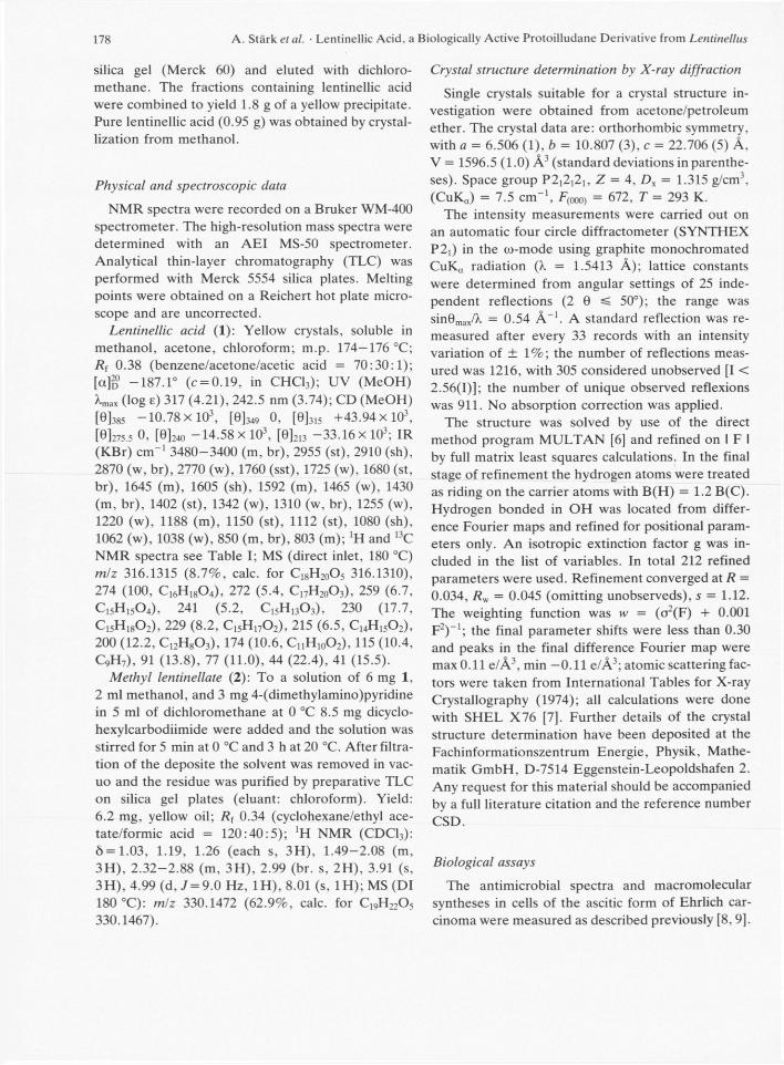
When grown in submerged culture isolates of Len-tinellus ursinus and L. omphalodes f rom Europe, U . S . A . , and Canada produced lentinellic acid in al-most equal amounts (50—80 mg/1 of culture). This finding suggests that the biosynthetic pathway lead-ing to lentinellic acid might be a suitable taxonomic character. A typical fermentation of L. ursinus is shown in Fig. 1. The antibiotic content of the culture is highest 13 days after inoculation. Under the same conditions L. omphalodes yielded the highest amounts of lentinellic acid (70 mg/1 of culture) after 16 days of fermentation.
Lentinellic acid (1), purified from the culture fluid as described in the experimental section, was ob-tained in form of yellow crystals. Esterification with methanol yielded the methyl ester 2 which exhibited enhanced antimicrobial activity.
According to the mass spectrum 1 has the molecu-lar formula C18H2o05. The mass spectrum is domi-nated by a ketene elimination from the molecular ion m/z 316 which leads to the base peak at mlz 274. Lentinellic acid exhibits two strong IR bands (KBr) in the carbonyl region at 1760 and 1680 cm - 1 . Two UV maxima (MeOH) at 242.5 and 317 nm point to the presence of an extended chromophore. In the 'H NMR spectrum (Table I) seven well separated multi-plets and two methyl singlets at ô = 0.98 and 1.14 can be assigned to the protons of partial structure A. The presence of a 3,4-disubstituted 1,1-dimethylcyclo-pentane ring in A is in agreement with the charac-teristic 1.9 Hz W-coupling between the a-protons of the two methylene groups. The structure 1 of len-
tinellic acid was finally solved by an X-ray analysis. The 13C NMR signals (Table I) were assigned by 2 D 13C-'H shift correlations and selective decoupling ex-periments. According to the chemical shifts of the olefinic carbons the lactone and carboxylic carbonyl groups exert a strong electron withdrawing effect whereas the cyclobutanone carbonyl is much less effective due to its out of plane conformation.
Lentinellic acid afforded crystals suitable for the X-ray structural determination by crystallization from acetone—petroleum ether. The structure was solved by direct methods using the SHEL X76 pro-gram and refined. Dispersion corrections and anisot-ropic temperature factors have been applied for the no-hydrogen atoms. The conformation and relative configuration of lentinellic acid is depicted in the stereoplot (Fig. 2). Selected structural parameters are given in Tables I I - V * . From the bond lengths of the double bonds a mesomeric interaction in the dienoic acid moiety is revealed and the carboxylic proton is partially chelated to the lactone carbonyl group. The molecules are bond in the crystal by Van der Waals forces only.
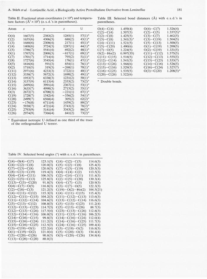
The absolute configuration of lentinellic acid (1) was not determined and is assumed to correspond to that of other natural protoilludane derivatives [10]. The CD spectrum of 1 is depicted in Fig. 3.
1 may be formed in the fungus by condensation of aldehyde 4 with a malonate unit. 4 can be derived
+ The numbering system is that used for the protoilludane skeleton [10d].
* The numbering used in Tables II—IV is indicated in Fig. 2. It differs from that given in formula 1.
10 15 Time (days)
Fig. 1. Time course of a fermentation of Len-tinellus ursinus 80163. Dry weight of the my-celium, pH, and lentinellic acid content of the culture fluid, determined photometrically after isolation from a 100 ml sample.
180 A. Stärk et al. • Lentinellic Acid, a Biologically Active Protoilludane Derivative from Lentinellus
2, R = CH3
Table I. 'H and 13C NMR data of lentinellic acid (1) (400 and 100.62 MHz, respectively; CDCI3 as solvent and internal standard).
H-atom ô multiplicity, J [Hz] C-atom ô multiplicity, J [Hz]
10a 2.02 ddd 12.6/7.2/1.9 8 82.60 Dtd 153/5/2.5** 10ß 1.33 dd 12.6/10.8 9 50.91 Dm 130 12 1.22 s 10 46.07 Tm 129 13 8.37 s 11 39.20* m 14 0.98*** s 12 19.34 Qqd 128/4/4 15 1 14*** s 13 142.60 Dd 170/2 CO2H 12.30 br. s 14 26.65 Qm 126 CO2H
15 29.20 Qm 124 16 118.02 d 1.5 17 165.18 dd 9/2 18 161.70 d 5.6
* Signals may be interchanged. •̂ 2-H.8-C — 2.5 Hz.
*** Assignment according to [10c],
0(5) 0(2)
Fig. 2. Stereoplot of lentinellic acid (1).
181 A. Stärk et al. • Lentinellic Acid, a Biologically Active Protoilludane Derivative from Lentinellus
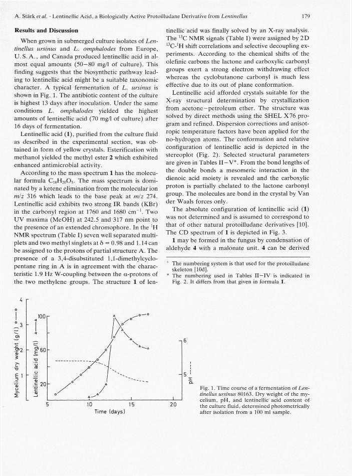
Table II. Fractional atom coordinates ( x 104) and tempera- Table III. Selected bond distances (A) with e.s.d.'s in ture factors ( Â 2 x 10³) (e.s.d.'s in parentheses). parentheses.
Atom X y z U 0 ( 4 ) - -C(4) 1.459(4) 0 ( 4 ) - -C(7) 1.326(4) C(2)- C(4) 1.507(5) C(2) - C(5) 1.337(5)*
200 250 350 400 X[nm] Fig. 3. Circular dichroism spectrum ( ) and UV spec-trum ( ) of lentinellic acid (1) in methanol.
from armillol (3) [10 b] by oxidation of two of its hydroxyl groups. Esters of 3 with orsellinic acid and related aromatic acids have recently been isolated from cultures of Armillariella mellea [10b, 11].
H0,C.
[01
X0,H
-21-1,0
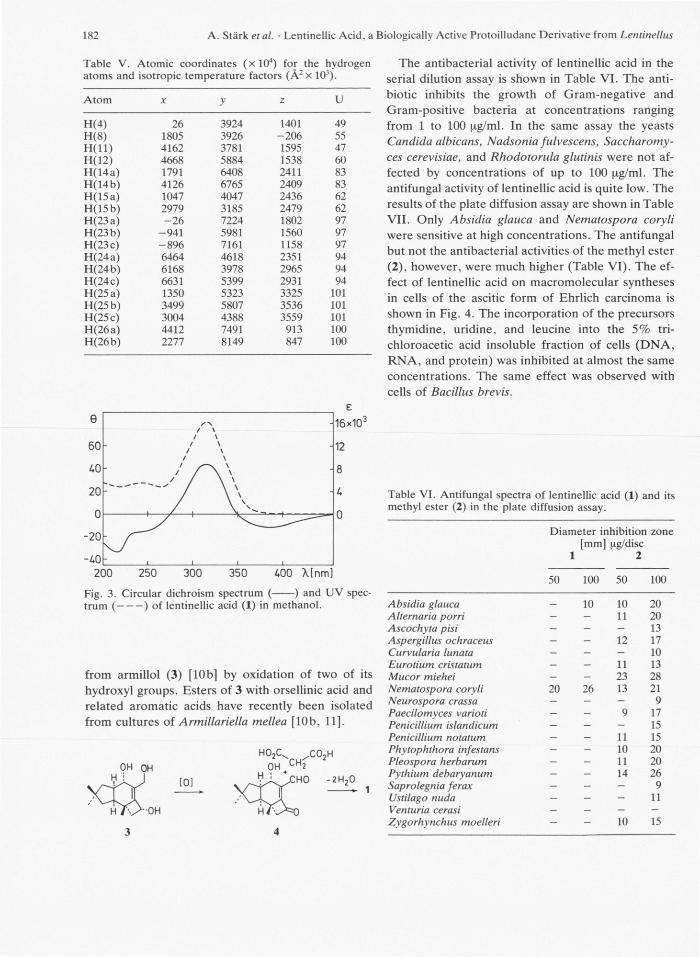
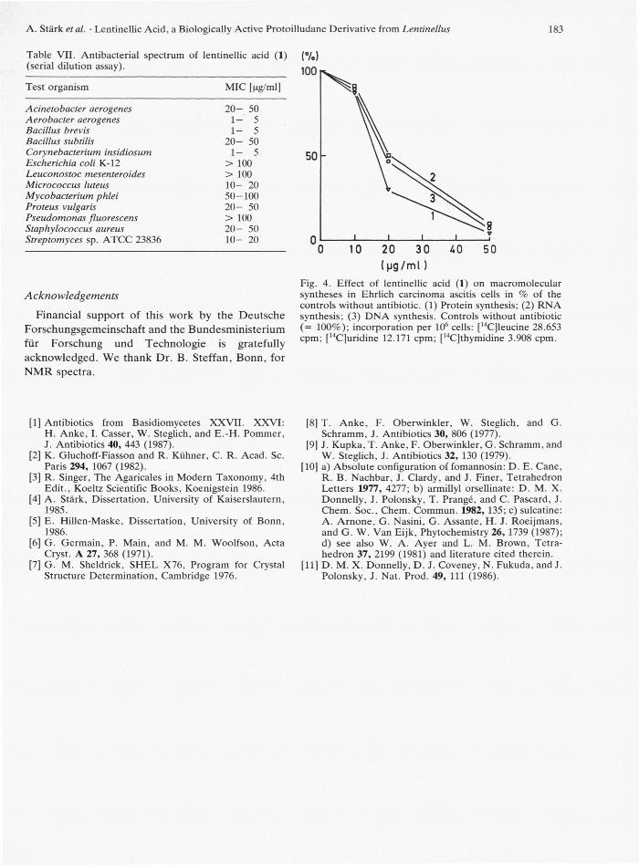
The antibacterial activity of lentinellic acid in the serial dilution assay is shown in Table VI. The anti-biotic inhibits the growth of Gram-negative and Gram-positive bacteria at concentrations ranging from 1 to 100 pg/ml. In the same assay the yeasts Candida albicans, Nadsonia fulvescens, Saccharomy-ces cerevisiae, and Rhodotorula glutinis were not af-fected by concentrations of up to 100 pg/ml. The antifungal activity of lentinellic acid is quite low. The results of the plate diffusion assay are shown in Table VII. Only Absidia glauca and Nematospora coryli were sensitive at high concentrations. The antifungal but not the antibacterial activities of the methyl ester (2), however, were much higher (Table VI). The ef-fect of lentinellic acid on macromolecular syntheses in cells of the ascitic form of Ehrlich carcinoma is shown in Fig. 4. The incorporation of the precursors thymidine, uridine, and leucine into the 5% tri-chloroacetic acid insoluble fraction of cells (DNA, RNA, and protein) was inhibited at almost the same concentrations. The same effect was observed with cells of Bacillus brevis.
Table VI. Antifungal spectra of lentinellic acid (1) and its methyl ester (2) in the plate diffusion assay.
Financial support of this work by the Deutsche Forschungsgemeinschaf t and the Bundesminis ter ium fü r Forschung und Technologie is gratefully acknowledged. We thank Dr . B. S te f fan , Bonn , for N M R spectra.
1 0 2 0 3 0 4 0 5 0
( p g / m l ) Fig. 4. Effect of lentinellic acid (1) on macromolecular syntheses in Ehrlich carcinoma ascitis cells in % of the controls without antibiotic. (1) Protein synthesis; (2) RNA synthesis; (3) DNA synthesis. Controls without antibiotic (= 100%); incorporation per 106 cells: [l4C]leucine 28.653 cpm; [I4C]uridine 12.171 cpm; [14C]thymidine 3.908 cpm.
[1] Antibiotics from Basidiomycetes XXVII. XXVI: H. Anke, I. Casser, W. Steglich, and E.-H. Pommer, J. Antibiotics 40, 443 (1987).
[2] K. Gluchoff-Fiasson and R. Kühner, C. R. Acad. Sc. Paris 294, 1067 (1982).
[3] R. Singer, The Agaricales in Modern Taxonomy, 4th Edit., Koeltz Scientific Books, Koenigstein 1986.
[4] A. Stärk, Dissertation, University of Kaiserslautern, 1985.
[5] E. Hillen-Maske, Dissertation, University of Bonn, 1986.
[6] G. Germain, P. Main, and M. M. Woolfson, Acta Cryst. A 27, 368 (1971).
[7] G. M. Sheldrick, SHEL X76, Program for Crystal Structure Determination, Cambridge 1976.
[8] T. Anke, F. Oberwinkler, W. Steglich, and G. Schramm, J. Antibiotics 30, 806 (1977).
[9] J. Kupka, T. Anke, F. Oberwinkler, G. Schramm, and W. Steglich, J. Antibiotics 32, 130 (1979).
[10] a) Absolute configuration of fomannosin: D. E. Cane, R. B. Nachbar, J. Clardy, and J. Finer, Tetrahedron Letters 1977, 4277; b) armillyl orsellinate: D. M. X. Donnelly, J. Polonsky, T. Prangé, and C. Pascard, J. Chem. Soc., Chem. Commun. 1982, 135; c) sulcatine: A. Arnone, G. Nasini, G. Assante, H. J. Roeijmans, and G. W. Van Eijk, Phytochemistry 26, 1739 (1987); d) see also W. A. Ayer and L. M. Brown, Tetra-hedron 37, 2199 (1981) and literature cited therein.
[11] D. M. X. Donnelly, D. J. Coveney, N. Fukuda, and J. Polonsky, J. Nat. Prod. 49, 111 (1986).