+ All Categories
Home > Documents > Isolated skin relapse of Philadelphia chromosome-positive ...

Isolated skin relapse of Philadelphia chromosome-positive ...

Date post: 20-Oct-2021
Category:
Upload: others
View: 3 times
Download: 0 times
Share this document with a friend
3
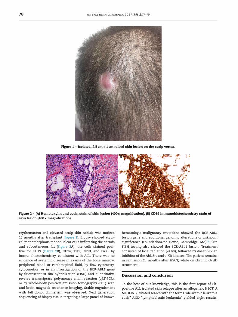
rev bras hematol hemoter. 2 0 1 7; 3 9(1) :77–79 www.rbhh.org Revista Brasileira de Hematologia e Hemoterapia Brazilian Journal of Hematology and Hemotherapy Case report Isolated skin relapse of Philadelphia chromosome-positive acute lymphoblastic leukemia after allogeneic stem cell transplant Masumi Ueda a,b , Carlos Silva a,b , Linda Baer a,b , Paolo F. Caimi a,b , Kevin Cooper a,b , Kord Honda a,b , Marcos de Lima a,b,a University Hospitals Seidman Cancer Center, Cleveland, United States b Case Western Reserve University, Cleveland, United States a r t i c l e i n f o Article history: Received 14 October 2016 Accepted 7 November 2016 Available online 30 December 2016 Introduction Leukemia cutis is a rare entity diagnosed in only 1–3% of T- and B-cell acute lymphoblastic leukemia (ALL). 1,2 Aleukemic leukemia cutis (ALC) is an even rarer diagnosis, occurring without leukemic cells in the blood or marrow, often preced- ing systemic disease. 2,3 The Philadelphia chromosome (Ph) is one of the most common chromosomal abnormalities in adult B-ALL patients and is associated with poor prognosis. Although the use of tyrosine kinase inhibitors (TKIs) target- ing the oncoprotein breakpoint cluster region-Abelson murine leukemia 1 (BCR-ABL1) has dramatically improved outcomes, allogeneic hematopoietic stem cell transplant (HSCT) is still recommended for all eligible patients, with relapse after HSCT remaining a major cause of treatment failure. 4 Herein we report a case of isolated skin relapse of Ph-positive pre-B cell ALL after allogeneic HSCT. Corresponding author at: University Hospitals Seidman Cancer Center, 11100 Euclid Avenue/SCC 1015, Cleveland, OH 44106, United States. E-mail address: [email protected] (M. de Lima). Case report A 26-year-old man received a matched related donor periph- eral blood HSCT for Ph-positive pre-B cell ALL in first remission. Prior to HSCT he had achieved complete molec- ular remission after two cycles of imatinib and the regimen rituximab with hyperfractionated cyclophosphamide, vin- cristine, doxorubicin, and dexamethasone alternating with methotrexate and cytarabine (R-HyperCVAD). Remission was consolidated with an allogeneic transplant from his human leukocyte antigen (HLA)-matched sibling using cyclophos- phamide and 12 Gy total body irradiation conditioning. Graft-versus-host disease (GvHD) prophylaxis consisted of tacrolimus and methotrexate. His post-HSCT course was com- plicated by chronic GvHD involving the lungs, liver, skin and lacrimal glands; he was treated with extracorporeal pho- topheresis, tacrolimus and prednisone. An isolated 2-cm http://dx.doi.org/10.1016/j.bjhh.2016.11.003 1516-8484/© 2016 Associac ¸˜ ao Brasileira de Hematologia, Hemoterapia e Terapia Celular. Published by Elsevier Editora Ltda. This is an open access article under the CC BY-NC-ND license (http://creativecommons.org/licenses/by-nc-nd/4.0/).
Transcript

C

Icl

MKa

b

a

A

R

A

A

I

LalwiiaAilarrrA

S

h1o

rev bras hematol hemoter. 2 0 1 7;3 9(1):77–79

www.rbhh.org

Revista Brasileira de Hematologia e HemoterapiaBrazilian Journal of Hematology and Hemotherapy

ase report

solated skin relapse of Philadelphiahromosome-positive acute lymphoblasticeukemia after allogeneic stem cell transplant

asumi Uedaa,b, Carlos Silvaa,b, Linda Baera,b, Paolo F. Caimia,b, Kevin Coopera,b,ord Hondaa,b, Marcos de Limaa,b,∗

University Hospitals Seidman Cancer Center, Cleveland, United StatesCase Western Reserve University, Cleveland, United States

r t i c l e i n f o

rticle history:

eceived 14 October 2016

ccepted 7 November 2016

vailable online 30 December 2016

tacrolimus and methotrexate. His post-HSCT course was com-

ntroduction

eukemia cutis is a rare entity diagnosed in only 1–3% of T-nd B-cell acute lymphoblastic leukemia (ALL).1,2 Aleukemiceukemia cutis (ALC) is an even rarer diagnosis, occurring

ithout leukemic cells in the blood or marrow, often preced-ng systemic disease.2,3 The Philadelphia chromosome (Ph)s one of the most common chromosomal abnormalities indult B-ALL patients and is associated with poor prognosis.lthough the use of tyrosine kinase inhibitors (TKIs) target-

ng the oncoprotein breakpoint cluster region-Abelson murineeukemia 1 (BCR-ABL1) has dramatically improved outcomes,llogeneic hematopoietic stem cell transplant (HSCT) is stillecommended for all eligible patients, with relapse after HSCT

4

emaining a major cause of treatment failure. Herein weeport a case of isolated skin relapse of Ph-positive pre-B cellLL after allogeneic HSCT.

∗ Corresponding author at: University Hospitals Seidman Cancer Centtates.

E-mail address: [email protected] (M. de Lima).ttp://dx.doi.org/10.1016/j.bjhh.2016.11.003516-8484/© 2016 Associacao Brasileira de Hematologia, Hemoterapiapen access article under the CC BY-NC-ND license (http://creativecom

Case report

A 26-year-old man received a matched related donor periph-eral blood HSCT for Ph-positive pre-B cell ALL in firstremission. Prior to HSCT he had achieved complete molec-ular remission after two cycles of imatinib and the regimenrituximab with hyperfractionated cyclophosphamide, vin-cristine, doxorubicin, and dexamethasone alternating withmethotrexate and cytarabine (R-HyperCVAD). Remission wasconsolidated with an allogeneic transplant from his humanleukocyte antigen (HLA)-matched sibling using cyclophos-phamide and 12 Gy total body irradiation conditioning.Graft-versus-host disease (GvHD) prophylaxis consisted of

er, 11100 Euclid Avenue/SCC 1015, Cleveland, OH 44106, United

plicated by chronic GvHD involving the lungs, liver, skin andlacrimal glands; he was treated with extracorporeal pho-topheresis, tacrolimus and prednisone. An isolated 2-cm

e Terapia Celular. Published by Elsevier Editora Ltda. This is anmons.org/licenses/by-nc-nd/4.0/).

78 rev bras hematol hemoter. 2 0 1 7;3 9(1):77–79

Figure 1 – Isolated, 2.5 cm × 1 cm raised skin lesion on the scalp vertex.

Figure 2 – (A) Hematoxylin and eosin stain of skin lesion (400× magnification). (B) CD19 immunohistochemistry stain of

skin lesion (400× magnification).

erythematous and elevated scalp skin nodule was noticed15 months after transplant (Figure 1). Biopsy showed atypi-cal monomorphous mononuclear cells infiltrating the dermisand subcutaneous fat (Figure 2A); the cells stained posi-tive for CD19 (Figure 2B), CD34, TDT, CD10, and PAX5 byimmunohistochemistry, consistent with ALL. There was noevidence of systemic disease in exams of the bone marrow,peripheral blood or cerebrospinal fluid, by flow cytometry,cytogenetics, or in an investigation of the BCR-ABL1 geneby fluorescent in situ hybridization (FISH) and quantitativereverse transcriptase polymerase chain reaction (qRT-PCR),

or by whole-body positron emission tomography (PET) scanand brain magnetic resonance imaging. Stable engraftmentwith full donor chimerism was observed. Next generationsequencing of biopsy tissue targeting a large panel of known

hematologic malignancy mutations showed the BCR-ABL1fusion gene and additional genomic alterations of unknownsignificance (FoundationOne Heme, Cambridge, MA).5 SkinFISH testing also showed the BCR-ABL1 fusion. Treatmentconsisted of local radiation (24 Gy), followed by dasatinib, aninhibitor of the Abl, Src and c-Kit kinases. The patient remainsin remission 25 months after HSCT, while on chronic GvHDtreatment.

Discussion and conclusion

To the best of our knowledge, this is the first report of Ph-positive ALL isolated skin relapse after an allogeneic HSCT. AMEDLINE/PubMed search with the terms “aleukemic leukemiacutis” AND “lymphoblastic leukemia” yielded eight results.

oter.

Oadtttrov

E

Toj

C

T

r

1

2

3

4

rev bras hematol hem

f these, only one case described ALC relapse of B-ALL afterllogeneic transplantation.3 Remarkably, our patient has noetectable systemic disease despite the availability of sensi-ive molecular tests. In addition, the translocation (9;22) andhe BCR-ABL1 gene were detected only in the skin, allowingargeted treatment of the relapse. Long-term prognosis of ALCecurrence after allogeneic HSCT and the potential benefitf tyrosine-kinase inhibitor therapy in addition to the graft-ersus-leukemia effects are unknown.

thical statement

his protocol was approved by the Institutional Review Boardf University Hospitals Cleveland Medical Center, and the sub-

ect gave written informed consent.

onflicts of interest

he authors declare no conflicts of interest.

5

2 0 1 7;3 9(1):77–79 79

e f e r e n c e s

. Campuzano-Garcia AE, Torres-Alvarez B, Castanedo-CazaresJP. Leukemia cutis in a patient with pre-B-cell acutelymphoblastic leukemia. JAAD Case Rep. 2015;1(6):364–7.

. Cho-Vega JH, Medeiros LJ, Prieto VG, Vega F. Leukemia cutis.Am J Clin Pathol. 2008;129(1):130–42.

. Ansell LH, Mehta J, Cotliar J. Recurrent aleukemic leukemiacutis in a patient with pre-B-cell acute lymphoblasticleukemia. J Clin Oncol. 2013;31(20):e353–5.

. Giebel S, Czyz A, Ottmann O, Baron F, Brissot E, Ciceri F, et al.Use of tyrosine kinase inhibitors to prevent relapse afterallogeneic hematopoietic stem cell transplantation forpatients with Philadelphia chromosome-positive acutelymphoblastic leukemia: a position statement of the AcuteLeukemia Working Party of the European Society for Blood andMarrow Transplantation. Cancer. 2016;122(19):2941–51.

. FoundationOne Heme Technical Information and TestOverview [cited 2016 July 24]. Available from:http://foundationone.com/docs/FM TechnicalOverview HEM-I-001-20150105.pdf.


Recommended