36
INFORMATIONEN ZUM SONNENVITAMIN D3

INFORMATIONENZUM SONNENVITAMIN D3

Die Wichtigkeit von Vitamin D3
Die fi nale Stufe von Vitamin D3 in der Zelle ist ein Hor-mon, welches Calcitriol genannt wird und zusammen mit dem Parathormon (PTH) zu den wichtigsten hormonellen Steuerelementen des Calcium- und Phosphathaushalts gehört. Das von der Nebenschilddrüse sezernierte Parathormon, welches beim Absinken des Calciumspie-gels freigesetzt wird, führt indirekt zur Aktivierung der Osteoklasten („Knochenfresszellen“) und zur Mobilisie-rung von Calcium und Phosphat aus dem Knochenge-webe. Die Folge ist ein erhöhter Calciumspiegel im Blut und ein erniedrigter Gehalt an Mineralien in den Knochen (Ostepenie, Osteoporose). Die Synthese und Ausschüt-tung von PTH wird durch Calcitriol gehemmt. Calcitriol vermindert somit die Ausscheidung von Calcium aus den Nieren und erhöht das zur Verfügung stehende Calcium durch Absorption im Dünndarm. Daraus folgt die erhöhte Osteoblastenaktivität, also die Fähigkeit gesunden neuen Knochen zu bilden.
Eine Studie (vgl. Choukroun et al 2014) belegt die Bedeu-tung von Vitamin D3 hinsichtlich des Knochenaufbaus, von welchem die Einheilung von Implantaten abhängt. 1,25-(OH)2-VitaminD3 (= Calcitriol) ist das wichtigste Hormon, welches in die Knochenbildung involviert ist und gleichzeitig die Entzündungsbereitschaft reduziert. i Ein Mangel an Vitamin D3 hemmt die Einheilung von Implan-taten und erhöht das Infektionsrisiko. ii
Nachgewiesen ist ebenfalls eine anti-entzündliche Wir-kung auf das Zahnfl eisch und den Zahnhalteapparat. Aktiviertes Vitamin D3 stimuliert die Bildung antimikro-bieller Peptide an Haut und Schleimhaut und hat somit eine antibakterielle und antientzündliche Wirkung iii
(Hieremath, 2013). Eine Studie aus dem Jahr 2016 (vgl. Woelber et al.) zeigt, dass durch eine kohlenhydratarme Ernährung mit gleichzeitig ausreichender Deckung des Bedarfs an Omega 3 Fettsäuren, Ballaststoff en, Vitamin C und D sowie Antioxidantien Zahnfl eisch- und Zahnbett-entzündungen verhindert werden können. Die Parodonti-tis muss daher heute nicht mehr chirurgisch behandelt werden, sondern kann mit der Deckung benötigter Vitamine und Mineralstoff en vorgebeugt und behandelt werden. Bereits eine Studie aus dem Jahr 2012 (vgl. F.R. Teles et al.) zeigte, dass Patienten mit hohem Vitamin D Spiegel deutlich weniger Zahnfl eischbluten, geringere Taschentiefen und weniger Zahnverlust verzeichneten. v
Neben der Wichtigkeit für den Kalziumstoff wechsel und somit auch für den Knochenaufbau hat Vitamin D3 immunologische und metabolische Eff ekte auf unseren Körper. Autoimmunerkrankungen wie Multiple Sklerose, Arthritis und Diabetes treten gehäuft bei niedrigem D3 Spiegel auf. vi Bei ausreichender Vitamin D3 Bildung wird die erworbene (im Falle von Autoimmunerkankungen überaktive) Immunantwort nach unten, und die angebo-rene unspezifi sche Immunantwort nach oben reguliert. Rezeptoren für Vitamin D3 sind in einigen Zelltypen unseres Immunsystems zu fi nden wie in T-Lymphozyten und T-Helfer Zellen. Die Ausschaltung dieser Rezeptoren führte in Versuchen zu Ausbrüchen von entzündlichen Darmerkrankungen. vii U.a. werden durch Vitamin D3 auch AMPs (antimikrobielle Proteine) gestärkt. Diese AMPs töten Mikroorganismen, also Bakterien und Viren, oft schneller und eff ektiver ab, als das erworbene Immunsystem mit der Aktivierung von spezialisierten Abwehrzellen.

Die belegte Gripperesistenz durch ausreichend Vitamin D3 beruht auf der Hemmung des NFκB Transkriptase-faktors. Der nukleäre Faktor kappa B ist ein Protein, welches durch Zellstress aktiviert wird und sowohl eine Entzündungskaskade als auch die Bildung freier Radikale hervorruft. Vitamin D3 spielt somit eine regulierende Rolle im Rahmen von Zellstress-Reaktionen, vorausgesetzt es ist ein ausreichender Vorrat von 25-Hydroxyvitamin D3 (Speicherform des Vitamin D3) vorhanden.Ebenso ist die vorbeugende Wirkung von Vitamin D hinsichtlich Herzinfarkt, Krebserkrankungen und chronischer Müdigkeit, ausgelöst durch die permanente Aktivierung des NFκB, gesichert. D3 hilft somit, die Patienten in den Parasympathikus zu bringen. Es sorgt für einen gesunden Schlaf und notwendige Entspannung.
Vitamin D3 wird zu 80% in der Haut gebildet. Für die Umwandlung des in der Haut vorkommenden 7-Dehydro-cholesterol wird UVB-Strahlung benötigt, um es durch Photolyse in das Prävitamin D3 umzuwandeln. viii Dieses Prävitamin wird durch thermische Isomerisierung in das Vitamin D3 (Cholecalciferol) überführt. Nach 8 Stunden sind 80% des Prävitamins in der Haut umgewandelt. Sobald das Vitamin D3 in die Blutbahn gelangt, wird es mithilfe des Vitamin-D-bindenden Proteins (DBP) zur Leber transportiert und dort zu 25-OH-Vitamin D3 (Calcidiol) hydroxyliert. Calcidiol ist eine Speicherform des Vitamins D3. Die Umwandlung zum aktiven Steroid-hormon Calcitriol erfolgt dann weiter in der Niere.Der Gehalt an 7-Dehydrocholesterolgehalt in der Haut nimmt im Alter zunehmend ab. Auch die Fähigkeit bei älteren Menschen D3 in der Haut zu bilden, ist im Gegen-satz zu einer 20- jährigen Person um den Faktor 3 reduziert.
Bei Anwendung von Sonnencreme oder Tagescreme mit Lichtschutzfaktor reicht ein LSF 8 aus, um die Vitamin D3 Produktion um mehr als 97% zu behindern. Nach wissen-schaftlichen Erkenntnissen des Karolinska-Institutes in Stockholm über 20 Jahre und mehr als 30.000 Probanden ist Sonnencreme nachweislich für die Entste-hung von Hautkrebs verantwortlich (Dr. Elizabeth Plourde: Sunscreens-Biohazard: Treat as hazardous waste). Zudem schädigt nanopartikuläres Titandioxid, welches in Son-nencreme enthalten ist, die DNA und fördert die Ent-stehung von Alzheimer, Epilepsie und Autismus. ix Das ebenfalls enthaltende nanopartikuläre Zinkoxid steht im Verdacht Darm- und Hirnstammzellen abzutöten. x
Da Sonnencreme durch das enthaltene Oxybenzone und Octinozate das Ökosystem der Korallenriffe bedroht, hat der US Bundesstaat Hawaii als erster amerikanischer Staat den Verkauf von Sonnenschutzmitteln verboten. xi Interessanterweise sprechen die Presseartikel die fatale Wirkung der beiden Supergifte auf die Korallen an, jedoch mit keinem Wort die Wirkung auf den Menschen, welcher sich diese Supergifte mehrmals am Tag auf eines der besten Resorptionsorgane (die menschliche Haut) mit einer Fläche von 1,5 – 2 Quadratmeter einreibt...
20% der Aufnahme an Vitamin D erfolgt durch die Nahrung. xii Fettreiche Fischarten wie Lachs und Hering
weisen einen hohen Anteil auf, ebenso Milch, Steinpilze, Shiitake-Pilze und Avocados. Generell ist jedoch ein zunehmender Verlust an Mineralien und Vitaminen in allen Obst- und Gemüsesorten zu verzeichnen. Durch ausge-laugte Böden, Luftverschmutzung, moderne Verarbei-tungsmethoden und Lagerung zeigte sich innerhalb 50 Jahren ein drastischer Verlust an wertvollen Inhaltsstoffen in unserer Nahrung. xiii Man müsste heute zehn Mal so viel Obst und Gemüse zu sich nehmen, um denselben Gehalt an Nährstoffen wie vor 50 Jahren zu erhalten.
Durch den heutigen Lebenswandel und überwiegenden Aufenthalt in geschlossenen Räumen weist die Mehrheit der Bevölkerung heute einen Vitamin-D-Mangel auf. Wichtig ist zu wissen, dass in den Ländern, die nördlich des 40. Breitengrades liegen (in Europa nördlich von Rom), in den Monaten Oktober bis März nicht ausreichend Vitamin D gebildet werden kann. Die Aufnahme von UV-B Strahlung hängt von der Bewölkung und vom Einfallswin-kel der Sonne ab. Ist der Winkel geringer als 45° ist der Weg für die Sonnenstrahlen durch die Ozonschicht zu lang, um noch Vitamin D produzieren zu können, da die Ozonschicht ein Teil der UV-Strahlung absorbiert. Auf der Website www.timeanddate.com kann man die Sonnenstunden mit Einfallswinkel an beliebigen Orten der Welt nachverfolgen. Zum Beispiel gab es am 11. Januar 2018 in Oslo (40. Breitengrad) zu keiner Tageszeit einen Sonneneinstrahlungswinkel von über 45°. In Tel Aviv hingegen (32. Breitengrad) konnte man am 11. Januar zwischen 9:28 Uhr und 16:03 Uhr optimal Vitamin D produzieren. Für Mobiltelefone ist eine App verfügbar (Dminder von Prof. Molick) welche präzise anzeigt wie viele I.E. Vitamin D zu welcher Tageszeit innerhalb welcher Zeit gebildet werden kann. Es gibt eine einfache Faustregel, die man sich hierzu merken kann: Wenn der Schatten länger als die Körpergröße ist, findet keine Vitamin D Produktion statt. Für die Produktion von Vitamin D ist ausschließlich die UV-B - Strahlung verant-wortlich, die den geringeren Anteil der UV- Strahlung ausmacht. Die längeren UV-A Strahlen dringen tiefer in die Haut ein und sind für mögliche Zellschädigung und Hautalterung verantwortlich.
Durch stressige Lebensumstände, die zur systemischen Azidose und dadurch zur Resorption von Calcium aus dem Knochen führt, um den Blut-PH-Wert auf 7,4 abzu-puffern, wird dem Körper ein ausreichend hoher D3-Spiegel simuliert, der ebenso für den Mangel an D3 verantwortlich ist. Da Vitamin D3 das Immunsystem unterstützt, kann ein Mangel vielfältige Auswirkungen haben. Neben Konzentrations- und Herz- Kreislauf-störungen kann es zu reduzierter Muskelstärke, Wachs-tumsstörungen, Osteomalazie, Immunschwäche, Schlaf-störungen, Nervosität, Depressionen, Zahnausfall und erhöhter Frakturanfälligkeit kommen. Auch Multiple Sklerose, Asthma und Krebs werden in Zusammenhang mit einem Mangel an Vitamin D3 gebracht. Eine Studie aus dem Jahr 2016 von Yehuda Shoenfeld (vgl. Lindqvist et al 2016) wies bereits darauf hin, dass Meidung des Sonnenlichts als Risikofaktor für einen frühzeitigen Tod auf gleicher Stufe mit dem Rauchen steht. xiv Auch wurde festgestellt, dass die Verbreitung von chronischen

Erkrankungen wie Diabetes und Multiple Sklerose mit der Distanzierung zum Äquator und somit der Sonnenein-strahlung und Aufenthalt im Freien ansteigt.
Die u.a. Übersichts-Metastudie zeigt, dass Patienten mit einem Serumlevel der Speicherform 25-OH-Vitamin D3 von über 60 ng/ml zu etwa 85% vor den meisten chroni-schen Erkrankungen geschützt sind!
Die im Blut getestete Speicherform 25-OH-D gibt Aus-kunft über einen Mangel an Vitamin D3. Auch Röntgenbil-der können Auskunft über Vitamin D3 Mangel geben: Bei Patienten mit schwerem Vitamin D3 Mangel sind die Pulpahörner asymmetrisch und verengt und erinnern optisch an einen Stuhl mit harter Lehne. Gesunde Pulpa ähnelt einem runden Bogen mit breiteren Pulpahörnern. xv
An einem Sonnentag am Äquator bildet der Mensch etwa 20.000 I.E. (Internationale Einheiten) D3. Die empfohlene Tagesdosis wurde in Deutschland erst im Jahre 2015 von 400 Einheiten auf 1.000 Einheiten erhöht. Nachdem Wissenschaftler bestätigten, dass die niedrigen Einnah-meempfehlungen für Vitamin D auf einen Rechenfehler um den Faktor 10 zurückzuführen sind (vgl Veugelers et al 2014) forderten sie die Vitamin D Tagesdosis von mindes-tens 7.000 I.E. bekannt zu geben. Wir gehen davon aus, dass eine schützende Dosis bei 20.000 I.E. pro Tag liegt und den Patienten optimal auf einen chirurgischen Eingriff vorbereiten sollte. Mit dieser Pauschaldosierung von
20.000 I.E./Tag erreichen wir bei einer Verordnung vier Wochen vor dem OP-Termin eine Blutkonzentration von rund 70 bis 120 ng D3 / ml. Dies entspricht etwa dem Vitamin D3-Level einer Person, welche in der Äquatorre-gion lebt. Damit ist der Patient optimal auf eine Operation vorbereitet. 85 % aller Deutschen liegen sogar noch unter dem deutschen Soll-Wert von 30 ng D3 / ml, was bedeu-tet, dass sie sich im „Immunlogischen Winterschlaf“ befi nden und nicht in der Lage sein werden, Knochen und Wunden vollständig und komplikationslos ausheilen lassen zu können.
Bei einer Langzeitanwendung ist es wichtig, die Einnahme von Vitamin D3 mit Vitamin K2-mk7 zu kombinieren, da Vitamin D3 das Vitamin K2 verbraucht und hohe Calcium-spiegel im Blut vermieden werden sollten.

Ein K2-Mangel kann sich u.a. in Herzbeschwerden äußern. Durch die Kombination von D3 mit K2-mk7 wird auch der bei Überdosierung von D3 auftretenden Hyperkalzämie vorgebeugt. Das Verhältnis von Vitamin D3 zu K2/mk7 sollte 10.000 I.E. D3 zu 100 µg K2-mk7 sein. Um Vitamin D in das aktive Vitamin D Hormon umzuwandeln und für den weiteren Transport im Körper, ist vor allem Magne-sium nötig. Ein Mangel an Magnesium würde den gesamten Haushalt von PTH, Kalzium und Vitamin D blockieren. Für die Proteinsynthese und Aktivierung einiger Gene ist zusätzlich Vitamin A in ausgewogener Konzentration zu Vitamin D erforderlich. Wenn das Verhältnis unausgeglichen ist, verhalten sich die Vitamine wie Gegenspieler und die Wirkung von Vitamin D wird beeinträchtigt.
Auch ein Zink-Mangel würde die Funktion von Vitamin D einschränken. Zink wird benötigt, um die Vitamin-D-Re-zeptoren, die sich an fast allen Zellen befi nden, zu bilden. Die Balance von D3 und K2 sowie der weiteren Co-Fakto-ren ist durch Dr. Klinghardt und Dr. Volz optimal im BASIC IMMUNE aufeinander abgestimmt. Die Einnahme sollte bereits 4 Wochen vor dem chirurgischen Eingriff begonnen werden.
Sogar die deutsche Olympiamannschaft im Segeln nimmt BASIC IMMUNE ein, um sich optimal auf die Olympiade vorzubereiten und stellt eine enorme Leis-tungssteigerung und schnellere Regenerationszeit fest:
Jan Jasper Wagner und Julian Authenried: „We tried out Basic Immune during and after the competition and could immediately identify constant and long-lasting energy, attention and just a general feeling of well-being, even after 6 days of hard work. Usually, right after the compe-tition the body needs some time to rest where all the systems shut down, but with Basic Immune this process could be reconciled. Furthermore, Basic Immune is incredible easy to transport and to take in!In my opinion, this is one of the great strengths of Basic Immune, as usually it needs a lot of discipline to force oneself to take in all the diff erent supplements”.
Es ist in unserer Zeit und gerade auch in unseren weit vom Äquator entfernten Lebensbereichen mit hohem Stresslevel nicht möglich, den für unsere Gesundheit notwendigen Vitamin D3 Spiegel durch ausreichenden Aufenthalt in der Sonne zu erreichen. Auch wenn die natürliche Sonneneinstrahlung optimal wäre, können wir heutzutage nicht mehr auf die Einnahme von Vitamin D verzichten, um uns vor akuten und chronischen Krankhei-ten zu schützen und optimale Langzeitprognosen für Keramikimplantate garantieren zu können.
Quellenangabe
i Choukroun, Joseph, et al. Two neglected biologic risk factors in bone grafting and implantology: high low-density lipoprotein cholesterol and low serum vitamin D. Journal of Oral Implantology, 2014, 40. Jg., Nr. 1, S. 110-114
ii Two Neglected Biologic Risk Factors in Bone Grafting and Implantology: High Low- Density Lipoprotein Cholesterol and Low Serum Vitamin D
iii Hieremath, V. e. Anti-inflammatory effect of vitamin D on gingivitis: a dose-response randomised control trial. Oral Health Prev Dent, 2013
iv Woelber, J. P. et al. “An Oral Health Optimized Diet Can Reduce Gingival and Periodontal Inflammation in Humans - a Randomized Controlled Pilot Study.” BMC Oral Health 17 (2017): 28.
v Teles, F.R. et al. “Relationships Among IL-6, TNF-Α, Adipokines, Vitamin D and Chronic Periodontitis.” Journal of periodontology 83.9 (2012): 1183–1191.
vi http://www.vitamind.net/interviews/coimbra-ms-autoimmun/
vii http://immunendokrinologie.de/html/vitamin_d.html
viii Environmental factors that influence the cutaneous production of vitamin D M F Holick The American Journal of Clinical Nutrition, Volume 61, Issue 3, 1 March 1995
ix Lin, W., Xu, Y., Huang, CC. et al. J Nanopart Res (2009) 11: 25. https://doi.org/10.1007/s11051-008-9419-7
x https://www.mayr-kuren.de/sonne-sonnenschutz.html
xi https://www.theguardian.com/travel/2018/may/03/hawaii-becomes-first-us-state-to-ban-sunscreens-harm ful-to-coral-reefs
xii Natalie J. Lauer: Gesundheit, Vitalität & Lebensfreude. Gesund mit veganer Ernährung. Hg. v. Eberhard J. Wormer u. Johann A. Bauer. Lingen, Köln 2015
xiii Mayer, Anne-Marie. (1997). Historical changes in the mineral content of fruits and vegetables. British Food Journal. 99. 207-211. 10.1108/00070709710181540.
xiv Journal of Internal Medicine Volume 280, Issue 4 Original Article Avoidance of sun exposure as a risk factor for major causes of death: a competing risk analysis of the Mela noma in Southern Sweden cohort
xv https://www.zm-online.de/news/zahnmedizin/wie-der-zahnarzt-einen-vitamin-d-mangel- diagnostiziert/
Literaturverzeichnis
• CHOUKROUN, Joseph, et al. Two neglected biologic risk factors in bone grafting and implantology: high low-density lipoprotein cholesterol and low serum vitamin D. Journal of Oral Implantology, 2014, 40. Jg., Nr. 1, S. 110-114.
• SCHULZE-SPÄTE, Ulrike, et al. Systemic vitamin D supplementation and local bone formation after maxillary sinus augmentation–a randomized, double-blind, placebo-controlled clinical investigation. Clinical oral implants research, 2015.
• BRYCE, G.; MACBETH, N. Vitamin D deficiency as a suspected causative factor in the failure of an immediately placed dental implant: a case report. Journal of the Royal Naval Medical Service, 2013, 100. Jg., Nr. 3, S. 328-332.
• COOPER, Lyndon F. Systemic effectors of alveolar bone mass and implications in dental therapy. Periodontology 2000, 2000, 23. Jg., Nr. 1, S. 103-109. Cannell JJ et al., „Epidemic influenza and vitamin D“, Epidemiol Infect. 2006 Dec;134(6):1129-40. (Grippe-Epidemie und Vitamin D) [Quelle als PDF]
• Berry DJ et al., „Vitamin D status has a linear association with seasonal infections and lung function in British adults“, Br J Nutr. 2011 Nov;106(9):1433-40. (Der Vitamin-D-Status hat einen linearen Zusam menhang mit saisonalen Infektionen und Lungenfunktion bei britischen Erwachsenen.) [Quelle als PDF]
• Litonjua AA. „Childhood asthma may be a consequence of vitamin D deficiency“, Curr Opin Allergy Clin Immunol. 2009 Jun;9(3):202-7. (Asthma in der Kindheit kann eine Folge von Vitamin-D-Mangel sein.) [Quelle als PDF]
• Forman JP et al., „Plasma 25-hydroxyvitamin D levels and risk of incident hypertension“, Hypertension. 2007 May;49(5):1063-9. (Plasma 25-Hydroxy-Vitamin-D-Spiegel und das Risiko von Bluthochdruck.) [Quelle als PDF]
• Anderson JL et al., „Relation of vitamin D deficiency to cardiovascular risk factors, disease status, and incident events in a general healthcare population.“, Am J Cardiol. 2010 Oct 1;106(7):963-8. (Zusam menhang zwischen Vitamin-D-Mangel und kardiovaskulären Risikofaktoren, Krankheits Status und Vorfallereignisse in der allgemeinen Bevölkerung, die Gesundheitsvorsorge betreibt.) [Quelle als PDF]
• Kuloğlu O et al., „Serum 25-hydroxyvitamin d level is associated with arterial stiffness, left ventricle hypertrophy, and inflammation in newly diagnosed hypertension.“, J Investig Med. 2013 Aug;61(6):989- 94. (Der Serum 25-Hydroxy-Vitamin-D-Spiegel ist mit arterieller Steifigkeit, linker Herzkammer Hypertrophie und Entzündungen bei neu diagnostiziertem Bluthochdruck assoziiert.) [Quelle als PDF]
• Ananthakrishnan AN. „Environmental risk factors for inflammatory bowel disease“, Gastroenterol Hepatol (N Y). 2013 Jun;9(6):367-74. (Umwelt-Risikofaktoren für entzündliche Darmerkrankungen) [Quelle als PDF]
• Miznerova E et al., „The prevalence and risk factors for osteoporosis in patients with inflammatory bowel disease“, Bratisl Lek Listy. 2013;114(8):439-45. (Die Prävalenz und Risikofaktoren für Osteopo rose bei Patienten mit chronisch entzündlichen Darmerkrankungen.) [Quelle als PDF]
• Szep Z et al., „Vitamin D deficiency is associated with type 2 diabetes mellitus in HIV infection“, AIDS. 2011 Feb 20;25(4):525-9. (Vitamin D-Mangel ist mit Typ-2-Diabetes mellitus bei HIV-Infektion) [Quelle als PDF]
• Gombart AF. „The vitamin D-antimicrobial peptide pathway and its role in protection against infec tion“, Future Microbiol. 2009 Nov;4(9):1151-65. (Der Vitamin-D-antimikrobielles Peptid Stoffwechsel weg und seine Rolle beim Schutz gegen eine Infektion.) [Quelle als PDF]
• Dale BA et al., „Oral antimicrobial peptides and biological control of caries“, BMC Oral Health. 2006 Jun 15;6 Suppl 1:S13. (Orale antimikrobielle Peptide und biologische Bekämpfung von Karies.) [Quelle als PDF]

• Sabbagh Z et al., „Vitamin D status is associated with disease activity among rheumatology outpa tients“, Nutrients. 2013 Jun 26;5(7):2268-75. (Vitamin-D-Status wird mit der Krankheitsaktivität bei ambulanten Rheumatologie Patienten verbunden.) [Quelle als PDF]
• Di Rosa M et al., „Vitamin D status is associated with disease activity among rheumatology outpa tients“, Crit Rev Oncol Hematol. 2013 Aug 10. pii: S1040-8428(13)00167-4. (Vitamin D3 Mangel und Darmkrebs) [Quelle als PDF]
• Rasheed H „Serum ferritin and vitamin d in female hair loss: do they play a role?“ Skin Pharmacol Physiol. 2013;26(2):101-7. (Serum-Ferritin und Vitamin D bei weiblichem Haarausfall: spielen sie eine Rolle?) [Quelle als PDF]

Studien
• CHOUKROUN, Joseph, et al. Two neglected biologic risk factors in bone grafting and implantology: high low-density lipoprotein cholesterol and low serum vitamin D. Journal of Oral Implantology, 2014, 40. Jg., Nr. 1, S. 110-114.
• SCHULZE-SPÄTE, Ulrike, et al. Systemic vitamin D supplementation and local bone formation after maxillary sinus augmentation–a randomized, double-blind, placebo-controlled clinical investigation. Clinical oral implants research, 2015.
• BRYCE, G.; MACBETH, N. Vitamin D deficiency as a suspected causative factor in the failure of an immediately placed dental implant: a case report. Journal of the Royal Naval Medical Service, 2013, 100. Jg., Nr. 3, S. 328-332.
• COOPER, Lyndon F. Systemic effectors of alveolar bone mass and implications in dental therapy. Periodontology 2000, 2000, 23. Jg., Nr. 1, S. 103-109.
• Cannell JJ et al., „Epidemic influenza and vitamin D“, Epidemiol Infect. 2006 Dec;134(6):1129-40. (Grippe-Epidemie und Vitamin D) [Quelle als PDF]
• Berry DJ et al., „Vitamin D status has a linear association with seasonal infections and lung function in British adults“, Br J Nutr. 2011 Nov;106(9):1433-40. (Der Vitamin-D-Status hat einen linearen Zusam menhang mit saisonalen Infektionen und Lungenfunktion bei britischen Erwachsenen.) [Quelle als PDF]
• Litonjua AA. „Childhood asthma may be a consequence of vitamin D deficiency“, Curr Opin Allergy Clin Immunol. 2009 Jun;9(3):202-7. (Asthma in der Kindheit kann eine Folge von Vitamin-D-Mangel sein.) [Quelle als PDF]
• Forman JP et al., „Plasma 25-hydroxyvitamin D levels and risk of incident hypertension“, Hypertension. 2007 May;49(5):1063-9. (Plasma 25-Hydroxy-Vitamin-D-Spiegel und das Risiko von Bluthochdruck.) [Quelle als PDF]
• Anderson JL et al., „Relation of vitamin D deficiency to cardiovascular risk factors, disease status, and incident events in a general healthcare population.“, Am J Cardiol. 2010 Oct 1;106(7):963-8. (Zusam menhang zwischen Vitamin-D-Mangel und kardiovaskulären Risikofaktoren, Krankheits Status und Vorfallereignisse in der allgemeinen Bevölkerung, die Gesundheitsvorsorge betreibt.) [Quelle als PDF]
• Kuloğlu O et al., „Serum 25-hydroxyvitamin d level is associated with arterial stiffness, left ventricle hypertrophy, and inflammation in newly diagnosed hypertension.“, J Investig Med. 2013 Aug;61(6):989- 94. (Der Serum 25-Hydroxy-Vitamin-D-Spiegel ist mit arterieller Steifigkeit, linker Herzkammer Hypertrophie und Entzündungen bei neu diagnostiziertem Bluthochdruck assoziiert.) [Quelle als PDF]
• Ananthakrishnan AN. „Environmental risk factors for inflammatory bowel disease“, Gastroenterol Hepatol (N Y). 2013 Jun;9(6):367-74. (Umwelt-Risikofaktoren für entzündliche Darmerkrankungen) [Quelle als PDF]
• Miznerova E et al., „The prevalence and risk factors for osteoporosis in patients with inflammatory bowel disease“, Bratisl Lek Listy. 2013;114(8):439-45. (Die Prävalenz und Risikofaktoren für Osteopo rose bei Patienten mit chronisch entzündlichen Darmerkrankungen.) [Quelle als PDF]
• Szep Z et al., „Vitamin D deficiency is associated with type 2 diabetes mellitus in HIV infection“, AIDS. 2011 Feb 20;25(4):525-9. (Vitamin D-Mangel ist mit Typ-2-Diabetes mellitus bei HIV-Infektion) [Quelle als PDF]
• Gombart AF. „The vitamin D-antimicrobial peptide pathway and its role in protection against infec tion“, Future Microbiol. 2009 Nov;4(9):1151-65. (Der Vitamin-D-antimikrobielles Peptid Stoffwechsel weg und seine Rolle beim Schutz gegen eine Infektion.) [Quelle als PDF]
• Dale BA et al., „Oral antimicrobial peptides and biological control of caries“, BMC Oral Health. 2006 Jun 15;6 Suppl 1:S13. (Orale antimikrobielle Peptide und biologische Bekämpfung von Karies.) [Quelle als PDF]
• Sabbagh Z et al., „Vitamin D status is associated with disease activity among rheumatology outpa tients“, Nutrients. 2013 Jun 26;5(7):2268-75. (Vitamin-D-Status wird mit der Krankheitsaktivität bei ambulanten Rheumatologie Patienten verbunden.) [Quelle als PDF]
• Di Rosa M et al., „Vitamin D status is associated with disease activity among rheumatology outpa tients“, Crit Rev Oncol Hematol. 2013 Aug 10. pii: S1040-8428(13)00167-4. (Vitamin D3 Mangel und Darmkrebs) [Quelle als PDF]
• Rasheed H „Serum ferritin and vitamin d in female hair loss: do they play a role?“ Skin Pharmacol Physiol. 2013;26(2):101-7. (Serum-Ferritin und Vitamin D bei weiblichem Haarausfall: spielen sie eine Rolle?) [Quelle als PDF]

Two Neglected Biologic Risk Factors in Bone Grafting andImplantology: High Low-Density Lipoprotein Cholesteroland Low Serum Vitamin DJoseph Choukroun1*Georges Khoury, DDS2
Fouad Khoury, MD, PhD3
Philippe Russe, DDS4
Tiziano Testori, MD5
Yataro Komiyama, DDS, PhD6
Gilberto Sammartino, MD, PhD7
Patrick Palacci, DDS8
Mustafa Tunali, DDS, PhD9
Elisa Choukroun10
Following a failure of a bone graft or an implant placement, the hypothesis of a biological abnormality is rarely
considered as a possible cause. A systematic search of peer-reviewed literature for dyslipidemia or vitamin D
deficiency may explain this lack of consideration. Excess low-density lipoprotein cholesterol (dyslipidemia) is
responsible for a slower bone metabolism or lower dental implant osseointegration. In addition, vitamin D is a
key factor for linking innate and adaptive immunity. Both of these factors are compromised under the
conditions of vitamin D deficiency. Therefore, vitamin D deficiency slows implant osseointegration and increases
the risk of graft infection. Vitamin D is also involved in immune function and therefore allergic reactions.
Key Words: cholesterol, LDL cholesterol, vitamin D, failures, implants, bone grafts, infections, immunedefense, osseointegration
INTRODUCTION
The search for a biological anomaly
labeled as a risk factor before oral
surgery is limited to disease states such
as diabetes. However, it seems in recent
years that cholesterol and vitamin D
levels should be more systematically investigated.Good cholesterol (high-density lipoprotein [HDL]) andbad cholesterol (low-density lipoprotein [LDL]) needto be included in this investigation because bothcould have a negative effect on bone growth andosseointegration (high LDL or low HDL). Vitamin D isone the most important hormones involved in bonegrowth. In addition, vitamin D also plays a role inreducing the effects of inflammation and helpsimprove the body’s natural immune reactions.
DYSLIPIDEMIA
LDL cholesterol and bone metabolism
Cholesterol is transported in the plasma predomi-nantly as cholesteryl esters associated with lipopro-
1 Pain Clinic, Nice, France.2 University Paris VII, Paris, France.3 Munster University, Olsberg, Germany.4 Private practice, Reims, France.5 Galeazzi Institute, Universita di Milano, Milan, Italy.6 Branemark Osseointegraion Center, Tokyo, Japan.7 University Frederico 2, Napoli, Italy.8 Branemark Osseointegration Center, Marseille, France.9 Haidarpasa Hospital, Istanbul, Turkey.10 University of Nice, France.* Corresponding author, e-mail: [email protected]: 10.1563/AAID-JOI-D-13-00062
110 Vol. XL/No. One/2014
LITERATURE REVIEW

328J Royal Naval Medical Service 2014, Vol 100.3
Case ReportVitamin D defi ciency as a suspected causative factor in the failure of an immediately placed dental implant: a case report
Surg Lt Cdr G Bryce, Wg Cdr N MacBeth
Abstract
Aim To discuss the infl uence of Vitamin D defi ciency in the osseointegration process of a dental implant by way of a case report.
Summary A 29-year-old soldier attended clinic with a fractured mandibular premolar (tooth 44) that was traumatised following head trauma related to the detonation of an Improvised Explosive Device (IED) whilst serving on operational duty. The tooth was deemed unsalvageable and was extracted with immediate placement of a dental implant. The patient experienced no problems but at assessment, fi ve months post-operatively, no osseo-integration of the implant was found. Concurrent medical investigations revealed that he was severely Vitamin D defi cient and that this may have contributed to the implant failure.
ConclusionVitamin D defi ciency may play a role in the failure of osseointegration in dental implants. The assessment of vitamin D status in patients who have been in long-term hospital care or rehabilitation should be considered, prior to the placement of dental implants.
Case report A 29-year-old soldier was referred to the Centre for Restorative Dentistry with a painful right mandibular fi rst premolar (LR4) that had been fractured following head trauma relating to the explosion of an Improvised Explosive Device (IED). LR4 suffered a crown fracture that led to an irreversible pulpitis.
Associated injuries included: fractures to second, third and fourth lumbar vertebrae; left third to eighth ribs, left clavicle, left radius, left femur, and right medial malleolus. He also suffered a left pneumothorax and a minor traumatic brain injury. His right ankle had been fused as a component of his stabilisation treatment and the patient had spent approximately twelve months confi ned, predominantly indoors, within a rehabilitative facility. The patient was a non-smoker, did not drink alcohol and was motivated to maintaining good oral health.
The examination of the LR4 (Figures 1, 2) found prominent enamel crazings extending vertically on the tooth surface.
The tooth gave normal responses to pulpal nerve and periodontal ligament tests. The radiographic examination (with a periapical radiograph and subsequent Computerized Tomography (CT) scan) found several fracture lines extending both horizontally and obliquely through the tooth (Figure 3). The LR4 was diagnosed as having a vertical root fracture and was not considered restorable.
Figure1: Facial view of patient’s dentition. LR4 marked with arrow.

329 Case Report
Following discussion and dental implant planning on NobelGuide® software, the patient opted to have the tooth extracted with defi nitive replacement using an implant-supported crown. All treatment was undertaken followed strict surgical protocols. The LR4 was extracted atraumatically in two fractured parts using periotomes. Curettage of the socket was undertaken, with the cortical plate perforated using a Nobel Biocare® precision drill. A tapered implant (Nobel Replace® RP 4.3x10) was positioned in the extraction site and torqued to achieve primary stability. Xenograft material (Bio-oss® collagen) was placed on the buccal aspect, prior to apposition of the fl ap using 6.0 Ethilon® non-resorbable sutures. A post-operative radiograph was taken (see fi gure 4) and the Tooth 44 space was provisionally restored using an immediate resin-bonded cantilever bridge (RBB).
The patient was reviewed fi ve months post-operatively in order to undertake the second-stage surgery to expose the implant. In the interim period, osteopaenia of his fused right ankle had led to an underlying diagnosis of Vitamin D defi ciency (<10nmol/L by tandem mass spectrometry)
(1) and he had been prescribed oral Vitamin D supplements (Dekristol®). A fl ap was raised but, although no infl ammatory tissue was visualized, the implant was found to be mobile and was removed. The examination of the extracted implant found there to be minimal bony deposition on the fi xture and provided good visualisation of the incomplete osseointegration process (see fi gure 5). The RBB was repositioned and the failure of the implant was discussed with the patient. Following discussion about potential treatment options (including the placement of a further dental implant), the patient decided to accept the RBB as a longer-term restorative measure.
Vitamin DThe immediate placement of dental implants in sockets following tooth extractions has been shown to have a success rate that is high and similar to that of delayed placement (2, 3). Failure of dental implants to integrate adequately has been related to both general and local factors
Figure 2: Pre-operative view of fractured tooth LR4.
Figure 3: Periapical radiograph and section through CT with arrows indicating oblique fracture of root.
Figure 4: Post-operative radiograph of dental implant placement.
Figure 5: Explanted dental implant.

330J Royal Naval Medical Service 2014, Vol 100.3
(4) (Table 1). More recently, there have been investigations into the role that mineral and vitamin deficiencies (for example magnesium and vitamin D), may play in dental implant osseointegration (5-7).Vitamin D is primarily manufactured in the skin, following exposure to solar Ultraviolet-B (UV-B) (wavelengths of 290-315 nm) irradiation. It can also be absorbed via the ingestion of vitamin D-rich foods (oily fish, egg yolks). The exposure of skin to UV-B irradiation initiates the C-photolysis of 7-dehydrocholesterol to previtamin D3 (8). The previtamin D3 undergoes two sequential hydroxylations, in the kidney and liver, prior to reaching its biologically active form, 1,25 dihydroxyvitamin D (1,25[OH]2D) (8).
1,25 (OH)2D is involved in the regulation of bone resorption, formation and mineralisation (8-10). 1,25 (OH)2D can exert an effect on the skeleton by direct interaction with osteoblasts and can also act indirectly, by influencing parathyroid hormone (PTH) production. These interactions, combined with the influence on the intestine to increase calcium and phosphate absorption, help regulate the homeostasis of both calcium and phosphate within the body.
Reduced levels of 1,25 (OH)2D can lead to impaired absorption of calcium and phosphorus from the small intestine (9-11) and so to levels that are deficient for the requirements of both skeletal and extra-skeletal health. In addition, reduced 1,25 (OH)2D encourages increased osteoclast activity, which can result in bone resorption and decreased bone mineral density. 1,25 (OH)2D deficiency may be age-related, but can also result from reduced exposure to UV-B light, fat malabsorption conditions and reduced intake of dietary vitamin D (9).
Although contrasting guidelines as to the cut-off values for
vitamin D deficiency exist, it is generally agreed that 1,25 (OH)2D serum levels can be classified as sufficient ( >50 ng/ml), inadequate (30-50nmol/L), or deficient (<30nmol/L) (1). The prevalence of vitamin D deficiency is higher than once suspected (9), with service personnel, especially those who serve as submariners, found to be at particular risk (12, 13). For hospital in-patients, the numbers affected by 1,25 (OH)2D deficiency can increase to between 70% and 100% (14). 1,25 (OH)2D deficiency is most commonly associated with childhood bone deformation conditions such as rickets, but may also be associated with bone pain, malignancies, autoimmune disorders, osteomalacia and increased risk of fracture within the adult population (15-17).
Clinical studies into the effects of vitamin D deficiency in adults have generally focused on the increased risk of osteoporosis, osteomalacia and fragility fracture within older populations (14). When studies have investigated osseointegration within osteoporotic patients, there have been conflicting results, with some groups finding impaired integration (18, 19) and others finding no difference (20). However, these studies have not assessed the vitamin D status of their groups and it is impossible to determine a direct correlation between vitamin D deficiency and impaired osseointegration. In-vivo rat models, used to test the effects of vitamin D deficiency on the osseointegration of titanium implants, found that significantly inferior osseointegration occurred when compared to rats with normal serum Vitamin D levels (6). However, currently, there have been very few studies examining the direct relationship of 1,25 (OH)2D deficiency to the success or failure of integration of dental implants.
DiscussionVitamin D deficiency is more prevalent than previously thought, with some studies indicating that 70% to 100% of
Table 1: General and local factors relating to dental implant failure.
331 Case Report
in-patients may be deficient (21, 22). The patient in the case we present had undergone a twelve-month rehabilitation period that confined him indoors; this potentially offers an explanation for his low vitamin D serum level.
The failure of a dental implant to osseointegrate can be the result of a number of different systemic and local factors. General factors such as heavy smoking, diabetes mellitus and chemotherapy have been linked with implant failure. Local factors contributing to failure include: mismanagement of the surgical site; radiotherapy; failure to achieve primary stability; over-heating of the alveolar bone during placement; and the quality and quantity of alveolar bone (4).
In this case, aside from his physical injuries, the patient was an otherwise suitable candidate for implant placement. The implant fixture was placed using an optimal surgical technique into Type II bone (good density for osseointegration) that was free of infection, and a favourable outcome was expected. Although it is impossible to state that his vitamin D deficiency was the sole cause of the implant failure, it may have acted as a contributing factor.
In light of recent research investigating the prevalence of vitamin D deficiency within medical in-patients, at-risk individuals should have their vitamin D levels
checked prior to dental implant placement. It is worth noting that 1,25(OH) 2D can be normal or even, in certain cases, elevated in patients who are vitamin D deficient (21). Chemiluminescence protein-binding assays or radioimmunoassay of serum 25(OH)D (the major circulating metabolite of vitamin D) should be employed to measure Vitamin D status (21, 22).
Patients with impaired Vitamin D levels can be managed in two ways: they can be exposed to UV-B rays (increasing the sub-cutaneous synthesis of vitamin D), or managed by dietary loading using supplements. The patient in the case we present was placed on a course of supplemental vitamin D (Dekristol®) tablets, and his serum vitamin D level was monitored until repeated normal values were obtained. Subsequent dental implant placement remains an option for the patient, should he choose to pursue this restorative route.
ConclusionThis case report identifies severe Vitamin D deficiency as a factor that may have contributed to the failure of a dental implant to osseointegrate successfully. Assessment of the Vitamin D status of patients who are long-term in-patients or undergoing prolonged rehabilitative care, is indicated prior to the surgical placement of dental implants.
References1. Medicine Io. Dietary Reference Intakes For Calcium And Vitamin D. Washington, DTNAP; 2011.2. Chen ST, Wilson Jr TG, Hämmerle CH. Immediate or early placement of implants following tooth extraction: review of biologic
basis, clinical procedures, and outcomes. Int J Oral Maxillofac Imp 2004;19 Suppl:12-25.3. Lindeboom JA, Tjiook Y, Kroon FH. Immediate placement of implants in periapical infected sites: a prospective randomized study
in 50 patients. Oral Surgery, Oral Med, Oral Path, Oral Rad, and Endodont 2006;101(6):705-10.4. Van Steenberghe D, Jacobs R, Desnyder M, et al. The relative impact of local and endogenous patient-related factors on implant
failure up to the abutment stage. Clin Oral Imp Res 2002;13(6):617-22.5. Belluci MM, Giro G, del Barrio RAL, et al. Effects of magnesium intake deficiency on bone metabolism and bone tissue around
osseointegrated implants. Clin Oral Imp Res 2011;22(7):716-21.6. Del Barrio RAL, Giro G, Belluci MM, et al. Effect of severe dietary magnesium deficiency on systemic bone density and removal
torque of osseointegrated implants. Int J Oral Maxillofac Imp 2010;25(6):1125-30.7. Kelly J, Lin A, Wang CJ, et al. Vitamin D and bone physiology: demonstration of vitamin D deficiency in an implant osseointegration
rat model. J Pros 2009;18(6):473-8.8. MacLaughlin JA, Anderson R, Holick MF. Spectral character of sunlight modulates photosynthesis of previtamin D3 and its
photoisomers in human skin. Science 1982;216(4549):1001-3.9. Holick MF. High prevalence of vitamin D inadequacy and implications for health. Mayo Clin Proc; 2006;81(3):353-73.10. Holick MF. Vitamin D: the underappreciated D-lightful hormone that is important for skeletal and cellular health. Cur Opin
Endocrinol, Diab Obes 2002;9(1):87-98.11. Holick MF. Sunlight and vitamin D for bone health and prevention of autoimmune diseases, cancers, and cardiovascular disease. Am
J Clin Nut 2004;80(6):1678S-88S.12. Baker A, Wood C, Wood A, et al. Changes in vitamin D and matrix metalloproteinase-9 in submariners during a submerged patrol.
Occ Environ Med 2014;71(2):104-8.13. Duplessis CA, Harris EB, Watenpaugh DE, et al. Vitamin D supplementation in underway submariners. Aviat Space and Environ
Med 2005;76(6):569-75.14. Eriksen EF, Glerup H. Vitamin D deficiency and aging: implications for general health and osteoporosis. Biogerontol 2002;3(1-
2):73-7.15. Peacock M. Effects of calcium and vitamin D insufficiency on the skeleton. Osteoporosis Int 1998;8(8):S045-S51.16. Holick MF. Vitamin D: importance in the prevention of cancers, type 1 diabetes, heart disease, and osteoporosis Am J Clin Nut
2004;79(3):362-71.

332J Royal Naval Medical Service 2014, Vol 100.3
17. Passeri G, Pini G, Troiano L, et al. Low vitamin D status, high bone turnover, and bone fractures in centenarians. J Clin Endocrinol Metabol 2003;88(11):5109-15.
18. Baxter J, Fattore L. Osteoporosis and osseointegration of implants. J Pros 1993;2(2):120-5.19. Mori H, Manabe M, Kurachi Y, et al. Osseointegration of dental implants in rabbit bone with low mineral density. J Oral Maxillofac
Surg 1997;55(4):351-61.20. Becker W, Hujoel PP, Becker BE, et al. Osteoporosis and implant failure: an exploratory case-control study. J Periodontol
2000;71(4):625-31.21. Gómez-Alonso C, Naves-Díaz ML, Fernández-Martín JL, et al. Vitamin D status and secondary hyperparathyroidism: the importance
of 25-hydroxyvitamin D cut-off levels. Kid Int 2003;63:S44-S8.22. McKenna MJ. Differences in vitamin D status between countries in young adults and the elderly. Am J Med 1992;93(1):69-77.
AuthorsSurgeon Lieutenant Commander (D) G Bryce BDS MSc(Endo) MEndoRCS(Edin) MRD RCPSG RN. Specialist Registrar in Restorative [email protected] [email protected] Wing Commander N MacBeth BDS MSc (Cons)) MFDS MGDS FDS (Rest Dent) RAFConsultant in Restorative DentistryCentre For Restorative DentistryEvelyn Woods Rd, Aldershot

Systemic vitamin D supplementation and local bone formation after maxillary sinus augmentation – a randomized, double-blind, placebo-controlled clinical investigation
Ulrike Schulze-Späte Thomas Dietrich Christina Wu Kun Wang Hatice Hasturk Serge Dibart
AbstractObjectives
Maxillary sinus augmentation procedures with bone replacement grafts aimed to increase bone height in the posterior maxilla. During healing, bone particles are partially resorbed and replaced by the patient‘s own bone. Vitamin D plays an essential role in calcium homeostasis and is critical for bone formation and remodeling.
Materials and methods
This randomized, double-blind, placebo-controlled clinical investigation studied whether oral supplementation with vitamin D3 (5000 IU) combined with calcium (600 mg) impacts bone formation and remodeling after maxillary sinus augmentation compared to a placebo medication containing calcium alone (n = 10/group). Bone cores were harvested at the time of implant placement (6–8 months) for histological analysis.
Results
Serum 25-hydroxyvitamin D (25-OHD) levels were comparable between both groups at the baseline (P = nonsignifi-cant [n.s.]). Vitamin D3+ calcium supplementation improved significantly serum 25-OHD levels (placebo vs. vitamin D3 group: 25-OHD ng/ml: 31.13 ± 7.06 vs. 61.11 ± 20.42, P ≤ 0.01); however, no statistically significant difference in bone formation or graft resorption was detected between groups. However, in the vitamin D3 group, a significant associa-tion was found between increased vitamin D levels and number of bone-resorbing osteoclasts around graft particles suggesting that local bone remodeling might be more pronounced when serum vitamin D levels were improved (r = 0.92, P ≤ 0.05).
Conclusions
Vitamin D3+ calcium supplementation improves serum vitamin D levels and potentially impacts local bone remodeling on a cellular level. However, no statistically significant difference in bone formation or graft resorption was detected between groups.
Nutrients 2010, 2, 408-425; doi:10.3390/nu2040408
nutrients ISSN 2072-6643
www.mdpi.com/journal/nutrients Review
Nonclassical Vitamin D Actions Armin Zittermann * and Jan F. Gummert Clinic for Thoracic and Cardiovascular Surgery, Heart and Diabetes Center North Rhine-Westphalia, Ruhr University Bochum, Georgstrasse 11, 32545 Bad Oeynhausen, Germany; E-Mail: [email protected]
* Author to whom correspondence should be addressed; E-Mail: [email protected];
Tel.: +49-5731-97-1912; Fax: +49-5731-97-2020.
Received: 23 February 2010; in revised form: 17 March 2010 / Accepted: 22 March 2010 / Published: 25 March 2010
Abstract: It is becoming increasingly clear that vitamin D has a broad range of actions in the human body. Besides its well-known effects on calcium/phosphate homeostasis, vitamin D influences muscle function, cardiovascular homeostasis, nervous function, and the immune response. Vitamin D deficiency/insufficiency has been associated with muscle weakness and a high incidence of various chronic diseases such as cardiovascular disease, cancer, multiple sclerosis, and type 1 and 2 diabetes. Most importantly, low vitamin D status has been found to be an independent predictor of all-cause mortality. Several recent randomized controlled trials support the assumption that vitamin D can improve muscle strength, glucose homeostasis, and cardiovascular risk markers. In addition, vitamin D may reduce cancer incidence and elevated blood pressure. Since the prevalence of vitamin D deficiency/insufficiency is high throughout the world, there is a need to improve vitamin D status in the general adult population. However, the currently recommended daily vitamin D intake of 5–15 µg is too low to achieve an adequate vitamin D status in individuals with only modest skin synthesis. Thus, there is a need to recommend a vitamin D intake that is effective for achieving adequate circulating 25-hydroxyvitamin D concentrations (>75 nmol/L).
Keywords: vitamin D; cancer; cardiovascular; mortality; ultraviolet B radiation; diet
OPEN ACCESS
Nutrients 2010, 2
409
1. Introduction
Vitamin D has long been known for its effects on calcium and bone metabolism. Severe vitamin D deficiency causes a lack of bone mineralization, which manifests as rickets in children and osteomalacia in adults. There is also accumulating evidence that insufficient vitamin D status contributes to the bone disease osteoporosis. Adequate vitamin D supplementation can reduce the risk of osteoporotic fractures by approximately 20% [1]. However, it is now becoming increasingly clear that vitamin D has a much broader range of actions in the human body than believed before. Its physiological effects are not only limited to bone. Various other chronic diseases that are frequently observed in modern societies are probably at least in part caused by inadequate vitamin D supply. The present article describes the potential clinical relevance of nonclassical vitamin D actions. It refers to randomized, controlled clinical trials (RCTs) or meta-analyses of RCTs whenever it is possible. Results from non-RCTs are also presented in fields where no RCTs are available yet. Although the article primarily refers to the literature of the last four years, some useful older data are also included. Note that this article should provide evidence for nonclassical vitamin D actions. It is not a systemic review of the available literature.
2. Vitamin D Metabolism
Vitamin D is unique among vitamins in that humans can produce it themselves in their skin provided they have sufficient exposure to ultraviolet radiation B (290–315 nm). Vitamin D is also found naturally in small amounts in milk and eggs, and in relatively large amounts in fatty fish such as herring and mackerel. Nevertheless, skin synthesis of vitamin D usually contributes 80% to 90% to vitamin D supply in free-living persons. This assumption is based on the fact that in healthy young adults circulating 25(OH)D concentrations usually lie between 30–80 nmol/L [2], dietary vitamin D intake is usually below 5 µg daily [3], and 1 µg vitamin D increases circulating 25(OH)D concentrations by approximately 1–3 nmol/L [4,5]. The exact amount of vitamin D production in human skin depends on the geographic latitude, season, time of day, as well as on the weather conditions (cloudiness), amount of air pollution and surface reflection. In addition, clothing habits, lifestyle, and workplace (e.g., indoor versus outdoor), sunscreen use, and sun avoidance practices have a strong impact on vitamin D synthesis. It is also noteworthy that skin type determines a person’s effectiveness in producing vitamin D. The darker the skin is pigmented, the more ultraviolet radiation is absorbed by melanin and the less vitamin D is produced [6,7]. Migrant populations and their descendants often have skin types that do not fit to the ambient ultraviolet environment. To achieve a similar effect on vitamin D production compared to a fair-skinned person, the exposure time to ultraviolet radiation in a dark-skinned person living in Europe or North America must be up to six times longer [8].
Vitamin D can be produced very effectively by humans when ultraviolet radiation B (UVB) from sunlight or artificial sources reaches skin cells. A whole body exposure to UVB radiation of 15 to 20 minutes daily is able to produce up to 250 µg vitamin D (10,000 IU) [9,10]. Once in the circulation, vitamin D is converted by a hepatic hydroxylase into 25-hyroxyvitamin D (25(OH)D). The circulating 25(OH)D level is an indicator of vitamin D status. This level reflects both, ultraviolet exposure and dietary vitamin D intake. As needed, 25(OH)D is converted in the kidney to its active hormonal form
Nutrients 2010, 2
410
1,25-dihydroxyvitamin D3 (calcitriol) in a process which is usually tightly controlled by parathyroid hormone. In spite of this, inadequate vitamin D supply lowers the circulating level of this important hormone [11]. Circulating calcitriol is also adversely affected by a reduced number of viable nephrons, high serum concentrations of fibroblast growth factor-23, and high levels of inflammatory cytokines [12,13].
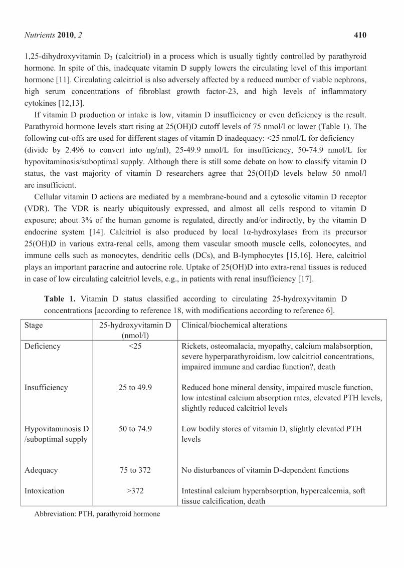
If vitamin D production or intake is low, vitamin D insufficiency or even deficiency is the result. Parathyroid hormone levels start rising at 25(OH)D cutoff levels of 75 nmol/l or lower (Table 1). The following cut-offs are used for different stages of vitamin D inadequacy: <25 nmol/L for deficiency (divide by 2.496 to convert into ng/ml), 25-49.9 nmol/L for insufficiency, 50-74.9 nmol/L for hypovitaminosis/suboptimal supply. Although there is still some debate on how to classify vitamin D status, the vast majority of vitamin D researchers agree that 25(OH)D levels below 50 nmol/l are insufficient.
Cellular vitamin D actions are mediated by a membrane-bound and a cytosolic vitamin D receptor (VDR). The VDR is nearly ubiquitously expressed, and almost all cells respond to vitamin D exposure; about 3% of the human genome is regulated, directly and/or indirectly, by the vitamin D endocrine system [14]. Calcitriol is also produced by local 1α-hydroxylases from its precursor 25(OH)D in various extra-renal cells, among them vascular smooth muscle cells, colonocytes, and immune cells such as monocytes, dendritic cells (DCs), and B-lymphocytes [15,16]. Here, calcitriol plays an important paracrine and autocrine role. Uptake of 25(OH)D into extra-renal tissues is reduced in case of low circulating calcitriol levels, e.g., in patients with renal insufficiency [17].
Table 1. Vitamin D status classified according to circulating 25-hydroxyvitamin D concentrations [according to reference 18, with modifications according to reference 6].
Stage 25-hydroxyvitamin D (nmol/l)
Clinical/biochemical alterations
Deficiency Insufficiency Hypovitaminosis D /suboptimal supply Adequacy Intoxication
<25
25 to 49.9
50 to 74.9
75 to 372
>372
Rickets, osteomalacia, myopathy, calcium malabsorption, severe hyperparathyroidism, low calcitriol concentrations, impaired immune and cardiac function?, death Reduced bone mineral density, impaired muscle function, low intestinal calcium absorption rates, elevated PTH levels, slightly reduced calcitriol levels Low bodily stores of vitamin D, slightly elevated PTH levels No disturbances of vitamin D-dependent functions Intestinal calcium hyperabsorption, hypercalcemia, soft tissue calcification, death
Abbreviation: PTH, parathyroid hormone
Nutrients 2010, 2
411
3. Worldwide Vitamin D Status
A recent review [19] summarized human vitamin D status according to region of the world. Six regions of the world were reviewed - Asia, Europe, Middle East and Africa, Latin America, North America, and Oceania–through a survey of published literature. Based on the articles referred to in this review, it was concluded that insufficient vitamin D status is prevalent in every of the six regions studied. Depending on the region, between 50% and more than 90% of people had 25(OH)D concentrations below 50 nmol/L. Low vitamin D status is most common in regions such as South Asia and the Middle East. Data demonstrate that insufficient vitamin D status is widespread and is re-emerging as a major health problem globally. Urbanization in combination with modern and also traditional lifestyles such as indoor working, indoor leisure time activities, and traditional Islamic clothing, and in combination with the aging process (institutionalization) is an important risk factor for vitamin D insufficiency/deficiency in large parts of the adult population. In highly urbanized areas, individual daily sun exposure is usually too low to achieve a 25(OH)D level of 75 nmol/L. Due to the fact that the vast majority of foods naturally contain no or only modest amounts of vitamin D, diet is not able to close the gap in vitamin D supply. It is noteworthy that urbanization and industrialization has long been known as a major cause of childhood rickets in western countries [7]. Rickets is now on the increase in many developing countries, and is also re-emerging as an important health problem in countries with strong sun avoidance policies and cultures requiring modest dress.
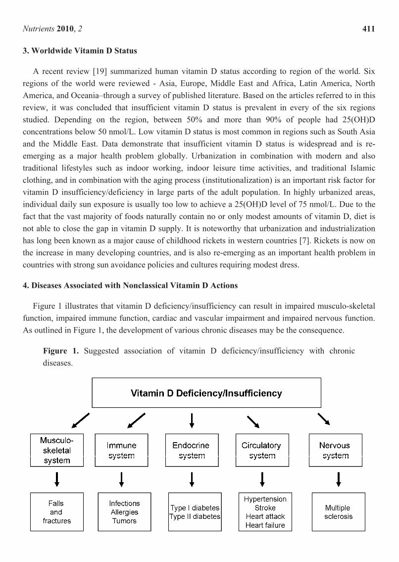
4. Diseases Associated with Nonclassical Vitamin D Actions
Figure 1 illustrates that vitamin D deficiency/insufficiency can result in impaired musculo-skeletal function, impaired immune function, cardiac and vascular impairment and impaired nervous function. As outlined in Figure 1, the development of various chronic diseases may be the consequence.
Figure 1. Suggested association of vitamin D deficiency/insufficiency with chronic diseases.
Nutrients 2010, 2
412
4.1. Vitamin D and Muscle Strengthening
Vitamin D deficiency causes reduced aktomyosin content of myofibrils, low calcium content of mitochondria, reduced calcium uptake into the sarcoplasmic reticulum, and low serum levels of muscle enzymes [3]. The importance of vitamin D-repletion for adequate muscle function was underscored in a recent study in institutionalized people ≥60 years of age with insufficient vitamin D status [20]: This RCT demonstrated that six-month supplementation (December to May) of oral vitamin D (3,750 µg once a month during the first two months, followed by 2,250 µg once a month for the last four months) was able to improve lower limb muscle strength by 16–24%. Data support results of a recently performed meta-analysis of randomized controlled trials (RCTs), indicating that daily doses of 17.5 to 20 µg supplemental vitamin D are able to prevent falls in elderly adults [21]. The relative risk of falls was reduced by approximately 20% if the achieved serum 25(OH)D concentrations is 60 nmol/l or more. In contrast to “high dose” supplemental vitamin D, low dose daily supplemental vitamin D (5 to 15 µg) is not able to prevent falls. Thus, doses of supplemental vitamin D of less than 17.5 µg or serum 25-hydroxyvitamin D concentrations of less than 60 nmol/L may not reduce the risk of falling among older individuals. It is noteworthy that in elderly people the risk of falling predicts the risk of developing osteoporotic fractures. Therefore, the effects of vitamin D on muscle strength may contribute to the preventive effect of vitamin D on osteoporotic fractures. There is also evidence that adequate vitamin D supply is important for muscle function in children. Already more than 50 years ago, Ronge [22] has demonstrated that children who have hands and face exposed to UVB radiation in their classroom at school for 3–5 hours during wintertime show better endurance performance compared to a control group without UVB exposure. Endurance performance was assessed by bicycle ergometry. In that study, a similar positive effect on endurance performance was seen in children who received a single vitamin D bolus of 6.25 mg vitamin D in February.
4.2. Infections
There is mounting evidence for a pivotal role of vitamin D in the immune system. Calcitriol is able to induce the differentiation of monocytes into macrophages. In addition, calcitriol increases the activity of macrophages and facilitates their cytotoxic activity. Macrophages represent the first unspecific defence line of the immune system. It is well known that the prevalence of infections such as pneumonia is high in infants with rickets [3]. The use of vitamin D (or cod liver oil) as a treatment of infections have been practised for over 150 years. As early as 1903, Niels Finsen was awarded the Nobel Prize for Medicine and Physiology for his theory to cure Lupus vulgaris (skin-tuberculosis) using phototherapy. In 2007, Schauber et al. [23] published data demonstrating that vitamin D is able to stimulate synthesis of the anti-microbial peptide cathelicidin in human skin cells to enhance innate immunity. A meta-analysis of observational studies has demonstrated that patients with tuberculosis have lower circulating 25(OH)D concentrations compared to healthy controls [24]. Ecological studies also support a preventive role of vitamin D in influenza: the seasonal and latitudinal distribution of outbreaks of influenza A in the world in 1967–1975, and weekly consultation rates for illnesses diagnosed clinically as influenza or influenza-like in England 1968-1970 were inversely associated with solar UVB radiation [25]. Very recently, it has been demonstrated in an RCT that supplementation with 30 µg vitamin D daily reduces the risk of wintertime influenza A in Japanese
Nutrients 2010, 2
413
nursery school children [26]. Some epidemiological data support the assumption that vitamin D may reduce the susceptibility to respiratory tract infections [27,28]. In addition, vitamin D users of the RECORD trial [29], an RCT with approximately 3,500 participants who received 20 µg vitamin D or placebo, reported a lower tendency for infections and antibiotic use in March compared to vitamin D nonusers. In another RCT in individuals with baseline circulating concentrations below 50 nmol/L, supplementation with 20 µg or 50 µg vitamin D daily for three years significantly reduced upper respiratory tract infections compared to placebo [30]. In contrast, a daily vitamin D supplement of 50 µg for 12 weeks did not prevent upper respiratory tract infections in individuals with baseline circulating 25(OH)D concentrations above 50 nmol/L [31]. Consequently, there is currently insufficient data to conclusively state that vitamin D supplementation could result in lowered infection [32]. One factor that has to be considered in future studies is baseline 25(OH)D concentration. In addition, the relation between vitamin D supplementation, local calcitriol, and local cathelicidin production has to be investigated more detailed. Interestingly, oral intake of activated vitamin D in rickets patients for four weeks significantly increased human cathelicidin expression in neutrophils compared to age-matched healthy controls without administration of activated vitamin D [33], indicating a critical role of adequate calcitriol availability for regulation of the innate immune response.
4.3. Allergies
Activation of the adaptive immune system is complex. Generally, it is of importance that specific pathways of the specific immune system are adequately suppressed in order to avoid autoimmune diseases or allergic reactions. Regulatory T cells are crucial for the maintenance of immunological tolerance. Their major role is to shut down T cell-mediated immunity toward the end of an immune reaction and to suppress auto-reactive T cells. A strong Th2 predominance leads to pathologic conditions such as overproduction of IgE and allergic diseases, whereas a strong Th1 predominance leads to autoimmunity and severe allograft rejection. Of clinical importance is the fact that DCs may induce naïve T cells in an immunogenetic direction but also in a tolerogenic direction, depending on the state of their maturation and their cell surface receptor. Tolerogenic DCs generally are semimature. There is accumulating evidence that vitamin D modulates the adaptive immune system [16]. Calcitriol appears to generate tolerogenic DCs in vivo, as demonstrated in models of transplantation and autoimmune disease. DCs appear to be key targets of calcitriol. Calcitriol arrest the differentiation and maturation of DCs, maintaining them in an immature state. Calcitriol is able to enhance the secretion by DCs of the anti-inflammatory and anti-allergic cytokine IL-10.
At present, the vitamin D hypothesis of allergies takes two forms: Some argue that vitamin D deficiency may cause allergic reactions whereas others argue that vitamin D excess leads to an increased allergy risk. Wjst is a representative of the latter hypothesis. He argues that the increase in allergies in Bavaria after 1960 coincided with vitamin D supplementation intervention programs to prevent rickets in childhood. Moreover, both, adherence to these programs and prevalence of allergies in children seem to be lower in farming communities in Bavaria [34]. The farm protection is observed mainly during the first year of life [35], when vitamin D supplementation is also recommended. Wjst’s hypothesis is based on the assumption that vitamin D may lead to Th2 predominance and increased IgE production. Generally, his hypothesis is supported by findings that children whose mothers'

Nutrients 2010, 2
414
concentration of 25(OH)-vitamin D in late pregnancy was >75 nmol/l had an increased risk of eczema on examination at nine months and asthma at age nine years compared to children whose mothers' concentration was <30 nmol/L [36]. In addition, vitamin D supplementation during infancy was associated with a higher allergy risk [37,38], and the prevalence of allergic rhinitis increased across quartile groups of 25(OH)D serum levels in adults of NHANES III [39].
It is, however, noteworthy that several other epidemiological studies support the vitamin D deficiency hypothesis of allergic reactions [40-44]. Moreover, administration of calcitriol to blood cells of healthy persons and steroid-resistant asthmatic patients enhanced subsequent responsiveness to dexamethasone for induction of IL-10 [43]. Very few intervention trials are available so far. In a small, randomized, double-blind, placebo-controlled trial, vitamin D2 supplementation (25 µg/day) significantly improved skin symptoms in children with winter-related atopic dermatitis [45]. In a study in heart failure patients, vitamin D3 supplementation (50 µg/day) was able to increase blood levels of the anti-allergic cytokine IL-10 [46]. However, the effect on allergic reactions has not been elucidated in that earlier investigation.
In total, it cannot be ruled out that vitamin D deficiency as well as vitamin D excess may increase the risk of allergic reactions. This assumption is supported by recent findings. Hyppönen et al. [47] observed a biphasic effect of vitamin D with both low and high 25(OH)D levels associated with elevated IgE concentrations in participants of the 1958 British birth cohort. Compared with the reference group with the lowest IgE concentrations [25(OH)D 100–125 nmol/L], adjusted IgE concentrations were 29% higher for participants with the 25(OH)D < 25 nmol/L, and 56% higher for participants with 25(OH)D > 135 nmol/L.
4.4. Cancer
Since vitamin D is a key regulator of various cellular metabolic pathways, it is important for cellular maturation, differentiation, and apoptosis [3]. In 2008, the WHO published a report from the International Agency for Research on cancer [48] that came to the conclusion that there is (i) consistent epidemiological evidence for an inverse association between 25(OH)D and colorectal cancer and colorectal adenomas, (ii) suggested epidemiological evidence for an inverse association between 25(OH)D and breast cancer, (iii) insufficient evidence for an inverse association between 25(OH)D and other types of cancer, and (iv) the need for new randomized controlled trials (RCTs). One such RCT has already been published [49]: In a four-year, population-based study, where the primary outcome was fracture incidence, and the principal secondary outcome was cancer incidence, 1179 community-dwelling women were randomly assigned to receive 1500 mg supplemental calcium/d alone (Ca-only), supplemental calcium plus 27.5 µg vitamin D/d (Ca + D), or placebo. Cancer incidence was 60–77% lower in the Ca + D women and 43% lower in the Ca-only group than in the placebo control subjects (P < 0.03). Gorham et al. [50] have estimated that in North America, Europe, and East Asia approximately 32% of colon cancer and approximately 26% of breast cancer can be prevented with 50 µg vitamin D daily and 3–10 min daily of noon sunlight seasonality, when weather permits. Garland et al. [51] estimated that raising the minimum year-around serum 25(OH)D level to 100–150 nmol/L would prevent approximately 58,000 new cases of breast cancer and 49,000 new cases of colorectal cancer each year, and three fourths of deaths from these diseases in the United States and Canada. Such intakes also are expected to reduce case-fatality rates of patients who have

Nutrients 2010, 2
415
breast, colorectal, or prostate cancer by half. Nevertheless, there is also some concern that cancer risk is not only enhanced in individuals with deficient/insufficient vitamin D status, but also if 25(OH)D concentrations rise above 80 nmol/L [52], a concentration several vitamin D researchers consider adequate. However, this increase in cancer risk has only been observed in observational studies after multivariable adjustments have been made for confounding factors. This kind of exploratory data analysis has been criticized by some researchers [53].
4.5. Diabetes Mellitus
In vitro and in vivo studies suggest that vitamin D can prevent pancreatic beta-cell destruction and reduces the incidence of autoimmune diabetes. This may at least in part be due to a suppression of proinflammatory cytokines such as tumor necrosis factor (TNF)-α. Recently, the relationship between UVB irradiance, the primary source of circulating vitamin D in humans, and age-standardized incidence rates of type 1 diabetes mellitus in children aged <14 years, was analyzed according to 51 regions of the world [54]. Incidence rates were generally higher at higher latitudes and were inversely associated with UVB irradiance. As early as 2001, Hyppönen et al. [55] has demonstrated in a birth cohort study that vitamin D supplementation was associated with a decreased frequency of type 1 diabetes. In contrast, children suspected of having rickets during the first year of life had a three times higher relative risk compared with those without such a suspicion. Meanwhile, a meta-analysis of four case-control studies has shown that the risk of type 1 diabetes is reduced by 29% in infants who are supplemented with vitamin D compared to those who are not supplemented [56]. There is also some evidence of a dose-response effect, with those using higher amounts of vitamin D being at lower risk of developing type 1 diabetes. Finally, timing of supplementation might also be important for the subsequent development of type 1 diabetes. In a recent RCT [57], the majority of patients with latent autoimmune diabetes in adults increased their concentrations of plasma C–peptide levels in fasting state after 1 year of treatment with activated vitamin D, whereas only a minority of patients treated with insulin alone maintained stable fasting C-peptide levels.
In 2007, Pittas et al. [58] conducted a systemic review and meta-analysis for observational studies and clinical trials in adults with outcomes related to glucose homeostasis in type 2 diabetes mellitus. Observational studies show a relatively consistent association between low vitamin D status and prevalent type 2 diabetes, with an odds ratio of 0.36 among non-Blacks for highest versus lowest 25-hydroxyvitamin D. Evidence from RCTs with vitamin D and/or calcium supplementation suggests that combined vitamin D and calcium supplementation may have a role in the prevention of type 2 diabetes only in populations at high risk (i.e,. glucose intolerance). Whereas vitamin D supplementation did not improve glycemic control in diabetic subjects with normal serum 25(OH)D levels [59], administration of 100 µg vitamin D3 improved insulin sensitivity in vitamin D deficient and insulin resistant South Asian women [60]. Insulin resistance was most improved when endpoint serum 25(OH)D reached ≥ 80 nmol/L. Optimal vitamin D concentrations for reducing insulin resistance were shown to be 80–119 nmol/L.
Nutrients 2010, 2
416
4.6. Cardiovascular Disease
Globally, cardiovascular disease (CVD) is the number one cause of death. In 2005, CVD was responsible for approximately 30% of deaths worldwide. CVD includes various illnesses such as coronary heart disease (CHD), peripheral arterial disease, cerebrovascular disease such as stroke, and congestive heart failure. There is accumulating evidence that the vitamin D hormone calcitriol exerts important physiological effects in cardiomyocytes, vascular smooth muscle cells, and the vascular endothelium. The mechanisms have been reviewed in detail elsewhere [61]. Hypertension is a key risk factor for CVD. A recently published systematic review and meta-analysis came to the conclusion that vitamin D produces a fall in systolic blood pressure of −6.18 mm Hg and a nonsignificant fall in diastolic blood pressure of −2.56 mm Hg in hypertensive patients. No reduction in blood pressure is seen in studies examining patients who are normotensive at baseline [62]. Since these studies had small sample sizes, future studies should investigate their generalizability.
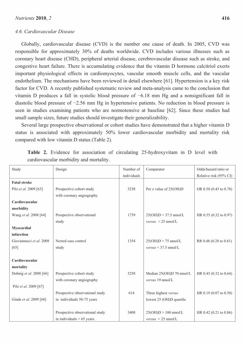
Several large prospective observational or cohort studies have demonstrated that a higher vitamin D status is associated with approximately 50% lower cardiovascular morbidity and mortality risk compared with low vitamin D status (Table 2).
Table 2. Evidence for association of circulating 25-hydroxyvitam in D level with cardiovascular morbidity and mortality.
Study Design Number of
individuals
Comparator Odds/hazard ratio or
Relative risk (95% CI)
Fatal stroke
Pilz et al. 2009 [63]
Cardiovascular
morbidity
Wang et al. 2008 [64]
Myocardial
infarction
Giovannucci et al. 2008
[65]
Cardiovascular
mortality
Dobnig et al. 2008 [66]
Pilz et al. 2009 [67]
Ginde et al. 2009 [68]
Prospective cohort study
with coronary angiography
Prospective observational
study
Nested case control
study
Prospective cohort study
with coronary angiography
Prospective observational study
in individuals 50-75 years
Prospective observational study
in individuals > 65 years.
3258
1739
1354
3258
614
3408
Per z value of 25(OH)D
25(OH)D > 37.5 nmol/L
versus < 25 nmol/L
25(OH)D > 75 nmol/L
versus < 37.5 nmol/L
Median 25(OH)D 70 nmol/L
versus 19 nmol/L
Three highest versus
lowest 25 (OH)D quartile
25(OH)D > 100 nmol/L
versus < 25 nmol/L
OR 0.58 (0.43 to 0.78)
HR 0.55 (0.32 to 0.97)
RR 0.48 (0.28 to 0.81)
HR 0.45 (0.32 to 0.64)
HR 0.19 (0.07 to 0.50)
HR 0.42 (0.21 to 0.86)

Nutrients 2010, 2
417
The Women’s Health Initiative (WHI) calcium/vitamin D (CaD) trial could however not demonstrate a reduction in cardiovascular mortality by daily supplementation of 1,000 mg calcium and 10 µg vitamin D [69]. Meanwhile it is clear that an amount of 10 µg vitamin D is far too low to result in a meaningful increase in serum 25(OH)D levels (see before) and that a daily calcium supplement of 1,000 mg increases the risk for cardiovascular events in healthy older women. Both, the supplemental calcium in the vitamin D arm of the WHI study and the low amount of vitamin D might have countermanded its cardiovascular benefits. In line with this assumption, a recent meta-analysis of seven randomized trials showed a slight but statistically nonsignificant reduction in CVD risk (relative risk: 0,90; 95% CI: 0.77 to 1.05) with vitamin D supplementation at moderate to high doses (approximately 25µg/d) but not with calcium supplementation (relative risk: 1,14; 95% CI: 0.92 to 1.41) or a combination of vitamin D and calcium supplementation (relative risk: 1.04; 95% CI: 0.92 to 1.18) [70].
In line with a potential beneficial effect of vitamin D on CVD risk, a daily vitamin D supplement of 83 µg could improve some traditional and nontraditional cardiovascular risk markers in healthy overweight and obese subjects with mean 25(OH)D concentrations of 30 nmol/L who attended a weight-reduction program [71].
4.7. Multiple Sclerosis
Multiple sclerosis (MS) is a demyellinating disease of the central nervous system that is debilitating and can be fatal. Manifestation of the disease is typically between the age of 20 and 40. In Europe and North America, regions with higher UVB radiation have low rates of MS and vice versa [3]. In Israel, MS prevalence depends on the country of origin. The prevalence is high in people who were born in a country with low UVB irradiance [72], indicating that vitamin D status during the period of early life is of importance for MS susceptibility. MS disease activity shows inverse fluctuations according to season and vitamin D status [73]. In a prospective, nested case-control study among more than seven million US military personnel [74], MS prevalence was lower in those people who had circulating 25-hydroxyvitamin D concentrations between 100 and 150 nmol/L compared with those who had 25-hydroxyvitamin D concentrations below 63 nmol/L. However, this association was only seen in Whites and not in Blacks, indicating that genetic factors play an important role in the pathogenesis of MS. Therefore, the recent finding is of importance that expression of the MS-associated MHC class II allele HLA-DRB1*1501 is regulated by Vitamin D [75].
5. Mortality
As mentioned before, vitamin D status is an important independent predictor of CVD and specific types of cancer. In addition, vitamin D status predicts CVD and cancer mortality. Both, CVD and cancer are the most important causes of mortality in developed countries. In 2007, Autier and Gandini [76] published a meta-analysis of randomized controlled trials (RCTs) on vitamin D and mortality that were not primarily designed to assess mortality. The authors found out that in middle-aged and elderly patients with low serum concentrations of 25-hydroxyvitamin D (25(OH)D) vitamin D supplementation was linked to lower all-cause mortality compared to no vitamin D supplementation.

Nutrients 2010, 2
418
Daily dose of vitamin D ranged between 10 µg and 50 µg. Risk reduction was 7% during a mean follow-up of 5.7 years.
Based on the aforementioned meta-analysis, several large prospective cohort studies were recently published on all-cause mortality and vitamin D status (Table 3). They demonstrate a consistent increase in mortality risk in patients with insufficient or deficient 25(OH)D concentrations. However, low 25(OH)D was not an independent predictor for mortality in patients with advanced disease [77,78]. One may speculate that in this case, vitamin D supplementation is unable to reverse the already existing severe pathophysiologic derangements.
Table 3. Evidence for association of circulating 25-hydroxyvitamin D level or vitamin D supplementation with all-cause mortality.
Study Design Number of individuals
Comparator Hazard ratio or relative risk (95% CI)
Autier and Gandini, 2007 [76] Dobnig et al. 2008 [66] Kuroda et al. 2009 [77] Ng et al. 2008 [78]
Ginde et al. 2009 [68] Pilz et al. 2009 [67]
Meta-analysis of 18 vitamin D supplementation studies Prospective cohort study with coronary angiography Prospective observational study in postmenopausal women Prospective cohort study in patients with colorectal cancer Prospective observational study in individuals > 65 years. Prospective observational study In individuals 50-75 years
57,311 3,258 1,232 304 3,408 614
Supplemented versus unsupplemented Median 25(OH)D 70 nmol/L versus 19 nmol/L ≥ 50 nmol/L versus < 50 nmol/L Mean 41 nmol/L versus 100 nmol/L 25(OH)D > 100 nmol/L versus < 25 nmol/L Three highest quartiles versus lowest quartile
RR 0.93 (0.87 to 0.99) HR 0.48 (0.37 to 0.63) HR 0.46 (0.27 to 0.79) HR 0.52 (0.29 to 0.94) HR 0.55 (0.34to 0.88) HR 0.51 (0.28 to 0.93)
6. Conclusions
In 2003, a review article had summarized the association of insufficient vitamin D status with various diseases such as myopathy, CVD, cancer, diabetes mellitus, MS, and infections [8]. Meanwhile, evidence has accumulated that vitamin D may indeed play an important role in the etiology of many of these diseases. Meta-analyses of RCTs demonstrate that vitamin D improves muscle function and seems to reduce blood pressure in hypertensive patients. In addition, some RCTs demonstrate that vitamin D reduces cancer incidence, and improves glucose homeostasis in patients with type 2 diabetes and cardiovascular risk markers in overweight people [49,60,71]. The most exiting result is however the fact that vitamin D may reduce mortality rate. This latter finding fits well together with the fact that severe deficiency of several other vitamins such as retinol, thiamine, niacin, and ascorbic acid is also associated with enhanced mortality. Nevertheless, additional large RCTs are needed to confirm whether or not vitamin D is able prolong survival in individuals with inadequate vitamin D status. In this context, the effect of vitamin D in deficient and insufficient individuals should be investigated separately.
Some aforementioned beneficial data on glucose homeostasis and cardiovascular risk markers were not confirmed by recent RCTs [59,81]. All these RCTs performed so far were relative small in sample

Nutrients 2010, 2
419
seize [59,60,71,81]. In addition, individual medication and baseline circulating 25(OH)D concentrations may have influenced study results. Therefore, additional research is necessary to clarify whether or not vitamin D supplementation is indeed effective in secondary prevention and also in tertiary prevention of chronic diseases. But we should be aware of the fact that many chronic diseases are of multi-factorial origin. Vitamin D is certainly only one factor among others. In addition, there may be individual differences with respect to the metabolic pathways that are disturbed in vitamin D deficient persons. Therefore, we should not be too enthusiastic that future RCTs will show clear beneficial vitamin D effects. For example, the meta-analysis by Autier and Gandini was based on more than 55,000 individuals. None of the single studies included in this analysis showed a significant vitamin D effect on mortality, indicating that huge sample seizes are probably needed to demonstrate a clear vitamin D effect. Even so, the consequences on a population scale may be important because of the large number of people who are affected.
The effect of vitamin D on MS, type 1 diabetes, infections, and allergies is less clear at present. Although newborns usually receive vitamin D supplements for preventing rickets, possible adverse effects of deficient vitamin D concentrations during fetal development such as increased susceptibility for type I diabetes and MS have to be considered as well. It is noteworthy that many women of childbearing age worldwide are vitamin D insufficient or even deficient [19,82]. With respect to MS, type 1 diabetes, and allergies, more birth cohort studies are needed.
Despite some uncertainties with respect to vitamin D and health, there is general agreement that currently a high percentage of people worldwide have low vitamin D status [19,83]. The recommended daily vitamin D intake of 5–15 µg is too low to achieve an adequate vitamin D status in people with only modest UVB exposure. Generally, treating vitamin D deficiency is easy to perform, safe, and inexpensive. Sources of vitamin D could include a combination of food fortification, supplements, and natural and artificial UV-B irradiation, if properly acquired. It has been calculated that 1 µg vitamin D increases circulating 25(OH)D levels by approximately 1 nmol/L [4]. Thus, a daily intake of approximately 50 µg vitamin D would be necessary for increasing the circulating 25(OH)D level from 25 nmol/L to 75 nmol/L. In order to achieve a 25(OH)D concentration above 75 nmol/L in almost all individuals of a group with mean baseline 25(OH)D concentrations of 38 nmol/L, daily supplementation with up to 100 µg vitamin D is necessary [5]. In otherwise healthy adults, the risk of vitamin D intoxication is extremely rare [3,84]. Vitamin D intoxications such as hypercalcemia do not occur until oral vitamin D intake and serum 25(OH)D concentrations exceed 250 µg/day (approximately 3–5 µg/kg body weight) [84] and 372 nmol/L [6], respectively. A daily amount of up to 250 µg vitamin D is similar to the amount that is produced by daily whole body exposure to UVB radiation [10].
References
1. Bischoff-Ferrari, H.A.; Willett, W.C.; Wong, J.B.; Stuck, A.E.; Staehelin, H.B.; Orav, E.J.; Thoma, A.; Kiel, D.P.; Henschkowski, J. Prevention of nonvertebral fractures with oral vitamin D and dose dependency: a meta-analysis of randomized controlled trials. Arch. Intern. Med. 2009, 169, 551-561.
2. McKenna, M.J. Differences in vitamin D status between countries in young adults and the elderly. Am. J. Med. 1992, 93, 69-77.
Nutrients 2010, 2
420
3. Zittermann, A. Vitamin D in preventive medicine – are we ignoring the evidence? Br. J. Nutr. 2003, 89, 552-572.
4. Vieth, R. Vitamin D and cancer mini-symposium: the risk of additional vitamin D. Ann. Epidemiol. 2009, 19, 441-445.
5. Aloia, J,F,; Patel, M.; Dimaano, R.; Li-Ng, M.; Talwar, S.A.; Mikhail, M.; Pollack, S.; Yeh, J.K. Vitamin D intake to attain a desired serum 25-hydroxyvitamin D concentration. Am. J. Clin. Nutr. 2008, 87, 1952-1958.
6. Holick, M.F. Vitamin D deficiency. N. Engl. J. Med. 2007, 357, 266-281. 7. Holick, M.F. McCollum Award Lecture, Vitamin D: New horizons for the 21st century. Am. J.
Clin. Nutr. 1994, 60, 619-630. 8. Clemens, T.L.; Adams, J.S.; Henderson, S.L.; Holick, M.F. Increased skin pigment reduces the
capacity of skin to synthesise vitamin D3. Lancet 1982, 1, 74-76. 9. Krause, R.; Bohring, M.; Hopfenmüller, W.; Holick, M.F.; Sharma, A.M. Ultraviolet B and blood
pressure. Lancet 1998, 352, 709-710. 10. Stamp, T.C.; Haddad, J.G.; Twigg, C.A. Comparison of oral 25-hydroxycholecalciferol, vitamin
D, and ultraviolet light as determinants of circulating 25-hydroxyvitamin D. Lancet 1977, 1, 1341-1343.
11. Zittermann, A.; Frisch, S.; Berthold, H.K.; Götting, C.; Kuhn, J.; Kleesiek, K.; Stehle, P.; Koertke, H.; Koerfer, R. Vitamin D supplementation enhances the beneficial effects of weight loss on cardiovascular risk markers. Am. J. Clin. Nutr. 2009, 89, 1321-1327.
12. Zittermann, A.; Koerfer R. Protective and toxic effects of vitamin D on vascular calcification: clinical implications. Mol. Aspects Med. 2008, 29, 423-432.
13. Antoniucci, D.M.; Yamashita, T.; Portaloe, A.A. Dietary phosphorus regulates serum fibroblast growth factor-23 concentrations in healthy men. J. Clin. Endocrinol. Metab. 2006, 91, 3144-3149.
14. Bouillon, R.; Carmeliet, G.; Verlinden, L.; van Etten, E.; Verstuyf, A.; Luderer, H.F.; Lieben, L.; Mathieu, C.; Demay, M. Vitamin D and human health: lessons from vitamin D receptor null mice, Endocr. Rev. 2008, 29, 726-776.
15. Hewison, M.; Burke, F.; Evans, K.N.; Lammas, D.A. Extra-renal 25-hydroxyvitamin D3-1alpha-hydroxylase in human health and disease, J. Steroid Biochem. Mol. Biol. 2007, 103, 316-321.
16. Zittermann, A.; Tenderich, G.; Koerfer, R. Vitamin D and the adaptive immune system with special emphasis to allergic reactions and allograft rejection. Inflamm. Allergy Drug Targets 2009, 8, 161-168.
17. Gallieni, M.; Kamimura, S.; Ahmed, A.; Bravo, E.; Delmez, J.; Slatopolsky, E.; Dusso, A. Kinetics of monocyte 1 alpha-hydroxylase in renal failure. Am. J. Physiol. 1995, 268, F746-F753.
18. Zittermann, A.; Gummert, J.F.; Boergermann, J. Vitamin D deficiency and mortality. Curr. Opin. Nutr. Metab. Care 2009, 2, 634-639.
19. Mithal, A.; Wahl, D.A.; Bonjour, J.P.; Burckhardt, P.; Dawson-Hughes, B.; Eisman, J.A.; El-Hajj Fuleihan, G.; Josse, R.G.; Lips, P.; Morales-Torres, J. IOF Committee of Scientific Advisors. (CSA) Nutrition Working Group. Global vitamin D status and determinants of hypovitaminosis D. Osteoporos Int. 2009, 20, 1807-1820.
20. Moreira-Pfrimer, L.D.; Pedrosa, M.A.; Teixeira, L.; Lazaretti-Castro, M. Treatment of vitamin D deficiency increases lower limb muscle strength in institutionalized older people independently of
Nutrients 2010, 2
421
regular physical activity: a randomized double-blind controlled trial. Ann. Nutr. Metab. 2009, 54, 291-300.
21. Bischoff-Ferrari, H.A.; Dawson-Hughes, B.; Staehelin, H.B.; Orav, J.E.; Stuck, A.E.; Theiler, R.; Wong, J.B.; Egli, A.; Kiel, D.P.; Henschkowski, J. Fall prevention with supplemental and active forms of vitamin D: a meta-analysis of randomised controlled trials. BMJ 2009 Oct 1;339:b3692. doi: 10.1136/bmj.b3692.
22. Ronge, H.E. Increase of physical effectiveness by systematic ultraviolet radiation. Strahlentherapie 1952, 88, 563-566, [article in German].
23. Schauber, J.; Dorschner, R.A.; Coda, A.B.; Büchau, A.S.; Liu, P.T.; Kiken, D.; Helfrich, Y.R.; Kang, S.; Elalieh, H.Z.; Steinmeyer, A.; Zügel, U.; Bikle, D.D.; Modlin, R.L.; Gallo, R.L. Injury enhances TLR2 function and antimicrobial peptide expression through a vitamin D-dependent mechanism. J. Clin. Invest. 2007, 117, 803-811.
24. Nnoaham, K.E.; Clarke, A. Low serum vitamin D levels and tuberculosis: a systematic review and meta-analysis. Int. J. Epidemiol. 2008, 37, 113-119.
25. Cannell, J.J.; Vieth, R.; Umhau, J.C.; Holick, M.F.; Grant, W.B.; Madronich, S.; Garland, C.F.; Giovannucci, E. Epidemic influenza and vitamin D. Epidemiol. Infect. 2006, 134, 1129-1140.
26. Urashima, M.; Segawa, T.; Okazaki, M.; Kurihara, M.; Wada, Y.; Ida, H. Randomized trial of vitamin D supplementation to prevent seasonal influenza A in schoolchildren. Am. J. Clin. Nutr. 2010, [Epub ahead of print].
27. Laaksi, I.; Ruohola, J.P.; Tuohimaa, P.; Auvinen, A.; Haataja, R.; Pihlajamäki, H.; Ylikomi, T. An association of serum vitamin D concentrations < 40 nmol/L with acute respiratory tract infection in young Finnish men. Am. J. Clin. Nutr. 2007, 86, 714-717.
28. Ginde, A.A.; Mansbach, J.M.; Camargo, C.A. Association between serum 25-hydroxyvitamin D level and upper respiratory tract infection in the Third National Health and Nutrition Examination Survey. Arch. Intern. Med. 2009, 169, 384-390.
29. Avenell, A.; Cook, J.A.; Maclennan, G.S.; Macpherson, G.C. Vitamin D supplementation to prevent infections: a sub-study of a randomised placebo-controlled trial in older people (RECORD trial, ISRCTN 51647438). Age Ageing 2007, 36, 574-577.
30. Aloia, J.F.; Talwar, S.A.; Pollack, S.; Yeh, J. A randomized controlled trial of vitamin D3 supplementation in African American women. Arch. Intern. Med. 2005, 165, 1618-1623.
31. Li-Ng, M.; Aloia, J.F.; Pollack, S.; Cunha, B.A.; Mikhail, M.; Yeh, J.; Berbari, N. A randomized controlled trial of vitamin D3 supplementation for the prevention of symptomatic upper respiratory tract infections. Epidemiol. Infect. 2009, 137, 1396-1404.
32. Yamshchikov, A.V.; Desai, N.S.; Blumberg, H.M.; Ziegler, T.R.; Tangpricha, V. Vitamin D for treatment and prevention of infectious diseases: a systematic review of randomized controlled trials. Endocr. Pract. 2009, 15, 438-449.
33. Misawa, Y.; Baba, A.; Ito, S.; Tanaka, M.; Shiohara, M. Vitamin D(3) induces expression of human cathelicidin antimicrobial peptide 18 in newborns. Int. J. Hematol. 2009, Nov 28. [Epub ahead of print]
34. Wjst, M. Another explanation for the low allergy rate in the rural Alpine foothills. Clin. Mol. Allergy 2005, 3, 7.

Nutrients 2010, 2
422
35. Von Mutius, E. Influences in allergy: epidemiology and the environment. J. Allergy Clin. Immunol. 2004, 113, 373-379.
36. Gale, C.R.; Robinson, S.M.; Harvey, N.C.; Javaid, M.K.; Jiang, B.; Martyn, C.N.; Godfrey, K.M.; Cooper, C.; and the Princess Anne Hospital Study Group. Maternal vitamin D status during pregnancy and childhood outcomes. Eur. J. Clin. Nutr. 2008, 62, 68-77.
37. Hyppönen, E.; Sovio, U.; Wjst, M.; Patel, S.; Pekkanen, J.; Hartikainen, A.L.; Järvelinb, M.R. Infant vitamin d supplementation and allergic conditions in adulthood: northern Finland birth cohort 1966. Ann. N. Y. Acad. Sci. 2004, 1037, 84-95.
38. Bäck, O.; Blomquist, H.K.; Hernell, O.; Stenberg, B. Does vitamin D intake during infancy promote the development of atopic allergy? Acta Derm. Venereol. 2009, 89, 28-32.
39. Wjst, M.; Hyppönen, E. Vitamin D serum levels and allergic rhinitis. Allergy 2007, 62, 1085-1086.
40. Zittermann, A.; Dembinski, J.; Stehle, P. Low vitamin D status is associated with low cord blood levels of the immunosuppressive cytokine interleukin 10. Pediatr. Allergy Immu. 2004, 15, 242-246.
41. Camargo, C.A.; Rifas-Shiman, S.L.; Litonjua, A.A.; Rich-Edwards, J.W.; Weiss, S.T.; Gold, D.R.; Kleinman, K.; Gillman, M.W. Maternal intake of vitamin D during pregnancy and risk of recurrent wheeze in children at 3 y of age. Am. J. Clin. Nutr. 2007, 85, 788-795.
42. Camargo, C.A.; Clark, S.; Kaplan, M.S.; Lieberman, P.; Wood, R.A. Regional differences in EpiPen prescriptions in the United States: the potential role of vitamin D. J. Allergy Clin. Immunol. 2007, 120, 131-136.
43. Oren, E.; Banerji, A.; Camergo, C.A. Vitamin D and atopic disoerdes in an obese population screened for vitamin D deficiency. J. Allergy Immunol. 2008, 121, 533-534.
44. Erkkola, M.; Kaila, M.; Nwaru, B.I.; Kronberg-Kippilä, C.; Ahonen, S.; Nevalainen, J.; Veijola, R.; Pekkanen, J.; Ilonen, J.; Simell, O.; Knip, M.; Virtanen, S.M. Maternal vitamin D intake during pregnancy is inversely associated with asthma and allergic rhinitis in 5-year-old children. Clin. Exp. Allergy 2009, 39, 875-882.
45. Sidbury, R.; Sullivan, A.F.; Thadhani, R.I.; Camargo, C.A. Randomized trial of vitamin D supplementation for winter-related atopic dermatitis in Boston: a pilot study. Br. J. Dermatol. 2008, 159, 245-247.
46. Schleithoff, S.S.; Zittermann, A.; Tenderich, G.; Berthold, H.K.; Stehle, P.; Koerfer, R. Vitamin D Supplementation Improves Cytokine Profile In Patients With Congestive Heart Failure: A Double-Blind, Randomized, Placebo-Controlled Trial. Am. J. Clin. Nutr. 2006, 83, 754-759.
47. Hyppönen, E.; Berry, D.J.; Wjst, M.; Power C. Serum 25-hydroxyvitamin D and IgE - a significant but nonlinear relationship. Allergy 2009, 64, 613-620.
48. World Health Organization. International Agency for Research on Cancer. Vitamin D and cancer. IARC Working Group Reports; WHO Press: Geneva, Switzerland 2008; Volume 5, p. 148.
49. Lappe, J.M.; Travers-Gustafson, D.; Davies, K.M.; Recker, R.R.; Heaney R.P. Vitamin D and calcium supplementation reduces cancer risk: results of a randomized trial. Am. J. Clin. Nutr. 2007, 85, 1586-1591.
50. Gorham, E.D.; Mohr, S.B.; Garland, F.C.; Garland, C.F. Vitamin D for Cancer Prevention and Survival. Clin. Rev. Bone Miner Metab. 2009, 7, 159-175.
Nutrients 2010, 2
423
51. Garland, C.F.; Gorham, E.D.; Mohr, S.B.; Garland, F.C. Vitamin D for cancer prevention: global perspective. Ann. Epidemiol. 2009, 19, 468-483.
52. Tuohimaa, P.; Tenkanen, L.; Ahonen, M.; Lumme, S.; Jellum, E.; Hallmans, G.; Stattin, P.; Harvei, S.; Hakulinen, T.; Luostarinen, T.; Dillner, J.; Lehtinen, M.; Hakama, M.. Both high and low levels of blood vitamin D are associated with a higher prostate cancer risk: a longitudinal, nested case-control study in the Nordic countries. Int. J. Cancer 2004, 108,104-108.
53. Zittermann, A.; Grant, W.B. 25-hydroxyvitamin D levels and all-cause mortality. Arch. Intern. Med. 2009, 169, 1075-1076.
54. Mohr, S.B.; Garland, C.F.; Gorham, E.D.; Garland, F.C. The association between ultraviolet B irradiance, vitamin D status and incidence rates of type 1 diabetes in 51 regions worldwide. Diabetologia 2008, 51, 1391-1398.
55. Hyppönen, E.; Läärä, E.; Reunanen, A.; Järvelin, M.R.; Virtanen, S.M. Intake of vitamin D and risk of type 1 diabetes: a birth-cohort study. Lancet 2001, 358, 1500-1503.
56. Zipitis, C.S.; Akobeng, A.K. Vitamin D supplementation in early childhood and risk of type 1 diabetes: a systematic review and meta-analysis. Arch. Dis. Child. 2008, 93, 512-517.
57. Li, X.; Liao, L.; Yan, X.; Huang, G.; Lin, J.; Lei, M.; Wang, X.; Zhou, Z. Protective effects of 1-alpha-hydroxyvitamin D3 on residual beta-cell function in patients with adult-onset latent autoimmune diabetes (LADA). Diabetes Metab. Res. Rev. 2009, 25, 411-416.
58. Pittas, A.G.; Lau, J.; Hu, F.B.; Dawson-Hughes, B. The role of vitamin D and calcium in type 2 diabetes. A systematic review and meta-analysis. J. Clin. Endocrinol. Metab. 2007, 92, 2017-2029.
59. Jorde R, Figenschau Y. Supplementation with cholecalciferol does not improve glycemic control in diabtetic subjects with normal serum 25-hydroxyvitamin D levels. Eur. J. Nutr. 2009, 48, 349-354.
60. von Hurst, P.R.; Stonehouse, W.; Coad, J. Vitamin D supplementation reduces insulin resistance in South Asian women living in New Zealand who are insulin resistant and vitamin D deficient - a randomised, placebo-controlled trial. Br. J. Nutr. 2009, 1-7, [Epub ahead of print].
61. Zittermann, A.; Koerfer R.Vitamin D in the prevention and treatment of coronary heart disease. Curr. Opin. Clin. Nutr. Metab. Care. 2008, 11, 752-757.
62. Witham, M.D.; Nadir, M.A.; Struthers, A.D. Effect of vitamin D on blood pressure: a systematic review and meta-analysis. J. Hypertens. 2009, 27, 1948-1954.
63. Pilz, S.; Dobnig, H.; Fischer, J.E.; Wellnitz, B.; Seelhorst, U.; Boehm, B.O.; März, W. Low vitamin D levels predict stroke in patients referred to coronary angiography. Stroke 2008, 39, 2611-2613.
64. Wang, T.J.; Pencina, M.J.; Booth, S.L.; Jacques, P.F.; Ingelsson, E.; Lanier, K.; Benjamin, E.J.; D'Agostino, R.B.; Wolf, M.; Vasan, R.S. Vitamin D deficiency and risk of cardiovascular disease. Circulation 2008, 117, 503-511.
65. Giovannucci, E.; Liu, Y.; Hollis, B.W.; Rimm, E.B. 25-hydroxyvitamin D and risk of myocardial infarction in men: a prospective study. Arch. Intern. Med. 2008, 168, 1174-1180.
66. Dobnig, H.; Pilz, S.; Scharnagl, H.; Renner, W.; Seelhorst, U.; Wellnitz, B.; Kinkeldei, J.; Boehm, B.O.; Weihrauch, G.; Maerz, W. Independent association of low serum 25-hydroxyvitamin D and
Nutrients 2010, 2
424
1,25-dihydroxyvitamin D levels with all-cause and cardiovascular mortality. Arch. Intern. Med. 2008, 168, 1340-1349.
67. Pilz, S.; Dobnig, H.; Nijpels, G.; Heine, R.J.; Stehouwer, C.D.; Snijder, M.B.; van Dam, R.M.; Dekker, J.M. Vitamin D and mortality in older men and women. Clin. Endocrinol. 2009, 71, 666-672.
68. Ginde, A.A.; Scragg, R.; Schwartz, R.; Camargo, C.A. Prospective Study of Serum 25-Hydroxyvitamin D Level, Cardiovascular Disease Mortality, and All-Cause Mortality in Older U.S. Adults. J. Am. Geriatr. Soc. 2009, 57, 1595-1603.
69. LaCroix, A.Z.; Kotchen, J.; Anderson, G.; Brzyski, R.; Cauley, J.A.; Cummings, S.R.; Gass, M.; Johnson, K.C.; Ko, M.; Larson, J.; Manson, J.E.; Stefanick, M.L.; Wactawski-Wende, J. Calcium plus vitamin D supplementation and mortality in postmenopausal women: the Women's Health Initiative calcium-vitamin D randomized controlled trial. J. Gerontol. A Biol. Sci. Med. Sci. 2009, 64, 559-567.
70. Wang, L.; Manson, J.E.; Song, Y.; Sesso, H.D. Systematic review: Vitamin D and calcium supplementation in prevention of cardiovascular events Ann. Intern. Med. 2010, 152, 315-323.
71. Zittermann, A.; Frisch, S.; Berthold, H.K.; Götting, C.; Kuhn, J.; Kleesiek, K. ; Stehle, P.; Koertke, H; Koerfer, R. Vitamin D supplementation enhances the beneficial effects of weight loss on cardiovascular risk markers. Am. J. Clin. Nutr. 2009, 89, 1321-1327.
72. Chaudhuri, A. Why we should offer routine vitamin D supplementation in pregnancy and childhood to prevent multiple sclerosis. Med. Hypotheses. 2005, 64, 608-618.
73. Embry, A.F.; Snowdon, L.R.; Vieth R. Vitamin D and seasonal fluctuations of gadolinium-enhancing magnetic resonance imaging lesions in multiple sclerosis. Ann. Neurol. 2000, 48, 271-272.
74. Munger, K.L.; Levin, L.I.; Hollis, B.W.; Howard, N.S.; Ascherio, A. Serum 25-hydroxyvitamin D levels and risk of multiple sclerosis. JAMA 2006, 296, 2832-2838.
75. Ramagopalan, S.V.; Maugeri, N.J.; Handunnetthi, L.; Lincoln, M.R.; Orton, S.M.; Dyment, D.A.; Deluca, G.C.; Herrera, B.M.; Chao, M.J.; Sadovnick, A.D.; Ebers, G.C.; Knight, J.C. Expression of the multiple sclerosis-associated MHC class II Allele HLA-DRB1*1501 is regulated by vitamin D. PLoS Genet. 2009, 5, e1000369.
76. Autier, P.; Gandini, S. Vitamin D supplementation and total mortality: a meta-analysis of randomized controlled trials. Arch. Intern. Med. 2007, 167, 1730-1737.
77. Kuroda, T.; Shiraki, M.; Tanaka, S.; Ohta, H. Contributions of 25-hydroxyvitamin D, co-morbidities and bone mass to mortality in Japanese postmenopausal women. Bone 2009, 44, 168-172.
78. Ng, K.; Meyerhardt, J.A.; Wu, K.; Feskanich, D.; Hollis, B.W.; Giovannucci, E.L.; Fuchs, C.S. Circulating 25-hydroxyvitamin d levels and survival in patients with colorectal cancer. J. Clin. Oncol. 2008, 26, 2984-2991.
79. Wang, A.Y.; Lam, C.W.; Sanderson, J.E. Wang, M.; Chan, I.H.; Lui, S.F., Sea, M.M.; Woo, J. Serum 25-hydroxyvitamin D status and cardiovascular outcomes in chronic peritoneal dialysis patients: a 3-y prospective cohort study. Am. J. Clin. Nutr. 2008, 87, 1631-1638.
80. Heist, R.S.; Zhou, W.; Wang, Z. Liu, G.; Neuberg, D.; Su, L.; Asomaning, K.; Hollis, B.W.; Lynch, T.J.; Wain, J.C.; Giovannucci, E.; Christiani, D.C. Circulating 25-hydroxyvitamin D,
Nutrients 2010, 2
425
VDR polymorphisms, and survival in advanced non-small-cell lung cancer. J. Clin. Oncol. 2008, 26, 5596-5602.
81. Jorde, R.; Sneve, M.; Torjesen, P.; Figenschau, Y. No improvement in cardiovascular risk factors in overweight and obese subjects after supplementation with vitamin D for 1 year. J. Intern. Med. 2009, [epub ahead of print].
82. Holmes, V.A.; Barnes, M.S.; Alexander, H.D.; McFaul, P.; Wallace, J.M. Vitamin D deficiency and insufficiency in pregnant women: a longitudinal study. Br. J. Nutr. 2009, 102, 876-881.
83. Standing Committee of European Doctors. Vitamin D nutritional policy in Europe. Available online: http://cpme.dyndns.org:591/adopted/2009/CPME_AD_Brd_241009_179_final_EN.pdf (assessed on 24 March 2010).
84. Zittermann, A.; Koerfer, R. Protective and toxic effects of vitamin D on vascular calcification: Clinical implications. Mol. Aspects Med. 2008, 29, 423-432.
© 2010 by the authors; licensee Molecular Diversity Preservation International, Basel, Switzerland. This article is an open-access article distributed under the terms and conditions of the Creative Commons Attribution license (http://creativecommons.org/licenses/by/3.0/).

SWISSBIOHEALTH®SWISS BIOHEALTH AGBrückenstrasse 15 . 8280 Kreuzlingen / Schweiz
Tel. +41 71 678 2000Fax +41 71 678 [email protected]
www.swiss-biohealth.com


