Human PET Studies of Metabotropic Glutamate Receptor Subtype 5 with 11 C-ABP688 Simon M. Ametamey 1 , Valerie Treyer 2 , Johannes Streffer 3 , Matthias T. Wyss 2 , Mark Schmidt 4 , Milen Blagoev 1 , Samuel Hintermann 4 , Yves Auberson 4 , Fabrizio Gasparini 4 , Uta C. Fischer 3 , and Alfred Buck 2 1 Center for Radiopharmaceutical Science of ETH, PSI, and USZ, Department of Chemistry and Applied Biosciences of ETH, Zurich, Switzerland; 2 PET Center, Division of Nuclear Medicine, University of Zurich, Zurich, Switzerland; 3 Division of Psychiatric Research, University of Zurich, Zurich, Switzerland; and 4 Novartis Institutes for Biomedical Research Basel, Novartis Pharma AG, Basel, Switzerland 3-(6-Methyl-pyridin-2-ylethynyl)-cyclohex-2-enone-O- 11 C-methyl- oxime ( 11 C-ABP688), a noncompetitive and highly selective antag- onist for the metabotropic glutamate receptor subtype 5 (mGluR5), was evaluated for its potential as a PET agent. Methods: Six healthy male volunteers (mean age, 25 y; range, 21–33 y) were studied. Brain perfusion ( 15 O-H 2 O) was measured immediately before each 11 C-ABP688 PET scan. For anatomic coregistration, T1-weighted MRI was performed on each subject. Arterial blood samples for the determination of the arterial input curve were obtained at predefined time points, and 11 C-ABP688 uptake was assessed quantitatively using a 2-tissue-compartment model. Results: An initial rapid uptake of radioactivity followed by a gradual clearance from all examined brain regions was observed. Relatively high radioactivity concentrations were observed in mGluR5-rich brain regions such as the anterior cingulate, medial temporal lobe, amygdala, caudate, and putamen, whereas radioactivity up- take in the cerebellum and white matter, regions known to contain low densities of mGluR5, was low. Specific distribution volume as an outcome measure of mGluR5 density in the various brain re- gions ranged from 5.45 6 1.47 (anterior cingulate) to 1.91 6 0.32 (cerebellum), and the rank order of the corresponding specific dis- tribution volumes of 11 C-ABP688 in cortical regions was temporal . frontal . occipital . parietal. The metabolism of 11 C-ABP688 in plasma was rapid; at 60 min after injection, 25% 6 0.03% of ra- dioactivity measured in the plasma of healthy volunteers was intact parent compound. Conclusion: The results of these studies in- dicate that 11 C-ABP688 has suitable characteristics and is a prom- ising PET ligand for imaging mGluR5 distribution in humans. Furthermore, it could be of great value for the selection of appropri- ate doses of clinically relevant candidate drugs that bind to mGluR5 and for PET studies of patients with psychiatric and neurologic dis- orders. Key Words: metabotropic glutamate receptor subtype 5; 11 C- ABP688; positron emission tomography; specific distribution volume J Nucl Med 2007; 48:247–252 Glutamate is considered the major excitatory neuro- transmitter in the central nervous system. Metabotropic glutamate receptors (mGluRs) are G-protein–coupled recep- tors that activate intracellular secondary messenger systems when bound by the physiologic ligand glutamate. In the central nervous system, mGluRs modulate glutamatergic neurotransmission and are recognized potential therapeutic targets (1,2). Eight mGluR subtypes have been identified and classified into 3 groups. Group I mGluRs (mGluR1 and mGluR5) are coupled to phospholipase C and upregulate or downregulate neuronal excitability (3). Group II (mGluR2 and mGluR3) and group III (mGluR4, mGluR6, mGluR7, and mGluR8) inhibit adenylate cyclase and hence reduce synaptic transmission. During the last few years, mGluRs have been the focus of intensive investigation. Their possible involvement in a variety of disease states (2), including anxiety (4,5), depres- sion (5), schizophrenia (6), Parkinson’s disease (7), drug addiction or withdrawal (8), and various pain states (9–11), has been reported. Imaging techniques such as PET offer the possibility to visualize and study mGluR5 under physiologic and patho- logic conditions. Although mGluR5 antagonists have been used successfully in vitro to label mGluR5 (12,13), imaging studies of mGluR5 have been hampered by the lack of a tracer that is suitable for in vivo imaging (14–16). Hamill et al. recently reported on the successful imaging of mGluR5 in a rhesus monkey brain with 3 different ligands (17), but no report on the human use of any of these tracers has yet been published. More recently, we reported on 3-(6-methyl- pyridin-2-ylethynyl)-cyclohex-2-enone-O- 11 C-methyl-oxime ( 11 C-ABP688) (Fig. 1) as a potential PET agent for mGluR5 (18). Ex vivo and in vivo studies using rodents showed specific binding in mGluR5-rich brain regions—in contrast to the 3 ligands disclosed by Hamill et al. (17)—with negligible uptake in the cerebellum, a brain region known to be devoid of mGluR5. The specificity of 11 C-ABP688 binding to mGluR5 was further substantiated using mGluR5 knockout mice. Received Sep. 5, 2006; revision accepted Nov. 3, 2006. For correspondence or reprints contact: Simon M. Ametamey, PhD, Center for Radiopharmaceutical Science of ETH, PSI, and USZ, ETH-H ¨ onggerberg, D-CHAB IPW HCI H427, Wolfgang-Pauli-Strasse 10, CH-8093 Zurich, Switzerland. E-mail: [email protected]COPYRIGHT ª 2007 by the Society of Nuclear Medicine, Inc. MGLUR5 IMAGING IN HUMANS • Ametamey et al. 247 by on January 5, 2019. For personal use only. jnm.snmjournals.org Downloaded from
Transcript
Human PET Studies of Metabotropic GlutamateReceptor Subtype 5 with 11C-ABP688
Simon M. Ametamey1, Valerie Treyer2, Johannes Streffer3, Matthias T. Wyss2, Mark Schmidt4, Milen Blagoev1,Samuel Hintermann4, Yves Auberson4, Fabrizio Gasparini4, Uta C. Fischer3, and Alfred Buck2
1Center for Radiopharmaceutical Science of ETH, PSI, and USZ, Department of Chemistry and Applied Biosciences of ETH, Zurich,Switzerland; 2PET Center, Division of Nuclear Medicine, University of Zurich, Zurich, Switzerland; 3Division of Psychiatric Research,University of Zurich, Zurich, Switzerland; and 4Novartis Institutes for Biomedical Research Basel, Novartis Pharma AG, Basel,Switzerland
3-(6-Methyl-pyridin-2-ylethynyl)-cyclohex-2-enone-O-11C-methyl-oxime (11C-ABP688), a noncompetitive and highly selective antag-onist for the metabotropic glutamate receptor subtype 5 (mGluR5),was evaluated for its potential as a PET agent. Methods: Sixhealthy male volunteers (mean age, 25 y; range, 21–33 y) werestudied. Brain perfusion (15O-H2O) was measured immediatelybefore each 11C-ABP688 PET scan. For anatomic coregistration,T1-weighted MRI was performed on each subject. Arterial bloodsamples for the determination of the arterial input curve wereobtained at predefined time points, and 11C-ABP688 uptake wasassessed quantitatively using a 2-tissue-compartment model.Results: An initial rapid uptake of radioactivity followed by a gradualclearance from all examined brain regions was observed. Relativelyhigh radioactivity concentrations were observed in mGluR5-richbrain regions such as the anterior cingulate, medial temporallobe, amygdala, caudate, and putamen, whereas radioactivity up-take in the cerebellum and white matter, regions known to containlow densities of mGluR5, was low. Specific distribution volume asan outcome measure of mGluR5 density in the various brain re-gions ranged from 5.45 6 1.47 (anterior cingulate) to 1.91 6 0.32(cerebellum), and the rank order of the corresponding specific dis-tribution volumes of 11C-ABP688 in cortical regions was temporal. frontal . occipital . parietal. The metabolism of 11C-ABP688in plasma was rapid; at 60 min after injection, 25% 6 0.03% of ra-dioactivity measured in the plasma of healthy volunteers was intactparent compound. Conclusion: The results of these studies in-dicate that 11C-ABP688 has suitable characteristics and is a prom-ising PET ligand for imaging mGluR5 distribution in humans.Furthermore, it could be of great value for the selection of appropri-ate doses of clinically relevant candidate drugs that bind to mGluR5and for PET studies of patients with psychiatric and neurologic dis-orders.
Glutamate is considered the major excitatory neuro-transmitter in the central nervous system. Metabotropicglutamate receptors (mGluRs) are G-protein–coupled recep-tors that activate intracellular secondary messenger systemswhen bound by the physiologic ligand glutamate. In thecentral nervous system, mGluRs modulate glutamatergicneurotransmission and are recognized potential therapeutictargets (1,2). Eight mGluR subtypes have been identifiedand classified into 3 groups. Group I mGluRs (mGluR1 andmGluR5) are coupled to phospholipase C and upregulate ordownregulate neuronal excitability (3). Group II (mGluR2and mGluR3) and group III (mGluR4, mGluR6, mGluR7,and mGluR8) inhibit adenylate cyclase and hence reducesynaptic transmission.
During the last few years, mGluRs have been the focusof intensive investigation. Their possible involvement in avariety of disease states (2), including anxiety (4,5), depres-sion (5), schizophrenia (6), Parkinson’s disease (7), drugaddiction or withdrawal (8), and various pain states (9–11),has been reported.
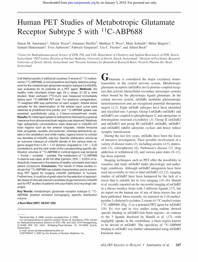
Imaging techniques such as PET offer the possibility tovisualize and study mGluR5 under physiologic and patho-logic conditions. Although mGluR5 antagonists have beenused successfully in vitro to label mGluR5 (12,13), imagingstudies of mGluR5 have been hampered by the lack of atracer that is suitable for in vivo imaging (14–16). Hamillet al. recently reported on the successful imaging of mGluR5in a rhesus monkey brain with 3 different ligands (17), butno report on the human use of any of these tracers has yetbeen published. More recently, we reported on 3-(6-methyl-pyridin-2-ylethynyl)-cyclohex-2-enone-O-11C-methyl-oxime(11C-ABP688) (Fig. 1) as a potential PET agent for mGluR5(18). Ex vivo and in vivo studies using rodents showedspecific binding in mGluR5-rich brain regions—in contrastto the 3 ligands disclosed by Hamill et al. (17)—withnegligible uptake in the cerebellum, a brain region knownto be devoid of mGluR5. The specificity of 11C-ABP688binding to mGluR5 was further substantiated using mGluR5knockout mice.
Received Sep. 5, 2006; revision accepted Nov. 3, 2006.For correspondence or reprints contact: Simon M. Ametamey, PhD, Center
for Radiopharmaceutical Science of ETH, PSI, and USZ, ETH-Honggerberg,D-CHAB IPW HCI H427, Wolfgang-Pauli-Strasse 10, CH-8093 Zurich,Switzerland.
E-mail: [email protected] ª 2007 by the Society of Nuclear Medicine, Inc.
MGLUR5 IMAGING IN HUMANS • Ametamey et al. 247
by on January 5, 2019. For personal use only. jnm.snmjournals.org Downloaded from
The present study investigated the utility of 11C-ABP688for imaging mGluR5 in humans.
MATERIALS AND METHODS
Radiosynthesis of 11C-ABP688The 11C labeling of ABP688 was achieved by O-11C-methylation
of desmethyl-ABP688 with 11C-methyl iodide as described previ-ously (18) but with slight modifications. Briefly, 11C-methyl iodidewas produced with a PET TRACE system (GE Healthcare) at theUniversity Hospital in Zurich from 11C-CO2 in a 2-step reactionsequence involving catalytic reduction of 11C-CO2 to 11C-methaneand subsequent gas-phase iodination according to the standard pro-cedure (19). In a typical procedure, the sodium salt of desmethyl-ABP688 obtained by stirring desmethyl-ABP688 with sodiumhydride in anhydrous N,N-dimethylformamide was reacted with11C-methyl iodide at 90�C for 5 min and the product purified bysemipreparative high-performance liquid chromatography (LunaC18 10-mm column [Phenomenex]; 250 · 10 mm; mobile phase,ethanol:0.1% phosphoric acid [50:50]; flow rate, 6 mL/min). Theproduct peak was collected, passed through a Millex-FG sterilefilter (Millipore), and formulated for human application.
Study PopulationSix healthy male volunteers (mean age, 25 y; range, 21–33 y)
were studied. None of the subjects had a history of neurologic orpsychiatric disorders. The study was approved by the ethics com-mittee of Canton Zurich, the Swiss Federal Office for RadiationProtection, and SwissMedic. Written consent was obtained fromeach volunteer.
MRIFor anatomic coregistration, a 3-dimensional spoiled gradient-
echo T1-weighted whole-brain image (voxel size, 0.94 · 0.94 ·1.5 mm) was acquired for each subject on a 1.5-T Signa InfinityTwinSpeed MRI system (GE Healthcare).
PETThe PET studies were performed on a whole-body Discovery
LS PET/CT scanner (GE Healthcare) in 3-dimensional mode. Thisis a scanner with an axial field of view of 14.6 cm and a recon-structed in-plane resolution of 7 mm. Before positioning of the vol-unteers on the scanner, catheters were placed in an antecubitalvein for tracer injection and in the radial artery for blood sam-pling. Before the PET study, a low-dose CT scan was acquired forcorrection of photon attenuation.
Transaxial images of the brain were reconstructed using filteredbackprojection (matrix, 128 · 128; 35 slices; voxel size, 2.34 ·2.34 · 4.25 mm).
15O-H2O PET. Before each study with 11C-ABP688, cerebralperfusion (cerebral blood flow, or CBF) was evaluated using 15O-H2O. For each PET measurement, 400–500 MBq of 15O-H2Owere injected intravenously using an automatic injection devicethat delivers a predefined dose of 15O-H2O over 20 s. After thebolus had arrived in the brain, a series of eighteen 10-s scans wasinitiated. The time course of arterial radioactivity was assessed bycontinuous sampling of blood drawn from the radial artery.
11C-ABP688 PET. After injection of 300–350 MBq of radioli-gand as a slow bolus over 2 min, a series of 20 scans was initiatedin 3-dimensional mode (10 · 60 s and 10 · 300 s; 60-min totalstudy duration).
For determination of the arterial input curve, arterial bloodsamples were collected every 30 s for the first 6 min and then atincreasing intervals until the end of the study (60 min after injec-tion). An aliquot of each sample was measured in a g-counter.Plasma samples were analyzed to correct for radiolabeled metab-olites. Samples of plasma (200 mL) were diluted with 2.5 mL ofwater. For assessment of tracer–to–radiolabeled metabolite ratios,the solution was passed through Sep-Pak tC18 cartridges (Waters),and each was eluted with 5 mL of water. 11C radioactivity trappedon the cartridges and aliquots of the eluted solution—correspondingto parent compound and radiolabeled metabolites, respectively—was measured in a g-counter.
Data AnalysisAll calculations were performed using the dedicated software
PMOD (PMOD Technologies) (20). Decay-corrected time–activitycurves were generated for the following cerebral regions: anteriorcingulate, medial temporal lobe, parietal cortex, cerebellum, andwhite matter. Values for the specific distribution volume (DV) werecalculated from the estimated rate constants.
15O-H2O PET. Quantitative parametric maps representing re-gional CBF were calculated using the integration method describedby Alpert et al. (21).
11C-ABP688 PET. Tissue time–activity curves were generatedfrom various brain structures by applying manually drawn vol-umes of interest to the dynamic datasets. Two-tissue-compartmentmodels were fitted to the time–activity curves, resulting in the rateconstants K1, k2, k3, and k4. K1 is a rate constant describing thetransfer of radioligand from plasma to the compartment consistingof free and nonspecifically bound ligand; k2 is a rate constantdescribing the reverse. The rate constants k3 and k4 denoteradioligand binding to the receptor and dissociation from thereceptor, respectively. The metabolite-corrected arterial activitywas used as the input function for calculating the model curve, andleast-squares optimization was performed using the Levenberg–Marquardt algorithm. Additionally, a voxelwise analysis was usedto calculate quantitative maps of the model rate constants. Toreduce the number of fit parameters, a global value of K1/k2(nondisplaceable compartment) for a study was obtained in a firststep by a coupled fit of the regional time–activity curves with K1/k2 as a common parameter (22). In a second step, the 2-tissue-compartment model was fitted to the time–activity curve of eachindividual voxel using the global K1/k2 value as a fixed modelparameter (23). The specific DVs (K1/k2) · (k3/k4) served as theoutcome parameter directly related to cerebral mGluR5 density.All data were processed using the quantitative PMOD software.
FIGURE 1. Structure of 11C-ABP688.
248 THE JOURNAL OF NUCLEAR MEDICINE • Vol. 48 • No. 2 • February 2007
by on January 5, 2019. For personal use only. jnm.snmjournals.org Downloaded from
11C-ABP688 was obtained from 11C-methyl iodide withan overall decay-corrected radiochemical yield of 35% 6
8% (n 5 14). The radiochemical purity was greater than98%, and the specific radioactivity ranged from 70 to 95GBq/mmol at the time of injection. The total amount ofstable ABP688 in the formulated solution was always lessthan 5 mg.
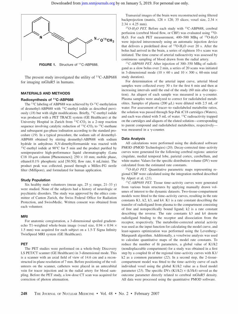
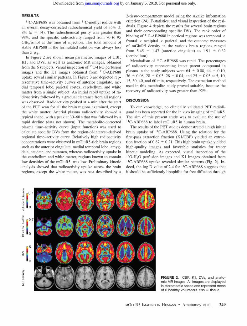
In Figure 2 are shown mean parametric images of CBF,K1, and DVs, as well as anatomic MR images, obtainedfrom the 6 subjects. Visual inspection of 15O-H2O perfusionimages and the K1 images obtained from 11C-ABP688uptake reveal similar patterns. In Figure 3 are depicted rep-resentative time–activity curves of anterior cingulate, me-dial temporal lobe, parietal cortex, cerebellum, and whitematter from a single subject. An initial rapid uptake of ra-dioactivity followed by a gradual clearance from all regionswas observed. Radioactivity peaked at 4 min after the startof the PET scan for all the brain regions examined, exceptthe white matter. Arterial plasma radioactivity showed atypical shape, with a peak at 30–60 s that was followed by arapid decline (data not shown). The metabolite-correctedplasma time–activity curve (input function) was used tocalculate specific DVs from the region-of-interest–derivedregional time–activity curve. Relatively high radioactivityconcentrations were observed in mGluR5-rich brain regionssuch as the anterior cingulate, medial temporal lobe, amyg-dala, caudate, and putamen, whereas radioactivity uptake inthe cerebellum and white matter, regions known to containlow densities of the mGluR5, was low. Preliminary kineticanalysis showed that radioactivity uptake across the brainregions, except the white matter, was best described by a
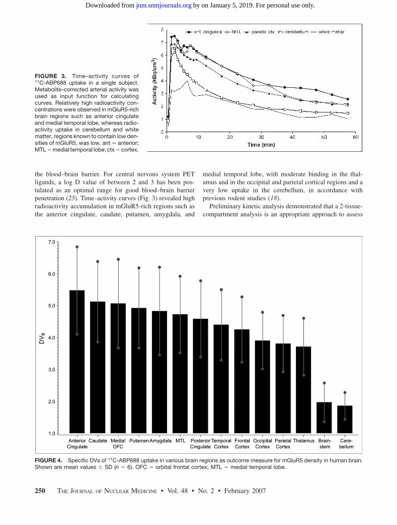
2-tissue-compartment model using the Akaike informationcriterion (24), F-statistics, and visual inspection of the resi-duals. Figure 4 depicts the results for several brain regionsand their corresponding specific DVs. The rank order ofbinding of 11C-ABP688 in cortical regions was temporal .
frontal . occipital . parietal, and the outcome measuresof mGluR5 density in the various brain regions rangedfrom 5.45 6 1.47 (anterior cingulate) to 1.91 6 0.32(cerebellum).
Metabolism of 11C-ABP688 was rapid. The percentagesof radioactivity representing intact parent compound inplasma in the study subjects were 64 6 0.08, 44 6 0.10,36 6 0.08, 28 6 0.03, 28 6 0.04, and 25 6 0.03 at 5, 10,15, 30, 40, and 60 min, respectively. The extraction methodused in this metabolite study proved suitable, because therecovery of radioactivity was greater than 92%.
DISCUSSION
To our knowledge, no clinically validated PET radioli-gand has been reported for the in vivo imaging of mGluR5.The aim of this present study was to evaluate the use of11C-ABP688 to label mGluR5 in human brain.
The results of the PET studies demonstrated a high initialbrain uptake of 11C-ABP688. Using the relation for thefirst-pass extraction fraction (K1/CBF) yielded an extrac-tion fraction of 0.87 6 0.21. This high brain uptake yieldedhigh-quality images and favorable statistics for tracerkinetic modeling. As expected, visual inspection of the15O-H2O perfusion images and K1 images obtained from11C-ABP688 uptake revealed similar patterns (Fig. 2). In-deed, the log D value of 2.4 for 11C-ABP688 suggests thatit should be sufficiently lipophilic for free diffusion through
FIGURE 2. CBF, K1, DVs, and anato-mic MR images. All images are displayedin stereotactic space and represent meanof 6 healthy volunteers. tiss 5 tissue.
MGLUR5 IMAGING IN HUMANS • Ametamey et al. 249
by on January 5, 2019. For personal use only. jnm.snmjournals.org Downloaded from
the blood–brain barrier. For central nervous system PETligands, a log D value of between 2 and 3 has been pos-tulated as an optimal range for good blood–brain barrierpenetration (25). Time–activity curves (Fig. 3) revealed highradioactivity accumulation in mGluR5-rich regions such asthe anterior cingulate, caudate, putamen, amygdala, and
medial temporal lobe, with moderate binding in the thal-amus and in the occipital and parietal cortical regions and avery low uptake in the cerebellum, in accordance withprevious rodent studies (18).
Preliminary kinetic analysis demonstrated that a 2-tissue-compartment analysis is an appropriate approach to assess
FIGURE 3. Time–activity curves of11C-ABP688 uptake in a single subject.Metabolite-corrected arterial activity wasused as input function for calculatingcurves. Relatively high radioactivity con-centrations were observed in mGluR5-richbrain regions such as anterior cingulateand medial temporal lobe, whereas radio-activity uptake in cerebellum and whitematter, regions known to contain low den-sities of mGluR5, was low. ant 5 anterior;MTL 5 medial temporal lobe; ctx 5 cortex.
FIGURE 4. Specific DVs of 11C-ABP688 uptake in various brain regions as outcome measure for mGluR5 density in human brain.Shown are mean values 6 SD (n 5 6). OFC 5 orbital frontal cortex; MTL 5 medial temporal lobe.
250 THE JOURNAL OF NUCLEAR MEDICINE • Vol. 48 • No. 2 • February 2007
by on January 5, 2019. For personal use only. jnm.snmjournals.org Downloaded from
the specific DV of 11C-ABP688 as a measure of receptordensity. Except for white matter, all brain regions examinedwere best described by a 2-tissue-compartment model. Thepattern of parametric images of the specific DVs wasdifferent from the perfusion studies with 15O-H2O and theK1 parametric images, indicating that this outcome mea-sure of mGluR5 density is not perfusion-related. The rankorder of the specific DVs obtained was in accordance withthe reported distribution pattern of mGluR5 in rodents andhumans (26–28). In a recent publication, Hamill et al. (17)described 3 potential PET ligands and their characterizationin rhesus monkey brain. In this study, all 3 candidatesexhibited selective uptake in mGluR5-rich regions such asthe frontal cortex, striatum, and surprisingly the cerebel-lum, a brain region that has been shown to have a low or noexpression of mGluR5 in rodents and humans (26–28). Inthe present study, the lowest DV was clearly in the cerebel-lum, a finding that is consistent with a low mGluR5 density.However, the exact fraction of specific DV in the cerebel-lum would have to be determined with a blocking study.Whether such a blocking study, which would require sub-stantial doses of a blocking agent, can be performed onhumans is not clear. An alternative could be to perform invitro autoradiography using postmortem human brain tis-sue. Nevertheless, the higher specific DVs obtained in brainregions known to contain high densities of mGluR5, thelower cerebellar uptake, and the good agreement of the pres-ent data with previous rodent studies (18) suggest that radio-activity uptake in these brain regions reflects the pattern ofmGluR5 distribution in humans.
In the radiometabolite analysis, the extraction methodproved suitable, because the recovery of radioactivity wasgreater than 92%. Radiolabeled metabolites were more hy-drophilic than the parent compound and thus were unlikelyto cross the blood–brain barrier. Previous animal studiesshowed that more than 95% of radioactivity in rat brain30 min after injection consisted of unchanged 11C-ABP688.Although the radiolabeled metabolites were not identified,the metabolite profile suggests a similar metabolic fate of11C-ABP688 in both rodents and humans.
CONCLUSION
An efficient radiosynthesis for producing 11C-ABP688 ingood radiochemical yield and with high specific radioac-tivity for application in humans was developed. The highestuptake of 11C-ABP688 was in mGluR5-rich regions such asthe anterior cingulate, medial temporal lobe, putamen, andcaudate. The specific DVs calculated from several regionsof interest also agreed well with prior animal data. Takentogether, these data suggest that 11C-ABP688 has suitablecharacteristics for exploring mGluR5 distribution in hu-mans using PET. Furthermore, 11C-ABP688 could be ofgreat value for the selection of appropriate doses of clin-ically relevant candidate drugs that bind to the mGluR5
and for PET studies on patients with psychiatric and neuro-logic disorders.
ACKNOWLEDGMENTS
We acknowledge the support of Pius August Schubiger,the head of the Center for Radiopharmaceutical Science ofETH, PSI, and USZ; Rene Amstutz, the head of DiscoveryTechnologies; and Graeme Bilbe, the head of the Neuro-science Department at the Novartis Institutes for Biomed-ical Research. We also thank Cyril Burger, Christoph Hock,and Andrea Bettio for their contributions.
REFERENCES
1. Pin JP, Duvoisin R. The metabotropic glutamate receptors: structure and
functions. Neuropharmacology. 1995;34:1–26.
2. Spooren WP, Gasparini F, Salt TE, Kuhn R. Novel allosteric antagonists shed
light on mGluR5 and CNS disorders. Trends Pharmacol Sci. 2001;22:331–337.
3. Gereau RW, Conn PJ. Roles of specific metabotropic glutamate receptor sub-
types in regulation of hippocampal CA1 pyramidal cell excitability. J Neurophysiol.
1995;74:122–129.
4. Spooren WP, Vassout A, Neijt HC, et al. Anxiolytic-like effects of the prototypi-
cal metabotropic glutamate receptor 5 antagonist 2-methyl-6-(phenylethynyl)-
pyridine in rodents. J Pharmacol Exp Ther. 2000;295:1267–1275.
5. Tatarczynska E, Klodzinska A, Chojnacka-Wojcik E, et al. Potential anxiolytic-
and antidepressant-like effects of MPEP, a potent, selective and systemically
active mGlu5 receptor antagonist. Br J Pharmacol. 2001;132:1423–1430.
6. Ohnuma T, Augood SJ, Arai H, McKenna PJ, Emson PC. Expression of the
human excitatory amino acid transporter 2 and metabotropic glutamate receptors
3 and 5 in the prefrontal cortex from normal individuals and patients with
schizophrenia. Brain Res Mol Brain Res. 1998;56:207–217.
7. Rouse ST, Marino MJ, Bradley SR, Awad H, Wittmann M, Conn PJ. Distribution
and roles of metabotropic glutamate receptors in the basal ganglia motor circuit:
implications for treatment of Parkinson’s disease and related disorders. Pharmacol
Ther. 2000;88:427–435.
8. Chiamulera C, Epping-Jordan MP, Zocchi A, et al. Reinforcing and locomotor
stimulant effects of cocaine are absent in mGluR5 null mutant mice. Nat
Neurosci. 2001;4:873–874.
9. Walker K, Bowes M, Panesar M, et al. Metabotropic glutamate receptor subtype
5 (mGluR5) and nociceptive function. I. Selective blockade of mGluR5 in models
of acute, persistent and chronic pain. Neuropharmacology. 2001a;40:1–9.
10. Walker K, Reeve A, Bowes M, et al. MGluR5 and nociceptive function II.
MGluR5 functionally expressed on peripheral sensory neurones mediate inflam-
2007;48:247-252.J Nucl Med. Hintermann, Yves Auberson, Fabrizio Gasparini, Uta C. Fischer and Alfred BuckSimon M. Ametamey, Valerie Treyer, Johannes Streffer, Matthias T. Wyss, Mark Schmidt, Milen Blagoev, Samuel C-ABP688
11Human PET Studies of Metabotropic Glutamate Receptor Subtype 5 with
http://jnm.snmjournals.org/content/48/2/247This article and updated information are available at:
Information about subscriptions to JNM can be found at:
http://jnm.snmjournals.org/site/misc/permission.xhtmlInformation about reproducing figures, tables, or other portions of this article can be found online at:
(Print ISSN: 0161-5505, Online ISSN: 2159-662X)1850 Samuel Morse Drive, Reston, VA 20190.SNMMI | Society of Nuclear Medicine and Molecular Imaging
is published monthly.The Journal of Nuclear Medicine


![Molecular Analysis of the Histamine H 3-Receptor · III 2.4.2 [³H]JNJ-7753707 and [35 S]GTP γS binding: 45Quantitative analysis of receptor-to-G protein stoichiometries 2.4.3 Steady-state](https://static.unterlagen.site/doc/80x56/5fd46fbb0be1866eec555122/molecular-analysis-of-the-histamine-h-3-receptor-iii-242-hjnj-7753707-and.jpg)


