Journal of large-scale research facilities, 2, A61 (2016) http://dx.doi.org/10.17815/jlsrf-2-109 Published: 16.03.2016 GALAXI: Gallium anode low-angle x-ray instrument Forschungszentrum Jülich, Jülich Centre for Neutron Science * Instrument Scientists: - Emmanuel Kentzinger, Jülich Centre for Neutron Science (JCNS) and Peter Grünberg Institut (PGI), JARA-FIT, Forschungszentrum Jülich GmbH, D-52425 Jülich, Germany, phone: +49(0) 2461 61 3139, email: [email protected]- Margarita Krutyeva, Jülich Centre for Neutron Science (JCNS) and Institut for Complex Systems (ICS), Forschungszentrum Jülich GmbH, D-52425 Jülich, Germany, phone: +49(0) 2461 61 1838, e-mail: [email protected]- Ulrich Rücker, Jülich Centre for Neutron Science (JCNS) and Peter Grünberg Institut (PGI), JARA-FIT, Forschungszentrum Jülich GmbH, D-52425 Jülich, Germany, phone: +49(0) 2461 61 6896, e-mail: [email protected]Abstract: The high brilliance laboratory small angle X-ray scattering instrument GALAXI, which is operated by JCNS, Forschungszentrum Jülich, permits the investigation of chemical correlations in bulk materials or of structures deposited on a surface at nanometre and mesoscopic length scales. The instrument is capable to perform GISAXS experiments in reection at grazing incidence as well as SAXS experiments in transmission geometry. The X-ray ux on sample is comparable or higher than the one obtained at a comparable beamline at a second generation synchrotron radiation source. 1 Introduction Small Angle X-ray Scattering (SAXS) permits the investigation of chemical correlations in bulk materi- als at nanometre and mesoscopic length scales. When the X-ray beam is sent under grazing incidence on a at surface, reectometry and Grazing Incidence Small Angle X-ray Scattering (GISAXS) can be performed, allowing the determination of the chemical depth-prole of multilayer thin lms and the in-plane correlations at nanometre and mesoscopic length scales. The diractometer of GALAXI is based on the JUSIFA anomalous SAXS instrument formerly installed at the beamline B1 of the DORIS storage ring at HASYLAB, DESY Hamburg, Germany. After the shutdown of DORIS, the instrument * Cite article as: Jülich Centre for Neutron Science. (2016). GALAXI: Gallium anode low-angle x-ray instrument. Journal of large-scale research facilities, 2, A61. http://dx.doi.org/10.17815/jlsrf-2-109 1
Transcript
Journal of large-scale research facilities, 2, A61 (2016) http://dx.doi.org/10.17815/jlsrf-2-109
Published: 16.03.2016
GALAXI:Gallium anode low-angle x-ray instrument
Forschungszentrum Jülich, Jülich Centre for Neutron Science *
Instrument Scientists:- Emmanuel Kentzinger, Jülich Centre for Neutron Science (JCNS) and Peter Grünberg Institut (PGI),
- Margarita Krutyeva, Jülich Centre for Neutron Science (JCNS) and Institut for Complex Systems(ICS), Forschungszentrum Jülich GmbH, D-52425 Jülich, Germany,phone: +49(0) 2461 61 1838, e-mail: [email protected]
- Ulrich Rücker, Jülich Centre for Neutron Science (JCNS) and Peter Grünberg Institut (PGI), JARA-FIT,Forschungszentrum Jülich GmbH, D-52425 Jülich, Germany,phone: +49(0) 2461 61 6896, e-mail: [email protected]
Abstract: The high brilliance laboratory small angle X-ray scattering instrument GALAXI, which isoperated by JCNS, Forschungszentrum Jülich, permits the investigation of chemical correlations inbulk materials or of structures deposited on a surface at nanometre and mesoscopic length scales. Theinstrument is capable to perform GISAXS experiments in re�ection at grazing incidence as well as SAXSexperiments in transmission geometry. The X-ray �ux on sample is comparable or higher than the oneobtained at a comparable beamline at a second generation synchrotron radiation source.
1 Introduction
Small Angle X-ray Scattering (SAXS) permits the investigation of chemical correlations in bulk materi-als at nanometre and mesoscopic length scales. When the X-ray beam is sent under grazing incidenceon a �at surface, re�ectometry and Grazing Incidence Small Angle X-ray Scattering (GISAXS) can beperformed, allowing the determination of the chemical depth-pro�le of multilayer thin �lms and thein-plane correlations at nanometre and mesoscopic length scales. The di�ractometer of GALAXI isbased on the JUSIFA anomalous SAXS instrument formerly installed at the beamline B1 of the DORISstorage ring at HASYLAB, DESY Hamburg, Germany. After the shutdown of DORIS, the instrument
*Cite article as: Jülich Centre for Neutron Science. (2016). GALAXI: Gallium anode low-angle x-ray instrument. Journalof large-scale research facilities, 2, A61. http://dx.doi.org/10.17815/jlsrf-2-109
Journal of large-scale research facilities, 2, A61 (2016) http://dx.doi.org/10.17815/jlsrf-2-109
has been moved to the Research Centre of Jülich and has been reinstalled with a new X-ray source anda new position sensitive detector.
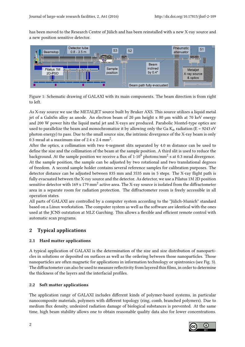
Figure 1: Schematic drawing of GALAXI with its main components. The beam direction is from rightto left.
As X-ray source we use the METALJET source built by Bruker AXS. This source utilizes a liquid metaljet of a GaInSn alloy as anode. An electron beam of 20 µm height x 80 µm width at 70 keV energyand 200 W power hits the liquid metal jet and X-rays are produced. Parabolic Montel-type optics areused to parallelize the beam and monochromatize it by allowing only the Ga Kα radiation (E = 9243 eVphoton energy) to pass. Due to the small source size, the intrinsic divergence of the X-ray beam is only0.3 mrad at a maximum size of 2.4 x 2.4 mm2.After the optics, a collimation with two 4-segment slits separated by 4.0 m distance can be used tode�ne the size and the collimation of the beam at the sample position. A third slit is used to reduce thebackground. At the sample position we receive a �ux of 1·109 photons/mm2·s at 0.3 mrad divergence.At the sample position, the sample can be adjusted by two rotational and two translational degreesof freedom. A second sample holder contains several reference samples for calibration purposes. Thedetector distance can be adjusted between 835 mm and 3535 mm in 5 steps. The X-ray �ight path isfully evacuated between the X-ray source and the detector. As detector, we use a Pilatus 1M 2D positionsensitive detector with 169 x 179 mm2 active area. The X-ray source is isolated from the di�ractometerarea in a separate room for radiation protection. The di�ractometer room is freely accessible in alloperation states.All parts of GALAXI are controlled by a computer system according to the "Jülich-Munich" standardbased on a Linux workstation. The computer system as well as the software are identical with the onesused at the JCNS outstation at MLZ Garching. This allows a �exible and e�cient remote control withautomatic scan programs.
2 Typical applications
2.1 Hard matter applications
A typical application of GALAXI is the determination of the size and size distribution of nanoparti-cles in solutions or deposited on surfaces as well as the ordering between those nanoparticles. Thosenanoparticles are often magnetic for applications in information technology or spintronics (see Fig. 3).The di�ractometer can also be used to measure re�ectivity from layered thin �lms, in order to determinethe thickness of the layers and the interfacial pro�les.
2.2 Soft matter applications
The application range of GALAXI includes di�erent kinds of polymer-based systems, in particularnanocomposite materials, polymers with di�erent topology (ring, comb, branched polymers). Due tomedium �ux density, undesired radiation damage of biological substances is prevented. At the sametime, high beam stability allows one to obtain reasonable quality data also for lower concentrations.
http://dx.doi.org/10.17815/jlsrf-2-109 Journal of large-scale research facilities, 2, A61 (2016)
The instrument was successfully used for structural studies of proteins in crowded environment andalso fragments of DNA.
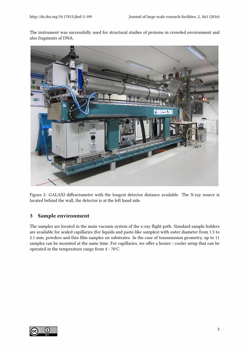
Figure 2: GALAXI di�ractometer with the longest detector distance available. The X-ray source islocated behind the wall, the detector is at the left hand side.
3 Sample environment
The samples are located in the main vacuum system of the x-ray �ight path. Standard sample holdersare available for sealed capillaries (for liquids and paste-like samples) with outer diameter from 1.5 to2.1 mm, powders and thin �lm samples on substrates. In the case of transmission geometry, up to 11samples can be mounted at the same time. For capillaries, we o�er a heater / cooler setup that can beoperated in the temperature range from 4 - 70°C.
Journal of large-scale research facilities, 2, A61 (2016) http://dx.doi.org/10.17815/jlsrf-2-109
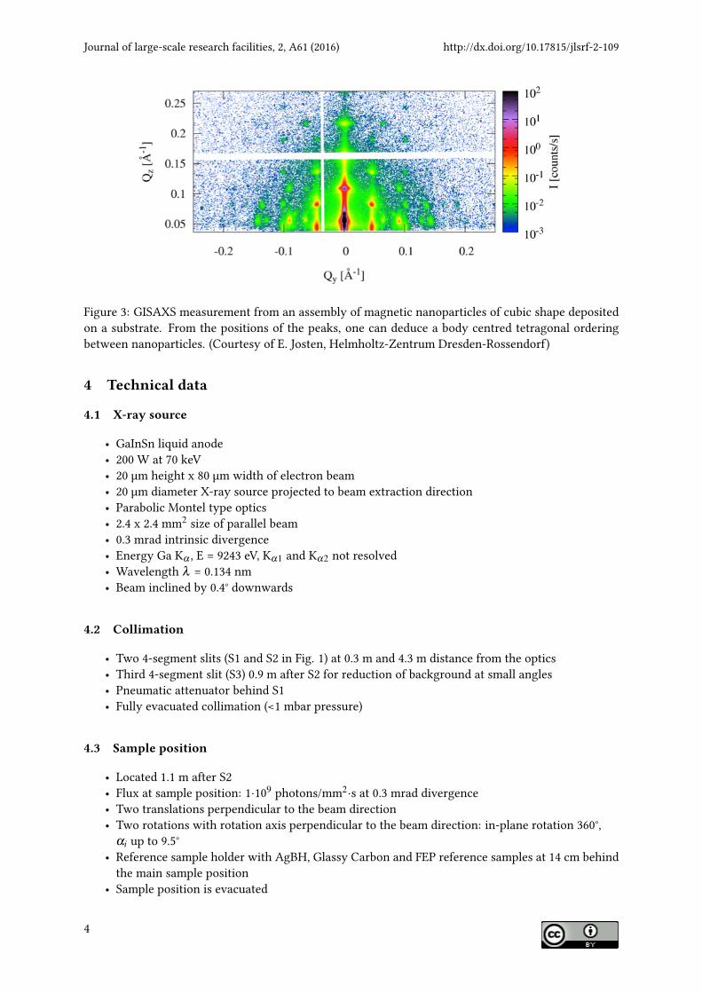
Figure 3: GISAXS measurement from an assembly of magnetic nanoparticles of cubic shape depositedon a substrate. From the positions of the peaks, one can deduce a body centred tetragonal orderingbetween nanoparticles. (Courtesy of E. Josten, Helmholtz-Zentrum Dresden-Rossendorf)
4 Technical data
4.1 X-ray source
• GaInSn liquid anode• 200 W at 70 keV• 20 µm height x 80 µm width of electron beam• 20 µm diameter X-ray source projected to beam extraction direction• Parabolic Montel type optics• 2.4 x 2.4 mm2 size of parallel beam• 0.3 mrad intrinsic divergence• Energy Ga Kα , E = 9243 eV, Kα1 and Kα2 not resolved• Wavelength λ = 0.134 nm• Beam inclined by 0.4° downwards
4.2 Collimation
• Two 4-segment slits (S1 and S2 in Fig. 1) at 0.3 m and 4.3 m distance from the optics• Third 4-segment slit (S3) 0.9 m after S2 for reduction of background at small angles• Pneumatic attenuator behind S1• Fully evacuated collimation (<1 mbar pressure)
4.3 Sample position
• Located 1.1 m after S2• Flux at sample position: 1·109 photons/mm2·s at 0.3 mrad divergence• Two translations perpendicular to the beam direction• Two rotations with rotation axis perpendicular to the beam direction: in-plane rotation 360°,
αi up to 9.5°• Reference sample holder with AgBH, Glassy Carbon and FEP reference samples at 14 cm behind
the main sample position• Sample position is evacuated
http://dx.doi.org/10.17815/jlsrf-2-109 Journal of large-scale research facilities, 2, A61 (2016)
4.4 Detectors
4.4.1 Small-Angle detector
• Pilatus 1M 2D-position sensitive detector located in the small-angle scattering region• Active area: 169 mm width x 179 mm height• Distance between pixels: 0.172 mm horizontal and vertical• Sample to detector distances available: 835 mm, 1285 mm, 1735 mm, 2635 mm, 3535 mm• Evacuated detector tube• Q-range: 4·10−2 . . . 8 nm −1
4.4.2 Single detectors and monitors
• Ionization chambers after optics and between S2 and S3• PIN diodes after S1 and after sample position• Calibrated PIN diode available at the sample position


![Text M25, M30, M40 Version B - Auß · PDF fileZündkerzen, Anode, Propeller, Kraftstofffilter, Ölfilter, Kohlebürsten, Starterseil, ... 17 [2] Fernsteuerungsseilzüge am Motor anschließen](https://static.unterlagen.site/doc/80x56/5a791ce77f8b9a7b548d2f79/text-m25-m30-m40-version-b-zndkerzen-anode-propeller-kraftstofffilter.jpg)


