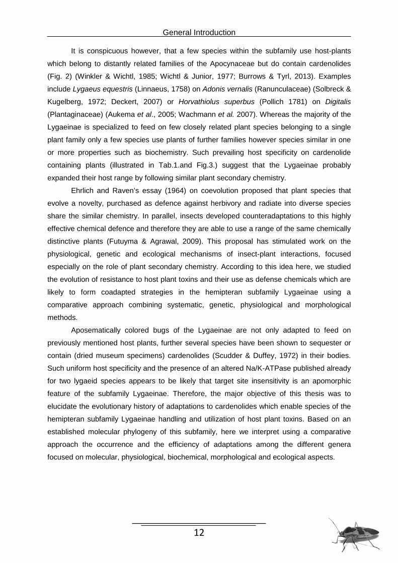
Evolution of adaptations to cardiac glycosides in the hemipteran subfamily Lygaeinae Dissertation Zur Erlangung der Würde des Doktors der Naturwissenschaften des Fachbereichs Biologie, der Fakultät für Mathematik, Informatik und Naturwissenschaften, der Universität Hamburg vorgelegt von Christiane Bramer aus Lübben Hamburg, 2014
Transcript
Evolution of adaptations to cardiac glycosides in the hemipteran
subfamily Lygaeinae
Dissertation
Zur Erlangung der Würde des Doktors der
Naturwissenschaften des Fachbereichs Biologie, der Fakultät für Mathematik,
Informatik und Naturwissenschaften,
der Universität Hamburg
vorgelegt von
Christiane Bramer
aus Lübben
Hamburg, 2014
“Milkweed bugs (Lygaeinae) are the butterflies of the bug world –
black on red associated with poison may deter predators, but it
attracts scientists!” Jeffrey R. Aldrich
Table of contents
General introduction 6
Chapter 1: Evolution of resistance traits: How Lygaeinae (Heteroptera, 15
Lygaeidae) cope with toxic host plant cardenolides
Chapter 2: Metabolic alteration of cardiac glycosides in Lygaeinae: 32
detoxification or optimized uptake?
Chapter 3: Stepwise evolution of storage compartments for defensive toxins 47
in the Lygaeinae (Heteroptera: Lygaeidae)
Chapter 4: Deterrent effect of cardenolides: effects of diet on defence of 64
Oncopeltus fasciatus against the golden orb web spider Nephila
senegalensis
General discussion and outlook 75
Zusammenfassung 86
Summary 89
References 92
Eidesstattliche Versicherung und Aufführung der Inanspruchnahme fremder Hilfen
Der in Kapitel 1 durchgeführte Enzym - Assay wurde von Herrn Dr. Georg Petschenka
durchgeführt, die DNA - Sequenzanalysen der Na/K-ATPase dieser Studie
wurde von Frau Vera Wagschal durchgeführt.
Die in Kapitel 2 durchgeführte MALDI-TOF-MS Analyse wurde am Department für
Organische Chemie an der Universität Hamburg erstellt.
Die in Kapitel 4 vorgestellten Daten wurden von Herrn Christian Schweitzer erhoben.
Hiermit erkläre ich an Eides statt, dass ich die vorliegende Dissertationsschrift selbst verfasst
und keine anderen als die angegebenen Quellen und Hilfsmittel benutzt habe.
Hamburg, den 14.03.2014
6
General Introduction
In evolutionary terms, the so-called ‘arms race’ between plants and insects has given rise to
highly specialized chemical interactions. Noxious phytochemicals that are repulsive,
unpalatable or poisonous are one of the primary defence mechanisms of plants against
insect herbivores. Such plant secondary compounds usually display detrimental effects on
non-adapted insect herbivores and consequently restrict their host range via feeding or
oviposition choices (Awmack & Leather, 2002). Therefore, adapted herbivores in several
insect orders have evolved different ways to overcome these plant defence barriers
(Vaughan & Jungreis, 1977; Schoonhoven et al., 2005; Dobler et al., 2011). Including
mechanisms of detoxification (Scott & Wen, 2001; Li et al., 2002,2007; Després et al., 2007),
possession of impermeable guts (Scudder & Meredith, 1982b; Petschenka et al., 2013) or
the avoidance of noxious plant parts (Dussourd & Eisner, 1987, Després et al., 2007) are
most efficient and widespread adaptations to exploit a high variety of chemicals encountered
in their food. Moreover, some specialized insects not only avoid poisoning by the toxin,
further they acquire and store them in various tissues, glands or compartments
(sequestration) where they act as defensive compounds for insect’s own benefits (Opitz &
Müller; 2009). These naturally occurring substances have either beneficial or toxic effects,
depending on dosage or biological activity.
One class of secondary plant compounds (allelochemicals), are the cardiac
glycosides (CGs) (Fig.1) which have frequently been studied with regard to plant insect
interactions.
Fig. 1. Chemical structure of cardiac glycosides. A steroid backbone (here representing ouabain) of four fused rings is connected at C17 to a 5-membered mono-unsaturated lactone ring in the case of cardenolides (R2) or to a 6-membered double-unsaturated lactone ring in bufadienolides. Cardenolides occur as free genins or with sugar moieties glycosidically bound at C3 (R1). Rhamnose attached at C3 yields ouabain (g-strophantin), three molecules digitoxose3 yield digoxin.
General Introduction
7
All cardiac glycosides (CGs) share as common features the basic steroid skeleton (aglycone
or genin) with a lactone ring in β position at C17 and are characterized by a 14β-hydroxyl
group and sugar moieties (glycoside) attached through an OH of carbon 3β (Fig. 1). A wide
variety of sugar molecules are known to occur in natural CGs but only a few such as glucose,
rhamnose and fructose are widespread among plants. Representative examples of common
CGs in biochemical research are illustrated in Figure 1: digoxin with three molecules of
digitoxose linked to the aglycone and ouabain which has a single rhamnose molecule. The
typical AB cis and AB trans conformation is a feature of medically important cardenolides in
the plant families Apocynaceae. Depending on the structure of the aglycon, the CGs can be
divided into two groups: cardenolides and bufadienolides. Cardenolides have a 5-membered
mono-unsaturated γ-lactone ring, at the C17 position whereas bufadienolides possess a 6-
membered double-unsaturated δ-lactone ring (Fig. 1).
With a high diversity of chemical forms (>500), these secondary metabolites are erratically
distributed among 14 angiosperm families that include both cardenolide- and bufadienolide-
containing species (Tab. 1)( Malcolm, 1991; Luckner & Wichtl, 2000).
Tab. 1. Plant families that include cardiac glycoside (cardenolide or bufadienolide) containing species. Cardenolides have been recorded from 62 genera of 10 plant families (Malcolm, 1991; Luckner & Wichtl, 2000; Hänsel & Sticker, 2007). Bufadienolides are found in 11 genera and 6 families (Krenn & Kopp, 1998; Steyn & van Heerden, 1998). The family names have been adapted to the current taxonomic classification by the KEW world checklists of selected Plant Families (as of October 18th 2013).
General Introduction
8
Bufadienolides have been recordet from only 11 genera of 6 plant families but have been
proved in only few animal families. In the animal kingdom, these substances are most
wiespread in toads such as Bufonidae but also occur in snakes, fireflies, and other insects
(Krenn & Kopp, 1998; Steyn & van Heerden, 1998).
Cardenolides on the other hand have a larger distribution among a total of 62 genera
in 10 plant families (Malcolm, 1991; Luckner & Wichtl, 2000; Hänsel & Sticker, 2007) which
have been found in a wide range of habitats among the world and particularly in tropical and
temperate regions (Agrawal et al. 2012). The most prominent occurrence of cardenolides is
reported from the dogbane family Apocynaceae (including the former Asclepiadaceae) which
produce cardenolides as an abundant toxic principle (Agrawal et al. 2012). In general,
cardinolides are an important class of naturally occurring drugs whose actions include both
beneficial and toxic effects on the heart in vertebrates and insects as neurotoxins (Scholz &
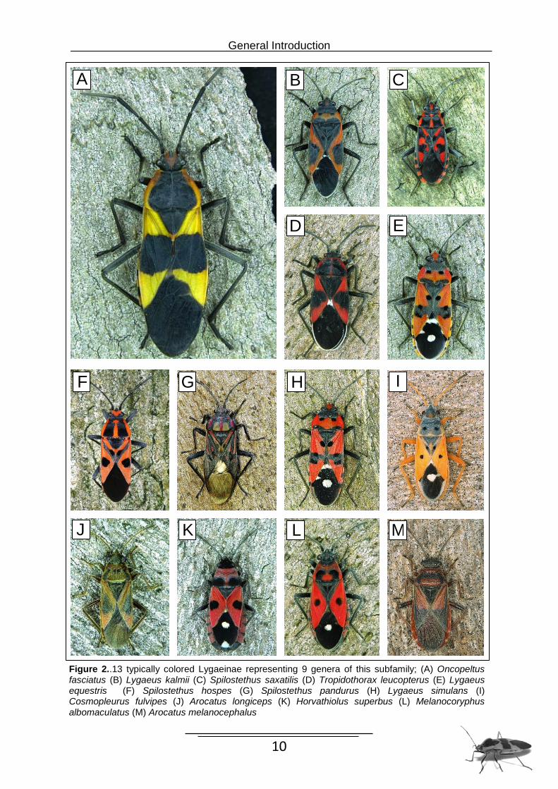
Figure 3. Common cardenolide-rich plant species used as host plants by several species of lygaeinae. (A) Nerium oleander; Apocynaceae, (B) Digitalis purpurea; Plantaginaceae, (C) Calotropis procera; Apocynaceae, (D) Gomphocarpus physocarpus; Apocynaceae, (E) Asclepias syriaca; Apocynaceae, (F) Adonis vernalis; Ranunculaceae
A
C
B
D
E F
General Introduction
12
It is conspicuous however, that a few species within the subfamily use host-plants
which belong to distantly related families of the Apocynaceae but do contain cardenolides
Approximately 80 % of the Lygaeinae from at least five continents use plants of the
milkweed family Apocynaceae as primary hosts (Scudder & Duffey, 1972). Whereas the
Chapter 1: Evolution of resistant traits
17
majority is specialized to feed on few closely related plant species belonging to a single plant
family, only a few species of the aposematically colored bugs utilize further families but with
the same chemically properties. Moreover, several species have been shown to sequester or
contain (in dried museum specimens) cardenolides (Scudder & Duffey, 1972). Gene
sequence analyses suggested that in addition to O. fasciatus, the small milkweed bug
(Lygaeus kalmii) also possesses a Na/K-ATPase with a reduced sensitivity to cardenolides
(Dobler et al., 2012; Zhen et al. 2012). This, together with the typical black and red
aposematic coloration, render it likely that the use of sequestered cardenolides as defensive
compounds and the possession of target site insensitivity of Na/K-ATPase are basal features
of this hemipteran group. In a comparative approach we tested 10 species of the Lygaeinae
for the coadapted traits target site insensitivity and cardenolide sequestration. The parallel
construction of a molecular phylogeny of 20 Lygaeinae and 4 outgroup species (2 lygaeids,
one species each of Oxycarenidae and Pyrrhocoridae) enabled us to address the following
questions: 1) is the ability to tolerate and to sequester cardenolides a basal feature of the
Lygaeinae, 2) if basal, are target site insensitivity and cardenolide sequestration then
maintained in species which no longer have to cope with cardenolides, and 3) is the
secondary use of non-apocynaceous cardenolide plants like Adonis vernalis
(Ranunculaceae), Digitalis purpurea (Plantaginaceae) or Urginea maritima (Asparagaceae)
likely due to a preadaptation to cardenolides?
To approach these questions we performed a comparative analysis of sequestration
of two radioactively labeled cardenolides, which represent a polarity range of toxic
compounds present in the natural diet of the insects. Through in vitro spectrophotometric
assays we tested nervous tissues of seven Lygaeinae and one outgroup species for target
site insensitivity against the cardenolide ouabain.
Mutations conferring resistance to cardenolides were shown only in a few studies
(Holzinger et al., 1992; Labeyrie & Dobler, 2004; Dobler et al., 2012; Zhen et al., 2012;
Petschenka et al., 2013) demonstrating that due to specific amino acid substitutions several
insect species are resistant to ouabain. In the present study, we also sequenced Na/K-
ATPase genes of 11 species of Lygaeinae (plus 2 outgroups) and analyzed the resulting
amino acid sequences for critical substitutions to understand the molecular basis of
resistance.
Our integrative analysis of two probably coadapted traits in a phylogenetic framework
affords reconstructing the evolution of adaptations to cardenolides in the Lygaeinae.
Chapter 1: Evolution of resistant traits
18
Material and Methods
Specimens for sequestration assays and in vitro analysis of Na/K-ATPase
Adults of several species of the Lygaeinae were obtained both from the field and from
laboratory cultures. As outgroups we used species of the families Phyrrhocoridae, Berytidae
and Lygaeidae (Ischnorhynchinae). For feeding assays the species were reared in the
laboratory from the egg stage on a diet of husked sunflower seeds (Helianthus annuus L.)
and water. Colonies were kept under artificial light at a 16 h/8 h light/dark cycle at 26°C (C.
fulvipes, H. superbus, O. fasciatus, S.pandurus) or 30°C (L. equestris, L. kalmii, L. simulans).
All further species including Arocatus longiceps, A. melanocephalus, Kleidocerys
resedae (Lygaeidae), Metatropis rufescens (Berytidae), and Pyrrhocoris apterus
(Pyrrhocoridae) were field collected individuals. A. longiceps (Berlin, Germany) and A.
melanocephalus (Kallinchen, Germany) were maintained on sunflower seeds and seeds of
Platanus (2 days) under ambient conditions (A. l.) or at 26°C (A. m., 16 h/8 h light/dark
cycle). K. resedae, M. rufescens, and P. apterus (Hamburg/Hamburg/Straupitz, Germany)
were maintained on sunflower seeds (26°C, 16 h/8 h light/dark cycle) (2-3 days).
Tropidothorax leucopterus (Grießheim, Germany) was reared on sunflower seeds and
cut branches of Vincetoxicum hirundinaria (26°C, 16 h/8 h light/dark cycle).
Sequestration Assay
To assess the ability to sequester cardenolides, we fed 9 species of the Lygaeinae of 6
genera (see Tab.1.) and the three outgroup species (K. resedae, Lygaeidae; M. rufescens,
Berytidae; and P. apterus, Pyrrhocoridae) with radioactively (3H) labeled cardenolides.
As plants typically produce several cardenolides with a wide polarity range we used
the polar [3H]-ouabain and the relatively apolar cardenolide [3H]-digoxin (both Perkin Elmer
LAS GmbH, Rodgau, Germany). Both cardenolides which probably do not occur in the
natural host plants of Lygaeinae were used due to their commercial availability. Individuals
were immobilized with a lasso made of dental floss (see insert in Fig. 2) and their proboscis
was manually introduced into a droplet (2 µl) of 5% sucrose solution containing 5 µM 3H-
cardenolide dissolved in ethanol on parafilm (final concentration of ethanol 17.7%). After
feeding, specimens were kept for 10 days at 26°C and supplied with water and sunflower
seeds ad libitum to allow for gut clearance of cardenolides. The incubation period of 10 days
was chosen as we observed that keeping treated individuals for only 3 days before analysis
Chapter 1: Evolution of resistant traits
19
produced less reliable results (see supplemental figure 1 and discussion) which might be due
to incomplete gut clearance of cardenolides. After 10 days, individuals were frozen in liquid
nitrogen and homogenized with a pestle (glass or stainless steel). To evaluate stored
cardenolides, samples were extracted with 1 ml methanol (3 x) by vortex stirring. After
centrifugation, an aliquot (200 µl) of the pooled supernatants was added to 3 ml scintillation
cocktail (Ultima Gold, Perkin Elmer) to quantify radioactivity (amount of [3H]-cardenolide
taken up) with a liquid scintillation counter (Wallac 1409). In addition, the residual
radioactivity on the Parafilm used as feeding support and the radioactivity of the drinking
solution (2 µl aliquots) were determined to calculate the percentage of radioactivity actually
stored by the hemipteran specimens. To do so, the residual radioactivity (in disintegrations
per minute (dpm)) on the parafilm support was subtracted from the initial total radioactivity of
the drinking solution (= total amount ingested by the hemipteran specimen). Dpm values
obtained by extraction of the specimens were then divided by these values to calculate the
percentage of cardenolides stored after 10 days. All feeding experiments (each species was
tested for ouabain and digoxin, separately) were repeated 3 to 13 times (see Fig. 2 for
number of replications) using individual specimens.
DNA sequences for molecular systematics
Our taxonomic sampling included twenty species of Lygaeinae selected to represent the
presumed phylogenetic breadth within this subfamily. Some species were represented by
more than one individual (indicated by Roman numerals). In addition, DNA was sequenced
from K. reseda and Belonochilus numenius, species belonging to closely related taxa
(Ischnorhynchinae and Orsillinae) and P. apterus and Oxycarenus lavaterae (Pyrrhocoridae
and Oxycarenidae) as representatives of distant relatives (Henry, 1997) (Tab.1). The target
sequences were 1714bp from the 3’ half of the mitochondrial genes for cytochrome oxidase
subunit I and II (COI/II) including the intermittent tRNA leucine gene (tRNALeu) and 507bp of
the large nuclear ribosomal subunit (28S rDNA). DNA extraction was performed using either
the Qiagen DNeasy Tissue kit or a DNA extraction system for dry museum material
described by Gilbert et al., (2007). The target sequences were amplified by standard
polymerase chain reaction (PCR) protocols. To generate homologous sequences for the 28S
rDNA fragment, we used the primers described by Muraji & Tachikawa, (2000). Amplification
of the target gene region COI/II was achieved by amplifying two or three smaller overlapping
fragments using primers previously reported by Maus et al. (2001) and Weller et al., (2004).
Complementary strands of a single individual were edited and aligned using Sequencer 4.6
(Gene Codes Corporation, Ann Arbor, MI).
Chapter 1: Evolution of resistant traits
20
Table 1. List of species used in the different treatments. The coloured names represent the species used as outgroup, members of the Lygaeidae (green), Oxycanedidae (orange), Pyrrhocoridae (blue).
Species Phylogeny Sequestration Assays Sequencen Kleidocerys resedae X X X X Belonochilus numenius X Oxycarenus lavaterae X Arocatus longiceps I X Arocatus longiceps II X X X X Arocatus longiceps III X Arocatus aenescens X Arocatus melanocephalus I X X X Caenocoris nerii X X Cosmopleurus fulvipes X X X Graptostethus izzardi X Graptostethus servus X Haemobaphus concinnus X Horvatiolus superbus X X X Lygaeus equestris I X Lygaeus equestris II X X X X Lygaeus kalmii I X Lygaeus kalmii II X X X X Lygaeus simulans I X Lygaeus simulans II X X Melanocoryphus albomaculatus X X Metatropis rufescens X Oncopeltus fasciatus I X X Oncopeltus fasciatus II X X X Spilostethus hospes X Spilostethus macilentus X Spilostethus saxatilis I X Spilostethus saxatilis II X Spilostethus pandurus I X X Spilostethus pandurus II X X Spilostethus pandurus III X X Tropidothorax leucopterus I X X Tropidothorax leucopterus II X X X Tropidothorax leucopterus III X Pyrrhocoris apterus X X X
Phylogenetic analyses
Phylogenetic reconstructions were carried out using maximum likelihood (ML) analysis and
Bayesian inference. Prior to likelihood analyses, best-fit models of nucleotide substitution
were selected with likelihood ratio tests as implemented in Modeltest v3.7 (Posada &
Crandall, 1998). Models of evolution and parameters were estimated for each gene position
separately. ML analyses was performed with TreeFinder (Jobb, 2008) using the model of
Chapter 1: Evolution of resistant traits
21
sequence evolution (GTR+G) for all partitions. Tree searches were started from five trees
derived by a random walk of 10 nearest neighbour interchange (NNI) steps around a center
tree (neighbour joining tree) generated by PAUP. The robustness of the ML tree was
evaluated by bootstrap analyses with 1000 replicates using the same program.
Bayesian analysis was conducted with MrBayes v3.0b4 (Huelsenbeck & Ronquist,
2001), fitting a GTR+I+G model to each of the four data partitions. Two independent runs
were carried out with four parallel Markov Chain Monte Carlo (MCMC) chains of 1 million
generations and trees sampled every 200 generations.
The topologies derived from ML searches were evaluated under the fourfold GTR+G
model with parameters estimated from each tree and compared by the approximately
unbiased (AU) test (Shimodaira, 2002) as implemented in Treefinder using 50000 RELL
bootstrap replicates.
In vitro assay of Na/K-ATPase
To test for the occurrence of target site insensitivity to cardenolides we assayed Na/K-
ATPase of 7 Lygaeinae and one lygaeid outgroup (Tab.1) in vitro. Na/K-ATPase assays were
performed as described in Petschenka et al. (2013). Briefly, brains and thoracic ganglia of
hemipteran specimens (killed and stored at -80°C) were dissected under deionized water,
pooled (see supplemental Table. 2 for numbers of individuals used), and homogenized in
deionized water (500 µl) using an all glass grinder (Wheaton). Extracts were frozen at -80°C,
lyophilized and stored frozen until use. Prior to assay, lyophilisates were reconstituted by
adding 1000 µl water, vortex stirring and incubation for 10 min in a chilled ultrasonic bath.
Undissolved residues were removed by centrifugation at 5,000 x g (3 min). Cardenolide
sensitivity of Na/K-ATPase was determined by quantification of inorganic phosphate released
from ATP by Na/K-ATPase at different concentrations of ouabain (a water soluble
cardenolide widely used in biochemical studies) over a period of 20 min at 37°C. To test for
linearity of Pi release over the period of incubation we measured a time course of Pi release
(reactions stopped after 0, 5, 10, 15, and 20 min) using a Na/K-ATPase preparation of O.
fasciatus at all reaction conditions and found that Pi release was always linear (see
Petschenka et al., 2013). Linearity under all conditions ensures that ouabain inhibition curves
are not biased by non-linear Pi release over time under different incubation regimes.
Chapter 1: Evolution of resistant traits
22
Sequencing of Na/K-ATPase genes
For amino acid substitution screening we analyzed the nervouse tissues of 11 different
species of the Lygaeinae (Tab.1) and two outgroup species (K. resedae, Lygaeidae and P.
apterus, Pyrrhocoridae). The nervous tissue was dissected in RNA later (Qiagen, Hilden,
Germany) or water and homogenized. Sequencing of Na/K-ATPase genes was performed as
described in Petschenka et al. (2013). Sequence fragments were assembled with Sequencer
4.6 (Gene Codes., Ann Arbor, MI) and compared with sequences deposited in GenBank
using the BLAST algorithm.
Results
Phylogeny of Lygaeinae Analysis of the combined dataset
The final 2221 bp alignment consisted of 1714 bp of the mitochondrial COI/II and tRNALeu
genes and of 507 bp of the nuclear 28S gene obtained for 35 individuals. The unconstrained
maximum likelihood tree generated with a GTR+G model fitted to each gene partition (Fig. 1)
and the tree derived from a Bayesian analysis did not differ, except for minimal conflicts in
well supported clades with ML bootstrap values >50% and posterior probabilities (PP) >0.50.
Phylogenetic relationships
In all analyses, the subfamily Lygaeinae was recovered as a monophyletic group. Of its
eleven genera, all individuals of the same species from different populations cluster together
and are supported by high bootstrap values. The genera Arocatus, Graptostethus and
Spiostethus with the inclusion of Haemobaphus concinnus are represented by several
species and are recovered as monophyletic. Overall, three clusters including Arocatus +
Caenocoris, Graptostethus + Tropidothorax and Horvathiolus + Melanocoryphus are
recovered as monophyletic, too. In all analyses the subfamily Lygaeinae is split into two well
supported sister groups. The smaller one consists of a monophyletic genus Arocatus (A.
aenescens, A. rusticus, A. longiceps and A. melanocephalus) with Caenocoris nerii at the
bottom of this clade. In the second major group, Cosmopleurus fulvipes appears with strong
Chapter 1: Evolution of resistant traits
23
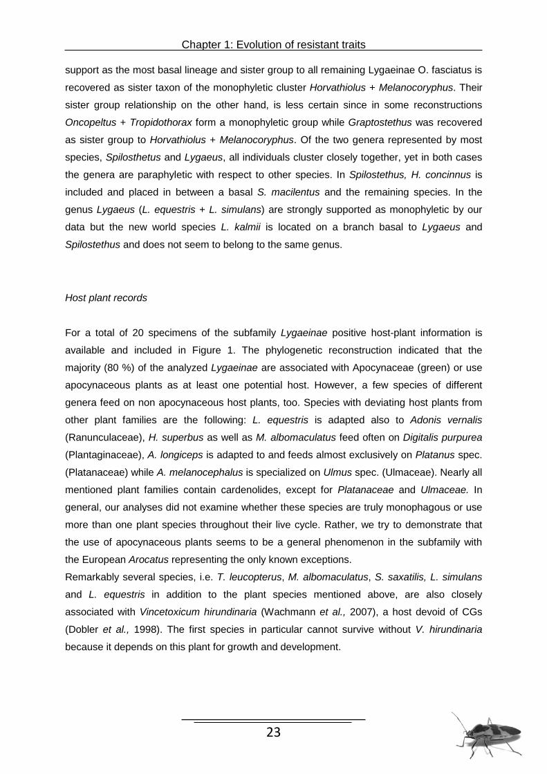
support as the most basal lineage and sister group to all remaining Lygaeinae O. fasciatus is
recovered as sister taxon of the monophyletic cluster Horvathiolus + Melanocoryphus. Their
sister group relationship on the other hand, is less certain since in some reconstructions
Oncopeltus + Tropidothorax form a monophyletic group while Graptostethus was recovered
as sister group to Horvathiolus + Melanocoryphus. Of the two genera represented by most
species, Spilosthetus and Lygaeus, all individuals cluster closely together, yet in both cases
the genera are paraphyletic with respect to other species. In Spilostethus, H. concinnus is
included and placed in between a basal S. macilentus and the remaining species. In the
genus Lygaeus (L. equestris + L. simulans) are strongly supported as monophyletic by our
data but the new world species L. kalmii is located on a branch basal to Lygaeus and
Spilostethus and does not seem to belong to the same genus.
Host plant records
For a total of 20 specimens of the subfamily Lygaeinae positive host-plant information is
available and included in Figure 1. The phylogenetic reconstruction indicated that the
majority (80 %) of the analyzed Lygaeinae are associated with Apocynaceae (green) or use
apocynaceous plants as at least one potential host. However, a few species of different
genera feed on non apocynaceous host plants, too. Species with deviating host plants from
other plant families are the following: L. equestris is adapted also to Adonis vernalis
(Ranunculaceae), H. superbus as well as M. albomaculatus feed often on Digitalis purpurea
(Plantaginaceae), A. longiceps is adapted to and feeds almost exclusively on Platanus spec.
(Platanaceae) while A. melanocephalus is specialized on Ulmus spec. (Ulmaceae). Nearly all
mentioned plant families contain cardenolides, except for Platanaceae and Ulmaceae. In
general, our analyses did not examine whether these species are truly monophagous or use
more than one plant species throughout their live cycle. Rather, we try to demonstrate that
the use of apocynaceous plants seems to be a general phenomenon in the subfamily with
the European Arocatus representing the only known exceptions.
Remarkably several species, i.e. T. leucopterus, M. albomaculatus, S. saxatilis, L. simulans
and L. equestris in addition to the plant species mentioned above, are also closely
associated with Vincetoxicum hirundinaria (Wachmann et al., 2007), a host devoid of CGs
(Dobler et al., 1998). The first species in particular cannot survive without V. hirundinaria
because it depends on this plant for growth and development.
Chapter 1: Evolution of resistant traits
24
Chapter 1: Evolution of resistant traits
25
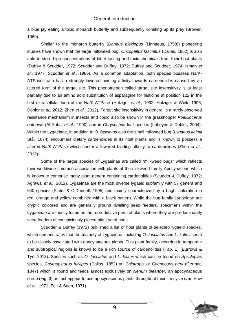
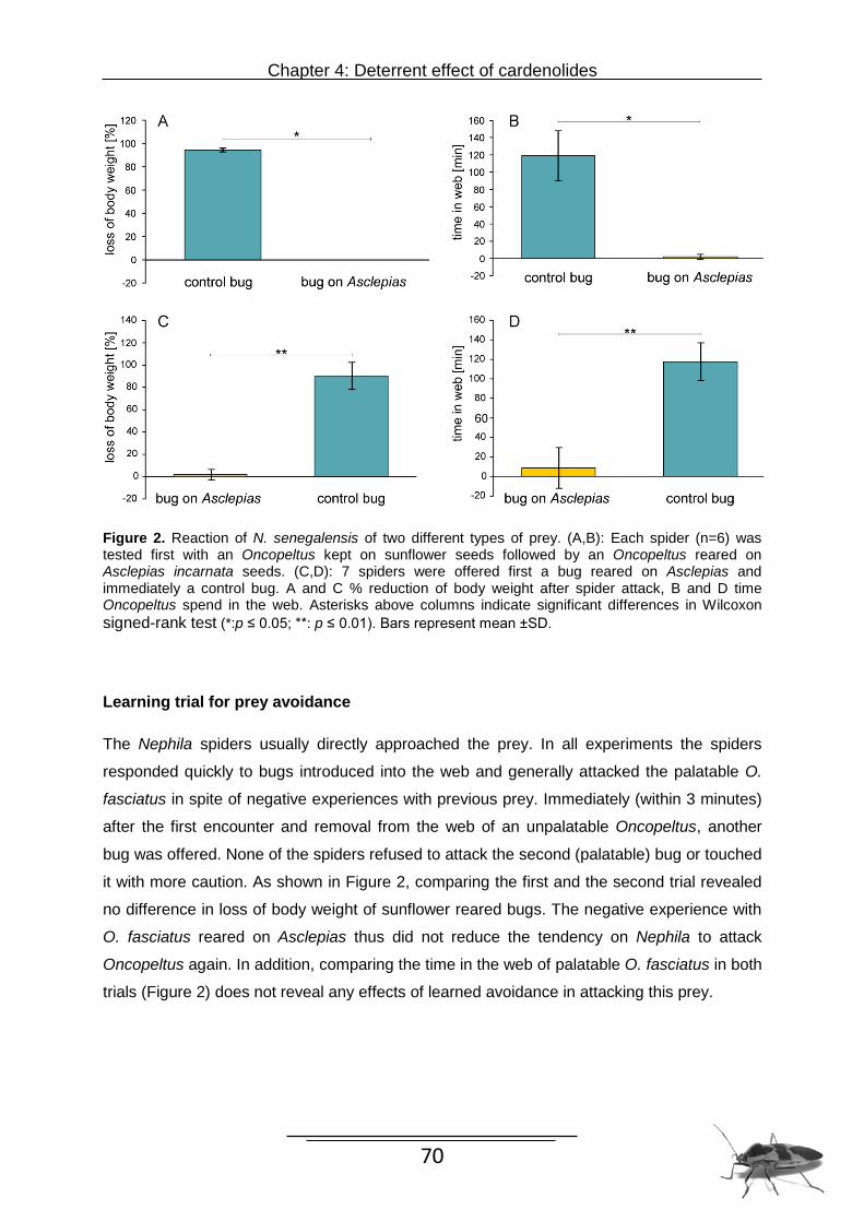
Figure 1. Maximum likelihood tree of the subfamily Lygaeinae based on the combined dataset of COI, COII, tRNALeu and 28S genes. Values above branches indicate ML bootstrap support values (>50%) in 1,000 replicates. The black branches represent two lygaeid species from subfamilies other than the Lygaeinae (Ischnorhynchinae, Orsillinae) and P. apterus (Pyrrhocoridae) as well as O. lavaterae (Oxicarenidae) which were used as outgroups. The green branches indicate that the respective species use at least one apocynaceous plant as host whereas the red branches refer to a use of non apocynaceous plants as hosts. Capital letter A marks the evolutionary origin of target site insensitivity and cardenolide sequestration. Capital letter B displays the loss of the ability to sequester cardenolides. Evidence for sequestration was determined here or obtained from the literature. The single letter behind the bug pictures indicates the amino acids at positions 122 for each tested species (Histidine: H; Asparagine: N). Bug photographs illustrate representative species.
Amino acid substitutions in the Na/K-ATPase gene
Our current genetic screen of the Na/K-ATPase α subunit of 11 Lygaeinae and P. apterus
(Pyrrhocoridae) as well as K. resedae (Ischnorhynchinae) as outgroup can only reveal with
certainty the identity of the amino acid at position 122 of the protein. This position is well
supported as decisive for cardenolide binding as has been previously shown for the monarch
butterfly D. plexippus and other insects (Holziger & Wink, 1996; Labeyrie & Dobler, 2004;
Dobler et al., 2012; Zhen et al., 2012; Dalla et al., 2013). All members of the subfamily
Lygaeinae investigated here possess an amino acid substitution of asparagine for histidine at
position 122 (N122H) in the first extracellular loop of the Na/K-ATPase which is essential for
ouabain binding. Both outgroup species however, had the conserved asparagine residue
representing the original condition. Mapping the character states on the phylogeny of the
Lygaeinae yields a uniform pattern of a resistance conferring substitution even in species
which are not confronted with cardenolides due to a CG free diet.
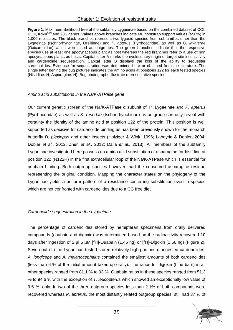
Cardenolide sequestration in the Lygaeinae
The percentage of cardenolides stored by hemipteran specimens from orally delivered
compounds (ouabain and digoxin) was determined based on the radioactivity recovered 10
days after ingestion of 2 µl 5 µM [3H]-Ouabain (1.46 ng) or [3H]-Digoxin (1.56 ng) (Figure 2).
Seven out of nine Lygaeinae tested stored relatively high portions of ingested cardenolides.
A. longiceps and A. melanocephalus contained the smallest amounts of both cardenolides
(less than 6 % of the initial amount taken up orally). The ratios for digoxin (blue bars) in all
other species ranged from 81.1 % to 93 %. Ouabain ratios in these species ranged from 51.3
% to 94.6 % with the exception of T. leucopterus which showed an exceptionally low value of
9.5 %, only. In two of the three outgroup species less than 2.1% of both compounds were
recovered whereas P. apterus, the most distantly related outgroup species, still had 37 % of
Chapter 1: Evolution of resistant traits
26
the initially imbibed ouabain and 5.6 % digoxin. These results suggest that throughout the
sequestering Lygaeinae (all species without Arocatus) digoxin as apolar cardenolide is
favored over ouabain.
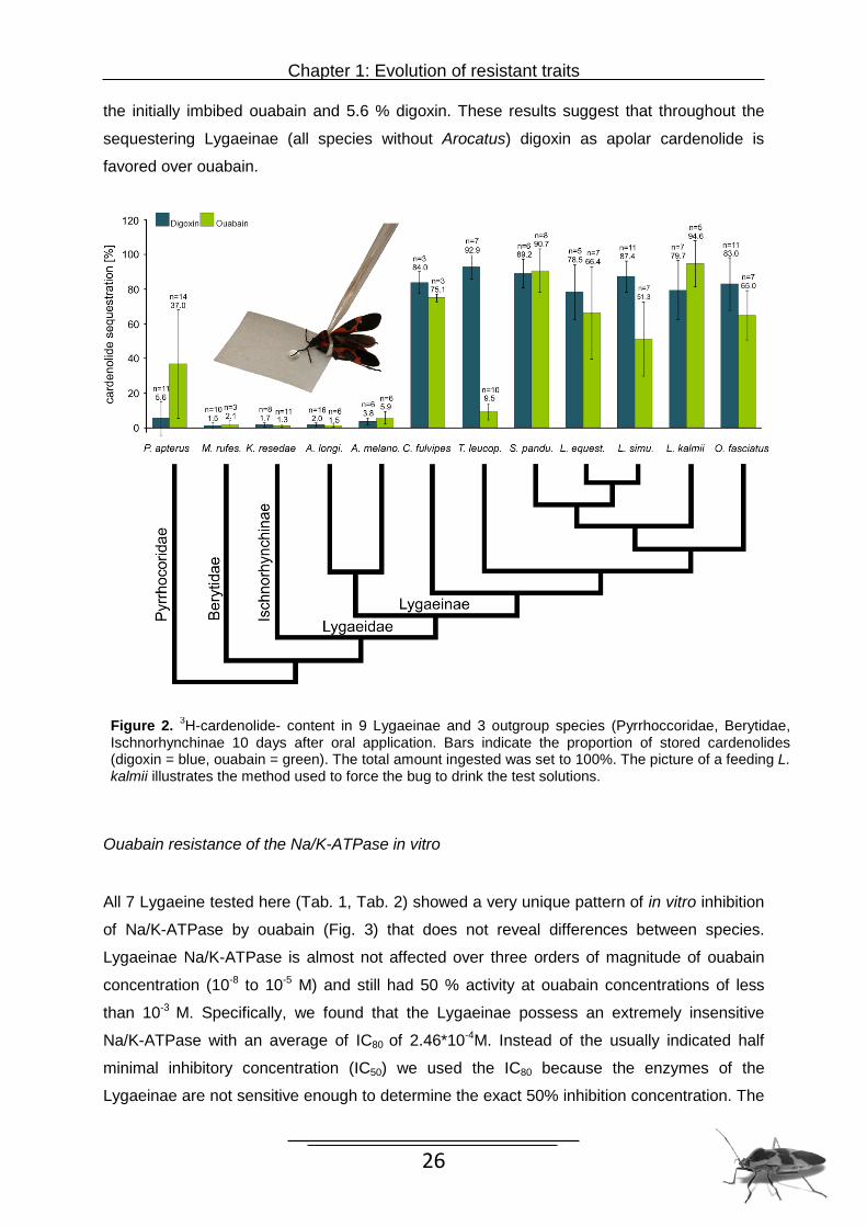
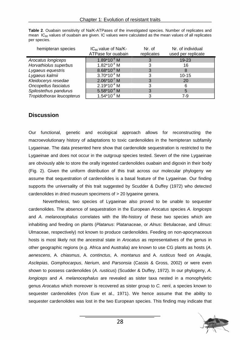
Ouabain resistance of the Na/K-ATPase in vitro
All 7 Lygaeine tested here (Tab. 1, Tab. 2) showed a very unique pattern of in vitro inhibition
of Na/K-ATPase by ouabain (Fig. 3) that does not reveal differences between species.
Lygaeinae Na/K-ATPase is almost not affected over three orders of magnitude of ouabain
concentration (10-8 to 10-5 M) and still had 50 % activity at ouabain concentrations of less
than 10-3 M. Specifically, we found that the Lygaeinae possess an extremely insensitive
Na/K-ATPase with an average of IC80 of 2.46*10-4M. Instead of the usually indicated half
minimal inhibitory concentration (IC50) we used the IC80 because the enzymes of the
Lygaeinae are not sensitive enough to determine the exact 50% inhibition concentration. The
Figure 2. 3H-cardenolide- content in 9 Lygaeinae and 3 outgroup species (Pyrrhoccoridae, Berytidae, Ischnorhynchinae 10 days after oral application. Bars indicate the proportion of stored cardenolides (digoxin = blue, ouabain = green). The total amount ingested was set to 100%. The picture of a feeding L. kalmii illustrates the method used to force the bug to drink the test solutions.
Chapter 1: Evolution of resistant traits
27
individual IC80 values of all individuals are presented in Tab.2. Our analysis furthermore
revealed a dramatic difference regarding sensitivity to ouabain in the outgroup species K.
resedae (Ischnorhynchinae) compared to the Lygaeinae. The enzyme of K. resedae is highly
sensitive and showed with an IC80 of 2.06*10-7M already an inhibition at ouabain
concentrations as low as 10-7 M. Together with the Na/K-ATPase sequence data we
conclude that target site insensitivity to cardenolides probably is a unique feature of all
Lygaeinae.
Figure 3. In vitro sensitivity of the Lygaeinae Na/K-ATPases to ouabain. Each curve illustrates the inhibition of the enzyme over a magnitude of six different ouabain concentrations (10-8 to 10-3 M). The single data points show the average of the replicates per species which are alos represented by different colors. For comparison the inhibition curve of D. plexippus Na/K-ATPase is included (data taken from Petschenka et al. 2013).
Chapter 1: Evolution of resistant traits
28
Table 2. Ouabain sensitivity of Na/K-ATPases of the investigated species. Number of replicates and mean IC80 values of ouabain are given. IC values were calculated as the mean values of all replicates per species.
hemipteran species IC80 value of Na/K-ATPase for ouabain
Nr. of Nr. of individual replicates used per replicate
Arocatus longiceps 1.89*10-4 M 3 19-23 Horvathiolus superbus 1.82*10-4 M 3 16 Lygaeus equestris 8.68*10-5 M 3 8 Lygaeus kalmii 3.70*10-4 M 3 10-15 Kleidocerys resedae 2.06*10-7 M 3 20 Oncopeltus fasciatus 2.19*10-4 M 3 6 Spilostethus pandurus 5.58*10-4 M 3 5 Tropidothorax leucopterus 1.54*10-4 M 3 7-9
Discussion Our functional, genetic and ecological approach allows for reconstructing the
macroevolutionary history of adaptations to toxic cardenolides in the hemipteran subfamily
Lygaeinae. The data presented here show that cardenolide sequestration is restricted to the
Lygaeinae and does not occur in the outgroup species tested. Seven of the nine Lygaeinae
are obviously able to store the orally ingested cardenolides ouabain and digoxin in their body
(Fig. 2). Given the uniform distribution of this trait across our molecular phylogeny we
assume that sequestration of cardenolides is a basal feature of the Lygaeinae. Our finding
supports the universality of this trait suggested by Scudder & Duffey (1972) who detected
cardenolides in dried museum specimens of > 20 lygaeine genera.
Nevertheless, two species of Lygaeinae also proved to be unable to sequester
cardenolides. The absence of sequestration in the European Arocatus species A. longiceps
and A. melanocephalus correlates with the life-history of these two species which are
inhabiting and feeding on plants (Platanus: Platanaceae, or Alnus: Betulaceae, and Ulmus:
Ulmaceae, respectively) not known to produce cardenolides. Feeding on non-apocynaceous
hosts is most likely not the ancestral state in Arocatus as representatives of the genus in
other geographic regions (e.g. Africa and Australia) are known to use CG plants as hosts (A.
aenescens, A. chiasmus, A. continctus, A. montanus and A. rusticus feed on Araujia,
Asclepias, Gomphocarpus, Nerium, and Parsonsia (Cassis & Gross, 2002) or were even
shown to possess cardenolides (A. rusticus) (Scudder & Duffey, 1972). In our phylogeny, A.
longiceps and A. melanocephalus are revealed as sister taxa nested in a monophyletic genus Arocatus which moreover is recovered as sister group to C. nerii, a species known to
sequester cardenolides (Von Euw et al., 1971). We hence assume that the ability to
sequester cardenolides was lost in the two European species. This finding may indicate that
Chapter 1: Evolution of resistant traits
29
physiological adaptations which are necessary to sequester cardenolides are costly and are
therefore reduced when not needed.
Arocatus which moreover is recovered as sister group to C. nerii, a species known to
sequester cardenolides (Von Euw et al., 1971). We hence assume that the ability to
sequester cardenolides was lost in the two European species. This finding may indicate that
physiological adaptations which are necessary to sequester cardenolides are costly and are
therefore reduced when not needed. The lower, but unequivocal presence of ouabain (digoxin 5.6 %, only) in the outgroup
species P. apterus (Fig. 2) is not necessarily in disagreement with a monophyletic origin of
sequestration in the Lygaeinae as P. apterus also is an aposematic species and might derive
toxins from its host plants as well. Moreover, the outgroup species do not possess
cardenolide resistant Na/K-ATPases (see sequencing and physiological data) which might be
a prerequisite for cardenolide sequestration.
Testing the resistance traits which prevent self-intoxication in the sequestering
Lygaeinae, we found that uninhibited Na/K-ATPase activity under ouabain stress is a
common feature in the subfamily Lygaeinae. Our in vitro investigations of Lygaeinae Na/K-
ATPase of brain and thoracic ganglia showed a relative uniform pattern of strong insensitivity
towards oubain (Fig. 3). All seven species investigated here possess the same cardenolide
insensitive form of Na/K-ATPase i.e. a much less ouabain-sensitive Na/K-ATPase (IC80 =
2.46*10-4M) than the outgroup species Kleidoceris resedae (IC80 = 2.06*10-7M) and a slightly
higher insensitivity than the monarch butterfly (IC80 = 2.32*10-5M) (Petschenka et al., 2013).
The characteristics of Na/K-ATPase inhibition by ouabain described here, resembles the one
described by Moore & Scudder (1986). In our in vitro investigations we focused on nervous
Na/K-ATPase, only, as the nervous tissue is a rich source of Na/K-ATPase facilitating in vitro
assays. Moreover, the nervous system was shown to be the main site of Na/K-ATPase
expression in other insect orders (Lepidoptera: Petschenka et al. 2012) rendering the
nervous system most relevant for toxicological interpretation.
The presence of cardenolide resistant Na/K-ATPases in all Lygaeinae tested
suggests that target site insensitivity of Na/K-ATPase, like the ability to sequester
cardenolides, also is a basal feature of the Lygaeinae. Both species, A. longiceps and A.
melanocephalus, which are not exposed to dietary cardenolides and do not store the toxins,
still possessed a modified, cardenolide insensitive Na/K-ATPase.
Responsible for a uniform insensitivity in the Lygaeinae are amino acid substitutions
at positions of the enzyme which are known to be involved in binding of ouabain (Fig.1). Our
preliminary molecular investigations of the Na/K-ATPase of several Lygaeinae demonstrated
that an amino acid substitution of asparagine for histidine at position 122 (N122H) is present
in the first extracellular loop of the Na/K-ATPase of all species and is at least partly
Chapter 1: Evolution of resistant traits
30
responsible for insensitivity in the in vitro enzyme assays (Dobler et al., 2012; Dalla et al.,
2013). Further amino acid substitutions at positions which are known to be involved in
ouabain binding of Na/K-ATPase in the Lygaeinae lead to a further increased insensitivity
(Dobler et al., 2012, Dalla et al., 2013). Previous studies could show that up to eight different
substitutions may be responsible for the lower cardenolide binding characteristics in CG
adapted O. fasciatus and L. kalmii (Dobler et al., 2012; Zhen et al., 2012). Whether all of
these substitution arepresent in all the Lygaeinae investigated here could not yet be
unequivocally clarified. The possession of several Na/K-ATPase gene copies in the
Lygaeinae (Zhen et al., 2012; Dobler et al., 2012; Dobler et al., unpublished data) renders
this investigation difficult. Evidence from genetic modifications of the Drosophila Na/K-
ATPase show that accumulation of substitutions at positions forming the ouabain binding
pocket in mammals (Croyle et al., 1997; Qiu et al., 2005; Yatime et al., 2013) lead to
increased insensitivity of the enzyme (Dobler et al., 2012; Dalla et al., 2013). Two combined
substitutions, either Q111T-N122H as observed in O. fasciatus or Q111V-N122H as present
in D. plexippus have been shown to reduce the inhibition by ouabain strongly, whereas the
enzyme occurring in K. resedae represents the typical sensitive form. As the enzyme of
Lygaeinae however, is far more resistant to ouabain in vitro than the enzyme of the monarch
butterfly this increased resistance is likely due to at least a third substitution at an additional
position. Enzyme assays performed with the single mutation T797A discovered in L. kalmii
showed a 250-fold increased resistance (Dalla et al., 2013) while a combination of four
substitutions (Q111T, N122H, F786N, T797A) introduced into the Drosophila Na/K-ATPase
gene leads to enzyme characteristics closely similar to the Na/K-ATPase inhibition curves
observed here (Dalla & Dobler, unpubl. data). Therefore, it is likely that due to combinations
of at least four potentially important substitutions at different positions the strongly increased
resistance in the Lygaeinae can be explained.
The worldwide host association of Lygaeinae with plants of the milkweed family
Apocynaceae, suggests a very old relationship between these two taxa. Remarkably, several
Lygaeinae use plant species as hosts which belong to non-related families but are known to
produce cardenolides as well. Examples include L. equestris which is well known to feed on
Wichtl, 1986; Deckert, 2007), or H. superbus which often is associated with Digitalis
purpurea a Plantaginaceae (Péricart, 1998; Wachmann et al., 2007, Petschenka pers. obs.).
Furthermore, S. pandurus uses Urginea maritima as a host (Vivas, 2012), an Asparagaceae
known to contain bufadienolides which are structurally related to cardenolides and have the
pharmacodynamical activity. Most likely these Lygaeinae also sequester cardenolides from
these sources which has been shown for L. equestris (Petschenka, 2010, unpublished data)
collected from A. vernalis. Species like Tropidothorax leucopterus, Melanocoryphus
Chapter 1: Evolution of resistant traits
31
albomaculatus, Spilostethus saxatilis, Lygaeus simulans and Horvathiolus superbus, in
addition to the plant species mentioned above, are also closely associated with Vincetoxicum
hirundinaria (Wachmann et al., 2007). The current use of plants from non-apocynaceaous
families most likely represents host shifts which were facilitated by the preadaptation of the
Lygaeinae to CGs. As they, like many other species of the Lygaeinae, can use a variety of
plants as nutritional resources the availability of a certain class of chemicals may predict host
plant associations more strongly than the supply of nutrition. Given that Lygaeinae are the
most species rich lineage within the Lygaeidae (sensu Henry, 1997) with more than 500
species compared to the sister groups Orsilinae (250 species) and Ischnorhynchinae (75
species), the adaptations to dietary CGs may represent a key innovation of this group. In
general, it is assumed that species that are specialized in their food utilization are often more
diverse than taxa including more generalist feeders because the rate of evolution is thought
to be higher among specialists (Whitlock, 96).
Taken together, results from three comparative approaches revealed that
sequestration and target site mutation as resistance traits in the Lygaeinae are ancestral
adaptations and have apparently originated at the basis of the subfamily. Even species, who
do not normally encounter dietary cardenolides and do not store the toxins, still possess a
modified insensitive Na/K-ATPase. These results lead us to conclude that target site
insensitivity and sequestration of cardenolides are basal and plesiomorphic characters of the
Lygaeinae which derive from an originally host plant use.
32
Chapter 2
Metabolic alteration of cardiac glycosides in Lygaeinae: detoxification or optimized uptake?
Abstract
Several Lygaeinae (Heteroptera) are morphologically and physiologically adapted to
sequester cardenolides for their own protection against predation. When fed on Asclepias
seeds, a high concentration of polar cardenolides was found in Oncopetus fasciatus, despite
a wide polarity range of cardenolides was available. These hints to a selective sequestration
or a metabolic alteration process of cardenolides. Previous studies could show that different
polar metabolites arise through change of apolar cardenolides, however, little is known about
the details of the metabolic processes. We therefore performed a comparative study in which
10 different species of Lygaeinae plus one outgroup were tested for metabolic alteration of
two purified [3H]-labeled cardenolides: the apolar digoxin and the polar ouabain by using
different treatments. Both treatments yielded always the same metabolites. Ouabain was
always recovered unchanged whereas digoxin alteration seems to follow a uniform
mechanism which is, at least in part, associated with the selective accumulation of
cardenolides in the glycoside storage compartment. Further, we elucidated the metabolic
alteration of digoxin in O. fasciatus over a period of 170 h and identified the structure of the
storable metabolite. As MALDI analysis indicates this metabolite arises by at least one
amination step. Metabolic alteration of apolar cardenolides is a general phenomenon in the
Lygaeinae which may have evolved as an adaptation to handling and accumulating
cardenolides derived from their host plants.
Chapter 2: Metabolic alteration
33
Introduction
Specializing on plants with toxic secondary compounds requires in herbivores traits to
overcome these chemical defences. These counter-mechanisms include for instance
sequestration (and compartmentalization) of toxic compounds used for own defenses or the
metabolic modification of the molecules to avoid specific binding to targets. Whereas the
former mechanism requires an active form of toxins, the latter mechanism represents a
detoxification mechanism which might lead to excretion of substances via the Malpighian
tubules (Després et al., 2007). Biotransformation is one of the major weapons against many
classes of toxic allelochemicals to resist intoxication. Specialized Lygaeinae (Heteroptera) for
instance possess a wide spectrum of adaptations to cope with toxic host plant cardenolides
with sequestration being one of them. Cardenolides in general bind to and inhibit the
ubiquitous Na/K-ATPase responsible for maintaining cellular potentials (Lingrel et al., 1990;
Jorgensen et al., 2003; Horisberger, 2004). To cope with these toxic compounds Lygaeinae
possess amino acid substitutions in the target site of the Na/K-ATPase which lower the
binding affinity for crdenolides (Dobler et al. 2012, Zhen et al. 2012 and Chapter 1).
The phenomenon of cardenolide sequestration, per se, involves the uptake, transfer,
and concentration of cardenolides in the storage compartments. Physiological studies on
permeability of insect guts suggest that polar cardenolides such as ouabain require an
energy dependent transport and presumed intestinal carrier in the gut epithelium which allow
polar cardenolides to enter the haemolymph. Conversely, apolar cardenolides can be
expected to cross the gut passively due to their physiochemical properties (Wright, 1960).
Further, there are two opposing mechanisms in uptake and accumulation of cardenolides in
the species. The uptake of apolar cardenolides occurred far more rapidly (77 % in 30 min)
than the uptake of polar ones (3 % in 30 min.) (Yoder et al., 1976; Duffey et al., 1978;
Scudder & Meredith, 1982b; Detzel & Wink, 1995). In the reverse situation, the results of
Duffey et al., (1978) showed that the uptake of polar cardenolides from the haemolymph into
the glycoside storage compartments is faster than the transfer of metabolized apolar
cardenolides. Nevertheless, the mechanisms of sequestration, transport and accumulation of
cardenolides in the haemolypmph or the integument are not known in detail.
Interestingly, in Oncopeltus fasciatus a high concentration of polar- and an absence
of apolar cardenolides was detected (in several studies) (Duffey & Scudder, 1974; Yoder et
al., 1976; Duffey et al., 1978; Moore & Scudder, 1985; Scudder et al., 1986). Next to the
sequestration and target-site mutation, Lygaeinae also possess the ability of metabolic
alteration of cardenolides. They apparently use biotransformation not for detoxification but
rather to transform cardenolides into storable forms. Studies on accumulation and distribution
of cardenolides in O. faciatus (Duffey & Scudder, 1974; Moore & Scudder, 1985) illustrate
Chapter 2: Metabolic alteration
34
that the ratios of cardenolide concentration differ among several insect tissues. Whereas
wings, gut and haemolymph are characterized by low cardenolide content, the glycoside
storage compartment represents the greatest storage capacity of the body. In vivo and in
vitro evidence using indicator cardenolides which cover a wide polarity range of natural
occurring plant compounds, indicated a metabolic alteration of apolar cardenolides (Duffey &
Scudder, 1974; Scudder & Meredith, 1982b). However, these reports did not address the
metabolic mechanisms in detail.
Thus, it was of interest to examine the metabolic process in O. fasciatus in detail and
to compare it among several species of this cardenolide adapted subfamily. Using thin-layer-
chromatography the cardenolide profiles after ingestion or injection of polar ouabain or apolar
digoxin were determined in these species. Finally, this study was undertaken to identify the
cardenolide derivative present in the defensive secretion stored in the glycoside
compartment. This may elucidate the mechanism by which certain Lygaeinae are able to
handle glycosides because their toxicity has been correlated with polarity. We elucidated the
metabolite structure to obtain information on enzymes required for apolar cardenolide
metabolism in the Lygaeinae and their occurrence in the insect body tissues. This ultimately
allows to deduce the role of metabolism in the ability to cope with large amounts of
cardenolides.
Material and Method Insect handling Adult Lygaeinae were obtained both from the field and from laboratory cultures. The species
Oncopeltus fasciatus, Horvathiolus superbus, Lygaeus equestris, L. simulans, L. kalmii,
Spilostethus pandurus and Cosmopleurus fulvipes were reared in the laboratory from the egg
stage and raised on husked sunflower seeds (Helianthus annuus L.) and water which was
available from cotton wicks in plastic tubes. All species were reared in a climatic chamber at
16 h/8 h light/dark at 26°C (C. fulvipes, H. superbus, O. fasciatus, S.pandurus) or 30°C (L.
equestris, L. kalmii, L. simulans). Tropidothorax leucopterus (from Grießheim, Germany) was
reared on sunflower seeds and cut branches of Vincetoxicum hirundinaria and was also kept
at 26°C, 16 h/8 h light/dark cycle. For Arocatus melanocephalus (from Kallinchen, Germany),
A. longiceps (from Berlin, Germany) and Kleidocerys resedae (from Kamburg, Germany)
field collected individuals were used.
Chapter 2: Metabolic alteration
35
Treatment of species for different analyses
A) Metabolite profile 72 h after injection
For this treatment six species (A. melanocephalus, O. fasciatus, T. leucopterus, L. simulans,
L. equestris, C. fulvipes) were used and each of them were injected with a polar or an apolar
cardenolide. A solution containing [3H]-ouabain (polar) or [3H]-digoxin (apolar) (Perkin Elmer
LAS GmbH, Rodgau, Germany) and 1.125 % NaCl (1:4) in water was laterocranial injected
between fifth and sixth abdominal segment by a fine capillary syringe (Hamilton 701 NR;
ga22S/51mm/pst3; ROTH GmbH+Co). The specific activity of radiolabelled cardenolides was
6 Ci/mmol for [3H]-ouabain and 7.08 Ci/mmol for [3H]-digoxin. Before injection, species were
cooled for 5 min at -20°C to immobilize them in order to prevent injury or puncture of the gut
during injection. After injection species were kept for 72 h on water and sunflower seeds (as
previously described).
A) Ingestion of cardenolide solutions by forced drinking
The Lygaeinae and K. resedae used in this experiment were forced to drink cardenolide
solutions (composed of 5 µM cardenolide in water with 17.7 % ethanol and 5 % sucrose) by
fixing them with a lasso of dental floss. The proboscis was manually introduces into a 2 µl
droplet containing cardenolide on parafilm. After feeding, the species were again kept for 72
h on water and sunflower seeds.
B) Metabolite profile over a period of 170 h Before starting the experiments fifth instar larvae were separated from adults to ensure that
all individuals had approximately the same age. In this treatment 75 adults of the large
milkweed bug, O. fasciatus were injected with 2 µl of the nonpolar digoxin as described
above to determine metabolite profiles over a period of 170 hours. Species were kept as
described above over a period of 170 hours. Within this period three individuals each (two
females, one male) were frozen separately at 25 different times.
C) Accumulation of different polar cardenolides in the glycoside storage compartment
Six adult O. fasciatus each were injected as previously described with [3H]-ouabain or the
apolar [3H]-digoxin diluted in 1.125 % NaCl and kept on water and sunflower seeds for 72 h.
Chapter 2: Metabolic alteration
36
Detection of metabolites of cardenolides in the examined Lygaeinae
All species of treatment A, B and C given drinking solution or treated by injection were frozen
after the end of the treatment. Individuals of each species were separately frozen in liquid
nitrogen, homogenized with a glass pestle and extracted with 1 ml 100 % methanol. The
sample was shaken for one hour and then centrifuge at 8000 rpm. Supernatant was
transferred and methanol evaporated overnight. Dried residual sample was dissolved in 50 µl
methanol. Species of treatment D were squeezed through slight pressure at specific points of
the body surface whereby the release of defence fluid out of the storage compartment was
initiated as previously described by Duffey & Scudder (1974). Crude methanol extracts and
the fluid of defence droplets were separated by thin-layer chromatography (TLC) (pre-coated
TLC sheets with silica gel 60, ALUGRAM® SIL G) and analyzed using a multichannel
radioactivity detector (Rita-32a, Raytest). The plates were developed in filter-paper-lined,
saturated chambers with ethyl acetate-methanol-water (30:4:3). As control for cardenolides
or cardenolide derivates we performed a second TLC by using kedde reagent (1:1 mixture of
5% 3,5-dinitrobenzoic acid in MeOH and 2 N KOH) for the specific detection of the lactone of
the cardenolides.
Structure analysis by MALDI-TOF- MS
O. fasciatus individuals were injected with a solution of 3 µl digoxin (10-6 M dissolved in
ethanol and water 1:3) and kept on sunflower seeds impregnated with digoxin by applying
the compound directly as a solution in chloroform-methanol (2:1). After six days the defence
fluid was collected from the insects as described above and dissolved in water. To check for
presence of metabolite in the defence fluid, TLC separation was performed as control. The
sampled fluid (2 µl) was spotted on a TLC sheet and developed as described previously.
MALDI-TOF (Matrix-associated laser desorption ionization-time of flight mass spectrometry)
analyses were performed on an ultrafleXtreme instrument (BrukerDaltonics, Bremen,
Germany) equipped with a smartbeam-II laser with a repetition rate of 1 kHz. The matrix (α-
cyano-4-hydroxycinnamic acid (4-HCCA) was dissolved in 2:1 water:acetonitrile with 0.1 %
trifluoroacetatic acid (TFA). The matrix was shaken and centrifuged for 1 to 2 min at 14 000
rpm. The clear liquid was decanted for use. Digoxin (Sigma) was used as standard for
instrument calibration and as internal mass standard. The calibration standard (1 mg/mL)
was prepared in 0.1 % TFA in water. The defence fluid sample was analyzed using a 10:1 (µl
each, matrix/sample) mixture. Samples were spotted on a MALDI target plate and allowed to
air dry. The spots were measured within the FlexControl software (version 3.3). The spectra
Chapter 2: Metabolic alteration
37
were processed using FlexAnalysis software (version 3.3). The MALDI-TOF MS analyses
were performed at the Department of Organic Chemistry, University of Hamburg.
Results Cardenolide alterations in the Lygaeinae
Body samples of all six Lygaeinae 72 hours after injection with [3H]-labelled ouabain showed
no alteration by the insects, since the radio TLC analysis revealed only a single cardenolide
identical in Rf value (Rf = 0.06-0.07; ouabain, Rf = 0.07) with the reference ouabain (Fig.1 A1-
E1). The same result (Rf = 0.07) was obtained 72 hours after imbibing [3H]-labelled ouabain.
All nine tested species had unchanged ouabain in the body fluid samples. Nevertheless, the
amount of ouabain recovered from the body samples of Arocatus species was less than 20
% of the amount ingested or injected previously.
In contrast, after injection or ingestion of [3H]-labelled digoxin the cardenolide was
converted into metabolites. In a total of six species (L. equestris, L. simulans, O. fasciatus, T.
leucopterus, C. fulvipes) digoxin was altered to at least two metabolites. Three separate
fractions could be recovered by TLC (Fig. 2 A2-E2): fraction I representing a very polar
cardenolide detected at Rf = 0.02-0.03, fraction II chromatographed in an intermediate
position at Rf = 0.10-0.13, and fraction III with Rf = 0.50 corresponded to unchanged digoxin.
In species of the genera Arocatus (A. melanocephalus, A. longiceps) the analysis showed
only two cardenolides including unchanged digoxin. The metabolite corresponded with an Rf
= 0.02 to fraction I of the other species. In additional analyses up to 144 hours after ingestion
of digoxin no second less polar metabolite appeared which was formed out of the unstable
polar product (fraction I). During this time the cardenolide concentration in the body sample
decreased linearly up to a detection limit after 130 hours. Chromatography of the body
sample in H. superbus indicated that even after 72h the radioactive label introduced with
digoxin occurred in a single peak coincident with the fraction II. Analyzing the metabolite
profile in K. resedae, there was no evidence for cardenolides in the body fluid after 72 hours.
Chapter 2 : Metabolic alteration
38
Chapter 2 : Metabolic alteration
39
Profile of digoxin metabolites in O. fasciatus
Chromatography of the body-samples showed that in adults of O. fasciatus injected with
[3H]-labelled digoxin the cardenolide was converted into at least two metabolites (fraction I
and II) in the insect compared to the unchanged standard (Fig.1 A-E). Immediately (30 min)
after injection of digoxin 70 % of this apolar cardenolide was transformed into a very polar
metabolite (fraction I) (Rf = 0.03; cf. digoxin, Rf =0.50) (Fig.2). Already after one hour a
second less polar metabolite (fraction II) (Rf = 0.12) appeared which was formed out of the
unstable polar product (first metabolite). The total amount of unchanged digoxin 12 hours
after injection was found to be 10 % of the total digoxin available. Metabolite amounts,
however, were found to be 90 % after 16 hours summing both fraction I and II. Over a period
of 66 hours most of the intermediate product (80%) was modified into a final stable product
(15%) and only a low amount of unchanged digoxin (5%) could be found. Finally after 163
hours digoxin and the first polar metabolite had completely vanished. Among the three
individuals analyzed no deviations in this time course could be detected.
Figure.1. A diagram representing the radioactive analysis of chromatograms of the body sample 72 h after ingestion of digoxin or ouabain in different species of Lygaeinae. Abscissa represents distance along the TLC plate: origin at 15 mm and solvent front at 180 mm. Ordinate shows the intensity in counts. A1) show the position of ouabain standard with an Rf = 0.07 and A2) show the standard of digoxin with Rf = 0.50. B1) represents the position of ouabain after 72h of H. superbus, C1 of T. leucopterus, D1 of L. equestris and E1 of A. melanocephalus. B2 shows the metabolized digoxin after 72h in H. superbus, C2 of T. leucopterus, D2 of L. equestris and E2 of A. melanocephalus.; The colours of the peaks represents the different compounds: Ouabain (green), Digoxin (blue), first metabolite (yellow) and second metabolite (red).
Chapter 2 : Metabolic alteration
40
Figu
re. 2
. Met
abol
ite p
rofil
e in
Onc
opel
tus
fasc
iatu
s ov
er 1
70 h
ours
afte
r inj
ectio
n of
dig
oxin
. Abs
ciss
a re
pres
ents
dis
tanc
e al
ong
the
TLC
pla
te,
orig
in a
t 15
mm
and
sol
vent
fron
t at 1
80 m
m. O
rdin
ate
show
s th
e in
tens
ity in
cou
nts.
A. A
t tim
e 0
100%
dig
oxin
(blu
e) w
as in
ject
ed. B
. Afte
r 30
min
70
% o
f the
inje
cted
dig
oxin
was
tran
sfor
med
into
a v
ery
pola
r met
abol
ite (y
ello
w).
C. A
fter 1
2 ho
urs
mos
t of t
he d
igox
in (b
lue)
dis
appe
ared
and
a s
econ
d le
ss p
olar
met
abol
ite (r
ed) a
ppea
red
that
was
form
ed o
ut o
f the
uns
tabl
e po
lar p
rodu
ct (y
ello
w).
D. A
fter 6
6 h
mos
t of t
he in
term
edia
te p
rodu
ct (y
ello
w)
was
mod
ified
into
the
seco
nd fi
nal p
rodu
ct (r
ed).
E. A
fter 1
63 h
dig
oxin
(blu
e) a
nd th
e fir
st p
olar
met
abol
ite (y
ello
w) h
ad c
ompl
etel
y va
nish
ed
Chapter 2 : Metabolic alteration
41
Uptake of cardenolides into the storage compartments in O. fasciatus
Chromatograms of TLC separation indicated that the uptake of polar ouabain from the
haemolymph into the storage space within 72 hours after injection was faster than the uptake
of metabolized digoxin. Fluid of the storage compartments contained 50-70 % ouabain
whereas only 1-3 % of digoxin was taken up after 72 h. However, in the defence fluid only
the second polar metabolite (fraction II with Rf = 0.12) could be detected.
Structure analysis of metabolite fraction II in the defence fluid of O. fasciatus
The defence fluid released from the storage compartments after the insect was treated with
digoxin was analyzed by MALDI- TOF-MS. A typical MALDI-TOF mass spectrum of the
cardenolide digoxin showed a single intense peak at m/z 803.415 [+Na+] corresponding to
the mass of digoxin with an additional sodium ion (Fig. 3). The measurement of the fluid
sample yielded several peaks of different masses (Fig. 4). Based on the mass of digoxin we
speculate that the intense peak at m/z 820.420 [+Na+] might be the metabolite of fraction II
identified by TLC separation of the defence fluid in addition to a sodium ion. This mass
coincides almost perfectly with the structure of the metabolite illustrated in Figure 5. We
assume that through amination an NH3 is introduced into the genin of digoxin at the C-20 of
the 5 membered lactone followed by a hydrogenation which entails the resolution of the
double bond. Mass spectra analyses and structure analyses identified two more metabolites.
Both mass spectra of m/z 782.489 with a protonated molecular ion and m/z 804.449 with an
added sodium ion are expected to be derivates of the metabolite of fraction //. The masses
[m/z 782.489 + H+] and [m/z 804.449 + Na+] agree with the assumed structures shown in
Figure 5. Through amination of NH3 at different positions of the genin of digoxin it is possible
that molecules of the same chemical formula could be present as derivates.
Chapter 2 : Metabolic alteration
42
Figure 3. MALDI-TOF mass spectrometric analysis with the cardenolide digoxin used as standard showing an intense fragmentation of m/z 803.415, corresponding to the mass of ionized digoxin
Figure 4. MALDI-TOF mass spectrum of defence fluid of O. fasciatus showing an intense peak of m/z 820.420, that is expected to be the metabolite identified as fraction // in thin-layer-chromatography, the two other marked spectra with m/z 782.489 and m/z 804.449 are expected to correspond to the mass of two metabolic derivatives
Chapter 2 : Metabolic alteration
43
Metabolites of Digoxin Exact Mass/Chemical Formula
782.47
C41H68NO13+
804.45
C41H67NNaO13+
820.45
C41H67NNaO14+
Figure 5. Structure and the according mass of metabolites of digoxin, the metabolite structure of C represents the expected metabolite found in fraction II of body fluid samples of Lygaeinae, A and B are expected to be derivates of metabolite shown in C.
Diskussion
This study was undertaken in order to clarify certain aspects of the metabolic alteration of
cardenolides in the Lygaeinae and to elucidate the mechanisms by which these species are
able to handle these toxic compounds. Similar to previous studies (Scudder & Meredith,
1982; Duffey & Scudder, 1974) ouabain does not appear to be metabolized in any of the
examined Lygaeinae. Labelled digoxin, however, was recovered from body samples as three
separate fractions which included two metabolites of digoxin as well as unchanged digoxin.
Detailed determination of the metabolite profile in O. fasciatus over a period of 170 hours
uncovered the metabolic transformation of apolar digoxin with regard to quantity and polarity
of the metabolites. Previous studies using the more apolar cardenolide digitoxin had already
reported the presence of two rather polar metabolites in O. fasciatus 24 hours after ingestion
(Scudder & Meredith, 1982b; Duffey & Scudder, 1974), yet did not uncover the
interdependence and the temporal appearance of the individual fractions. The fact that
digoxin was converted first to a more polar metabolite than ouabain but afterwards
transformed into a slightly more apolar cardenolide point to two enzyme catalyzed reactions
involved in digoxin metabolism. By using TLC and kedde reaction we confirmed the presence
A
B
C
Chapter 2 : Metabolic alteration
44
of a lactone in the metabolite representing fraction / and //, which primarily provide evidence
of a cardenolide or a cardenolide derivate.
The comparative studies testing digoxin uptake in several species of Lygaeinae
produced the same results and mostly uncovered the occurrence of two metabolites with the
same Rf values as found in O. fasciatus. Only in body extracts of H. superbus a single
metabolite corresponding to fraction // was detected which suggests a faster enzymatic
transformation than in other sequestering Lygaeinae. Testing the outgroup species K.
resedae, we found not cardenolides in body fluids after 72 hours. These findings suggest that
both cardenolides have not been taken up by the gut which in turn is an indication for the
possession of an impermeable gut.
Nevertheless, it is important to differentiate between cardenolide sequestering
species and those who do not sequester but take up cardenolides. In non-sequestering
Lygaeinae, i.e. A. longiceps and A. melanocephalus, we could only detect the first metabolite
(fraction I) in the extractions regardless of time. This metabolite disappeared rapidly, most
likely by excretion via the Malpigian tubules, which are suggested to be relevant for the
excretion of toxic plant compounds in general (Nation, 2001). It is tempting to speculate, that
the rapid excretion of metabolized cardenolides originated as an enzymatic detoxification
mechanism whereas the further transformation to the second metabolite represents an
adaptation to accumulation and storage of cardenolides. Since apolar cardenolides are
suspect to be more emetic than their polar counterparts (Blum, 1981) it appears plausible
that the transformation evolved to prevent selfintoxication by minimizing the mobility and
penetration ability of diffusible cadenolides in body tissues. In accordance with this Malcolm
(1991) presumed that polar cardenolides are easier to store.
Enzymes required for digoxin metabolism seem to be widespread in body tissues of
the Lygaeinae. The results presented here show that transformation of digoxin takes place at
least in the haemolymph or in associated tissues (e.g. the fat body), whereas Scudder &
Meredith (1982b) demonstrated a similar transformation already in the gut lumen. Since non-
sequestering species are able to produce digoxin metabolites (only fraction /), it might be that
involved enzymes are not specific for biotransformation of CGs, but rather they are engaged
in diverse biochemical functions such as detoxification of xenobiotics. Such detoxification
enzymes can sometimes be restricted to specific organs or tissues or vary according to
insect developmental stages and sex (Després et al., 2007). In contrast, the second
metabolic transformation of CGs occurs only in sequestering species and appears to be lost
in the non-sequestering Arocatus species.
In insects which do not efficiently sequester cardenolides, the gut is a first barrier for
ingested cardenolides at least for polar compounds (Scudder & Meredith, 1982b; Wink &
Schneider, 1990). Only few studies addressed the permeability of the gut to cardenolides.
Chapter 2 : Metabolic alteration
45
Physiological studies on permeability of O. fasciatus guts showed that the uptake of apolar
cardenolides occurred far more rapidly (77% in 30 min) than the uptake of polar ones (3% in
30 min) which require an energy dependent transport by presumed intestinal carriers in the
gut epithelium which allow these polar cardenolides to enter the haemolymph (Yoder et al.,
1976; Scudder & Meredith, 1982b; Detzel & Wink, 1995). The uptake of differing polar
carddenolides in the storage compartment however, showed that polar cardenolide will be
favoured. Digoxin derivates were concentrated in the defence fluid just above the detection
limit 72 h after O. fasciatus had been injected with digoxin solution. A very small percentage
(1-3 %) of the radioactively labeled metabolite (assigned to fraction //) could be found in the
fluid released out of the storage compartments. In the reverse situation, we found that the
uptake of polar CGs from the haemolymph into the storage space was about 20 times faster
(50-70% in 72h). These results suggest that gut uptake is unspecific and allows a broad
spectrum of cardenolides to enter the haemolymph whereas the passage into the storage
compartments seems to underlie a selective process. The preferential and far more rapid
uptake of originally polar CGs (e.g. ouabain) into the storage compartment, suggests either
that a selective carrier occurs in Lygaeinae or that selective transport of transformed
cardenolides from the place of metabolic alteration to the storage compartment is time-
consuming. In the former case, it might be plausible to assume that an ABC transporter acts
to carry CGs into the storage compartments as described for Chrysomelina larvae by Strauss
et al. (2013). Nevertheless, the sequestration of cardenolides is the result of the presence of
several barriers with various degrees of selectivity.
Sequestration of polar carenolides in general requires energetic costs for carriers and
possible transport mechanisms (from gut lumen to storage compartments) whereas in the
case of a unchanged apolar cardenolide the uptake would be less costly and there for more
advantages. Nevertheless, use all tested Lygaeinae the costly enzymatically metabolic
conversion to transform digoxin in derivates. The advantage of such a mechanism remains
unexplained and requires further examinations.
As a first hypothesis derived from the MALDI-TOF MS analysis we are able to
suggest an addition of NH3 followed by a hydrogenation at the lactone ring of digoxin. We
assume that the substitution happens at C-20 of the genin which results in a resolution of the
double bond. This hypothesis is supported by the exact molecular mass which would arisen
through amination of the digoxin and through the presence of predicted derivates of this
metabolite, which possess NH3 at various sites of the genin. A point to be clarified is, whether
the saturated lactone still can react to develop the violet colour as evidence for this amination
hypothesis.Moreover, it is necessary to perform further structure analyses to resolve the
mechanism which will provide further evidence for the details of the uptake, transportation
and concentration of cardenolides. Further, the relative importance of the metabolism of
Chapter 2 : Metabolic alteration
46
apolar cardenolides versus the apparent preference for the accumulation of the originally
polar cardenolides is not comprehensible up to know. To uncover the advantages of an
energy dependent metabolic process, complete elucidation of the associated enzymes
required for transformation is still needed.
47
Chapter 3
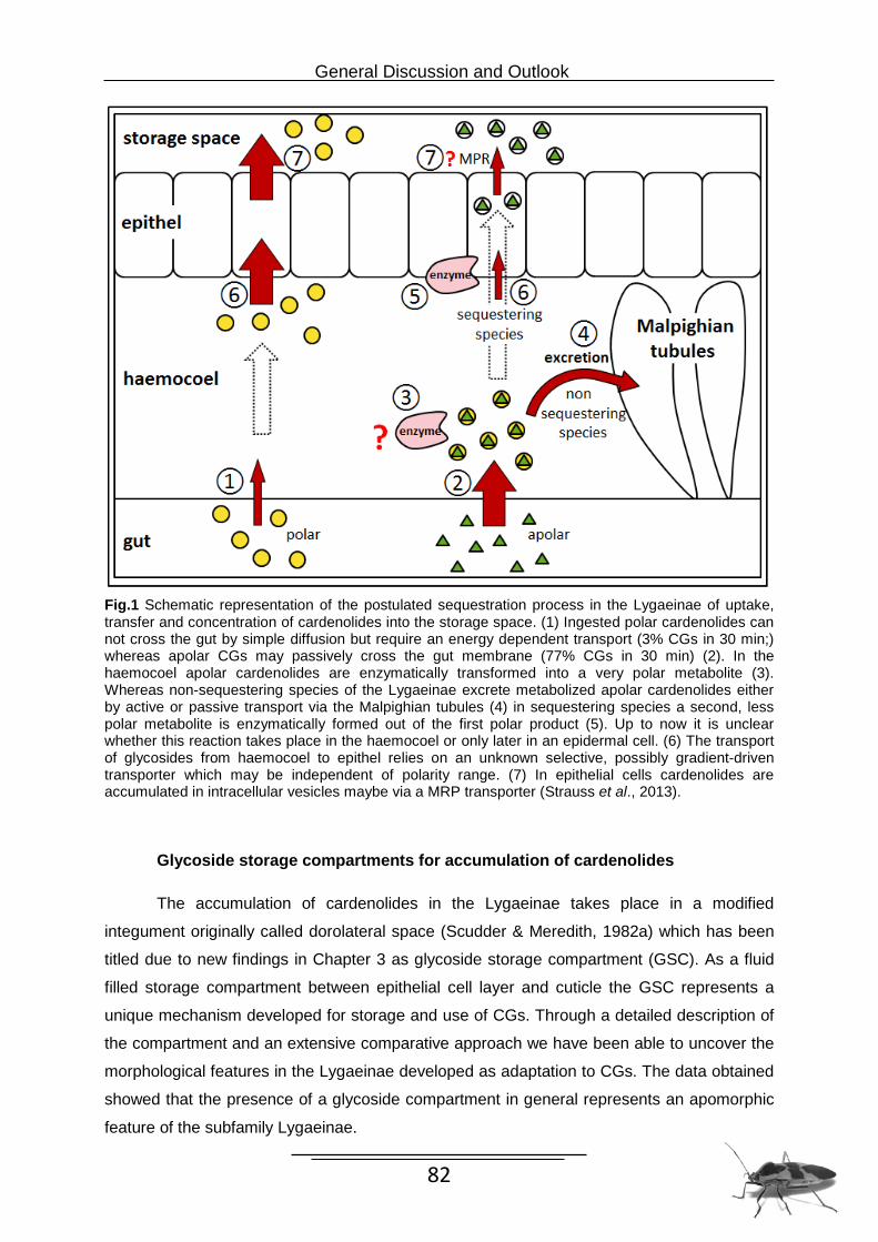
Stepwise evolution of storage compartments for defensive toxins in the Lygaeinae (Heteroptera: Lygaeidae)
Abstract
Although most species of the heteropteran subfamily Lygaeinae are known to accumulate
cardiac poisonous chemicals, in the case, cardenolides, from their host plants, little is known
about the morphological adaptations to these traits. Only the large milkweed bug Oncopeltus
fasciatus a representative of this group has previously been shown to store large amounts of
these plant toxins in a modified integument called the dorsolateral space. As additional
morphological adaptation special weak areas of the cuticle exist on thorax and abdomen
which rupture under pressure to release the cardenolide-rich droplets as a feeding deterrent
against predators. The present study reexamines this thoracic storage compartment of O.
fasciatus in detail and presents a comparative analysis of the integument as storage area for
defensive compounds in several species of Lygaeinae. By mapping the observed
morphological features on a recent phylogeny of the Lygaeinae we here report that the
adaptation for storage and release of plant compounds evolved in a stepwise manner.
Introduction
Heteropteran species are well-known as stink bugs because they possess metathoracic
scent glands (MTGs) excreting allelochemicals of an offensive odor (Remold, 1963; Staddon,
1979; Staddon & Daroogheh, 1981; Aldrich, 1988) which can be an effective anti-predator
defense (Remold, 1962; Schaefer, 1972; Staddon, 1979). Most representatives of the
hemipteran subfamily Lygaeinae, however, are known to have reduced MTGs (Schaefer,
1972; Scudder & Duffey, 1972), yet the chance of surviving a predator attack is increased.
This could be shown through predation assays in which species hold on toxin plants have
been tested directly against sunflower reared Lygaeinae (Sillen-Tullberg et al., 1982; Sillen-
Tullberg, 1985; Evens et al., 1986; Chapter 4). As alternative defense system Lygaeinae
bugs acquire toxic and emetic cardenolides (cardiac glycosides, CGs) by sequestration from
their food plants. The quality of being distasteful and poisonous is frequently combined with a
bright coloration of red or orange and black warning colors (Guilford, 1990).
Chapter 3: Stepwise evolution
48
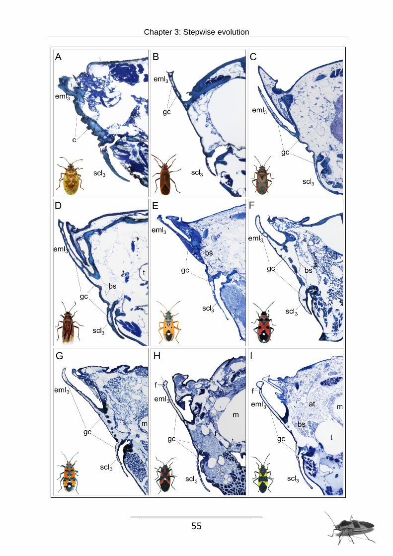
The large milkweed bug Oncopeltus fasciatus (DALLAS, 1852) is one of these brightly
patterned Lygaeinae which utilize sequestered CGs as chemical deterrents and for this
accumulates considerable quantities of these toxic compounds in specialized areas which
are called dorsolateral space (Scudder & Duffey, 1972; Scudder & Meredith, 1982a). These
structures have been shown primarily in the lateral parts of the integument in the meso- and
metathorax as well as in the sterna II to VII of the abdomen (Scudder & Meredith, 1982a). In
Heteroptera most representatives possess a duplication of the integument in the lateral parts
of the meso- and metathorax (Taylor, 1918) which will be called epimeral duplicature in the
following which is divided ventral in the supracoxal lobe and dorsal in the posterior epimeral
lobe. An investigation of these regions showed that the cuticle of the metathorax is extended
dorsally forming a pipe-like structure which releases the droplets through a slit between two
overlapping cuticular flanges. In addition the integument also has several discrete weak
points which can disrupt to release chemicals if the adult bug is squeezed. On release, these
repellent droplets are located on brightly colored areas where they can be immediately
spotted by a predator. Thus, concentration of cardenolides in the storage compartments and
release of a toxic CG containing fluid along the thorax and abdomen is probably a more
effective anti-predator strategy than the use of MTG chemicals. However, the release
mechanism of the defensive fluid is only known for O. fasciatus. According to the assumption
of Scudder and Duffey (1972) these structural specializations seen in the abdomen of O.
fasciatus created for cardenolide accumulation are also present in other Lygaeinae but are
not a universal characteristic of this subfamily. Further examinations of the sequestering
Lygaeinae, Lygaeus kalmii (SCHILLING, 1829) supposed to have found a two layered
integument very similar to that in the large milkweed bug (Scudder & Meredith, 1982a).
However, the exact 3-dimensional extensions of the dorsolateral space are not known and
comparative analyses of other species of Lygaeinae are also lacking.
Therefore, one aim of the present study was the detailed description of the glycoside
storage compartments (“dorsolateral space”) in the thoracic region of O. fasciatus using
histological sections and micro-computer tomography (µ-CT). Further ten additional
representatives of Lygaeinae and one closely related outgroup species were examined and
the findings compared with the storage compartment observed in Oncopeltus. The major
goals were to reconstruct the basic patterns of this specific defense mechanism in Lygaeinae
and to uncover the evolution of the morphological adaptations to the storage and release of
toxic CGs. This comparative approach revealed that a modification of a thickened double
layered integument is responsible for the origin of a uniform defense system combined with
the stepwise evolution of a toxin releasing system as anti-predator defense.
Chapter 3: Stepwise evolution
49
Material & Methods List of examined taxa
Adult specimens of 10 species of Lygaeinae from 8 genera and 1 closely related species of
the Ischnorhynchinae were examined. Species which were studied by serial cross sections
include Kleidocerys resedae (PANZER), 1797 (Germany: Hamburg, Martin-Luther-King-
Arocatus aenescens STÅL, 1874 (Australia: Queensland, old forestry camp, Bulburin FR), Caenocoris nerii (GERMAR, 1847) (Spain: La Gomera, between San Sebastiàn and
Chejelipes), Cosmopleurus fulvipes (DALLAS, 1852) (Marokko: Field near Chegaga),
Rheinwiesen). The species we investigated by SR-µCT were Arocatus longiceps STÅL,
1872 (Germany: Berlin, Cental Station) and O. fasciatus, in addition the latter species was
also examined with the SEM.
Methods
Histology
Specimens were dehydrated in an ethanol series and embedded in Araldite. Cross sections
of 1 µm thickness were produced using a Reichert-Jung Ultracut E microtome and glass
knifes. Sections were manually transferred to adhesion microscope slides (Histobond®+) and
stained with a mixture of toluidine blue O (MERCK) and pyronin G (SERVA) 4:1. The serial
sections were digitalised using a semiautomatic slide scanner composed of a Leica DM 5000
microscope and equipped with MetaMorph software. The digitalised sections were finished
with Adobe Photoshop CS6 (colour correction, unsharp masking) and aligned using Visage
Imaging™ Amira® 5.4 software.
Chapter 3: Stepwise evolution
50
Synchrotron Radiation based Micro-Computer Tomography (SR-µCT)
The SR-µCT scans were performed at Beamline BW2 of the German Electron Synchrotron
Facility (DESY, Hamburg; project number I-20070032) using a stable low photon energy
beam (8kV) and absorption contrast (see Beckmann et al. [2006, 2008], Friedrich et al.
[2008]). The specimens were dehydrated in an ethanol series, critical point dried (EmiTech
K850 Critical Point Dryer; Ashford, Kent, UK) and mounted with superglue on a metallic
holder. The samples were scanned in 180° rotation, resulting in an image stack with a
physical resolution of 4 µm (voxelsize: 1.98 µm). The high density resolution of the SR-µCT-
data allowed to discriminate different types of tissue (e.g., skeleton, muscles).
3-dimensional reconstructions
Based on both µCT-image stacks and histological sections the extension of the cardenolide
(or cardiac glycoside) storage compartment (CSC) of several species was reconstructed
three-dimensionally using Visage Imaging Amira 5.4 software. Due to the thin delimitation of
the CSC the structures had to be manually outlined with the “brush tool” using a Wacom
Cintiq 21UX interactive pen display. The obtained segmented image stacks were used for
the automatic creation of surface objects in Amira software.
Scanning Electron Microscopy (SEM)
For the examination of external skeleton structures the body of a specimen (with legs
dissected) was cleaned by ultrasonic bath and afterwards dehydrated in ascending
concentrations of ethanol. The specimen was fixed in formaldehyde-acetic acid- ethanol
(FAE), critical point dried (EmiTech K850 Critical Point Dryer, Ashford Kent) and glued to a
fine pin with nail polish. The sample was then coated with platinium (Polaron SC7650 Spulter
Coater) on both sides and examined in LEO 1525 SEM.
Results
Thoracic glycoside storage compartments of O. fasciatus
The glycoside storage compartment of adult O. fasciatus is an enormous, complexly
branched space underlying the cuticle of wide regions of the thorax. This compartment
originally described as dorsolateral space (Scudder & Meredith, 1982a) is now called as
glycoside storage compartment (GSC) due to new results concerning the actually locations
storing cardiac glycosides. Moreover, the new name designates the type of compounds
known to be accumulated in this compartment described in the recent study.
Chapter 3: Stepwise evolution
51
The glycoside compartment shows main concentrations in the posterolateral parts, i.e. the
epimeral regions (Fig.1.A-C). The compartment is located between the cuticle and the
epidermis, which is underlain by a thick basal lamina (Fig.2.I). The comparison of three
specimens revealed no distinct intraspecific variability. The location of the glycoside
containing spaces is identical. However, minor variation in size of some parts was
observable.
Three main compartments can be distinguished which are illustrated as pictures in
Fig.1. A-C and further are represented ba a supplementary 3D video. The anteriormost
compartment is located in the prothorax (Fig.1.A-C, represented in yellow). It is composed of
a circum-cervical ring just behind the head. This structure called as anterior protergal lobe
(apl) is a marginal evagination of the integument enclosing the neck around the prothorax
(structure by Govid & Dandy, 1970). Ventrally paired lobes widely spread in the prosternal
region to the edge of the coxa. The posterior half of the lateral prothoracic skeleton is
completely underlain by an extensive compartment. It is dorsally connected with paired, lap-
like extensions situated in the pronotum. A ventral interconnection with the precoxal room is
not developed. The prothoracic storage room is not connected to the following segmental
compartments.
The main part of the second, mesothoracic compartment fills the epimeral area. It
extends into almost every corner of the epimeron (em). The epimeron is folded in the central
region forming flattened cuticular duplicatures. Its ventral flange, the supracoxal lobe (scl),
covers the coxal joint, whereas the dorsal lobe masks the intersegmental membrane and the
spiracle. In cross section the lobes appear double layered with a narrow space between both
sheets of cuticle. The single connection to the body cavity is directly above the coxal joint
(Fig. 2.I). Almost the whole space in the cuticular lobes is occupied by extensions of the main
mesothoracic compartment. This compartment also expands along the dorsal pleural margin
on the anepisternum and enters the pleural wing process (pwp). A thin channel (vcb1) runs
ventral along the anterior rim of episternal (es) and sternal sclerites and interconnects the
spaces of both body halves. A second, posterior bridge (vcb2) is formed directly in front of the
mesocoxae. This channel is about twice time thicker as the anterior one. Two anteriorly
directed lobes are developed close to the mid line. A distinct dorsal interconnection of the
main compartments is not present. A dorsal clasp-like storage space is located below the
posterior rim of the mesoscutum but is completely delimited from the main compartments of
the mesothorax.
The third compartment, located in the mesothorax, is similar structured as its
mesothoracic counterpart. Both storage spaces are connected by a short bypass from the
posterolateral corner of the mesothorax with the anterodorsal metathoracic edge.
Chapter 3: Stepwise evolution
52
Figure 1. A-C) Thoracic glycoside storage compartments of O. fasciatus, 3D-reconstructed based on SRµCT-data. A, drawing of external features combined with reconstruction of the three biggest storage compartments in lateral view. B, posterolateral view. C, dorsal view. Distinct, unconnected compartments are differtently colored: yellow - prothoracic compartment, blue - meso- and metathoracic compartments,red - dorsal mesothoracic clasp. anterior protergal lobe (apa), coxal cleft (cc), dorsal connecting bridge 1 (dcb1), dorsal connecting bridge 2 (dcb2), epimeron (em), episternum (es), pleural wing process (pwp), supracocal lobe (scl), ventral connecting bridge 1 (vcb1), ventral connecting bridge 2 (vcb2),
A
scl1 scl2 scl3
es1
em1
apl
cc1
pwp3 em3 eml3 em2 esl2 esl3
B C
Chapter 3: Stepwise evolution
53
The lateral main elements of the metathoracic compartment differ only slightly from the
condition present in the mesothorax. Only the anepisternal part is wider distributed. Distinct
differences are observable in the interconnecting channels: neither an anterior nor a
posterior ventral connection is present but well-developed dorsal bridges (dcb1 + dcb2) fill
large areas under the metanotum, immediately behind the mesophragma (Fig. 1 B + C). The
larger one (dcb1) of these two connecting bridges has its origin in the pleural wing process
and runs from both sides along the mesophragma. The second conjunction (dcb2) arises
from the larger connecting bridge medially behind the wing base and runs along the
postscutellum as a narrowly centered deviating bridgeand merges with the parallel running
storage area (dcb1) at the end of the mesophragma.
Comparison of thoracic glycoside storage compartments in the Lygaeinae
By comparing the cross-sections respectively the SR-µCT scans of 10 different species of
Lygaeinae, we determined the extent of the glycoside storage compartment and can show
that this feature follows a uniform basic pattern. In general, it appears that all available
regions of the integument are used for cardenolide storage. The storage compartment in
further Lygaeinae shows the same construction and revealed no distinct variability as
described in O. fasciatus. Further, all investigated species show the same intersegmental
connections: a thin bridge (vcb1) anterior, and a bigger one posterior (vcb2) situated, which
ventral interconnect the space of the body halves. Equally present are both dorsal bridges
(dcb1 and dcb2) which run along under the metanotum. Examinations could not show that
further arias as described in O. fasciatus will be used to accumulate cardenolides.
The differentiating feature of the glycoside storage compartment within the
Lygaeinae is the structure of the lateral thoracic storage area. The main storage region as
previously shown (Fig.1 A-C) represents the epimeron of meso- and metathorax. In this
region is developed a special structure called as epimeral duplicature (ed) which arises
through evagination of the dorsal and ventral integument of the epimeron (see Fig.2 A-I). The
dorsal part of the epimeral duplicature forms the structure called epimeral lobe (eml) whereas
the ventral part builds the supraxocal lobe (scl). The cavity among the epimeral duplicature
represents the main part of the storage compartment. The glycoside storage compartment of
the epimeral duplicature in O. fasciatus is represented by a well and large developed space
with a cuticle thickness of 17.08 µm (Fig. 2I).
In comparison I could demonstrate that K. resedae representing the subfamily
Ischnorhynchinae exhibit no storage capacity due to a missing glycoside storage
compartment (Fig. 2.A). However, it has been shown that K. resedae exhibit a strong cuticle
Chapter 3: Stepwise evolution
54
which takes the whole region for the integument as storage space in O. fasciatus. Species of
the genera Arocatus and Caenocoris although have a compartment to store cardenolides
however, the capacity of storing substances is limited. The cuticular sides of the integuments
are still connected in some areas, which probably cause better stabilization but restrict the
space for fluid filling (Fig.2. B-D). Further the cuticle of the integument of those small bugs is
quite thick in contrast to the other genera of this lygaeid subfamily. Comparing the cuticle
thickness of the species we found that the cuticle of the genera Arocatus and Caenocoris is
1.5 times thicker relative to the cross-section of the body.
In the genera Cosmopleurus, Horvathiolus and Lygaeus I could observe
stabiliments, too, which cross the double layered integument in the basal region, yet do not
appear to reduce the storage capacity of the compartment (Fig. 2.E,F,G). The space
between the cuticle and basal lamina in the epimeral duplicature reaches the maximum
extension in the species of the genera Horvathiolus, Lygaeus, Spilostethus and
Tropidothorax as well as in O. fasciatus (Fig.2.F-I). Since the basal lamina borders the
storage space proximal directly to the body cavity, therefore species have the ability to
extend the storage compartment continuously into the body as shown in Figure 2. (F,G,I).
Abbreviations
apl anterior protergal lobe gc glycoside compartment as angled slit m midgut at adipose tissue ms median suture bs basal lamina pbp predetermined breaking point c cuticle prs posterior releasing sites dcb dorsal connecting bridge pwp pleural wing process drs dorsal releasing sites scl supracoxal lobe ed epimeral duplicature t trachea em epimeron vcb ventral connecting bridge emg epimeral wing groove eml epimeral lobe es episternum esg episternal wing groove esl episternal lobe f flange
Chapter 3: Stepwise evolution
55
Chapter 3: Stepwise evolution
56
Figure 2. Lateral part of histological cross sections through the metathoracic cuticular duplicature showing the conditions of the glycoside storage compartment and drop releasing systems in eight Lygaeinae and one Ischnorhynchinae. Note the differences in cuticule thickness and size of intercuticular space. A) Kleidocerys ressedae B) Caenocoris nerii C) Arocatus melanocephalus D) Arocatus aenescens E) Cosmopleurus fulvipes F) Horvathiolus superbus G) Lygaeus equestris H) Tropidothorax leucopterus I) Oncopeltus fasciatus.; adipose tissue (at), basal lamina (bs), cuticle (c), epimeral groove (emg), flange (f), glycoside compartment (gc), metepimeral lobe (eml3), midgut (m), supracoxal lobe (scl), trachea (t), Thoracic release sites of the Lygaeinae
By mechanical pressure on certain points of the surfaces of the adult O. fasciatus discrete
droplets of distasteful fluid (space fluid) are secreted (Fig.3 A+B). Without exception, the
integument of all segments of thorax and abdomen possesses an external modification of the
cuticle creating a weak point which can easily disrupt. The cuticle of the specific areas along
the dorsolateral margins of the body is somewhat thinner and breaks off at a predetermined
breaking point (pbp). Only in the area of the metathorax a second segmental orifice is
situated. In this region the cuticle forms a slit by two overlapping flanges for releasing
droplets without disrupting the cuticle (Fig.2.I, Fig.6.5 and Fig.5).The angled slit is situated
inferior of an epidermal lobe at the posterior margin of the metathoracic epimeron (Fig.4.6).
This region of secretion releasing we designated as posterior releasing side (prs).
By comparing twelve different species of the group we ascertained that there are
enormous differences in the characteristic of the releasing sites of the Lygaeinae. Like O.
fasciatus, T. leucopterus species also have both morphologically modified opening systems:
the predominant breaking point (pbp) situated distally on a lobe at the dorsal edge of the
metepisternum which we called dorsal releasing side (drs) and a slit formed by two
overlapping flanges at the dorsal margin of the metepimeron (prs) (Fig.2.H) very similar to
that in O. fasciatus. Gentle pressure on the thorax causes the release of droplets out of the
epimeral slit whereas strong pressure leads further to an opening of the breaking points (pbs)
in the metepisternum. An examination of six more species of Lygaeinae (H. superbus, S.
pandurus, S. saxatilis, L. equestris, L. simulans, C. fulvipes) could establish that strong
pressure of the thorax leads to the release of liquid only in the area of the metathoracic
dorsal releasing site. Histological sections clearly demonstrated that in H. superbus, S.
pandurus, L. equestris and C. fulvipes there is also a braking point of the metathoracic dorsal
releasing side. The breaking point occurs as a distally situated slit in the cuticle of the
epimeral lobe (Fig.4.6). This orifice is caused by a very thin region in the cuticle (Fig. 4.2)
and is most likely proximally closed by an underlying cell membrane. An analysis of the
region of the posterior releasing site (prs) in S. pandurus suggests that the cuticle of the
integument of the metepimeral lobe (eml3) is extremely thin but a slit formed by two cuticular
flanges is missing (Fig.4.3).
Chapter 3: Stepwise evolution
57
Figure 3. Lateral view of two different species of Lygaeinae showing releasing sites of glycoside compartments of thorax and abdomen. A). O. fasciatus excretes defence fluid of the storage compartment by seven segments through mechanical pressure. B). O. fasciatus ♀ releasing a droplet in the region indicated by the arrow of the prothorax. C). S. pandurus releasing a droplet of the pbp of the es2 and es3. D). The release of a droplet of distasteful glycoside-rich fluid from a tergo pleural weak point of the metathorax of C. fulvipes.
The region of the eml3 shows an additional cuticular modification (Fig.4.4) as determined in
the esl3 described for predominant breaking points. Despite strong pressure on the eml3 we
couldn’t observe release of glycoside-rich fluid out of this region in S. pandurus.
As a common feature between both opening systems (predominant breaking point
and the releasing system) we could observe that the slit of the eml3 as well as the preaking
point of the esl3 are located exactly in the region inferior to the end of the epimeral wing
grooves which serves as fixture for the edge of the wing. A further group species, C. nerii, A.
aenescense, A. rusticus, A. longiceps and A. melanocehalus, exhibited neither a slit as
mechanical opening nor thinly constructed weak points for predominant breaking points (pbp)
(Fig.4.1). Observations showed that pressure exerted on these species does not result in the
release of fluid.
Chapter 3: Stepwise evolution
58
Figure 4. Representation of all variants of the glycoside releasing site in the metathorax of the Lygaeinae. 1,2) Dorsolateral view of the thoracic transverse section through metepisternal lobe (esl3) of 1 A. melanocephalus without dorsal releasing sides (drs) and 2 C. fulvipes with a predetermined braking point (pbp) of the drs. 3,4,5) Dorsolateral view of the thoracic transverse section through metepimeral lobe (eml3) of 3. C. fulvipes with no posterior releasing site (prs), 4 S. pandurus showing a pbp of the prs and 5 O. fasciatus with an angled slit (as) of the prs formed by two overlapping flanges (f). 6) SEM of the dorsal part of the metathorax of O. fasciatus (lateral view); representing anteriorly the esl3 with the distally situated predetermined breaking point (pbp) of the drs and posterior the eml3 with the inferior situated (as) of the prs (Scale bars 1-5 50 µm, scale bar 6 100 µm); glycoside compartment (gc), basal lamina (bs), cuticle (c), adipose tissue (at), epimeral groove (emg)
Chapter 3: Stepwise evolution
59
Figure 5. Stepwise evolution of a storage- and releasing system for poisonous cardiac glycosides in the subfamily Lygaeinae. The top pictures display the three different forms of the storage compartment (A-C) and the three forms the metepimeral lobe (C-E) found in the subfamily. The phylogenetic tree based on combined data of COI, COII and 28S genes comprises ten genera of the Lygaeinae and Kleidocerys resedae as outgroup species. The coloration of the branches indicates the species clusters with the same morphological adaptations. Circled letters show the origins of new features: A) the origin of a storage compartment, B) the origin of a predominant breaking point (pbp) at the episternal lobe (esl3), C) the origin of a mechanical opening system at the epimeral lobe of the metathorax (eml3), D) origin of a predominant breaking point (pbp) at the metepimeral lobe (eml3). The black stars represent the species which were examined either by SR-µCT or by histological section series.
Discussion
The integument of insects not only acts as water barrier to prevent tissue desiccation but
also is very often involved in defense strategies against predators, parasites and disease
agents (Boucias & Pendland, 1998, Neville, 1975). As a physical barrier a strong exoskeleton
offers an efficient protection since it is first contacted by an attacking predator. Nevertheless,
a thinner cuticle can be advantageous in the insect’s defense strategy as well. In some
cases, mechanical defense mechanisms disappeared in order to focus prospectively on
chemical deterrence. One impressive demonstrations of such a case represents the defense
system of the subfamily Lygaeinae, shown in the present study.
Chapter 3: Stepwise evolution
60
The epimeral duplicature in Heteroptera such as in K. resedae generally consist of a
very strong cuticle to protect specimens as mechanical boundary (Larsén, 1945), whereas in
the Lygaeinae the cuticle thickness is reduced to get space for cardiac glycoside storage by
dietary sequestration from plants. As we have been able to show, the glycoside storage
compartment in O. fasciatus as well as in all examined species of Lygaeinae underlies vast
areas of thoracic regions of the external integument. Since nearly all available areas of
integument are used for glycoside storage, it seems almost impossible for predators to
overcome the chemical defense barrier.
The glycoside rich compartment arises as a result of extensive vacuolation of an
unusual epidermal cell layer (Scudder & Meredith, 1982a) which in turn enables the insect to
fill all areas of integument underlying the vacuolated tissue. Vacuolated epidermal cell layer
are examined only in a few insects including Schistocerca gregaria which used storage
vacuoles of a maturation pheromone (Neville, 1975). In popular leaf beetle (Chrysomela
populi), sequestered phenolglucoside are deposited in a reservoir of defensive glands which
include vesicles of defensive compounds (Strauss et al., 2013). With respect to the
occurrence of a similar structure, it might be possible that a mechanism as described in
Strauss et al., (2013) equally filled vesicles of the secretory epidermal cells in the lygaeinae
which via exocytosis are emitted in the glycoside storage compartment.
The basal lamina underlying the compartment serves as border between haemolymh
and glycoside fluid. The separate storage of glycoside fluid may enable the species to
decrease the concentration of glycosides in the haemolymph which despite an existing
insensitivity to the toxins might cause poisoning. Differences in the expansion of the storage
compartment in lateral and dorsal regions between species may depend on several factors,
including the type of diet, the age and the sex of the examined individuals. Because we also
know that the amount of glycosides in the females is higher than in male species and that
adults acquire about one-tenth of the cardenolides over a weak if they are only for a short
time on a glycoside diet as they are normally on glycosides over live time cycle.
Connecting bridges found between bilateral parts as well as different segments are
expected to work as pressure balance or as supply to refill depleted compartments.
Mechanical pressure resulting from the capture by a predator probably causes release of
glycoside-rich fluid from specialized cuticular structures. As already described by Scudder
and Meredith (1982a), the releasing sites in O. fasciatus can be found on the dorsolateral
margin of thorax and abdomen. However, this study shows that the prothoracic segment also
possesses a weak point which can rupture to release glycoside fluid. Since Scudder & Duffey
(1972) could not find any abdominal releasing sites they described a dichotomy in the
subfamily which divides bugs in two groups with regard to their glycoside use. In contrast, we
could show that all Lygaeinae use glycosides in the same manner as a special defense
Chapter 3: Stepwise evolution
61
strategy in which separate body parts are used as toxin reservoirs. The main difference
among the species could be observed when comparing the releasing sites.
The release of droplets can be compared with “reflex bleeding” of insects which
represents the release of haemolymph at a predefined spot. In according species the
haemolymph contains remarkable amounts of defensive substances which are used in
protection as well. As defensive action, at least five insect orders including mainly coleoptera
perform reflex bleeding where insects present haemolymph, through integumental rupture
(Wallace & Blum, 1971; Blum & Sannasi, 1974). Additional representatives are known from
Plecoptera, Homoptera (Cercopidae), Lepidoptera and Hymenoptera (Symphyta) (Peck,
2000; Boevé, & Schaffner, 2003; Capinera, 2008). However, in these cases the toxic
substances are dissolved in the haemolymph whereas in the Lygaeinae only specialized and
et al., 2011). The experiment performed with Chinese mantids (Tenoders ardifolia snensis)
demonstrated that mantids after rejection of a milkweed bug only when it had access to
Asclepias seeds have learned to reject aposematic bugs after encountering an unpalatable
conspecific. Field observations suggested that orb weaving spiders (Araneae) are able to
detect toxic Lygaeinae before biting into the prey and cut them out. Since the visual sense
plays a very minor role for orb weaving spiders, the aposematic color patterns of the bugs
can not serve as a warning signal. Moore et al., (1990) pointed out that many species relying
on toxins for defence also provide some odours as repellent signals. In this case might
olfactory senses explain the rejection of the aposematic bug by the blind spider.
Petschenka et al., (2011) illustrated that ouabain, (a cardenolide commonly used in
physiological research) per se deters potential enemies from otherwise suitable prey items,
Chapter 4: Deterrent effect of cardenolides
66
yet this cardenolide does not occur naturally in host plant species of O. fasciatus. We
therefore conducted predation assays with the golden orb web spider Nephila senegalensis
(Walckenaer) and tested the effect of a natural cardenolide composition obtained from the
host plant Asclepias incarnata. Furthermore, we investigated whether the spiders learn
avoidance of unpalatable prey by signals conveyed non-visually. Feeding on toxic host plant
species would thus confer a further advantage.
Material and methods
Plant and insect material
The milkweed seeds (Asclepias incarnata L.) for feeding-assays were received from the
botanical garden, in Hamburg. Adult male and female of the large milkweed bug O. fasciatus,
taken from a laboratory culture were kept under artificial light (16:8 h light-dark cycle) at
26°C. The insects were reared from the egg stage either on a diet of husked sunflower seeds
(Heliothus annuus L.) and water, or on milkweed seeds (A. incarnata). Specimens reared on
sunflower seeds were used as controls since this food source does not contain cardenolides.
Specimens on A. incarnata seeds derived Cardenolides from these host plant during larval
feeding. All used bugs were descendants derived from a number of couples obtained from
the Aquazoo, Düsseldorf. All N. senegalensis spiders used in this study were of the first
generation raised in captivity, the parental generation had been collected near Kapstadt,
South Africa in 2011. Due to the extreme sexual size dimorphism of Nephila only females
were used for the experiments. Spiders were placed in frames of acrylic glass (60 cm x 60
cm), with a rear panel and an open front and maintained on a diet of Calliphora flies. Animals
were kept under artificial light (12:12 h light-dark cycle) at 23-27°C and sprayed with water
every second day.
Predator-Assay
All 13 spiders used were naïve with regard to cardenolides and starved for two days before
beginning the test series because a hungry predator could be more willing to try aposematic
prey. The experiments consisted of two feeding trials in which consecutively two different
prey items were presented. For each trial a cardenolide rich and a cardenolide lacking O.
fasciatus were offered to the spiders in different order. The bugs were flipped into the webs
and the behavior of the spiders was recorded. Further, for each trial we noted the time until
the spider cut the prey out of the web (“time in web”) and as well as the residual weight of the
Chapter 4: Deterrent effect of cardenolides
67
prey. In the first trial, six spiders received a cardenolide rich Oncopeltus first. After
consumption or rejection of the prey an cardenolide rich Oncopeltus was hurled into the web.
In the second trial seven additional spiders were tested and were presented first with a
cardenolide rich Oncopeltus and afterwards with a cardenolide lacking one. We used these
two trials to check that the spiders behavior is not affected by the sequence (if the
cardenolide rich bug offered the spider first or second). We measured time in the web from
the attack of the spider to the excision of the insect and determined the weight before and
after excision.
Statistical analysis
The retention time in the web and the loss of weight after attack were separately analyzed for
both trials. All statistical comparisons used the Wilcoxen signed-rank test, which explicitly
incorporates paired data. Differences were accepted as statistically significant in all analyses
if p ˂ 0.05. To improve clarity, primary data on weight loss are illustrated as % values.
Statistical analysis was carried out with JMP 7.1 (SAS).
Results Attacking behaviour of N. senegalensis After introducing the prey into the web, the spiders responded in both trials at once to the
bug’s presence by vibrating the radii with their forelegs (plucking). (Fig. 1 A). A swift attack
after recognition of the prey was accompanied by directly grasping it with the fangs (Fig.1 B
and C). With few exceptions, naïve N. senegalensis released the cardenolide rich prey
(species reared on Asclepias) immediately upon contact (Fig.1 D). To do so, the spiders
either cut some of the threads around the prey or pulled the bug out by using their pedipalps.
In a single case, after completing a short test bite, the Nephila even backed away and
immediately begun to clean itself by wiping the fangs. The spider was instantly repelled and
acted confused and disoriented. After a while, the spider undertook numerous bites while
rotating the bug with the forelegs. This prey was released after a period of 56 minutes and
died from the bites, whereas for all other bugs in this trial the presence in the web lasted only
about one minute and the prey survived.
Chapter 4: Deterrent effect of cardenolides
68
A B
C D
E
F
Figure 1. Predation on Oncopeltus fasciatus reared (D) solely on Asclepias incarnata seeds or (E, F) exclusively on sunflower seeds (Helianthus annuus). (A) The spider is approaching the bug, and then grasps it immediately to inflict its bites (B, C). (D) Nephila releases the prey from the web by removing the silk enclosing the bug. (E) The spider spins loops of silk around the prey before translocating the completely enveloped bug to its feeding site (F).
Chapter 4: Deterrent effect of cardenolides
69
If the bugs were reared on sunflower seeds the Nephila spiders killed the bugs after
the first test bite by injection of poisonous saliva. After the bug was wrapped in silk by a few
loops, the spider brought the prey to the center of the orb and started feeding (Fig. E and F).
During feeding, which lasted 1.4-2.6 h, the spider twisted the bug and reduced the size to a
small chunk of dry remnants. All 13 bugs that we offered in both trials were treated similarly.
Effect of food plant This experiment using natural predators was conducted to test whether the insects
containing cardnolides have a deterrent effect for a higher survival. Our data suggest that
food plant species strongly affected the palatability of the prey for Nephila spiders and had
significant effects on the survival of the bugs. The Asclepias diet clearly ensured a higher
survival rate of Oncopeltus than when reared on nontoxic seeds (H. annuus). The weight
loss of the prey as a measure of consumption was significantly higher for O. fasciatus reared
on sunflower seeds than for those raised on A. incarnata seeds (Wilcoxon signed-rank test:
Figure 2A p = 0.0156 and Figure 2C p = 0.0078). The spiders consumed 94.6 % less of the
bugs reared on Asclepias compared to the control in treatment one and about 88.6 % less in
treatment two. In addition, we observed that of 13 Asclepias reared bugs that we offered the
various Nephila individuals, 12 survived. These results demonstrate that the cardenolide rich
bugs repelled the Nephila spiders significantly (Fig. 1 D).
An analysis of the time in the web also showed significant differences between individuals
fed on the nontoxic plant or the toxic hosts. Compared to the control, O. fasciatus reared on
cardenolide containing Asclepias remained significantly shorter in the web (Wilcoxon signed-
rank test: Figure 2B p = 0.0156 and Figure 2D p = 0.0078). While nontoxic O. fasciatus were
kept for 118 minutes in web by the spider, cardenolide rich O. fasciatus in contrast stayed
only 5.5 minutes in web. That’s on average, 95.3 % time difference between both
experiments. In addition, the time that the bugs were kept in the net did not differ significantly
between both trials for bugs reared on the same diet. Only in one experiment an Asclepias
reared bug was held 97.5 % longer in the web and 87.6 % more of it was consumed than of
all other individuals treated the same way.
Chapter 4: Deterrent effect of cardenolides
70
Figure 2. Reaction of N. senegalensis of two different types of prey. (A,B): Each spider (n=6) was tested first with an Oncopeltus kept on sunflower seeds followed by an Oncopeltus reared on Asclepias incarnata seeds. (C,D): 7 spiders were offered first a bug reared on Asclepias and immediately a control bug. A and C % reduction of body weight after spider attack, B and D time Oncopeltus spend in the web. Asterisks above columns indicate significant differences in Wilcoxon signed-rank test (*:p ≤ 0.05; **: p ≤ 0.01). Bars represent mean ±SD.
Learning trial for prey avoidance
The Nephila spiders usually directly approached the prey. In all experiments the spiders
responded quickly to bugs introduced into the web and generally attacked the palatable O.
fasciatus in spite of negative experiences with previous prey. Immediately (within 3 minutes)
after the first encounter and removal from the web of an unpalatable Oncopeltus, another
bug was offered. None of the spiders refused to attack the second (palatable) bug or touched
it with more caution. As shown in Figure 2, comparing the first and the second trial revealed
no difference in loss of body weight of sunflower reared bugs. The negative experience with
O. fasciatus reared on Asclepias thus did not reduce the tendency on Nephila to attack
Oncopeltus again. In addition, comparing the time in the web of palatable O. fasciatus in both
trials (Figure 2) does not reveal any effects of learned avoidance in attacking this prey.
Chapter 4: Deterrent effect of cardenolides
71
Discussion
During evolution insects have developed an impressive set of mechanisms to defend
themselves against natural enemies. Exploitation of chemical defences acquired through
feeding on toxic host plants might be the most widespread strategy for the insect`s own
benefit against predation. This study focuses on the potential benefits of cardenolide
sequestration from Asclepias host plants against an invertebrate predator (N. senegalensis).
We can show that the large milkweed bug O. fasciatus had a significantly lower mortality
then their sunflower-reared conspecifics. In the single case where a bug reared on Asclepias
seeds was killed, the spider undertook numerous test bites which may have been caused by
increasing hunger. Altogether, these results demonstrate that cardenolide sequestration is
advantageous and ensures the survival of the single individual.
Nevertheless, different predators vary in their sensitivity to the distastefulness of a
given insect. Generally, it appears that cardenolide containing Lygaeinae have a strong
deterrent effect on one of their main vertebrate predators, the insectivorous birds (Evans et
al., 1986; Sillen-Tullberg et al. 1982; Sillen-Tullberg, 1985; Tullberg et al., 2000) whereas in
case of mice as possible predators experimental data are lacking. However, could be shown
that cradenolide sequestration by monarch caterpillars causes protection of the adults from
mice (Glendinning, 1990). By their bitter taste and emetic effect cardenolides are expected to
induce a general negative reaction in vertebrate predators. Yet, there is only little
experimental evidence to suggest that cardenolides are effective against invertebrates
(Berenbaum & Miliczky, 1984; Malcolm, 1989; Petschenka et al., 2011).
In some instances, the deterrent effect of cardenolides differs between predators
possibly due to differences in taste perception. Berenbaum & Miliczky (1984) demonstrated
that cardenolides in O. fasciatus act as deterrents against Chinese mantids (Tenoders
ardifolia snensis), whereas Levey (1983) suggested that mantids did not distinguish between
cardenolide containing prey and prey lacking these toxins. On the other hand, it could be
shown that lygaeid species such as Spilostethus pandurus or Lygaeus kalmii were refused
by predatory bugs or ants (Levey, 1983; Jones, 1934). Malcolm (1989), however,
demonstrated that feeding on cardenolide containing Aphis nerii caused the orb-web spider
Zygiella x-notata (Clerck) to build disrupted webs, yet they did not reject the toxic prey. Part
of the difference in the behavior of these predators may also depend on the concentration of
unpalatable compounds sequestered from the host plants species. Cardenolide
concentrations in Asclepias in particular show extremely large variability between plant
species and among plant parts which can be responsible for differences in acceptability for
predators (Malcolm, 1991). Therefore, Petschenka et al. (2011) used Nephila spiders to test
Chapter 4: Deterrent effect of cardenolides
72
directly for the influence of ouabain against invertebrate predators without disturbing factors.
In this study, in addition, we provide evidence that cardenolides sequestered from a natural
host plant species, the swamp milkweed A. incarnata which has a low constitutive
cardenolide concentration (Roeske et al., 1976; Malcolm, 1991) still cause rejection by
Nephila spiders.
An observation, which could be important in this context, is the accumulation of
crdenolides in the integument as found in all lygaeinae bugs. A fluid filled compartment called
the glycoside storage compartment arises through a specialized layer of the epidermis where
sequestered cardenolides get enriched. In some cases it is the mechanical action of the
predator itself, which causes the release of the defence substances. Through mechanical
pressure on certain surfaces of the adult bugs, single discrete droplets of distasteful fluid are
secreted. Although a direct release of cardenolide fluid couldn’t be observed it seems
possible that the spider after tasting bitter repellents released the bug immediately upon
contact without visible injury. Nevertheless, it is known that rejection of distasteful or
otherwise obnoxious prey only occurs after a Nephila spider has bitten it (Robinson &
Robinson, 1973). Nephilidae generally thought to have a primitive prey capture strategy and
usually attack all types of prey by biting (Robinson & Myrik, 1971; Eberhard, 1982; Higgins,
2008), whereas the prey-catching behavior of other orb-weaving spiders differs: They do not
immediately kill the captured prey but rather they quickly wrap prey in silk for immobilization
(“wrap attack” strategy) (Olive, 1980). Observations showed that the first bite of Nephila upon
contact is a short-duration bite made to test the prey, whereas a second long bite will inject
the venom and kill a palatable prey. Obviously, Nephila spiders possess receptors which will
be stimulated by bitter tasting cardenolides and induce a negative response. Therefore, it
seems advantageous for Lygaeinae to be able to release distasteful fluid to outer parts of the
body to reduce the damage of an unavoidable bite. Despite of a test bite performed by the
spider, a survival rate of nearly 100 % illustrates the protective function of sequestered
cardenolides for the single prey. If bugs are attacked by a vertebrate predator a quail
respectively, the survival of the prey is much lower and the supposed chemical defence only
secures the bug from being eaten, not from being killed (Evans et al., 1986). Therefore, it is
advantageous that chemical defenses frequently co-occur with sounds or odours but often
with bright colorations, to advertise their unpalatability or toxicity that encouraged predators
to learn to avoid distasteful prey.
Avoidance learning is a widespread phenomenon which occurs in vertebrates as well
as in invertebrates (Berenbaum & Milictky, 1984; Lindström et al., 2001; Ham et al., 2006;
Skelhorn & Rowe; 2006). However, the preliminary examinations showed that N.
senegalensis isn’t able to learn to avoid O. fasciatus after a bad experience. In fact, for the
blind Nephila, there are no colourful bugs. However, it might be possible that several
Chapter 4: Deterrent effect of cardenolides
73
successful alerting signals stimulate the various sense of the spider and thus reflect the
chemical defence of the prey which in turn causes the learning behavior. The results of an
experiment by Rowe & Guilford (1996) illustrated that avoidance of an offered coloured prey
only showed up if an odour is also present.
Chemically defended insects in some cases release a disgusting odour when
attacked, e.g. ladybirds are not only aposematic rather they contain a bitter tasting toxin and
signal this by secreting a fluid that smells strongly of pyrazines (Moore et al., 1990). Indeed,
pyrazines seems to be a common alerting signal in insect defence which functions for
animals that rely on the olfactory sense. In order to test the influence of the spiders
behaviour through possible occurring pyrazines or similar defensive odours in O. fasciatus
reared on A. incarnate, further predation assays have to be performed in which two
cardenolide rich O. fasciatus become offered N. senegalensis spiders successive.
Performing this trial might explain the missing avoidance learning in the spiders so far.
The most essential feature of an alerting signal is that it has to be functional at a
distance that ensures the survival of the prey (Moore et al., 1990). Field observations with
the cross orb-weaving spider, Araneus diadematus, demonstrated that toxic Lygaeus
equestris have been rejected before contacting it (unpublished data). Besides Nephila also
Araneus isn’t able to visualize prey as being aposematic, however, it immediately removed
the bug from the web by cutting the silk. This behavior could be the result of deterrent
volatiles in combination with a learned avoidance which in turn of the spider provides an
advantage through reduction of silk and energy. Referring to Nephila spiders detecting an
alerting signal at an early stage could be a decisive advantage for both the spider which don’t
waste energy through performing test bites and for the bug that is protected before getting
damage.
Apart from the function of several stimuli the memories of a bad experienced prey
may only exist after the spiders were trained to associate through several repetitions. As it
was shown in the memorability test by Ham et al, (2006) birds generally remembered to avid
the prey with colour patterns they associate with unpalatabiliy, yet after a period of trays. To
ensure the Nephila had the ability to decide through learning processes to avoid unpalatable
Lygaeinae, several repetitions offering unpalatable bugs must be carried out in a separate
trial.
Further, it is a widespread phenomenon that nearly all Heteroptera possess scant
glands whose secretions primarily function in defence against predators (Aldrich, 1988). O.
fasciatus also possess these metathoracis scant glands (MTGs), yet in a reduced form
(Schaefer, 1972; Scudder & Duffery, 1972). The function of chemical compounds in MTGs
however, showed no influence in deterrence of the spider N. senegalensis because
cardenolide lacking bugs have been eaten immediately after contact.
Chapter 4: Deterrent effect of cardenolides
74
Taken together our results demonstrate that O. fasciatus benefit from sequestration of
cardenolides from the host plant Asclepias incarnata and obtain an effective defence against
the invertebrate predator N. senegalensis. But there was no effect of aposematic color signal
that implies avoidance learning and memorize retention. Several signals become directed
against different groups of predators and divers hunting strategies. Whereas vertebrate
predators such as birds were thought to be almost exclusively hunters by visual cues, some
predators mainly identify potential prey primarily by means of olfactory and gustatory signals
(Hostettler & Nentwig, 2006; Thomas et al., 2008). It is necessary to perform additional
assays in order to investigate whether besides an offensive hunting strategy avoidance
learning is a natural instinct in N. senegalensis.
75
General Discussion and Outlook
Herbivorous insects have to cope with a stunning array of toxic secondary plant compounds
which become ingested during the feeding process. In order to reduce or to overcome the
potential damage, adapted herbivores have evolved complementary adaptations as a
response to the chemical selection pressures. While some herbivores have developed
strategies to avoid dangerous plant chemicals, or physiological and biochemical resistance
mechanisms to overcome their toxic effect (Scudder & Meredith, 1982; Després et al., 2007;
Capinera, 2008; Petschenka et al., 2013), sequestering specialists, i.e. those that selectively
take up and accumulate plant chemicals, use these defensive substances for their own
benefit against predators or pathogens (Nishida, 2002; Hartmann, 2004; Després et al.,
2007; Opitz & Müller, 2009). In order to be effective, the defence compounds must be able to
interfere with molecular targets of cells, tissues or organs (Wink, 2009). Focusing on cardiac
glycosides (CGs), these bitter tasting and emesis provoking plant compounds cause toxic
effects in vertebrates and insects as neurotoxins (Malcolm, 1991; Wink, 2009).
Host plant use in the Lygaeinae
Some of the larger species of the hemipteran subfamily Lygaeinae are called
“milkweed bugs” which reflects their worldwide common association with plants of the
milkweed family Apocynaceae which is known to comprise many plant genera containing
CGs (Scudder & Duffey, 1972). Approximately 80 % of the Lygaeinae use an apocynaceous
plant as primary host. Whereas only a few species of herbivores utilize plants from more than
three plant families, the majority is specialized to feed on few closely related plant species
belonging to a single plant family or species similar in one or more properties such as
biochemistry (Schoonhoven et al., 2005).
Given that Lygaeinae are the most species rich lineage within the Lygaeidae (sensu
Henry, 1997) with more than 500 species compared to the sister groups Orsilinae (250
species) and Ischnorhynchinae (75 species), the adaptations to dietary CGs may represent a
key innovation of this group. In general, it is assumed that species that are specialized in
their food utilization are often more diverse than taxa including more generalist feeders
because the rate of evolution is thought to be higher among specialists (Whitlock, 96).
Some Lygaeinae bugs however, occur on plants of other CG containing taxa
including in families Ranunculaceae (Junior & Wichtl 1980; Winkler & Wichtl, 1986; Deckert,
2007), Asparagaceae (Vivas, 2012; Knittel et al., 2013) or they are associated with
Plantaginaceae (Péricart, 1998; Wachmann et al., 2007, Petschenka pers. obs.). The current
General Discussion and Outlook
76
use of plants from non-apocynaceaous families most likely represents host shifts which were
facilitated by the preadaptation of lygaeines to CGs. Remarkable however, species like
simulans and Horvathiolus superbus, which in addition to the plant species mentioned above,
are also closely associated with Vincetoxicum hirundinaria (Wachmann et al., 2007) an
apocynaceous host devoid of CGs (Dobler et al., 1998). The former species in particular
cannot survive without V. hirundinaria also known as Swallow-wort because they depend on
it for growth and development. It is of interest to find out why, despite elaborated adaptations
to GC in the Lygaeinae, particularly in T. leucopterus which possess a well-developed
defence system, so many species of the subfamily use V. hirudinaria as host. Members of
the genus Vincetoxicum contain the haemolytic glycoside vincetoxin and are highly
poisonous and emetic to humans and mammals (DiTommaso et al., 2004). A mixture of
steroid glycosides and phenanthroindolizidine alkaloids are characteristic constituents of 70
genera of Apocynaceae, including the species Vincetoxicum hirudinaria (Staerk et al., 2000,
2005; Muola et al., 2010). Further, the bitter tasting vincetoxin could be found next to CGs in
apocynaceous species including Asclepias syriaca and A. curassavica. Wink (2009)
classified the toxic effect of Vincetoxicum ingredients as strongly cytotoxic and neurotoxic.
Through physiological studies testing the effect of dissolved compounds of Vincetoxicum
officinale seeds on cardiac activity of frog hearts, Franzen (1929) could demonstrate that the
substances have the same influence on heart beat and rythm as compounds of Digitalis.
However, such biochemical studies focusing specifically on the mode of action of isolated
compounds of Vincetoxicum seeds have not been repeated. It is tempting to speculate, that
the structural properties of the vincetoxin molecule or related glycosides in the seeds are
similar to CGs in their influence on the function of the Na/K-ATPase. Nevertheless, it could
be shown that T. leucopterus, L. equestris, S. saxatilis species reared on V. hirudinaria are
not worse protected than bugs raised on a CG rich diet (Sillen-Tullberg, 1982, 1985; Sillen-
Tullberg et al., 2000; Svadova et al., 2010). It seems likely that due to the occurrence of
similar chemistry e.g. vincetoxin in the former host plant species the switch to CG lacking
Apocynaceae has been facilitated. Further, the presence of bitterness in the defensive
secretions in species feeding on V. hirudinaria might act as an alarm signal in predators
which is sufficient to get the same protection as the use of a CG diet. Nevertheless, the
reason for the switch to Vincetoxicum is still open to speculation. In the case of L. equestris
the second preferred host plant species is Adonis vernalis, a CG containing Ranunculaceae
with a restricted distribution range, whereas V. hirundinaria is distributed over wide areas of
continental Eurasia. The availability of a potential host plant may also explain current host
use and distribution for the European Arocatus longiceps which probably changed host plant
priority to a host plant (Platanus) that became widely distributed in Europe since the 17th
General Discussion and Outlook
77
century, starting from an original occurrence in southeastern Europe. A. longiceps is
common just as its host plant and originated from eastern Mediterranean regions, but has
spread rapidly westwards (Barndt, 1995; Wachmann et al., 2007).
Sequestration of – and Insensitivity against toxic cardenolides
Aposematically colored bugs of the Lygaeinae are not only adapted to feed on
previously mentioned host plants further they are characterized to store cardenolides a class
within the CGs for defensive purpose (Scudder & Duffey, 1972). It has to be mentioned that
indeed cardenolides could be detected in many of these bugs but sequestration per se and
adaptations against intoxication have only been found in a few species up to now (Dobler et
al., 2012; Zhen et al., 20012). Therefore in a first approach (Chapter 1) I compared the
uptake of two purified cardenolides, which represent a polarity range of these toxic
compounds present in the natural died of the insects. The data obtained showed that
cardenolide sequestration is restricted to the Lygaeinae and does not occur in outgroup
species, however, two species of Lygaeinae also proved to be unable to sequester
cardenolides. The absence of sequestration in the European Arocatus species A. longiceps
and A. melanocephalus correlates with the life-history of these two species which are living
and feeding on plants not known to produce cardenolides. Feeding on non-cardenolide
containing hosts is most likely not the ancestral state in Arocatus as representatives of the
genus in southern regions are well known to use CG plants as hosts or were even shown to
possess sequestered cardenolides (von Euw et al., 1971).
The presence of cardenolides in body parts of sequestering species necessitates
mechanisms to prevent intoxication. Coadapted species need to be insensitive because the
toxins should be in an active form to realize their impact on the next trophic level (Després et
al., 2007). As highly specifically acting substances, cardenolides binds to the Na/K-ATPase
directly inhibiting its action. One possible mechanism consists in a modification of the target
site in the Na/K-ATPase that has a strongly lowered binding affinity towards cardenolides.
This phenomenon called target site insensitivity was observed in the monarch butterfly
Danaus plexippus (Vaughan & Jungreis, 1977; Dobler et al., 2012; Zhen et al., 2012), in the
grasshopper Poekilocerus bufonius (Al-Robai et al., 1990), in Chrysochus leaf beetles
(Labeyrie & Dobler, 2004) and in the milkweed bug O. fasciatus (Moore & Scudder, 1986;
Dobler et al., 2012). How sequestering Lygaeinae avoid cardenolide intoxication is
demonstrated by our data (Chapter 1) on Na/K-ATPase activity under ouabain stress which
indicate that target site insensitivity towards cardenolides is a common feature in the
hemipteran subfamily Lygaeinae.
General Discussion and Outlook
78
Even species, who do not normally encounter dietary cardenolides and do not store
the toxins, still possess a modified insensitive Na/K-ATPase. These results lead us to
conclude that target site insensitivity and sequestration of cardenolides are basal and
plesiomorphic characters of the Lygaeinae which may stem from an originally host plant use.
Our in vitro investigations of the Na/K-ATPase of nervous tissues indicate that all
Lygaeinae tested showed a relative uniform pattern of strong insensitivity towards oubain, i.e.
a much higher ouabain-insensitive Na/K-ATPase (IC80 = 2.46*10-4M) than the outgroup
species Kleidoceris resedae (IC80 = 2.06*10-7M) and a slightly higher insensitivity than the
monarch butterfly (IC80 = 2.32*10-5M) (Petschenka et al., 2013). The preliminary molecular
investigations of the Na/K-ATPase of several lygaeinae demonstrated that an amino acid
substitution of asparagine for histidine at position 122 (N122H) is present in the first
extracellular loop of the Na/K-ATPase of all species and is at least partly responsible for
insensitivity in the in vitro enzyme assays (Dobler et al., 2012; Dalla et al., 2013). We have
evidence that the possession of further amino acid substitutions at positions which are known
to be involved in ouabain binding of Na/K-ATPase in the Lygaeinae lead to a further
increased insensitivity. As previous studies could show up to nine different substitutions may
be responsible for the lower cardenolide binding characteristics in CG adapted insects
(Dobler et al., 2012; Zhen et al., 2012; Dalla et al., 2013). The combined substitution of
Q111T-N122H in O. fasciatus and Q111V-N122H in D. plexippus are known to reduce the
inhibition by ouabain even more strongly compared to the wild-type occurring in K. resedae.
Both combinations do not differ significantly from each other, yet in combination with a
substitution of F786N and T797A in O. fasciatus may cause an even more strongly increased
insensitivity. Enzyme assays performed with the single mutation T797A discovered in L.
kalmii, a further Lygaeinae, showed a 250-fold increased resistance (Dalla et al., 2013).
Therefore, it is likely that due to combinations of at least four potentially important
substitutions at different positions the strongly increased resistance in the Lygaeinae can be
explained.
Metabolic alteration of cardenolides
It is interesting to note that in general in O. fasciatus a high concentration of polar and
an absence of apolar cardenolides was found (Duffey & Scudder, 1974; Yoder et al., 1976;
Duffey et al., 1978; Moore & Scudder, 1985). Similar results could be obtained in Syntomeida
epilais, Empyreuma pugione, and D. plexippus (Black, 1976; Seiber et al., 1980; Martin et al.,
1992). The very polar compounds detected represent products arising from metabolic
alteration in the species. Although apolar cardenolides may more emetic (Blum, 1981),
Malcolm (1991) presumed that more polar cardenolides exhibit a lower mobility in the
General Discussion and Outlook
79
haemocoel whereby they can better controlled and further they are easier to store. It appears
plausible to prevent selfintoxication by minimizing the mobility and penetration of diffusible
apolar cardenolides within the body tissues.
The study represented in Chapter 2 was undertaken in order to clarify certain aspects
of the metabolic alteration of cardenolides in the Lygaeinae and to take conclusions about
the mechanism by which these species are able to handle these toxic compounds.
Metabolism of ouabain was not determined in any examined Lygaeinae. Digoxin however,
was converted first to a metabolite (fraction I) more polar than ouabain but afterwards
changed to a poorly more apolar cardenolide (fraction II) prod to two enzyme catalyzed
reactions required for digoxin metabolism. According to our findings the storage of originally
polar cardenolides (ouabain) should be less costly than storage of apolar cardenolides
(digoxin) which necessitate an enzymatic conversion. The data presented here suggest the
occurrence of at least one, most likely two enzymatically catalyzed reactions (Fig.1) forming
a metabolic pathway to change apolar in rather polar cardenolides.
With regard to the toxic effects of cardenolides, it is important to differentiate between
cardenolide sequestering species and those who are not. In non-sequestering A. longiceps
and A. melanocephalus I could detect a highly polar metabolite (fraction I) in low amounts
which disappeared rapidly may by non-selective excretion via the Malpighian tubules (Fig.1).
In addition, excretion by the Malpighian tubules can be expected to reduce haemolymph
levels of cardenolides as described in Zonocerus variegatus (Rafaeli-Bernstein & Mordue,
1978), Drosophila melanogaster (Torrie et al., 2004), expected in Manduca sexta (Vaughan
& Jungreis, 1977) and in O. fasciatus (Meredith et al., 1984). The widespread occurrence of
this feature suggests that insect Malpighian tubules generally are able to excrete
cardenolides. Sequestering species in the Lygaeinae however, seem to possess a second
enzyme which causes a amination reaction, expected at C-20 of the metabolite (fraction I)
followed by a hydogenation. The metabolite of the postulated amination process (fraction II)
is characterized by a lower polarity and could finally be detect in the defensive secretions of
Lygaeinae bugs.
These results lead us to conclude that European Arocatus species not only lost the
ability for sequestration as shown in Chapter 1, but also the ability to transform cardenolides
into a storable form. It seems plausible to reduce a costly adaptation, such as an energy
dependent metabolism which might be necessary to sequester cardenolides, if there are no
cardenolides in the species diet anymore. Unexpected however, seems the maintaining of
the fist metabolism in this species, because the transformation process is expected to be
costly, too. These results suggest that involved enzymes are not specific for
biotransformation of CGs, but rather they are engaged in diverse biochemical functions.
General Discussion and Outlook
80
Generally, in species who are not adapted to use toxic compounds, it should be
favorable to deactivate the toxins and to excrete them. Detoxification may be aided by
specialized enzymes e.g. by polysubstrate monooxigenases (cytochrom P450
monooxigenase) that are rapidly induced by the presence of toxins. In general metabolic
alteration of allelochemicals is a common mechanism to avoid intoxication (Després et al.,
2007; Opitz & Müller, 2009). Cytochrom P450 enzymes transform toxins by oxidation into
more polar compounds that are excreted or further metabolized (Feyereisen, 2005).
These enzymes may be widely distributed in the tissues because polar metabolites
could be formed in Lygaeinae after ingestion as well as injection of apolar compounds. P450
gene expression, which have been found in insects’ guts and fat bodies (rich sources of
metabolism of substrates (Estela & Soulages, 2010)), may indicate that such an enzyme
could also be involved in metabolic alteration of cardenolides in Lygaeinae (Feyereisen,
1999). Nevertheless, in agreement with the results reported by Scudder & Meredith (1982)
metabolites could not be formed by isolated haemolymph, fat body or defence fluid so far.
With respect to the metabolic processes in the Lygaeinae we assume that alteration
of apolar cardenolides as observed in A. longiceps and A. melanocephalus originated as a
detoxification mechanism whereas further transformation as observed in O. fasciatus,
Horvathiolus superbus, Lygaeus equestris, L. simulans, L. kalmii, Spilostethus pandurus and
Cosmopleurus fulvipes represents an adaptation to accumulation and storage.
Uptake, transfer, and concentration of cardenolides
The phenomenon of cardenolide sequestration, per se, involves the uptake, transfer
to, and concentration of cardenolides in the storage regions. Physiological studies on
permeability of insect guts suggest that polar cardenolides such as ouabain require an
energy dependent transport and presumably intestinal carriers in the gut epithelium which
allow polar cardenolides to enter the haemolymph. Conversely, apolar cardenolides can be
expected to cross the gut passively due to their physiological properties (Wright, 1960). Up to
now the most comprehensive knowledge of sequestration processes in the Lygaeinae has
been obtained for O. fasciatus. Several studies comparing the CG profiles of the large
milkweed bug and its host plant species indicated that cardenolides of a wide polarity range
were taken up through the gut (Yoder et al., 1976; Duffey et al., 1978; Scudder & Meredith,
1982b) whereas Scudder et al. (1986) mentioned that individual cardenolides are selectively
sequestered and stored. The phenomenon of selective sequestration could also be observed
in D. plexippus (Brower et al., 1982), Aphis nerii (Rothschild et al., 1970), P. bufonius (Von
Euw et al., 1967; Nahrstedt, 1982) and the Lygaeinae Caenocoris nerii and Spilostethus
pandurus (von Euw et al., 1971).
General Discussion and Outlook
81
In O. fasciatus it was found (Fig.1) that the uptake from the gut into the haemolymphe
of apolar cardenolides occurred far more rapidly (77% in 30 min.) than the uptake of polar
ones (3% in 30 min.) (Yoder et al., 1976; Scudder & Meredith, 1982b). This result, exactly as
expected demonstrate that digoxin seem to cross the gut passively whereas, the uptake of
rather polar ouabain into the body cavity is only possible via a carrier mechanisms.
Nevertheless, the results by Yoder et al., 1976 and Scudder & Meredith, 1982b let us
assume that gut uptake is unspecific and allows a broad spectrum of cardenolides to enter
the haemolymph.
Further, the results presented here agree with those of Duffey et al. (1978) in so far
as the uptake of polar CGs from the haemolymph into the storage space (50-70% in 72h) is
faster than the transfer of metabolized apolar cardenolides (1-3% in 72h). My results clarify
that a selective process seems to underlie the passage into the storage compartment. It
might be expected that a transformation mechanism as shown for digoxin in Chapter 2 which
is costly offers an advantage for storing apolar cardenolides. However, our data suggest that
a selective carrier, a cardenolide carrier with substrate specificity might be present which
preferred the transport of original polar CGs (e.g. ouabain) into the storage compartment.
This glycoside storage compartment is a specialized layer of the epidermis where
sequestered cardenolides get enriched (Chapter 3). This special structure underlying the
storage compartment of sequestered compounds, include vacuoles suggested to store
cardenolides (Scuddee & Meredith, 1982a).
The study by Strauss et al., (2013) demonstrates that a selective transfer takes place
to regulate the uptake of plant compounds from the haemolymph into the secretory cells
which in turn contain vacuoles transporting compounds to the storage reservoir in
Chrysomelina larvae. With respect to the occurrence of a similar structure, it might be
possible that as described in Strauss et al., (2013) an ABC transporter in the Lygaeinae
transfers cardenolides into intracellular vesicles which are thereon delivered in the glycoside
storage compartment via exocytosis. Such a mechanism favors the re-fill of CGs after
release of stored fluid without the need to destroy or excrete epithel cells which entails costs
for regeneration of the cells. Further, the polarity dependent sequestration rates could be
explained by a selective import into the epithelium of the Lygaeinae as well as described in
Strauss et al., (2013). In such case a lowered binding affinity of metabolites to carrier would
be favor the passage of unchanged compounds which appears more plausible as described
in Scudder et al., (1986) that the uptake of CGs into the epithelial cells of the integument will
be achieved by passive diffusion.
General Discussion and Outlook
82
Fig.1 Schematic representation of the postulated sequestration process in the Lygaeinae of uptake, transfer and concentration of cardenolides into the storage space. (1) Ingested polar cardenolides can not cross the gut by simple diffusion but require an energy dependent transport (3% CGs in 30 min;) whereas apolar CGs may passively cross the gut membrane (77% CGs in 30 min) (2). In the haemocoel apolar cardenolides are enzymatically transformed into a very polar metabolite (3). Whereas non-sequestering species of the Lygaeinae excrete metabolized apolar cardenolides either by active or passive transport via the Malpighian tubules (4) in sequestering species a second, less polar metabolite is enzymatically formed out of the first polar product (5). Up to now it is unclear whether this reaction takes place in the haemocoel or only later in an epidermal cell. (6) The transport of glycosides from haemocoel to epithel relies on an unknown selective, possibly gradient-driven transporter which may be independent of polarity range. (7) In epithelial cells cardenolides are accumulated in intracellular vesicles maybe via a MRP transporter (Strauss et al., 2013).
Glycoside storage compartments for accumulation of cardenolides
The accumulation of cardenolides in the Lygaeinae takes place in a modified
integument originally called dorolateral space (Scudder & Meredith, 1982a) which has been
titled due to new findings in Chapter 3 as glycoside storage compartment (GSC). As a fluid
filled storage compartment between epithelial cell layer and cuticle the GSC represents a
unique mechanism developed for storage and use of CGs. Through a detailed description of
the compartment and an extensive comparative approach we have been able to uncover the
morphological features in the Lygaeinae developed as adaptation to CGs. The data obtained
showed that the presence of a glycoside compartment in general represents an apomorphic
feature of the subfamily Lygaeinae.
General Discussion and Outlook
83
Besides the Lygaeinae, many sequestering species store plant toxins in
defined compartments which include the cuticle, specialized tissues or glands where their
potential toxic effects are confined and accordingly concentrations in the haemolymphe can
be kept low (Blum et al., 1990; Karban & Agrawal, 2002; Opitz & Müller, 2009). However, in
some species the haemolymph contains remarkable amounts of defensive substances which
are used in protection as well. As defensive action, at least five insect orders including mainly
coleoptera perform reflex bleeding where insects present haemolymph, through integumental
known from Plecoptera, Homoptera (Cercopidae), Lepidoptera and Hymenoptera
(Symphyta) (Peck, 2000; Boevé, & Schaffner, 2003; Capinera, 2008). It is remarkable that
plant derived toxins are often deposited in the integument or cuticle. Toxins can be stored in
scales of the wings in Lepidoptera e.g. in Danainae (D. plexippus), Ctenuchidae (Syntomeida
epilais) (Roeske et al., 1975; Nickisch-Rosenegk et al., 1990) or in specialized exocrine
glands that are situated in thorax and abdomen of chrysomelid adults and larvae (Pasteels et
al., 1983, 89; Strauss et al., 2013). The exosceleton provides an ideal site for storage of toxic
compounds because it represents the barrier that a predator first is faced with.
Further, I could show that the reduction of cuticle thickness and the origin of a fluid
releasing system are features of a stepwise evolution of which the presence of a mechanical
toxin releasing system in O. fasciatus and Tropidothorax leucopterus represents the most
developed mechanism. In some cases it is the mechanical action of the predator itself which
causes the release of the defence substances. Through mechanical pressure on certain
points of the surfaces of the adult bugs, single discrete droplets of distasteful fluid are
secreted. Our data suggest however, that not all Lygaeinae possess the ability to emit
droplets of cardenolide-rich secretion upon disturbance. In a previous study Scudder &
Duffey (1972) described the occurrence of abdominal glycoside compartments in 57
Lygaeinae and justified a dichotomy due to the presence or absence of predefined releasing
sites. However, they neglected to examine further features relevant to assign Lygaeinae with
respect to how the insects use the chemicals. The present investigations pointed out how
these species can be divided into three categories: (1) Species which are incapable to emit
cardenolide-rich fluid at any point of body surface, (2) species whose ability is limited to
release droplets only in thoracic regions and (3) those that possess thoracic and abdominal
releasing sites. Species of the former category are well protected by confronting the
attacking predator with a solution of cardenolides of many single released droplets whereas
species without fluid exit points have reduced chances to survive an encounter of a predator
since distastefulness first get noticed when the bug was crushed. In this case conspecifics of
previously attacked prey are protected if a second prey will be released unharmed due to
avoidance learning.
General Discussion and Outlook
84
Cardenolides for defensive purpose
The process of learning and associating unpalatability with a certain kind of prey
becomes more effective if it is equipped with a bright coloration. Frequently, chemical
defences co-occur with conspicuous warning signals in insects. Most species of the
Lygaeinae are colored bright red, orange and yellow in combination with a black pattern, with
the exception of a few species from ground living genera (Slater & O’Donnell, 1995). The
benefit of being conspicuously colored is that naive predators learn to avoid aposematic prey
more quickly then cryptic prey (Gittleman & Harvey, 1980; Gambarele-Stille & Tullberg, 1999;
Speed, 2000; Ripii et al., 2001; Ham et al., 2006; Exnarova et al., 2006, 2008). Predators
however, whose visual sense plays a very minor role can not differentiate between defended
and non-defended prey on the basis of aposematic color patterns. Therefore, it is
advantageous that chemical defenses are also frequently associated with specific odours or
sounds which reveal their unpalatability or toxicity and encourage predators to learn to avoid
distasteful prey. Predation assays performed with the not visually oriented golden orb-web
spider Nephila senegalensis (Chapter 4) showed primarily that sequestered cardenolides
from the host plant species Asclepias incarnata in O. fasciatus cause rejection by the spider.
Furthermore, the results demonstrate that cardenolide sequestration of toxic host plant
species is advantageous and ensures the survival of the attacked individual, yet experiments
did not reveal any signs of avoidance learning so far. Moore et al., (1990) assumed that CGs
cannot serve as pre-sampling signals but rather act through their bitter taste as immediately
post-sampling repellents. Thus in order to present existing chemical defence some
aposematic insects release warning toxic fluid in combination with a strong odour (e.g.
Coccinella, Danaus, Zygaena) (Rothschild et al., 1984; Moore et al., 1990; Marples et al.,
1996; Harborne, 2001). It is necessary in that case to test for the influence of a possible
warning odour in defended bugs that stimulates the learn process in Nephila spiders.
The statement by Jeffry R. Aldrich that: ”Milkweed bugs (Lygaeinae) are the
butterflies of the bug world – black on red associated with poison may deter predators, but it
attracts scientists”, reflects only in part the actually importance of the bugs in studying plant-
insect-interactions. Indeed, the large milkweed bug O. fasciatus became widely used in
morphological, biochemical, physiological as well as molecular investigations. In general,
recent work in insect-host plant interactions has mostly concentrated on single species
relationships. With this thesis I have tried to do first steps towards a comparative approach to
analyze the evolution of adaptations to CG in the subfamily Lygaeinae. Taken together, we
showed that several Lygaeinae as mechanisms to prevent intoxication possess a
General Discussion and Outlook
85
modification of the target site of Na/K-ATPase that has a strongly lowered binding affinity
towards cardenolides. Besides these developed skilles, I could demonstrate that species of
different genera are able to store two different polar ingested cardenolides. Given the uniform
distribution of this trait all over our molecular phylogeny, it is likely that sequestration of
cardenolides is a basal feature of the Lygaeinae. As a first step to explain the transformation
mechanism in the Lygaeinae in detail, I cleared up that two separate reactions are involved
in the metabolic pathway to change apolar in rather polar cardenolides. Through
morphological investigations of thoracic compartments in several Lygaeinae and by mapping
the observed morphological features on a recent phylogeny of the Lygaeinae I reported that
the adaptation for storage and release of cardenolides evolved in a stepwise manner.
Further, by performing predation assays with the golden orb-web spider Nephila
senegalensis we showed primarily that sequestered cardenolides from the host plant species
in O. fasciatus cause rejection by the spider. Finay, I could demonstrate a uniform pattern in
terms of the resistance traits which might be due to a shared original host plant use. The
occurrence of CG associations including cardenolide resistant Na/K-ATPases and
cardenolide sequestration as probably basal features of the Lygaeinae as well as an frequent
usage of worldwide distributed Apocynaceae suggest a very old association between these
two taxa and adaptations may have evolved as basal features to cardenolides of
Apocynaceae. Apart from few species which left the former host plants, to our knowledge all
examined and aposematically colored species of Lygeinae use apocynaceous plants at least
as one host and are able to sequester defensive compounds for their own benefits.
Nevertheless, it is necessary to elucidate the metabolic alteration of apolar
cardenolides in detail which in turn, hopefully will provide information about the reaction sites
and the involved metabolic enzymes. Moreover, none of the speculated selective or non-
selective transporter utilized for cardenolide uptake or accumulation in the Lygaeinae have
yet been characterized or localized. Furthermore, it remains unclear how many amino acid
substitutions at which positions in the gene are responsible for the increased insensitivity in
the whole subfamily. And finally it seems important to include the few non aposematic
members of the subfamily in the analyses in order to clarify and to complete the story about
the evolution of adaptations to cardiac glycosides in the hemipteran subfamily Lygaeinae.
86
Zusammenfassung
Verschiedenste phytophage Insekten, zu denen unteranderem auch einige Heteropteren
zählen, haben den Umgang mit einer gewaltigen Menge sekundärer Pflanzenstoffe, die sich
in ihren Wirtspflanzen finden, zu meistern. Da Insekten jedoch sehr vielseitige Innovatoren
darstellen, sind sie in der Lage die verschiedensten pflanzlichen Abwehrstrategien durch
Anpassungen zu überwinden.
Eine Stoffklasse, innerhalb dieser zum Teil hochtoxischen Substanzen, sind die
Cardenolide, welche zu der Gruppe der Herzglykoside gezählt werden. Diese Herzgifte
kommen in verschiedensten Pflanzenfamilien vor und wirken als spezifische Inhibitoren des
tierischen Enzyms Na/K-ATPase, welches in eine Vielzahl physiologischer Funktionen
involviert ist. Demzufolge müssen Arten, die auf cardenolidhaltigen Pflanzen fressen und
diese sequestrieren, hochspezialisierte Anpassungen an diese Toxine aufweisen. Eines der
bekanntesten Beispiele in dieser Richtung ist die große Milchkrautwanze Oncopeltus
fasciatus (Heteroptera, Lygaeidae, Lygaeinae), die sich auf Samen verschiedenster
Asclepias Arten spezialisiert hat. Die zu den Hundsgiftgewächsen (Apocynaceae) gehörige
Pflanzengattung ist dafür bekannt hohe Mengen Cardenolide zu enthalten. Um sich vor der
Vergiftung zu schützen besitzt O. fasciatus daher eine veränderte Na/K-ATPase, welche
durch einen Aminosäure Austausch eine reduzierte Sensitivität gegenüber Herzglykosiden
aufweist (target site insensitivity). Neben dieser sich entwickelten Eigenschafft gehört O.
fasciatus zu den sequestrierenden Spezialisten, die die in der Lage sind selektiv
Pflanzentoxine aufzunehmen, zu speichern um sie höchstwahrscheinlich zur eigenen
Verteidigung zu verwenden. Nahezu alle Lygaeinae scheinen ebenfalls eine enge Bindung
an Pflanzen der Familie Apocynaceae aufzuweisen. Diese Eigenschaft in Kombination mit
der Warntracht, die bei den Lygaeinae zu finden ist, scheint möglicherweise mit der Nutzung
der Cardenolide als Verteidigungssubstanzen in Verbindung zu stehen.
In einer vergleichenden Studie präsentieren wir die Anpassungsmechanismen, die
sich bei den Lygaeinae entwickelt haben um eine Schädigung durch Cardenolide zu
vermeiden. Wir versuchten herauszufinden, ob target site insensitivity der Na/K-ATPase und
die Sequestration von Cardenoliden ein basales Merkmal der Hemipteren Unterfamilie sind.
Doch um dies zu bewerkstelligen, wurde vorerst ein molekularer Stammbaum der Lygaeinae
(20 Arten der Lygaeinae und 4 Außengruppen Taxa) konstruiert. Anschließend
sequenzierten wir die Na/K-ATPase Gene von insgesamt 13 Arten (inklusive Außengruppe)
und prüften die Aminosäuresequenz auf Substitutionen. Mit Hilfe eines physiologischen
Assays wurde dann direkt die Cardenolid abhängige Sensitivität der Na/K-ATPase ermittelt.
Es wurden 7 Arten der Lygaeinae und eine Außengruppenart (Kleidocerys resedae) auf ihre
Zusammenfassung
87
Sensitivität gegenüber des Herzglykosides Ouabain getestet. Diese Studie zeigte, dass die
Na/K-ATPase aller getesteten Arten mit Ausnahme der Außengruppe, hoch insensitiv
gegenüber Ouabain ist. Die molekularen Untersuchungen der Na/K-ATPase zeigten dass
alle getesteten Lygaeinae den gleichen Aminosäure Austausch an Position 122 aufweisen
(Histidin gegen Asparagin), welcher für die Bindung der Herzglykosiden eine wichtige Rolle
spielt. Anhand dieser Ergebnisse können wir vermuten, dass target site insensitivity ein
einheitliches Merkmal der Lygaeinae zu sein scheint und vermutlich an der Basis derer
entstanden ist.
Zusätzlich war es von großem Interesse herauszufinden, ob eine Aufnahme und
Einlagerung in mögliche Körpersegmente ebenfalls ein einheitliches Merkmal darstellt. Für
diesen Versuch haben wir die Aufnahme zweier unterschiedlich polarer, radioaktiv markierter
Crdenolide (Ouabain und Digoxin) verglichen. Sieben der neun getesteten Individuen zeigten
eine Fähigkeit zur Sequestration. Nur zwei europäisch beheimatete Arten aus der Gattung
Arocatus, deren Wirtspflanzen keine Cardenolide enthalten, waren nicht in der Lage
Cardenolide zu speichern. Betrachtet man jedoch das Einheitliche Vorhandensein dieses
Merkmales über den Stammbaum hinweg, so zeigt sich das Sequestration ebenfalls ein
grundlegendes Merkmal der Lygaeinae zu sein scheint.
Frühere vergleichende Studien haben gezeigt, dass O.fasciatus nicht exakt die
Cardenolide sequestriert wie sie in seiner Wirtspflanze vorhanden sind. Es zeigte sich ehr
eine Vorliebe für die Sequestration polarer Herzglykoside, die wie sich später herausstellte,
das Ergebnis einer stofflichen Veränderung darstellen. In dieser Doktorarbeit verglichen wir
verschiedene Arten der Unterfamilie an Hand ihrer Fähigkeit mit zwei unterschiedlich polaren
Cardenoliden umzugehen. Wir verwendeten Ouabain und Digoxin, zwei Substanzen, die
nicht natürlich in ihren Wirtspflanzen auftauchen, jedoch eine große polare Breite
repräsentieren. Weiterhin analysierten wir das Spektrum an Metaboliten bei O. fasciatus über
eine Zeitspanne von 170 Stunden nach der Injektion von Digoxin. Wir fanden heraus, dass
dieses Cardenolid in der Wanze in mindestens zwei Metabolite transformiert wird, während
Ouabain unverändert im Tier nachzuweisen ist. Mit Hilfe dieser Studie gelang es zu zeigen,
dass die Veränderung von unpolarem Digoxin ein gnerelles Phänomen der Unterfamilie ist,
welches möglicherweise als Anpassung an den Umgang und die Speicherung von
Cardenoliden aus den Wirtspflanzen entstanden ist. Mit Hilfe der MALDI-TOF-MS Analyse
Methode versuchten wir erste Schritte in der Aufklärung der Metaboliten, vorkommend in O.
fasciatus, zu unternehmen.
O. fasciatus ist bekannt dafür enorme Mengen Cardenolide in einem speziellen
Integument, welches als Dorsolateral-Raum bezeichnet wird, zu speichern. Als eine weitere
morphologische Anpassung an den Nutzen von Herzglykosiden, finden sich Öffnungsstellen
in der Cutikula von Thorax und Abdomen. Diese können durch ausgeübten Druck auf die
Zusammenfassung
88
Körperoberfläche Herzglykosid-haltige Flüssigkeitstropfen abgeben, die dann als
Verteidigung gegen Fraußfeinde eingesetzt werden. Die vorliegende Arbeit befasste sich mit
der Untersuchung des thorakalen Speicherraumes bei den Lygaeinae. Im Detail
untersuchten wir den Thorax von O. fasciatus und stellten anschließend vergleichende
Studien bezüglich des Integuments weiterer Lygaeinae an. Für diese Untersuchungen
nutzten wir zwei vergleichende Techniken: Histologische Serienschnitte und Mikro Computer
Tomographie (µ- CT). Die gefundenen morphologischen Eigenschafften wurden an Hand
des Stammbaums miteinander verglichen und es gelang uns zu zeigen, dass es sich bei der
Entstehung des Speicherraums und der Öffnungsstellen um eine schrittweise Evolution
innerhalb der Lygaeinae handelt.
Eine hohe Cardenolid Konzentration im Körper geht häufig einher mit einer
aposematischen Färbung des Sequestrierers. Diese Warntracht stellt für Prädatoren ein
deutliches Signal dar, welches dabei Hilft toxische von schmackhafter Beute zu
unterscheiden. Nymphen und Adulti der Milchkrautwanze sind darauf spezialisiert
vorwiegend auf Asclepias, einer Cardenolid-reichen Pflanze, zu fressen. In einer letzten
Studie testeten wir die Wirkung von sequestrierten Cardenoliden auf die Radnetzspinne
Nephila senegalensis. Der Prädatoren-Versuch zeigte sehr eindeutig, dass Wanzen, die auf
Asclepias gefressen haben signifikant weniger getötet bzw. gefressen wurden als die
Wanzen die auf Cardenolid-freiem Futter aufgezogen wurden. Diese Studie zeigt eindeutig
den Vorteil sequestrierender Arten.
89
Summary Phytophagous insects, including some heteroptera, have to cope with a stunning array of
toxic secondary plant compounds encountered in their natural host plants. Insects, however,
are the most versatile evolutionary innovators, as they are able to disarm multiple chemical
plant defences through evolution of complementary adaptations.
One such powerful class of chemicals are the cardenolides (cardiac glycosides; CGs),
distributed in several plant families. As high specifically acting substances, CGs bind to the
animal enzyme Na/K-ATPase, directly inhibiting its action. This ion-pump is involved in a
variety of physiological functions whereby species that feed on cardenolide-containing plants
have to be highly specialized. Most renown in this context is the large milkweed bug
Oncopeltus fasciatus (Heteroptera, Lygaeidae, Lygaeinae) being specialized on seeds of
Asclepias species (Apocynaceae) which contain high amounts of toxic cardenolides. It
possesses an altered form of the Na/K-ATPase which induces a reduced sensitivity by an
amino acid exchange (target site insensitivity). Besides these developed skills to avoid
dangerous plant chemicals, O. fasciatus belongs to sequestering specialists that selectively
take up and accumulate plant chemicals, frequently for their own benefit against predation.
Nearly all members of the subfamily Lygaeinae are also associated with apocynaceous
plants, and hence the warning coloration occurring in this taxon also appears to be correlated
with the use of cardenolides for defensive purposes.
In a comparative approach we were interested in identifying the adaptation which are
evolved in order to reduce or to overcome the extensive damage of toxic cardenolides. In an
initial investigation we tried to find out if target site insensitivity of Na/K-ATPase and
cardenolide sequestration as strategy to use defensive compounds, are basal features of the
hemipteran group. But to do so, we first constructed a molecular phylogeny of 20 Lygaeinae
and 4 outgroups which enabled us to test the assumption mentioned previously. Afterwards
we sequenced Na/K-ATPase genes of 13 species (including outgroup species) and analyzed
amino acid sequences for substitutions. In addition, by using a physiological assay which
allowed a direct measurement of Na/K-ATPase cardenolide sensitivity, seven Lygaeinae and
one outgroup species (Kleidocerys resedae) were tested on their sensitivity towards ouabain.
Molecular investigations of the Na/K-ATPase of several Lygaeinae demonstrated an amino
acid substitution at position 122 (N122H) in the first extracellular loop of the Na/K-ATPase, a
position which is critical for cardenolide binding.
Furthermore, this study revealed that Lygaeinae avoid cardenolide intoxication by the
possession of a relative insensitive Na/K-ATPase caused by amino-acid substitutions. We
could show that target-site-insensitivity is a common feature within the hemipteran subfamily
Summary
90
Lygaeinae. It is of interest to determine if Lygaeinae are able to selectively take up and
accumulate plant chemicals in body cavities. Therefore, we compared the uptake of two
purified radioactively labeled glycosides, ouabain and digoxin, by using feeding assays.
Seven out of nine species of the Lygaeinae tested are obviously able to store both ingested
cardenolides. Only European Arocatus species, whose lifestyle isn’t oriented to dietary
cardenolides, do not store the toxins. Given the uniform distribution of this trait all over our
molecular phylogeny, we assume that sequestration of cardenolides is a basal feature of the
Lygaeinae.
In general, in O. fasciatus there was found to be a high concentration of polar- and
an absence of apolar cardenolides. The very polar compounds detected represent products
arising from metabolic alteration in the species. We performed a comparative study in which
10 different Lygaeinae were tested for metabolic alteration of [3H]-labeled cardenolides
(ouabain and digoxin) which represent a relative wide polarity range. Furthermore, we
analyzed the alteration mechanism of digoxin in O. fasciatus in detail. Over a period of 170
hours after injection of digoxin, we found that the toxin was converted into individual fractions
depending on time, whereas ouabain appeared to be unaltered. Here we report that
metabolic alteration of apolar digoxin is a general phenomenon in the hemipteran subfamily
which might be evolved as an adaptation to handling and accumulation cardenolides derived
from host plant species. In addition, through use of MALDI-TOF-MS analysis we took the first
steps in analyzing the structure of the second metabolite recovered in O. fasciatus.
O. fasciatus is examined to store large amounts of sequestered cardenolides in an
unusual modified integument currently known as dorsolateral space. As additional
morphological adaptation, special weak areas of the cuticle exist on the thorax and abdomen
which rupture under pressure to release the cardenolide-rich droplets as a feeding deterrent
towards predators. The present study reexamines the thoracic storage compartment of the
large milkweed bug in detail and presents a comparative analysis of the integument as
storage space in several species of the Lygaeinae. Therefore we used two complementary
techniques: the histological section series and micro-computed tomography (µ-CT). By
mapping the observed morphological features onto the recent phylogeny of the subfamily we
here report that the adaptation for storage and release of plant compounds evolved in a
stepwise manner.
The cardenolide concentrations in Lygaeinae bugs are frequently associated with
their defensive or aposematic coloration, which is an essential warning signal for predators to
distinguish between toxic and tasty prey. Larvae and adults of O. fasciatus are specialized to
feed almost exclusively on milkweed species of the genus Asclepias, which contain high
concentrations of toxic cardenolides. In this study we tested whether O. fasciatus gained
improved protection by feeding on the toxic host plant vs. a nontoxic alternative. Therefore
Summary
91
we used naïve orb-weaving Nephila senegaensis (Araneae, Nephilidae) as predators. The
predation trials suggest that the cardenolide containing bugs were killed significantly less
than the bugs reared on a cardenolide free diet, which clarifies the effectiveness of the
defence due to sequestered CGs. Furthermore, we investigated whether the spiders learn
avoidance of unpalatable prey by signals conveyed non-visually.
92
Literature
AGRAWAL, A. A., PETSCHENKA, G., BINGHAM, R. A., WEBER, M. G., RASMANN, S. (2012). Toxic cardenolides: chemical ecology and coevolution of specializedplant-herbivore interactions. Tansley Review. 194, 28-45
ALDRICH, J. R. (1988). Chemical Ecology of the Heteroptera. Ann. Rev. Entomol.. 33, 211- 238
AL-ROBAI, A. A., KHOJA, S. M., AL-FIFI, Z. I. (1990). Properties of ouabain-resistant Na+/K+- Transporting ATPase from the excretory system of Poekilocerus bufonius. Insect Biochem. 20(7), 701-707
AUKEMA, B., BOS, F., HERMES, D., ZEINSTRA, P. (2005). Nieuwe en interessante Nederlandse wantsen II, met een geactualiseerde naamlijst (Hemiptera: Heteroptera). Nederlandse Faunistische Mededelingen. 23, 37–76
AWMACK., C. S., LEATHER, S. R. (2002). Host plant quality and fecundity in herbivorous insects. Annual Revies. 47, 817-844
BARNDT, D. (1995). Bemerkungenzum Vorkommen von Arocatus-Arten und Salda littoralis (LINNAEUS, 1758) (Heteroptera: Lygaeidae et Saldidae) in Brandenburg und Berlin. Märkische Ent. Nachr. 10(2). 187-194
BERENBAUM, M. R., MILICZKY, E. (1984). Mantids and milkweed bugs: Efficacy of aposematic coloration against invertebrate predators. Amrican Midland Naturalist. 111(1), 62-68
BLACK, D. W. (1976). Studies on cardiac glycoside storage in moths. Ph.D. thesis, University of Miami
BLUM, M. S., SANNASI, A. (1974). Reflex bleeding in the lampyrid Photinus pyralis: Defensive function. Journal of Insect Physiology. 20(3), 451-460
BLUM, M. S. (1981). Chemical defenses of arthropods. Academic Press, London, UK
BOEVÉ, J. L., SCHAFFNER, U. (2003). Why does the larval integument of some sawfly species disrupt so easily? The harmful hemolymph hypothesis. Oecologia. 134, 104-111 BOUCIAS, G. G., PENDLAND, J. C. (1998). Principles of insect pathology. Kluwer Academic Publishers, Boston, USA 7pp
BOWERS, M. D. (1993). Aposematic caterpillars: Lifestyles of the unpalatable and warningly colored. In: N. Stampand T. Casey (eds.) Caterpillars: Ecological and Evolutionary Constraints on Foraging. Chapman and Hall. pp. 331-371
BROWER, L. P., VAN BROWER, J., CORVINO, J. M. (1967). Plant poisons in a terrestrial food chain. Proceedings of the National Academy of Sciences of the United Stated of America. 57(4), 893-898
BROWER, L. P. (1969). Ecological chemistry. Sci. Amer. 220(2), 22-29
Literature
93
BROWER, L. P., SEIBER, J. N., NELSON, C. J., LYNCH, S. P., TUSKES, P. M. (1982). Plant- determined variation in the cardenolide content, thin-layer chromatography profiles, emetic potenc of monarch butterflies, Danaus plexippus reared on the milkweed, Asclepias eriocerpa in california. Journal of Chemical Ecology. 8(3), 578-633
BURROWS, G. E., TYRL, R. J. (2013). Toxic plants of North America. Wiley-Blackwell, 2nd ed.
CAPINERA, J. L. (2008). Encyclopedia of Entomolofy. Springer Science + Business Media B.V. USA. 2nd edition
CASSIS, G., GROSS. G. F. (2002). Zoological Catalogue of Australia. Heteroptera. Pentatomomorpha. Volume 27.3B. CSIRO, Canberra 732 pp
CROYLE, M. L., WOO, AL., LINGREL, J. B. (1997). Extensive random mutagenesis analysis of the Na+/K+-ATPase alpha subunit identifies known and previously unidentified amino acid residues that alter ouabain sensitivity—implications for ouabain binding. Eur J Biochem. 248, 488–495.
DALLA, S., SWARTS, H. G. P., KOENDERINK, J. B., DOBLER. S. (2013). Amino acid substitutions of Na,K-ATPase conferring decreased sensitivity to cardenolides in insects compared to mammals. Insect Biochemistry and Molecular Biology. 43, 1109-1115
DECKERT, J. (2007). Die Ritterwanze Lygaeus equestris LINNAEUS, 1758-das Insekt des Jahres 2007 in Deutschland und Österreich (Heteroptera, Lygaeidae). Entomologische Nachrichten und Berichte. 51(1), 1-5
DESPRÉS, L., DAVID, J-P., GALLET, C. (2007). The evolutionary ecology of insect resistance to plant chemicals. TRENDS in Ecology and Evolution. 22(6), 298-307
DETZEL, A., WINK, M. (1995). Evidence for a Carrier in Oncopeltus fasciatus (Dassas) (Insecta: Hemiptera). Zeitschrift für Naturforschung. C50 (1-2), 127-134.
DITOMMASO, A., LAWLOR, F. M., DARBYSHIRE, S. J. (2004). The Biology of Invasive Alien Plants in Canada. 2. Cynanchum rossicum (Kleopow) Borhidi [= Vincetoxicum rossicum (Kleopow) Barbar.] and Cynanchum louiseae (L.) Kartesz & Gandhi [= Vincetoxicum nigrum (L.) Moench]. Canadian Journal of Plant Science, 243-263
DOBLER, S., DALOZE, D., PASTEELS, J. M. (1998). Sequestration of plant compounds in a leaf beetle`s defensive secretion: cardenolides in Chrysochus. Chemoecology. 8, 111-118
DOBLER, S., PETSCHENKA, G., PANKOKE, H. (2011). Coping with toxic plant compounds – The insect’s perspective on iridoid glycosides and cardenolides. Phytochemistry, 72(13), 1593-1604
DOBLER, S., DALLA, S., WAGSCHAL, V., AGRAWAL, A. A. (2012). Community-wide convergent evolution in insect adaptation to toxic cardenolides by substitutions in the Na,K- ATPase. PNAS. 109(32), 13040-13045
DUSSOURD, D. E., EISNER, T. (1987). Vein-cutting behavior: insect counterploy to the latex defense of plants. Science. 237, 898-901
Literature
94
DUFFEY, S. S., SCUDDER, G. G. E. (1972). Cardiac glycosides in north American Asclepiadaceae, a basis for unpalatability in brightly coloured Hemiptera and Coleoptera. J. Insect Physiol., 18, 63-78
DUFFEY, S. S. & SCUDDER, G. G. E. (1974). Cardiac glycosides in Oncopeltus fasciatus (Dallas) (Hemiptera: Lygaeidae). I. The uptake and distribution of natural cardenolides in the body. Canadian Journal of Zoology, 52(2), 283-290
DUFFEY, S. S, BLUM, M. S., ISMAN, M. B., SCUDDER, G. G. E. (1978). Cardiac glycosides: a physiological system for their sequestration by the milkweed bug. J. Insect Physiol. 24, 639-645b
EBERHARD, W. G. (1982). Bihavioural characters for the higher classification of orb-weaving spiders. Evolution. 36, 1067-1095
EHRLICH, P. R., RAVEN, P. H. (1964). Butterflies and plants: A study in coevolution. Evolution. 18(4), 586–608.
ENDLER, J. A., MAPPES, J. (2004). Predator mixes and the conspicuousness of aposematic signals. The American Naturalist. 163(4), 531-547
ESTELA, L. A., SOULAGES, J. L. (2010). Insect fat body: energy, metabolism, and regulation. Annu Rev Entomol. 55, 207-225
EVANS, A. L., CASTORIADES, N., BADRUDDINE, H. (1986). Cardenolides in the defense of Caenocoris nerii (Hemiptera). OIKOS, 46, 325-329
EXNEROVA, A., STYS, P., FUCICOVA, E., VESELA, S., SVADOVA, K., PROKOPOVA, M., JAROSIK, A., FUCHS, R., LANDOVA, E. (2006). Avoidance of aposematic prey in European tits (Paridae): learned or innate?. Behavioral Ecology, 88(1),148-156
EXNEROVA, A., SVADOVA, K., FOUSOVA, P., FUCIKOVA, E., JEZOVA, D., NIEDERLOVA, A., KOPECKOVA, M., STYS, P. (2008). European birds and aposematic Heteroptera: review of comparative experiments. Bulletin of Insectology, 61 (1), 163-165
FEIR, D., SUEN, J. S. (1971). Cardenolides in milkweed plant and feeding by milkweed bug Hemiptera Lygaeidae. Ann. Entomol. Soc. 64, 1173-1174 FEYEREISEN, R. (1999). Insect P450 enzymes. Annual Review of Entomology. 44. 507-533
FRANZEN, G. (1929). Pharmakologische Untersuchungen über Semen Vincetoxici. Archiv f. experiment. Path. U. Pharmakol. 148, 211-217
FRICK, C., WINK, M. (1995). Uptake and sequestration of ouabain and other cardiac glycosides in Danaus plexippus (Lepidoptera: Danaidae): evidence for a carrier- mediated process. Journal of Chemical Ecology. 21(5), 557-575
FUTUYAMA, D. J., AGRAWAL, A. A. (2009). Macroevolution and the biological diversity of plants and herbivores. PNAS. 106(43), 18054-18061
GAMBARELE-STILLE, G., TULLBERG, B. S. (1999). Experienced chicks show biased avoidance of stronger signals: an experiment with natural colour variation in live aposematic prey. Evolutionary Ecology. 13, 579-589
Literature
95
GILBERT, M. T. P., MOORE, W., MELCHIOR, L., WOROBEY, M. (2007). DNA Extraction from dry museum beetles without conferring external morphological damage. PLOS ONE. 2(3). e272
GITTLEMAN, J. L., HARVEY, P. H. (1980). Why aren`t distasteful prey cryptic?. Nature, 286, 149-150
GLENDINNING, J. I. (1990). Responses of the three mouse species to deterrent chemicals in the monarch butterfly. II. Taste tests using intact monarchs. Chemoecology. 1, 124- 130
GOVID, C. K., DANDY, J. W. T. (1970). The thoracic mechanism of the milkweed bug, Oncopeltus fasciatus (Heteroptera: Lygaeidae). The Canadian Entomologist. 102(9), 1057-1074
GUILFORD, T. (1990). The evolution of aposematism. Pages 23-62 in D. L. Evans and J. O. Schmidt, eds. Insect defenses: adaptive mechanisms and strategies of prey and predators. State University of New York Press, Albany
HAM, A. D., IHALAINEN, E., LINDSTRÖM, L., MAPPES, J. (2006). Does colour matter? The importance of colour in avoidance learning, memorability and generalization. Behav Ecol Sociobiol. 60, 482-491
HANSEN, O. (1984). Interaction of Cardiac Glycosides with (Na+ + K+)- Activated ATPase. A Biochemical Link to Digitalis- Induced Inotropy. Pharmacological Reviews, 36(3), 143- 163
HÄNSEL, R., STICHER, O. (2007). Pharmakognosie-Phytopharmazie. Springer Medizin Verlag, 8. Auflage, München
HARBORNE, J. B. (2001). Twenty-five years of chemical ecology. Nat. Prod. Rep. 18, 361-379
HARTMANN, T. (2004). Plant-derived secondary metabolites as defensive chemicals in herbivorous insects: a case study in chemical ecology. Planta, 219, 1-4
HENRY, T. (1997). Phylogenetic Analysis of family groups within the infraorder Pentatomomorpha (Hemiptera: Heteroptera), with emphasis on the Lygaeidae. Ann. Entomol. Soc. Am. 90(3), 275-301
HIGGINS, L. (2008). Short Communications, Juvenile Nephila (Araneae, Nephilidae) use various attack strategies for novel prey. The Journal of Arachnology. 35, 530-534
HOLZIGER, F., FRICK, C., WINK, M. (1992). Molecular basis for the insensitivity of the Monarch (Danaus plexippus) to cardiac glycosides. FEBS, 314(3), 477-480
HOLZINGER, F., WINK, M. (1996). Mediation of cardiac glycoside insensitivity in the monarch butterfly (Danaus plexippus): Role of an amino acid substitution in the ouabain binding site of Na +,K +-ATPase. Journal of Chemical Ecology. 22(10), 1921-1937
HOSTETTLER, S., NENTWIG, W. (2006). Olfactory information saves venom during prey capture of the hunting spider Cupiennius salei (Araneae: Ctenidae). Functional ecology. 20(2), 369-375
Literature
96
HORISBERGER, J. D., KHAROUBI-HESSE, S. (2002) Functional differences between α subunit isoforms of the rat Na,K-ATPase expressed in Xenopus oocytes. The Journal of Physiology. 539, 669-680
HRISTOV, N., CONNER, W. E. (2005). Effectiveness of tiger moth (Lepidoptera, Arctiidae) chemical defenses against an insectivorous bat (Eotesucus focus). Chemoecology. 15, 105-113
HUELSENBECK, J. P., RONQUIST, F. (2001). MRBAYES: Bayesian inference of phylogeny. Bioinformatics 17:754-755. ISMAN, M. B., DUFFEY, S. S., SCUDDER, G. G. E. (1977). Variation in cardenolide content of the lygaeid bugs Oncopeltus fasciatus and Lygaeus kalmii kalmii and of their milkweed hosts (Asclepias spp.) in central California. J. Chem. Ecol. 3(6), 613-624
JOBB, G. (2008). Treefinder version of April 2008. Available at http://www.treefinder.de/. Accessed 31 June 2011 JONES, F. M. (1934). Further experiments on coloration and relative acceptability of insects to birds. Trans. R. Entomol. Soc. London. 82, 443-453
JORGENSEN, P. L., HÅKANSSON, K. O., KARLISH, S. J. D. (2003). Structure and mechanism of Na,K- ATPase: functional sites and their interactions. Annual Review of Physiology 65(1), 817-849.
JUNIOR, P., WICHTL, M. (1980). Phytochemistry, 19, 2193
KARBAN, R., AGRAWAL, A. A. (2002). Herbivore offense. Annual Review of Ecology and Systematics, 33, 641-664 KNITTEL, D. N., KAMMERER, D. R., STINTZING, F. C. (2013). Characterization of phenolic compounds and cardiac glycosides from red sea squill (Urginea maritima (L.) Baker) by RP-HPLC-DAD-MSn. Planta Med. 79(21),
KRENN, L., KOPP, B. (1998). Bufadienolides from animal and plant sources. Phytochemistry, 48(1), 1-29
LABEYRIE, E., DOBLER, S. (2004). Molecular Adaptation of Chrysochus Leaf Beetles to Toxic Compounds in Their Food Plants. Mol. Biol. Evol. 21(2), 218–221
LANGFORD, D. S., BOOR, P. J. (1996). Oleander toxicity: an examination of human and animal toxic exposures. Toxicology. 109(1), 1-13
LAURSEN, M., YATIME, L., NISSEN, P., NATALYA U., FEDOSOVA, N. U. (2013).Crystal structure of the high-affinity Na+,K+-ATPase– ouabain complex with Mg2+ bound in the cation binding site. PNAS. 110(27), 10958–10963.
LARSEN, O. (1945). Bisher unbeachete wichtige Züge im Bau des Metathorax bei den Heteropteren. K. fysiog. Sällsk. Lund. Förb. 12(13), 1-15
LEVEY, B. (1983). Plant allelochemicals and the evolution of host plant relationships in the genus Spilostethus. London
Literature
97
LI, W, PETERSEN, R. A., SCHULER, M. A., BERENBAUM, M. R. (2002).CYP6B cytochrome P450 monooxygenases from Papilio Canadensis and Papilio glaucus; potential contributions of sequence diver-gence to host plant associations. Insect Mol Biol. 11, 543–551 LI, X., SCHULER, M. A., BERENBAUM, M. R. (2007). Molecular mechanisms of metabolic resistance to synthetic and natural xenobiotics. Annu Rev Entomol 52, 231-253. LINDSTEDT, C., EAGER, H., IHALAINEN, E., KAHILAINEN, A., STEVENS, M., MAPPES, J. (2011). Direction and strength of selection by predators for the color of the aposematic wood tiger moth. Behavioral Ecology 22:580–587. LINDSTRÖM, L., ALATALO, R. V., LYYTINEN, A., MAPPES, J. (2001). Predator experience on cryptic prey affects the survival of conspicuous aposematic prey. Proc. R. Soc. Lond. B. 22. 268(1465), 357-361
LINGREL, J. B., ORLOWSKI, J., SHULL, M., PRICE, E. M. (1990). Molecular Genetics of Na,K- ATPase. Prog Nucleic Acid Res Mol Biol. 38, 37-89
LUCKNER, M., WICHTL, M. (2000). Digitalis. Wissenschaftliche Verlagsgesellschaft mbH Stuttgard
MALCOLM, S. B. (1989). Disruption of web structure and predatory behavior of a spider by plant-derived chemical defenses od an aposematic aphid. Journal of Chemical Ecology. 15(6), 1699-1716
MALCOLM, S. B. (1991). Cardenolide-mediated interactions between plants and herbivores In: Herbivores: their interactions with secondary metabolites (eds. Rosenthal A andBerenbaum MR). Academic Press, San Diego, California, USA.
MARPLES, N. M., VAN VEELEN, W., BRAKEFIELD, P. M. (1994). The relative importance of colour, taste and smell in the protection of an aposematic insect Coccinella septempunctata. Anim. Behav. 48, 967-974
MARPLES, N. M., ROPER, T. J. (1996). Effects of novel colour an smell on the response of naïve chicks towards food and water. Anim. Behav. 51, 1417-1424
MARTIN, R. A., LYNCH, S. P., BROWER, L. P., MALCOLM, S. B., VAN HOOK, T. (1992). Cardenolide content, emetic potency, and thin-layer chromatography profiles of monarch butterflies, Danaus plexippus, and their larval host-plant milkweed, Asclepias humistrata, in Florida. Chemoecology 3(1), 1-13
MAUS, C., PESCHKE, K., DOBLER, S. (2001). Pgylogeny of the genus Aleochara Inferred from mitochondrial cytochrome oxidase sequences (Coleoptera: Staphylinidae). Molecular Phylogenetica and Evolution. 18(2), 202-216
MEREDITH, J., MOORE, L., SCUDDER, G. G. E. (1984). Excretion of ouabain by Malpighian tubules of Oncopeltus fasciatus. Am J Physiol Regulatory Integrative Comp Physiol. 246, 705-715
Literature
98
MOORE, L. V., SCUDDER, G. G. E. (1985). Selective sequestration of Milkweed (Asclepias sp.) Cardenolides in Oncopeltus fasciatus (DALLAS) (Hemiptera: Lygaeidae). Journal of Chemical Ecology, 11(5)
MOORE, L. V., SCUDDER, G. G. E. (1986). Ouabain-resistant Na, K-ATPases and cardenolide tolerance in the large milkweed bug, Oncopeltus fasciatus. J. Insect Physiol. 32(1), 27-33
Moore, B. P., Brown, W. V., Rothschild, M. (1990). Methylalkylpyrazines in aposematic insects, their hostplants and mimics. Chemoecology. 1, 43-51
MUOLA, A., MUTIKAINEN, P., LILLEY, M., LAUKKANEN, L., SALMINEN, J.-P., LEIMU, R. (2010). Associations of plant fitness, leaf chemistry, and damage suggest selectionmosaic in plant-herbivoreinteractions. Ecology. 91(9), 2650-2659
MURAJI, M., TACHIKAWA, S. (2000). Phylogenetic analysis of water striders (Hemiptera: Gerroidea) based on partial sequences of mitochondrial and nuclear ribosomol RNA genes. Entomolo- gical. Science, 3, 615–626.
NAHRSTEDT, A. (1982). Strukturelle Beziehungen zwischen pflanzlichen uns tierischen Sekundärstoffen. Journal of Medical Plant Research. 44, 2-14
NATION, J. L. (2001). Insect physiology and biochemistry. CRC Press, Boca Raton, Florida, USA
NEVILLE, A. C. (1975). Biology of the arthropod cuticle. Springer, Berlin, Heidelberg, New York
NICKISCH-ROSENEGK, E., DETZEL, A., WINK, M., SCHNEIDER, D. (1990). Carrier-mediated uptake of digoxin by larvae of the cardenolide sequestering moth, Syntomeida epilais . Naturwissenschaften. 77(7), 336-338
NISHIDA, R. (2002). Sequestration of defensive substances from plants by Lepidoptera. Annu. Rev. Entomol. 47, 57-92
OLIVE, C. W. (1980). Foraging specialization in orb-weaving spiders. Ecology. 61(5), 1133-1144
OPITZ, S. E. W., MÜLLER, C. (2009). Plant chemistry and insect sequestration. Chemoecology. 19, 117-154
PASTEELS, J. M., ROWELL-RAHIER, M., BRAEKMAN, J. C., DUPONT, A. (1983). Salicin from host plant as precursor of salicylal- dehyde in defensive secretion of Chrysomeline larvae. Physiological Entomol 8, 307–314
PASTEELS, J. M., ROWELL-RAHIER, M., BRAEKMAN, J. C., DALOZE, D., DUFFEY, S. (1989). Evolution of exocrine chemical defense in leaf beetles (Coleoptera: Chrysomelidae). Experientia 45, 295-300
PARSONS, J. A.(1965). A digitalis-like toxin in the monarch butterfly, Danaus plexippus L. J. Physiol. 178, 290-303
Literature
99
PECK, D. C. (2000). Reflex Bleeding in Froghoppers (Homoptera: Cercopidae): Variation in Behavior and Taxonomic Distribution. Annals of the Entomological Society of America. 93(5). 1186-1194
PÉRICART, J. (1998). Hémiptéres Lygaeidae Euro-Méditerranéens. Volume 1. GénéralitésnSystématique: Premiére Partie. Faune de Frnce. 84, 1-468, 4 pls
PETSCHENKA, G., BRAMER, C., PANKOKE, H., DOBLER, S. (2011). Evience for a deterrent effect of vcardenolides on Nephila spiders. Basic and Applied Ecology, 12, 260-267
PETSCHENKA, G., OFFE, J., DOBLER, S. (2012). Physiological screening for target site insensitivity and localization of Na+/K+-ATPase in cardenolide-adapted Lepidoptera. Journal of Insect Physiology. 58(5), 607-612
PETSCHENKA, G., PICK, C., WAGSCHAL, V., DOBLER, S. (2013). Functional evidence for physiological mechanisms to cicumvent neurotoxicity of cardenolides in an adapted and a non-adapted hawk-moth species. Proceedings of The Royal Society B. 280, 20123089
POSADA, D., CRANDALL, K. A. (1998). MODELTEST: testing the model of DNA substitution. Bioinformatics. 14(9), 817-818
QIU, L. Y., KRIEGER, E., SCHAFTENAAR, G., SWARTS, H. G. P., WILLEMS, P. H. G. M., DE PONT, J. J. H. H. M., KOENDERINK, J. B. (2005) Reconstruction of the complete ouabain- binding pocket of Na,K- ATPase in gastric H,K-ATPase by substitution of only seven amino acids. J Biol Chem 280, 32349–32355.
RAFAELI-BERNSTEIN, A., MORDUE, W. (1978). The transport of the cardiac glycoside ouabain by the Malpighian tubules of Zonocerus variegatus. Physiological Entomology. 3, 59- 63
REMOLD, H. (1962). Über die biologische Bedeutung der Duftdrüsen bei den Landwanzen (Geocorisae). Z. Vergl. Physiol., 45, 636-694
REMOLD, H. (1963). Scent-glands of land-bugs, their physiology and biological function. Nature, 198, 764-768
RIIPI, M., ALATALO, R. V., LINDSTRÖM, L., MAPPES, J. (2001). Multiple benefits of grgariousness cover detectability costs in aposematic aggregations. Nature. 413. 512-514
ROBINSON, M. H., MYRIK, H. (1971). The predatory behaviour of the golden-web spider Nephila clavipes (Aranneae: Aranerdae). Psyche. 78, 123-139
ROBINSON, M. H., ROBINSON, B. (1973). Ecology and behaviour of the giant wood spider Nephila maculata (Fabricius) in New Guinea. Smiths. Contr. Zool. 149, 1-76
ROESKE, C. N., SEIBER, J. N., BROWER, L. P., MOFfiTT, C. M. (1975). Milkweed cardeno- lides and their comparative processing by monarch butterflies (Danaus plexippus). Recent Adv. Phytochem. 10, 93– 167
Literature
100
ROTHSCHILD, M., VON EUW, J., REICHSTEIN, T. (1970). Cardiac glycosides in the oleander aphid Aphis nerii. J. Insect Physiol. 16, 1141-1145
ROTHSCHILD, M., MOORE, B. P., BROWN. W. V. (1984). Pyrazines as warning odour components in the Monarch butterfly, Danaus plexippus, and in moths of the genera Zygaena and Amata (Lepidoptera). 23(4), 375-380
ROWE, C., GUILFORD, T. (1996). Hidden colour aversions in domestic chicks triggered by pyrazine odours of insect warning displays. Nature. 383, 520-522
ROWELL-RAHIER, M., PASTEELS, J. M. (1992). Third trophic level influences of plant allelochemicals in Herbivores: Their interactions with secondary plant metabolites, Volume 2: Evolutionary and Ecological Processeess. Academic Press, California, USA
SCHAEFER, C. W. (1972). Degree of metathoracic scent-gland development in the trichophorous Heteroptera (Hemiptera). Ann. Entomol. Soc. Am. 65, 810-r821
SCHOLZ, H., SCHMITZ, W. (1984). Positive inotropic effects of digitoxin- and digoxin- glucuronide in human isolated ventricular heart muscle preparations. Cardiac Glycoside Receptors and Positive Inotropy. 134-139
SCHOONHOVEN, L. M., VAN LOON, J. J. A., DICKE, M. (2005). Insect-plant biology. 2nd edn. University Press, Cambridge.
SCOTT, J. G., WEN, Z. (2001). Cytochromes P450 of insects: the tip of the iceberg. Pest Management science. 57(10). 958-967
SCOTT, J. G.(2008). 8 Insect cytochrome P450s: Thinking beyond detoxification. Research Signpost. 37, 117-124
SCUDDER, G. G. E., DUFFEY, S. S. (1972). Cardiag glycosides in the Lygaeinae (Hemiptera: Lygaeidae). Can. J. Zool. 50, 35-42
SCUDDER, G. G. E., MEREDITH, J. (1982a). Morphological basis of cardiac glycoside sequestration by Oncopeltus fasciatus (Dallas) (Hemiptera: Lygaeidae). Zoomorphology. 99, 87-101
SCUDDER, G. G. E., MEREDITH, J. (1982b). Permeability of the midgut of three insects to cardiac glycosides. J. Insect Physiol. 28(8), 689-694
SCUDDER, G. G. E., MOORE, L.V., ISMAN, M.B. (1986). Sequestration of cardenolides in Oncopeltus fasciatus: Morphological and physiological adaptations. Journal of Chemical Ecology, 12(5)
SEIBER, J. N., TUSKES, P. M., BROWER, L. P., NELSON, C. J. (1980). Pharmacodynamics of some individual milkweed cardenolides fed to larvae of the monarch butterfly (Danaus plexippus L.). Journal of Chemical Ecology. 6(2), 321-339
SHIMODAIRA, H., HASEGAWA, M. (1999). Multiple comparisons of log-likelihoods with
applications to phylogenetic inference. Mol. Biol. Evol. 16, 1114-6.
Literature
101
SHIMODAIRA, K. (2002). An approximately unibased test pf phylogenetic tree selection. Systematic Biology. 51, 492-508
SILLEN-TULLBERG, B., WIKLUND, C., JÄRVI, T. (1982). Aposematic coloration in adults and larvae of Lygaeus equestris and its bearing on müllerian mimicry: an experimental study on predation on living bugs by the great tit Parus major. OIKOS, 39, 131-136
SILLEN-TULLBERG, B. (1985). Higher survival of an aposematic than of a cryptic form of a distasteful bug. Oecologia, 67, 411-415
SILLEN-TULLBERG, B., GAMBERALE-STILLE, G., SOLBRECK, C. (2000). Effects of food plant and group size on predator defence: differences between two co-occuring aposematic Lygaeinae bugs. Ecological Entomology. 25, 220-225
SKELHORN, J., ROWE, C. (2006). Predator avoidance learning of prey with secreted or stored defences and the evolution of insect defences. Animal Behaviour. 72, 827-834
SLATER, J. A., O’DONNELL, J. E. (1995). A Cataloque of the Lygaeinae of the World. New York: New York Entomological Society, 410 pp
SOLBRECK , C., KUGELBERG, O. (1972). Field Observations on the Seasonal Occurrence of Lygaeus eguestris (L.) (Het., Lygaeidae) with Special Reference to Food Plant Phenology. End. Scand. 3, 189-210
SPEED, M. P. (1993). Müllerian mimicry and the psychology of predation. Animal Behaviour. 45, 571-580
SPEED, M. P. (2000). Warning signals, receiver psychology and predator memory. Animal Behaviour. 60, 269-278
STADDON, B. W. (1979). The scent glands of Heteroptera. Adv. Insect Physiol., 14, 351-418
STADDON, B. W., DAROOGHEH, H. (1981). Allometry of C6 and C8 Alk-2-enals and Alka-2,4- dienals in the metathoracic scent gland of Oncopeltus fasciatus. Comp. Biochem. Physiol., 68B, 593-598
STAERK, D., CHRISTENSEN, J.,LEMMICH, E., DUUS, J. O., OLSEN, C. E., JAROSZEWSKI, J. W. (2000). Cytotoyic activity of some phenanthroindolizidine N-oxide alkaloids from Cynanchum vincetoxicum. J Nat Prod. 63, 1584-1586
STAERK, D., NEZHAD, K. B., ASILI, J., EMAMI, S. A., AHI, A., SAIRAFIANPOUR, M., JAROSZEWSKI, J. W. (2005). Phenanthroindolizidine alkaloids from Vincetoxicum pumilum. Biochemical Systematics and Ecology. 33, 957-960
STEYN, P. S., VAN HEERDEN, F. R. (1998). Bufadienolides of plant and animal origin. Natural Product Reports.15(4), 397-413
STRAUSS, A. S., PETERS, S., BOLAND, W., BURSE, A. (2013). ABC transporter functions as a pacemaker for sequestration of plant glucosides in leaf beetles. eLIFE.01096
SVÁDOVA, K. H., EXNEROVÁ, A., KOPEÝKOVÝ, M., STYS, P. (2010). Predator dependent mimetic complexes: Do passerine birds avoid Central European red-and-black Heteroptera?. Eur. J. Entomol. 107, 349-355
Literature
102
TAYLOR, L. H. (1918). The Thoraic sclerites of Hemiptera and Heteroptera. The Entomological Society of America. 11(3), 225-254
THOMAS, R. S., GLEN, D. M., SYMONDSON, W. O. C. (2008). Prey detection through olfaction by the soil-dwelling larvae of the carabid predator Pterostichus melanarius. Soil Biology and Biochemistry. 40(1), 207-216
TORRIE, L. S., RADFORD, J. C., SOUTHALL, T. D., KEAN, L., DINSMORE, A. J., DAVIES, S. A., DOW, J. A. T. (2004). Resolution of the insect ouabain paradox. Proceedings of the National Academy of Sciences of the United States of America. 101, 13689-13693
VAUGHAN, G. L., JUNGREIS, A. M. (1977). Insensitivity of lepidopteran tissues to ouabain: physiological mechanisms for protection from cardiac glycosides, J. Insect Physiol. 23, 585-589
VIVAS, L. (2012). Algunos datos sobre ditribucion y biologia de Spilostethus furcula (Herrich- Schaeffer, 1850) (Hemiptera: Heteroptera: Lygaeidae) y clave para los ligeinos ibericos. Fotografia Biodiversida. 11, 59-74
VON EUW, J., FISHELSON, L., PARSONS, J. A., REICHSTEIN, T., ROTSCHILD, M. (1967). Cardenolides in a Grasshopper feeding on milkweeds. Nature. 214, 35-39
Von Euw, J., Reichstein, T., Rotschild, M. (1971). Heart Poisons (Cardiac Glycosides) in the Lygaeid bugs Caenocoris nerii and Spilostethus pandurus. Insect Biochem., 373-384
WACHMANN, E., MELBER, A., DECKERT, J. (2007).Wanzen Bd. 3 Pentatomorpha 1. Pp. 272, Verl. Geock & Evers,Keltern
WALLACE, J. B., BLUM, M. S. (1971). Reflex Bleeding: A highly refined defensive mechanism in Diabrotica Larvae (Coleoptera: Chrysomelidae). Annales of the Entomological Society of America, 64(5), 1021-1024
WELLER, S. J., SIMMONS, R. B., CARLSON, A. L. (2004). Empyreuma species and species limits: Evidence from morphology and molecules (Arctiidae: Arctiinae, ctenuchini). Journal of the Lepidopterists` Society. 58(1), 21-32
WHITLOCK, M. C. (1996). The red queen beats the jack- of-all-trades: the limitations on the evolution of phenotypic plasticity and niche breadth. Am. Nat. 148, 65-77
WICHTL, M., JUNIOR, P. (1977). Strophanthidindigitalosidu nd Strophanthidingulomethylosid (Desglucocheiro toxin), zwei neue Cardenolidglykoside aus Adonis vernalis L.. Arch. Pharm. 310, 905-910
WINK, M., SCHNEIDER, D. (1990). Fate of plant-derived secondary metabolites in three moth species (Synthomis mogadorensis, Synthomeida epilais and Creatonotus transiensis). J. Comp. Physiol. B 160, 389-400
WINK, M. (2009). Mode of action and toxycology of plant toxins and poisonous plants. Mitt. Julius Kühn-Instit. 421, 93-112
WINKLER, C., WICHTL, M. (1985). Neue Cardenolide aus Adonis vernalis. Planta Medica. 68- 70
Literature
103
WRIGHT, S. E. (1960). The metabolism of cardiac glycosides. In: American Lectures in Biochemistry and Biophysics (edited by Bladergroen W). Charles Thomas Publisher, Springfield, Illinois, USA
Yatime, L., Andersen, G. R. (2013). Structural insights into the oligomerization mode of the human receptor for advanced glycation end-products. FEBS J. 280(24), 6556- 6568
YODER, C. A., LEONARD, D. E., LERNER, J. (1976). Intestinal uptake of ouabain and digitoxin in the milkweed bug, Oncopeltus fasciatus. Experientia, 32, 1549-1550
ZHEN, Y., AARDEMA, M. L., MEDINA, E. M., SCHUMER, M., ANDOLFATTO, P. (2012). Parallel molecular evolution in an herbivore community. Science. 337(6102), 1634-1637
Danksagung
In erster Linie danke ich meiner Betreuerin, Prof. Dr. Susanne Dobler für die Aufnahme in Ihr Labor vor 8 Jahren und für die großartige Gelegenheit meine Dr. Arbeit bei Ihr zu machen. Sie war in der ganze Zeit eine nette Wegbegleiterin voller Begeisterung für meine Arbeit. Insbesondere Danke ich Ihr aber für die großartige Hilfsbereitschaft und sehr gute Betreuung.
Weiterhin Danke ich meinem ehemaligen Arbeitskollegen und gutem Freund Georg Petschenka für die Themenfindung und meiner dadurch entstandene Begeisterung für die Wanzen. Aber vor allem für die Unterstützung und Motivation während der gesamten Zeit in der Arbeitsgruppe Dobler. Großer Dank gilt insbesondere Dr. Jürgen Deckert, der durch die Beschaffung zahlreicher Wanzenarten viele Versuche erst möglich gemacht hat. Ich danke Ihm aber auch für die netten Inspirationen während der Kaffeerunden im Berliner Naturkundemuseum.
Ganz lieber Dank gilt auch Vera Wagschal für jedweilige Hilfe im Labor, für Ratschläge im Erstellen von Grafiken aber auch für die netten Pläuschen zwischendurch.
Herzlicher Dank, vor allem für die Hilfsbereitschaft, gilt der gesamten Arbeitsgruppe „Dobler“ mit der ich viel Spaß im Labor aber auch auf diversen Tagungen hatte.
Ich danke meiner liebsten Freundin Steffi für wöchentliche Motivationsschübe und inspirierende fachliche Austausche aber insbesondere dafür, dass sie immer für mich da ist.
Ich danke meinem guten Freund Martin für allerlei fachliche Unterstützung und eine lustige Zeit außerhalb der Uni.
Ich danke meinem Freund Robert für sein Verständnis für meine Arbeit und der wenigen Zeit die ich vor allem während des Schreibens hatte.
Aber vor allem Danke ich meinen Eltern für das Verständnis meiner Berufswahl, den jährlichen Insekten-Sammelausflügen und für jegliche Unterstützung während der gesamten Studienzeit.