Laser Zentrum Hannover, Germany Laser Zentrum Hannover, Germany BACHELOR- UND MASTERARBEITEN IM LZH UND NIFE: BIOMEDIZINISCHE OPTIK Prof. Alexander Heisterkamp [email protected]Dr. Tammo Ripken [email protected]26.01.2015
Transcript
Laser Zentrum Hannover, Germany Laser Zentrum Hannover, Germany
BACHELOR- UND MASTERARBEITEN IM LZH UND NIFE: BIOMEDIZINISCHE OPTIK
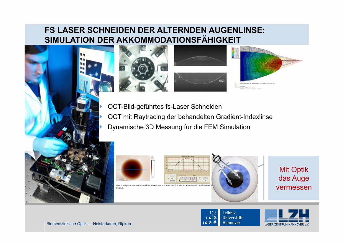
FS LASER SCHNEIDEN DER ALTERNDEN AUGENLINSE: SIMULATION DER AKKOMMODATIONSFÄHIGKEIT
Biomedizinische Optik — Heisterkamp, Ripken
} OCT-Bild-geführtes fs-Laser Schneiden } OCT mit Raytracing der behandelten Gradient-Indexlinse } Dynamische 3D Messung für die FEM Simulation
Mit Optik das Auge
vermessen
Interferometrische Vermessung der Brechkraft einer Augenlinse vor und nach der Applizierung von Laserschnitten
Emanuel Särchen 25.01.2016
Die Phasenkamera Sid4Bio der Firma Pasics soll verwendet werden, um die Brechkraft einer Augenlinse zu vermessen. Die Experimente werden in einem Freistrahlaufbau durchgeführt. Es wurden bereits Vorexperimente an Glaslinsen durchgeführt. Hierzu wird die Glaslinse in einer Wasserkammer platziert und mit einem telezentrischen Abbildungssystem auf den Sensor der Kamera abgebildet Die Beleuchtung erfolgt mit einer monochromatischen CW Beleuchtung im grünen Spektralbereich (532 nm).
Die ermittelte optische Weglänge, welche mit der Phasics aufgenommen wird, scheint realistisch zu sein. Ein kugelsymmetrisches Objekt ist erkennbar (Abb. 1).
Abb. 1: Aufgenommenes Phasenbild einer Glaslinse in Wasser (links), sowie ein Schnitt durch die Phasenwerte (rechts).
Des Weiteren wurde eine extrahierte Schweinelinse mit dem System vermessen. Um eine Brechungsindexanpassung zwischen der Schweinelinse (n = 1,38 – 1,42) und dem Umgebungsmedium zu gewährleisten wurde Silikonöl (n=1,4) verwendet. Durch die Brechungsindexanpassung kann ein auswertbares Interferenzbild aufgenommen werden. Dies ist der Fall, wenn über den gesamten Bildbereich ein Interferenzmuster zu sehen ist (Abb. 2, rechts).
Abb. 2: Aufgenommenes Interferenzbild einer Schweinelinse in Silikonöl (links), sowie die Vergrößerung, welche das schachbrettartige Interferenzmuster zeigt (rechts).
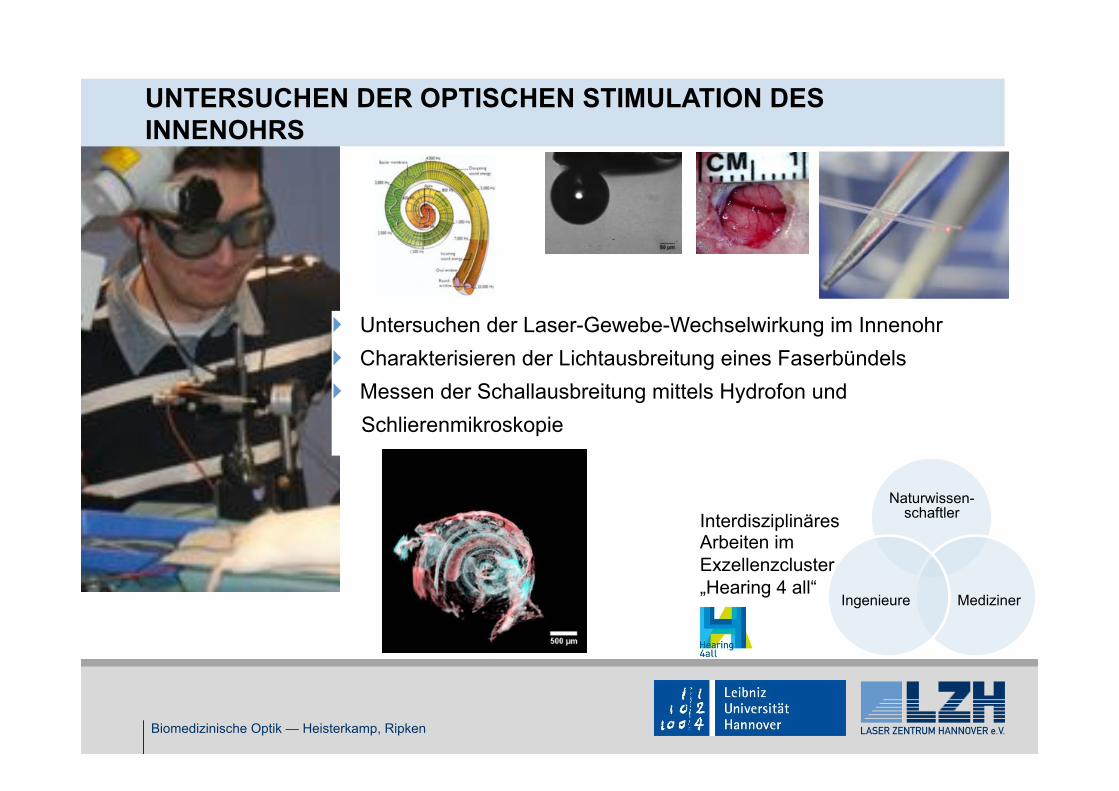
UNTERSUCHEN DER OPTISCHEN STIMULATION DES INNENOHRS
Biomedizinische Optik — Heisterkamp, Ripken
} Untersuchen der Laser-Gewebe-Wechselwirkung im Innenohr } Charakterisieren der Lichtausbreitung eines Faserbündels } Messen der Schallausbreitung mittels Hydrofon und Schlierenmikroskopie
Interdisziplinäres Arbeiten im Exzellenzcluster „Hearing 4 all“
2.4 dB cm21 (Le¼ 1.8 cm) with 5 × 10⁶ cells ml21 in the wavelengthrange 450–500 nm. The cell density of "1 × 10⁶ cells ml21 wasdetermined to be optimal for 4-cm-long hydrogels, for whichthe loss is less than 1 dB cm21 and the 1/e attenuation length(Le¼ 5.6 cm) is comparable to the length of the hydrogel. At thiscell density, a hydrogel with dimensions of 1 × 4 × 40 mm3
(0.16 cm3) could contain up to 160,000 cells and, without molecularabsorption, carry 70% of the light to its distal end.
Implantation of cell-containing hydrogel in vivoCell-containing hydrogels were implanted into a subcutaneouspocket in mice through a 1-cm-long skin incision on the back(Fig. 4a). The pigtail fibre was securely cemented onto the skull toestablish stable light coupling to the hydrogel while the animalwas awake and moving freely (Fig. 4b; Supplementary Movie S1).Light leaking out of the hydrogel to the surrounding tissue couldbe readily monitored through the thin skin layer (Fig. 4c). Theoptical intensity throughout the entire implant varied by no morethan 6 dB, which is slightly higher than the 1 dB cm21 measuredin air and is due to the contact with the tissue (index, 1.34–1.41;Fig. 4d). By comparison, when only a multimode fibre wasimplanted without hydrogel, the 1/e light intensity was constrainedto a small region with a diameter of 2–3 mm as seen through theskin (Fig. 4c,d). This result represents a 40-fold increase of the illu-mination area with the light-guiding scaffold.
The hydrogels and surrounding tissues were harvested at days 3and 8 after implantation (n¼ 3). Fluorescence microscopy with cellviability probes showed that "80% of the embedded cells werefound live in the hydrogels in vitro after photo-crosslinking, andmore than 70% and 65% of the embedded cells in the implantedhydrogels remained viable after 3 and 8 days, respectively(Fig. 4e), which was consistent with measurements with hydrogelsin a culture dish in vitro (Fig. 4f, Supplementary Fig. S3). The
decreases in optical transmittance at 3 and 8 days in vitro andin vivo were less than 1 dB cm21 (Fig. 4g). Histology suggestedthere were no major immune-cell infiltrations, but the formationof connective tissues around the implants, which is a typical mildreaction to foreign bodies, was observed in all, but not in sham-surgery, animals (Fig. 4h). The newly formed tissues were moder-ately vascularized. The hydrogel implants as a whole came off thesurrounding tissues easily during tissue collection, indicating alack of adhesion between the tissues and hydrogels.
In vivo sensing of nanotoxicityWe applied fibre-optic cell-containing hydrogel implants for themeasurement of the toxicity of quantum dots in vivo. To sense cel-lular toxicity we used an intrinsic cellular cytotoxicity sensor—heat-shock-protein 70 (hsp70)24—which is activated when cells are undercytotoxic stress, such as from heavy metal ions and reactive oxygenspecies, and green fluorescent protein (GFP) under the hsp70 pro-moter. Cadmium is a widely used heavy metal in quantum dots,but can cause cytotoxic effects when released as a result of degra-dation of the quantum dots. The magnitude of green fluorescencefrom these sensor cells in vitro increased with a sublethal doseof CdCl2 up to 1 mM, but saturated at higher concentrations of1–5 mM (Supplementary Fig. S4). Two types of cadmium-contain-ing quantum dots were tested: core-only CdTe and core/shellCdSe/ZnS nanoparticles. The sizes of the bare and shelledquantum dots were "3.2 and 5.2 nm, respectively, so they emitred fluorescence (605 nm), which is readily distinguishable fromthe green fluorescence signal. When the cells were encapsulated ina hydrogel in vitro, the sensor signal increased with the concen-tration of CdTe quantum dots in the medium, but no noticeablechange of green fluorescence was observed when CdSe/ZnSquantum dots were used (Fig. 5a,b). This result confirmed thedramatic role of the ZnS shell in reducing cellular toxicity.
0.5 50
5
10
15
20
Molecular weight (kDa)
Swel
ling
ratio
c
d
Lens
Twisted
Rolled
TIR
5 kDaa b
Atte
nuat
ion
(dB
cm−1
)
Wavelength (nm)400 600 800
0
5
300.5 kDa 10 kDa2 kDa 5 kDa
0.5 kDa
2 kDa 5 kDa
10 kDa
fe
0.5 kDa
1
20
2 10Hydrogel
Figure 2 | Characteristics of hydrogels. a, Photograph of PEG-based hydrogels prepared using 10% wt vol21 PEGDA solution with PEGDA molecular weightsof 0.5, 2, 5 and 10 kDa. Scale bar, 1 cm. b, Optical attenuation spectra of PEG hydrogels prepared with different molecular weights of PEGDA. c, Rectangular0.5 and 5 kDa hydrogels (thickness, 1 mm). Scale bar, 5 mm. d, Swelling ratios of PEG hydrogels. The swelling ratio was calculated by dividing the weight ofswollen hydrogel by the weight of dried hydrogel (n¼ 3). e, Mechanical flexibility of the PEG hydrogel (5 kDa, 10%). f, Demonstration of TIR within theslab hydrogel.
534 NATURE CHEMICAL BIOLOGY | VOL 10 | JULY 2014 | www.nature.com/naturechemicalbiology
REVIEW ARTICLE NATURE CHEMICAL BIOLOGY DOI: 10.1038/NCHEMBIO.1534
As the first use of these tools was in the control of neuronal activity in vivo, major efforts have been put into screening natural opsins and gen-erating a variety of chimeric rhodopsin versions with various dynamic responses and light sensitivities2 (Table 1). In the last five years, the study of freely moving animals in which the neuronal activity of specific cells can be controlled by light has provided truly unprecedented insights into neuronal connectivity and circuitry, cognition and behavior9–13. These tools have also been used in the field of developmental biology, enabling
precise mapping and control of the cardiac pacemaker14,15 or the auto-mated control of embryonic stem cell differentiation16.
These astonishing developments have motivated biologists to extend the optogenetic toolbox to soluble light-gated modules engi-neered from other natural light-sensitive proteins. Flavoproteins attracted particular interest because of their riboflavin-based chro-mophore, either flavin adenine dinucleotide (FAD) or flavin mono-nucleotide (FMN), which is naturally present in most cells. For some
Figure 1 | Using light to control proteins in living systems. (a) The high spatiotemporal resolution of light actuation makes it possible to address biological processes with a wide range of temporality (from seconds for enzymatic reaction to days for tissue renewal) at various spatial scales (from less than micrometers for organelles to centimeters for animals). Upon increasing the illumination requirements (e.g., smaller field and/or shorter light pulse to access better resolution in space and time), it becomes possible to control proteins with light (actuation) at smaller spatial and temporal scales. (b) Light gives control over protein function either directly by changing its active state (1) or by modifying its effective concentration via its rate of synthesis (2), rate of degradation (3) or compartmentalization (4). The timing of the response varies from few milliseconds to hours, depending on the chosen method.
a
b
d
Closed Open
Inactive Active
Inactive Active
Light
Light
Light
NH
Lys+
+
––
++
––
NH
Lys
Light
HNNH
O2C CO2
HN O
NH
O
HNNH
O2C CO2
HNO
NH
OS SCys Cys
N
N
NH
NR
O
OSH
Cys
NH
N
NH
NR
O
OS
Cys
Light
Lightc
e
f
Figure 2 | Light control of proteins with genetically encoded photoactuators. (a) Microbial rhodopsins can be expressed in neurons to regulate membrane potential; these proteins interact with the chromophore retinal, which is present in most cells. (b) The intracellular domain of vertebrate rhodopsins can be exchanged with the intracellular domain of specific GPCRs to photocontrol specific signaling cascade (IP3, DAG or cAMP). (c) Light-induced conformational changes in the LOV or in the CRY domain have been used to control protein localization, transcription or activity of a fused protein. (d) In rhodopsins, illumination drives isomerization of a double bond of the chromophore, thereby modifying its geometry. (e) In the case of the LOV domain, photoexcitation induces a covalent thioether bond between bound FMN and a highly conserved cystein residue of the LOV domain. (f) Phytochromes contain a covalently bound chromophore (bilin or biliverdin). Upon exposure to light, isomerization of the chromophore induces a conformational change in the protein, modifying its interaction properties.
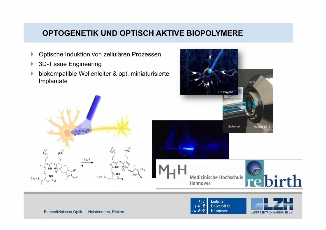
Ed Boyden
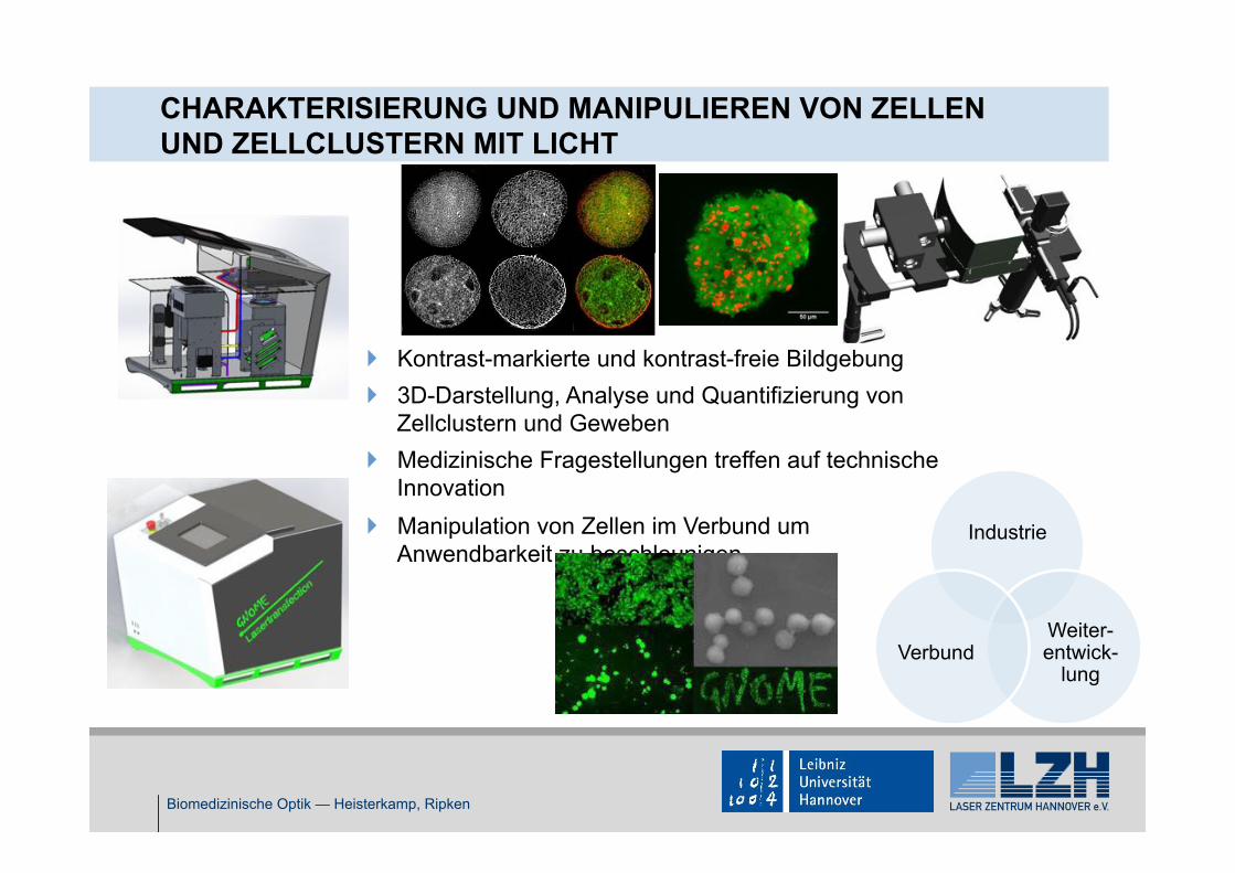
CHARAKTERISIERUNG UND MANIPULIEREN VON ZELLEN UND ZELLCLUSTERN MIT LICHT
Biomedizinische Optik — Heisterkamp, Ripken
} Kontrast-markierte und kontrast-freie Bildgebung } 3D-Darstellung, Analyse und Quantifizierung von
Zellclustern und Geweben } Medizinische Fragestellungen treffen auf technische
Innovation } Manipulation von Zellen im Verbund um
Anwendbarkeit zu beschleunigen Industrie
Weiter-entwick-
lung Verbund
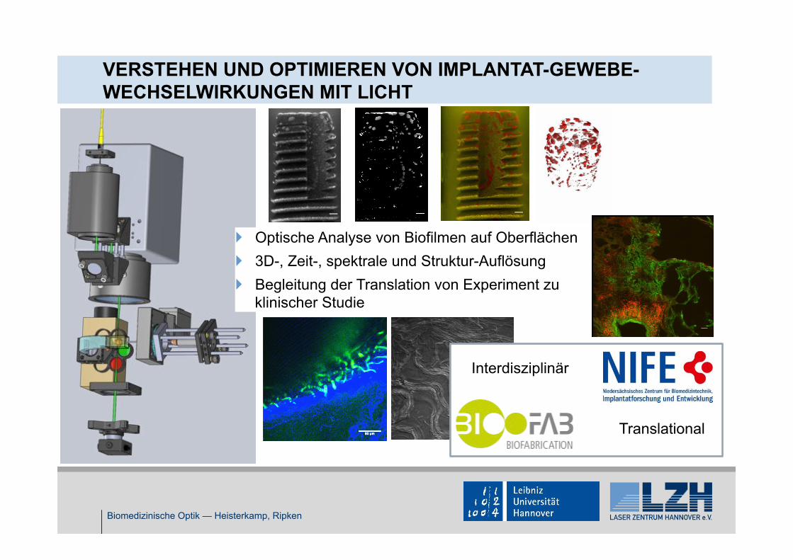
VERSTEHEN UND OPTIMIEREN VON IMPLANTAT-GEWEBE-WECHSELWIRKUNGEN MIT LICHT
Biomedizinische Optik — Heisterkamp, Ripken
} Optische Analyse von Biofilmen auf Oberflächen } 3D-, Zeit-, spektrale und Struktur-Auflösung } Begleitung der Translation von Experiment zu