This work has been digitalized and published in 2013 by Verlag Zeitschrift für Naturforschung in cooperation with the Max Planck Society for the Advancement of Science under a Creative Commons Attribution 4.0 International License. Dieses Werk wurde im Jahr 2013 vom Verlag Zeitschrift für Naturforschung in Zusammenarbeit mit der Max-Planck-Gesellschaft zur Förderung der Wissenschaften e.V. digitalisiert und unter folgender Lizenz veröffentlicht: Creative Commons Namensnennung 4.0 Lizenz. Local Anesthetic Binding to Thylakoid Membranes. Relation to Inhibition of Light-Induced Membrane Energization and Photophosphorylation Gabriele Günther and Henrik Laasch Institut für Ökologische Pflanzenphysiologie und Geobotanik Heinrich-Heine-Universität, Universitätsstraße 1, D-4000 Düsseldorf 1, Bundesrepublik Deutschland Z. Naturforsch. 46c, 79-86 (1991); received September 21, 1990 Chloroplast, Local Anesthetic, Dibucaine, Uncoupling, Photophosphorylation, Amine Bind ing The association of the lipophilic tertiary amine and local anesthetic dibucaine with osmoti- cally shocked chloroplasts of Spinacia oleracea L. cv. Monatol was investigated. Dibucaine, known as an effective inhibitor of thylakoid membrane energization and ATP synthesis, ex hibited three distinct binding classes with chloroplasts: partitioning in the lipid phase of the membranes, electrostatic screening of negative electrical charges on the thylakoid surface and light-induced association of an as yet unknown nature. Evidence is presented that the mecha nism of inhibition of the transthylakoid pH gradient, ApH, by dibucaine is distinct from ‘clas sical’ amine-type uncoupling: The inhibitory effect of dibucaine on ApH was independent of the initial strength of ApH. Light-induced dibucaine binding was independent of the volume of the intrathylakoid space and of the strength of ApH as varied by medium pH. Judged from a comparison of the data on dibucaine binding and on inhibition of ApH and photophosphory lation, dibucaine bound via partitioning in the membrane lipid phase is responsible for the un- coupler-like effects of the local anesthetic. A mechanism for the inhibition of thylakoid energi zation by local anesthetic amines is discussed. Introduction The investigation of local anesthetic effects has long been extended to problems beyond the ques tion of how anesthesia may block the excitation of nerve membranes. Local anesthetics, e.g. dibu caine and tetracaine, were shown to alter the lipid composition of plant membranes [1] and to be ef fective inhibitors of photosynthesis [2, 3]. A special focus of investigation was the inhibition by local anesthetics of energy transduction at ATP synthe sizing membranes in mitochondria [4, 5] and chlo roplasts [6, 7], In chloroplasts, dibucaine inhibited the light-induced transthylakoid pH gradient, ApH, and photophosphorylation [6]. However, this inhibition was unlike that exerted by ‘classical’ Abbreviations: Ab, amount of dibucaine bound to chlo roplast membranes; A f, concentration of free dibucaine in the medium; 9-AA, 9-aminoacridine; Chi, chlorophyll; ApH, transthylakoid pH gradient; Mes, 2-(N-mor- pholino)ethanesulfonic acid; PS, photosystem; Taps, 3-((tris-(hydroxymethyl)methyl)amino)-propanesulfonic acid; Tris, Tris-(hydroxymethyl)-methylamine; Hepes, N-2-hydroxyethylpiperazine-N'-2-ethanesulfonic acid; PAR, photosynthetically active radiation. Reprint requests to H. Laasch. Verlag der Zeitschrift für Naturforschung, D-7400 Tübingen 0939-5075/91/0100-0079 $01.30/0 uncouplers, since the pH-dependent photosynthet- ic control of electron transport at the cytochrome- b/f-complex [8] and at PS II [9] persisted in the presence of dibucaine despite a decreased ApH [10]. Further effects of dibucaine on primary reac tions of photosynthesis are summarized in [7], In the present study, we characterize the asso ciation of the lipophilic tertiary amine dibucaine with chloroplast membranes. Dibucaine was re garded as a model compound for local anesthetics and was chosen for binding studies since the pro- tonated (monocation) form exhibited strong flu orescence at room temperature which may be used for a determination of dibucaine concentration [11], Amine binding to chloroplast membranes was intensively studied with ‘classical’ amine-type un couplers, e.g. ammonia or methylamine [12, 13] and with fluorescent probes of light-induced thyla koid energization, e.g. acridines [14, 15], the latter being uncouplers themselves. Several of these stud ies evinced an involvement of membrane-bound protons in energy transduction at the thylakoid membrane [14, 16, 17]. As suggested previously, the existence of localized proton domains at the thylakoid membrane may also be indicated by the effects of local anesthetics on thylakoid energiza tion [7],
Transcript
This work has been digitalized and published in 2013 by Verlag Zeitschrift für Naturforschung in cooperation with the Max Planck Society for the Advancement of Science under a Creative Commons Attribution4.0 International License.
Dieses Werk wurde im Jahr 2013 vom Verlag Zeitschrift für Naturforschungin Zusammenarbeit mit der Max-Planck-Gesellschaft zur Förderung derWissenschaften e.V. digitalisiert und unter folgender Lizenz veröffentlicht:Creative Commons Namensnennung 4.0 Lizenz.
Local Anesthetic Binding to Thylakoid Membranes. Relation to Inhibition of Light-Induced Membrane Energization and PhotophosphorylationGabriele Günther and Henrik LaaschInstitut für Ökologische Pflanzenphysiologie und Geobotanik Heinrich-Heine-Universität, Universitätsstraße 1, D-4000 Düsseldorf 1, Bundesrepublik DeutschlandZ. Naturforsch. 46c, 7 9 -8 6 (1991); received September 21, 1990
Chloroplast, Local Anesthetic, Dibucaine, Uncoupling, Photophosphorylation, Amine Binding
The association o f the lipophilic tertiary amine and local anesthetic dibucaine with osmoti- cally shocked chloroplasts o f Spinacia oleracea L. cv. Monatol was investigated. Dibucaine, known as an effective inhibitor o f thylakoid membrane energization and ATP synthesis, exhibited three distinct binding classes with chloroplasts: partitioning in the lipid phase o f the membranes, electrostatic screening o f negative electrical charges on the thylakoid surface and light-induced association o f an as yet unknown nature. Evidence is presented that the mechanism o f inhibition o f the transthylakoid pH gradient, ApH, by dibucaine is distinct from ‘classical’ amine-type uncoupling: The inhibitory effect o f dibucaine on ApH was independent o f the initial strength o f ApH. Light-induced dibucaine binding was independent o f the volume o f the intrathylakoid space and o f the strength o f ApH as varied by medium pH. Judged from a comparison o f the data on dibucaine binding and on inhibition o f ApH and photophosphorylation, dibucaine bound via partitioning in the membrane lipid phase is responsible for the un- coupler-like effects o f the local anesthetic. A mechanism for the inhibition o f thylakoid energization by local anesthetic amines is discussed.
Introduction
The investigation of local anesthetic effects has long been extended to problems beyond the question of how anesthesia may block the excitation of nerve membranes. Local anesthetics, e.g . dibucaine and tetracaine, were shown to alter the lipid composition of plant membranes [1] and to be effective inhibitors of photosynthesis [2, 3]. A special focus of investigation was the inhibition by local anesthetics of energy transduction at ATP synthesizing membranes in mitochondria [4, 5] and chloroplasts [6, 7], In chloroplasts, dibucaine inhibited the light-induced transthylakoid pH gradient, ApH, and photophosphorylation [6]. However, this inhibition was unlike that exerted by ‘classical’
Abbreviations: A b, amount o f dibucaine bound to chloroplast membranes; A f, concentration o f free dibucaine in the medium; 9-AA, 9-aminoacridine; Chi, chlorophyll; ApH, transthylakoid pH gradient; Mes, 2-(N-mor- pholino)ethanesulfonic acid; PS, photosystem; Taps, 3-((tris-(hydroxymethyl)methyl)amino)-propanesulfonic acid; Tris, Tris-(hydroxymethyl)-methylamine; Hepes, N-2-hydroxyethylpiperazine-N'-2-ethanesulfonic acid; PAR, photosynthetically active radiation.Reprint requests to H. Laasch.
Verlag der Zeitschrift für Naturforschung, D-7400 Tübingen0939-5075/91/0100-0079 $01.30/0
uncouplers, since the pH-dependent photosynthet- ic control of electron transport at the cytochrome- b/f-complex [8] and at PS II [9] persisted in the presence of dibucaine despite a decreased ApH[10]. Further effects of dibucaine on primary reactions of photosynthesis are summarized in [7],
In the present study, we characterize the association of the lipophilic tertiary amine dibucaine with chloroplast membranes. Dibucaine was regarded as a model compound for local anesthetics and was chosen for binding studies since the pro- tonated (monocation) form exhibited strong fluorescence at room temperature which may be used for a determination of dibucaine concentration[11], Amine binding to chloroplast membranes was intensively studied with ‘classical’ amine-type uncouplers, e.g . ammonia or methylamine [12, 13] and with fluorescent probes of light-induced thylakoid energization, e.g . acridines [14, 15], the latter being uncouplers themselves. Several of these studies evinced an involvement of membrane-bound protons in energy transduction at the thylakoid membrane [14, 16, 17]. As suggested previously, the existence of localized proton domains at the thylakoid membrane may also be indicated by the effects of local anesthetics on thylakoid energization [7],
80 G. G ünther and H. Laasch • Binding o f D ibucaine to C hloroplasts
With this paper we tried to characterize different types of association of dibucaine with chloroplast membranes and to relate these to the effects of dibucaine on transmembrane ApH and ATP synthesis. Some characteristics of ‘classical’ amine- type uncoupling by ammonia were set against those of'selective' uncoupling by dibucaine.
Materials and Methods
Preparation o f chloroplasts
Intact chloroplasts were isolated from six-week- old leaves of glass-house grown Spinacia oleracea L. cv. Monatol. The isolation procedure was described by [7], Chloroplasts were stored at 0 C in the dark until use. The integrity of chloroplast envelopes was 85 to 95%. The chloroplasts were osmotically shocked immediately before use.
Binding o f dibucaine
In order to simplify terminology, any type of amine association with chloroplast membranes was termed binding. The concentration of dibucaine in binding assays was measured fluorometri- cally with excitation and emission wavelengths of 325 and 403 nm, respectively, with a halfband width of 2 or 10 nm, using a spectrofluorometer (F-2000, Hitachi). The pH of fluorescence assays was always adjusted to 8. Dibucaine was dissolved in methanol, the final solvent concentration in binding assays was < 1%. The standard reaction medium contained 20 mM Hepes/KOH, pH 8, 20 m M KCl, 1 m M MgCl2, 0.3 m sorbitol, and 200 U catalase (EC 1.11.1.6.). Unless stated otherwise, PS I electron flow was allowed by additions of 50 fiM dichlorophenolindophenol, 2 mM
Na-ascorbate and 5 |iM methylviologen. When the pH dependency of amine binding was studied, the chloroplast medium was composed as described for photophosphorylation experiments. When the salt concentration in the reaction medium was varied, isosmolarity of the medium was maintained by addition of sorbitol. Effects of salt concentration on dibucaine fluorescence were corrected. Measurements of dibucaine fluorescence were either carried out in the presence of chloroplasts or after chloroplast sedimentation by centrifugation.
Method a): Fluorescence after sedimentation of chloroplasts. A suspension of chloroplasts in a
translucent reaction vial was supplied with dibucaine in the dark and placed into a centrifuge (Minifuge E, Beckman) with a translucent cover. The samples were either illuminated with white light of 2500 | i E m “2-s_1, PAR, or kept in the dark for 90 s. Chloroplasts were then sedimented at 13,000 x g for 20 s. The fluorescence in the supernatant was measured. For a calculation of binding data, the fluorescence intensity was calibrated against amine concentration. The amount of amine bound was calculated from amine found in the supernatant and total amine added.
Method b): Fluorescence in the presence of chloroplasts: A chloroplast suspension equivalent to 25 |ig Chi • ml 1 was supplied with dibucaine in a stirred fluorescence cuvette. The time course of fluorescence emission was followed during a dark/ light (2000 |iE-m 2-s ', PAR) cycle.
Determination o f thylakoid osmotic space
The osmotic space was determined by incubation of osmotically shocked chloroplasts in a medium containing 20 mM Hepes/KOH, pH 8, 2 mM
MgCl2, 10 mM KC1 and sorbitol, varying between zero and 0.5 m. 20 kBq [?H ]-H 20 and 9 kBq [l4C]sorbitol (Amersham Buchler, Braunschweig) were added. Subsequently, the chloroplasts were centrifuged for 20 s at 13,000 x g through a layer of silicon oil mixture. AR 20 and AP 150 (Wacker, München), with 65 to 83% AR 20 depending on the sorbitol concentration applied, into a com partment containing 3 m HC104. The radioactivity in the HC104 compartment was determined by duallabel scintillation counting.
Displacement o f divalent cations
In a reaction medium containing 10 m M Hepes/ KOH, pH 8, 5 mM KC1, <0.1 m M MgCl2 and 75 mM sorbitol, chloroplasts were incubated for 30 s in the dark. Then the sorbitol concentration was raised to 0.3 m without changes of buffer and KC1. After 2 min in the dark or light, in the absence or presence of dibucaine, the samples were centrifuged for 20 s at 13,000 * g. The concentration of divalent cations in the supernatant was determined by addition of 50 |im Eriochrome Blue SE and 1 ml Tris/HCl, pH 9, to 0.4 ml of chlorophyll free supernatant and subsequent measurement of the Eriochrome Blue SE/cation fluores-
G. G ün ther and H. Laasch • Binding o f D ibucaine to C hloroplasts 81
cence with excitation and emission wavelengths of 515 and 594 nm, respectively, at a half-band width of 10 nm. Eriochrome Blue SE without bound divalent cations was not fluorescing at pH 9.
Photophosphorylation
ATP formation in the light was determined enzymatically, using hexokinase (EC 2.7.1.1.) and glu- cose-6-phosphate dehydrogenase (EC 1.1.1.49.). The reaction medium contained 0.3 M sorbitol, 30 mM KC1, 20 mM Hepes/KOH, pH 8, 1 mM
EDTA, 1 mM MnCl2, 2 m M K H 2P 0 4, 10 |iM dia- denosinepentaphosphate and 0.5 m M ADP. ATP synthesis was allowed for 2 min during illumination (2000 |iE -m _2-s_1, PAR). PS II + 1 electron flow was mediated by 20 |im methylviologen.
Determination o f light-induced ApH
5 |im 9-AA was added to osmotically shocked chloroplasts equivalent to 20 fig C hi-m l-1, in the presence of 10 |im methylviologen. The chloroplasts were illuminated with red light of 2000 |iE -m _2-s_1, PAR. Dibucaine was added after 9-AA and prior to illumination. 9-AA fluorescence was excited by light of 400 nm wavelength and was measured at 465 nm. The strength of ApH was calculated, using the equation ApH = log(AF-FR_1 • VE- V f 1) (AF= light-induced fluorescence quenching; FR = fluorescence remaining in the light; VE = external volume). An intrathyla- koid volume, F,, of 10 iil-mg“1 Chi was assumed.
Results
Comparison o f methods fo r binding assay
Binding of dibucaine to osmotically shocked chloroplasts was assayed by fluorometric determination of dibucaine concentrations after light or dark treatment and following sedimentation of the membranes (method a). For verification that neither dibucaine desorption from membranes during centrifugation nor dibucaine sorption to surfaces of reaction vials influenced the binding data obtained, an alternative approach was carried out. Dibucaine fluorescence in the presence of chloro- plast membranes in the dark and light was measured (method b). Dibucaine binding was quantified from fluorescence data for method a) by the
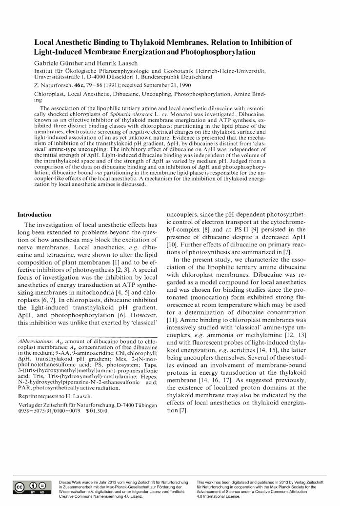
equation A J A {= (FD-FL)/FL (An, Af: concentrations of bound and free dibucaine; FD, FL: fluorescence in dark and light-treated samples after removal of chloroplasts). For method b), analogously, binding was quantified by the equation A J A f = F q/FR (F , Fr : fluorescence quenching and remaining fluorescence in the light in the presence of chloroplasts). In Fig. 1, the quotients A J A { determined by both methods are shown for dibucaine concentrations up to 100 jim. The data fitted well to a straightline with a slope of one, suggesting that both methods reveal equivalent data. Furthermore, the data may indicate that dibucaine bound to chloroplast membranes is non-fluores- cent. In the following, dibucaine binding was determined by method a).
Dibucaine binding in light and dark
The equilibration of dibucaine binding in the light or dark was generally completed after at most 60 s (data not shown). Therefore, a minimum incubation time of shocked chloroplasts with amine of 90 s was chosen. In Fig. 2 dibucaine bound to chloroplasts in dark and light, Ah, is shown in dependence of the free amine concentration present. In light-treated samples, PS I driven electron
A n • A f '1 (method b)
Fig. 1. Comparison o f methods for determination o f dibucaine binding. Dibucaine fluorescence was measured at pH 8 after sedimentation (method a) and in the presence (method b) o f osmotically shocked, light and dark treated chloroplasts. A n and A f are the concentrations o f bound and free amine, respectively. The*Chl concentration was 25 (ag m l-1, the dibucaine concentration was varied between 20 and 100 |iM.
82 G . G ün ther and H. Laasch • Binding o f D ibucaine to Chloroplasts
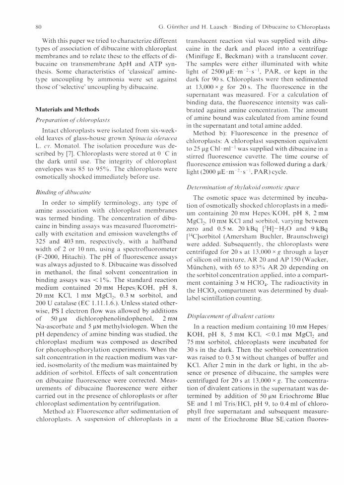
Fig. 2. Binding o f dibucaine to osmotically shocked thy- lakoid membranes in the light (O) and dark ( • ) at pH 8. The dashed line indicates ‘light-induced' binding, as calculated by a subtraction o f the amounts o f amine bound, A b, under light and dark conditions. A h is shown in dependence on the free amine concentration, A (. The Chi concentration was 100 lag ml"1.
transport was operated since the latter, in contrast to PS II dependent electron flow, was unaffected by dibucaine up to concentrations of 0.5 m M . Two major binding classes appear in the dark: in a concentration range of / l f < 3 0 ( i M a curvilinear dependence of bound amine, Ab, on A f appeared. Between A (= 30 and 300 |im, a linear relation of A b and A { was observed (not shown).
In the light, dibucaine binding exceeded the dark level. Light-dependent binding, estimated by subtraction of A b values in light and dark, was saturated at about 20 (im of free dibucaine (Fig. 2). The slopes of the linear branches of binding curves in light and dark coincided, suggesting that the underlying processes are not influenced by light. A partitioning of lipophilic dibucaine molecules [18] between membrane lipids and aqueous medium was assumed. Previously bound dibucaine re-emerged in the medium when chloroplast membranes were resuspended in the absence of free dibucaine (not shown). Hence, dibucaine binding was largely reversible.
Effects o f metal cations on dibucaine binding
When dibucaine was added to osmotically shocked chloroplasts in the dark in the presence of
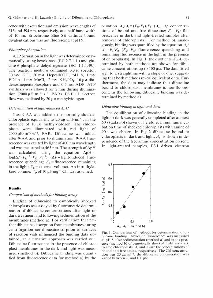
either high or low concentrations of salts, binding in high salt was lower than that in low salt media (Fig. 3). Under high-salt condition the curvilinear shape of the binding curve disappeared. The slopes of the linear branches of the curves, however, were unaffected. This indicates two binding classes in the dark: first, binding which is apparently saturated at free dibucaine concentrations of about 30 |4.m and may interact with salts and second, binding which predominates at A {> 30 |i m and is unaffected by salts. When the data obtained by subtraction of the curves in Fig. 3, were redrawn in a double-reciprocal plot, a straightline could be fitted to the data, suggesting a hyperbolic relationship between Ab and A f values of salt dependent binding (not shown). Apparent values for the ‘concentration’ of salt-sensitive binding sites and the dissociation constant of amine/membrane complexes were obtained with 280 nmol mg-1 Chi and 37 |iM, respectively.
The type of cations added was decisive for the strength of salt effects. Divalent were more effective in dibucaine displacement from chloroplasts than monovalent cations and the more tightly binding Ca2+ [19] was more effective than Mg2+ (Fig. 4). The anion used was always CL.
Fig. 3. Influence o f salt concentrations in the medium on dibucaine binding to osmotically shocked chloroplasts in the dark. For high salt condition (A ), 50 mM MgCl2 and 50 mM KC1 were added, for low salt conditions (■ ) 10 mM KC1. The dashed line shows ‘pure’ salt-dependent binding, as calculated by subtraction o f the related A h values. The Chi concentration was 100 ^g m l'1.
G. G ünther and H. Laasch ■ Binding o f D ibucaine to C hloroplasts 83
salt (mM)
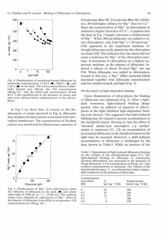
Fig. 4. Displacement o f membrane-bound dibucaine by increasing concentrations o f KCl ( • ) , MgCl2 ( A ) and CaCl2 ( ♦ ) in the dark. The total concentration o f dibucaine present was 100 |i m , the Chi concentration lOO^ig-ml“1 and the initial salt concentration 10 m M KC1. 3 min equilibration in the presence o f amine and salts were allowed before sedimentation o f the chloroplasts.
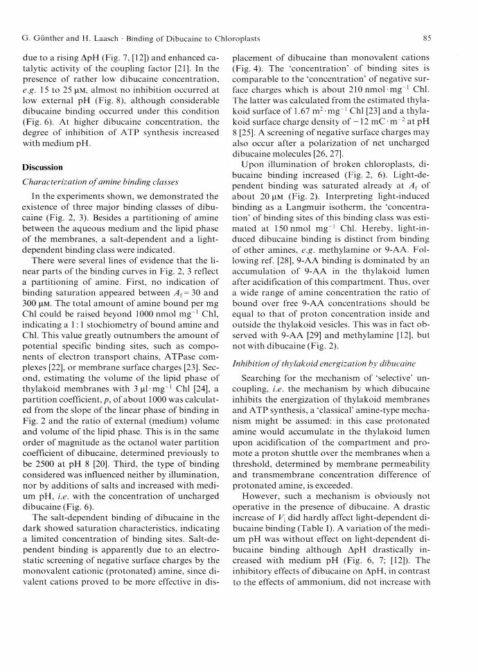
In Fig. 5 we show that, in reverse to the displacement of amine pictured in Fig. 4, dibucaine may displace divalent cations associated with chloroplast membranes. The concentration of divalent cations was monitored by fluorescence emission of
Dibucaine (^M)
Fig. 5. Displacement o f Mg:+ from chloroplasts under the influence o f dibucaine in the dark ( • ) and under white light o f 2500 ( iE - m '- s '1, PAR (O). The ordinate values were obtained by subtraction o f Mg2+ efflux in the absence o f dibucaine from efflux in its presence. Chi concentration was 100 jig-m l-1.
Eriochrome-Blue SE. Eriochrome Blue SE exhibited a 20-fold higher affinity for Mg:+ than for Ca2+. Since the concentration of Mg2+ in chloroplasts in addition is higher than that of Ca2+, it appears that the data in Fig. 5 largely represent a displacement of Mg2+. When 200 |!M dibucaine was added to intact chloroplasts, only little Mg2+ (<20 nmol mg“1 Chi) appeared in the suspension medium, although dibucaine easily penetrates the chloroplast envelope [10]. This indicates that the amine did not cause a leakiness for Mg2+ of the chloroplast envelope. A treatment of chloroplasts in a slightly hypotonic medium, in the absence of dibucaine, induced a release of about 30 nmol Mg2+ per mg Chi. When dibucaine was added to chloroplasts treated in this way, a Mg2+ efflux occurred which increased together with dibucaine concentration and was similar in dark and light (Fig. 5).
On the nature o f light-dependent binding
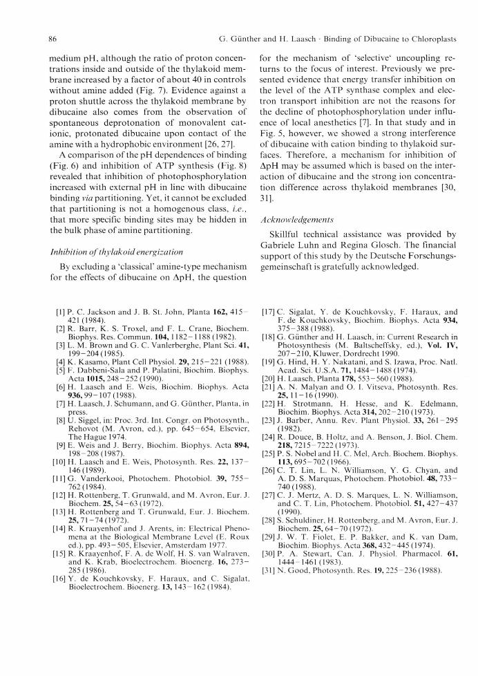
Upon illumination of chloroplasts the binding of dibucaine was enhanced (Fig. 2). After a light- dark transition, light-induced binding disappeared. Also an addition of nigericin to chloroplasts in the light inhibited light-dependent binding (not shown). This suggested that light-induced binding may be related to proton accumulation in the thylakoid lumen. Having in view the effect of "classical’ amine-type uncouplers, e.g. methyl- amine or ammonia [12, 13], an accumulation of protonated dibucaine in the thylakoid lumen in the light may be assumed. However, a ApH induced accumulation of dibucaine is challenged by the data shown in Table I. While an increase of the
Table I. Dependence o f light:induced dibucaine binding on the volume o f the intrathylakoid space (F,). The light-induced binding o f dibucaine to osmotically shocked chloroplasts was measured in the presence o f 50 (aM dibucaine, a Chi concentration o f 80 (ig-m l-1 and sorbitol concentrations as indicated. The volume o f the intrathylakoid space, was determined under comparable conditions in the presence o f dibucaine.
Concentration o f sorbitol [M]
Vi[(il-mg 1 Chi]
Amine bound [nm ol-m g-1 Chi]
0 18 1600.1 15 1600.2 11 1450.3 7 1400.5 4 130
84 G. G ün ther and H. Laasch ■ Binding o f D ibucaine to Chloroplasts
sorbitol concentration from zero to 0.5 M reduced the intrathylakoid volume, Vx, by a factor of four, the amount of dibucaine bound by a light-induced mechanism only decreased by 20% (Table I).
With increasing medium pH the strength of the light-induced ApH increases ([12]; see also Fig. 7). Assuming a ApH dependent accumulation of dibucaine to underly light-induced binding, the latter should increase with medium pH as well. When light-induced dibucaine binding was studied in dependence of medium pH, no variation was found over the range of pH considered (Fig. 6). Binding of amine in the dark, however, increased with the external pH. Since the latter is assumed to be largely produced by partitioning, an increase of the concentration of unprotonated (uncharged) amine species in the medium with rising pH may be responsible for increased dark binding.
Evidence against a dibucaine induced proton shuttle across the thylakoid membrane also appears from a comparison of the inhibitory effect of ammonia and dibucaine on ApH in dependence on medium pH (Fig. 7). While the inhibitory effect on ApH of ammonia, a classical amine-type uncoupler, increased with external pH as expected, the effect of dibucaine on ApH was constant over the range of pH considered. The pKa of ammonium of 9.3 is comparable to pKa=r 9.0 of dibucaine [20],
pH
Fig. 7. Inhibition o f light-induced transthylakoid ApH, in the absence o f ADP and F,, by 3 mM ammonia (A ) and 35 |iM dibucaine ( ♦ ) in dependence on medium pH. ApH in the absence o f uncoupler is shown by ( • ) . ApH was calculated from 9-AA fluorescence quenching.
Hence, the different effects of medium pH on ammonium and dibucaine may not be explained by different degrees of protonation at the pH values tested.
Photophosphorylation by thylakoid membranes increases with external pH (Fig. 8). This is largely
pHFig. 6. Dependence o f dibucaine binding in the dark (■ ) and under white light o f 2500 n E ir T 2 s_l, PAR (O) on medium pH. ‘Pure" light-dependent binding (A ) was obtained as described in Fig. 2. The Chi concentration was 8 0 n g -m l-1. PS II + 1 electron flow was mediated by 10 |iM methylviologen. The dibucaine concentration was 30 | i M .
Fig. 8. Influence o f medium pH on the inhibition o f photophosphorylation by 15 |!M (A ), 25 (■ ) and 35 |im ( ♦ ) dibucaine. Controls (absence o f amine) are shown by ( • ) . The Chi concentration was 25 (ig ml“1. Dibucaine was added 60 s before illumination o f chloroplast membranes.
G . G ünther and H. Laasch • Binding o f D ibucaine to C hloroplasts 85
due to a rising ApH (Fig. 7, [12]) and enhanced catalytic activity of the coupling factor [21]. In the presence of rather low dibucaine concentration, e.g. 15 to 25 (J.M, almost no inhibition occurred at low external pH (Fig. 8), although considerable dibucaine binding occurred under this condition (Fig. 6). At higher dibucaine concentration, the degree of inhibition of ATP synthesis increased with medium pH.
Discussion
Characterization o f amine binding classes
In the experiments shown, we demonstrated the existence of three major binding classes of dibucaine (Fig. 2, 3). Besides a partitioning of amine between the aqueous medium and the lipid phase of the membranes, a salt-dependent and a light- dependent binding class were indicated.
There were several lines of evidence that the linear parts of the binding curves in Fig. 2, 3 reflect a partitioning of amine. First, no indication of binding saturation appeared between A f = 30 and 300 fj-M- The total amount of amine bound per mg Chi could be raised beyond 1000 nmol mg“1 Chi, indicating a 1:1 stochiometry of bound amine and Chi. This value greatly outnumbers the amount of potential specific binding sites, such as components of electron transport chains, ATPase complexes [22], or membrane surface charges [23]. Second, estimating the volume of the lipid phase of thylakoid membranes with 3 |il mg_1 Chi [24], a partition coefficient,/), of about 1000 was calculated from the slope of the linear phase of binding in Fig. 2 and the ratio of external (medium) volume and volume of the lipid phase. This is in the same order of magnitude as the octanol water partition coefficient of dibucaine, determined previously to be 2500 at pH 8 [20]. Third, the type of binding considered was influenced neither by illumination, nor by additions of salts and increased with medium pH, i.e. with the concentration of uncharged dibucaine (Fig. 6).
The salt-dependent binding of dibucaine in the dark showed saturation characteristics, indicating a limited concentration of binding sites. Salt-de- pendent binding is apparently due to an electrostatic screening of negative surface charges by the monovalent cationic (protonated) amine, since divalent cations proved to be more effective in dis
placement of dibucaine than monovalent cations (Fig. 4). The ‘concentration’ of binding sites is comparable to the ‘concentration' of negative surface charges which is about 210 nmol mg“1 Chi. The latter was calculated from the estimated thylakoid surface of 1.67 m2-mg-1 Chi [23] and a thylakoid surface charge density of -1 2 mC • m 2 at pH 8 [25]. A screening of negative surface charges may also occur after a polarization of net uncharged dibucaine molecules [26, 27].
Upon illumination of broken chloroplasts, dibucaine binding increased (Fig. 2, 6). Light-dependent binding was saturated already at Af of about 20 |i m (Fig. 2). Interpreting light-induced binding as a Langmuir isotherm, the ‘concentration’ of binding sites of this binding class was estimated at 150 nmol mg“1 Chi. Hereby, light-in- duced dibucaine binding is distinct from binding of other amines, e.g. methylamine or 9-AA. Following ref. [28], 9-AA binding is dominated by an accumulation of 9-AA in the thylakoid lumen after acidification of this compartment. Thus, over a wide range of amine concentration the ratio of bound over free 9-AA concentrations should be equal to that of proton concentration inside and outside the thylakoid vesicles. This was in fact observed with 9-AA [29] and methylamine [12], but not with dibucaine (Fig. 2).
Inhibition o f thylakoid energization by dibucaine
Searching for the mechanism of ‘selective’ uncoupling, i.e. the mechanism by which dibucaine inhibits the energization of thylakoid membranes and ATP synthesis, a ‘classical’ amine-type mechanism might be assumed: in this case protonated amine would accumulate in the thylakoid lumen upon acidification of the compartment and promote a proton shuttle over the membranes when a threshold, determined by membrane permeability and transmembrane concentration difference of protonated amine, is exceeded.
However, such a mechanism is obviously not operative in the presence of dibucaine. A drastic increase of Vx did hardly affect light-dependent dibucaine binding (Table I). A variation of the medium pH was without effect on light-dependent dibucaine binding although ApH drastically increased with medium pH (Fig. 6, 7; [12]). The inhibitory effects of dibucaine on ApH, in contrast to the effects of ammonium, did not increase with
86 G. G ün ther and H. Laasch • Binding o f D ibucaine to C hloroplasts
medium pH, although the ratio of proton concentrations inside and outside of the thylakoid membrane increased by a factor of about 40 in controls without amine added (Fig. 7). Evidence against a proton shuttle across the thylakoid membrane by dibucaine also comes from the observation of spontaneous deprotonation of monovalent cationic, protonated dibucaine upon contact of the amine with a hydrophobic environment [26, 27].
A comparison of the pH dependences of binding (Fig. 6) and inhibition of ATP synthesis (Fig. 8) revealed that inhibition of photophosphorylation increased with external pH in line with dibucaine binding via partitioning. Yet, it cannot be excluded that partitioning is not a homogenous class, i.e., that more specific binding sites may be hidden in the bulk phase of amine partitioning.
Inhibition o f thylakoid energization
By excluding a ‘classical’ amine-type mechanism for the effects of dibucaine on ApH, the question
[1] P. C. Jackson and J. B. St. John, Planta 162, 415 — 421 (1984).
[2] R. Barr, K. S. Troxel, and F. L. Crane, Biochem. Biophys. Res. Commun. 104, 1182- 1188 (1982).
[3] L. M. Brown and G. C. Vanlerberghe, Plant Sei. 41, 199-204(1985).
[4] K. Kasamo, Plant Cell Physiol. 29, 215-221 (1988).[5] F. Dabbeni-Sala and P. Palatini, Biochim. Biophys.
Acta 1015, 248-252(1990).[6] H. Laasch and E. Weis, Biochim. Biophys. Acta
936, 99-107(1988).[7] H. Laasch, J. Schumann, and G. Günther, Planta, in
press.[8] U. Siggel, in: Proc. 3rd. Int. Congr. on Photosynth.,
Rehovot (M. Avron, ed.), pp. 645 -6 5 4 , Elsevier, The Hague 1974.
[9] E. Weis and J. Berry, Biochim. Biophys. Acta 894, 198-208(1987).
[10] H. Laasch and E. Weis, Photosynth. Res. 22, 137 — 146 (1989).
[12] H. Rottenberg, T. Grunwald, and M. Avron, Eur. J. Biochem. 25, 54 -63(1972).
[13] H. Rottenberg and T. Grunwald, Eur. J. Biochem. 25 ,71 -74 (1972 ).
[14] R. Kraayenhof and J. Arents, in: Electrical Phenomena at the Biological Membrane Level (E. Roux ed.), pp. 493 — 505, Elsevier, Amsterdam 1977.
[15] R. Kraayenhof, F. A. de Wolf, H. S. van Walraven, and K. Krab, Bioelectrochem. Bioenerg. 16, 2 7 3 - 285(1986).
[16] Y. de Kouchkovsky, F. Haraux, and C. Sigalat, Bioelectrochem. Bioenerg. 13, 143-162(1984).
for the mechanism of ‘selective1 uncoupling returns to the focus of interest. Previously we presented evidence that energy transfer inhibition on the level of the ATP synthase complex and electron transport inhibition are not the reasons for the decline of photophosphorylation under influence of local anesthetics [7], In that study and in Fig. 5, however, we showed a strong interference of dibucaine with cation binding to thylakoid surfaces. Therefore, a mechanism for inhibition of ApH may be assumed which is based on the interaction of dibucaine and the strong ion concentration difference across thylakoid membranes [30,31]-
A cknowledgemen ts
Skillful technical assistance was provided by Gabriele Luhn and Regina Glosch. The financial support of this study by the Deutsche Forschungsgemeinschaft is gratefully acknowledged.
[17] C. Sigalat, Y. de Kouchkovsky, F. Haraux, andF. de Kouchkovsky, Biochim. Biophys. Acta 934, 375-388(1988).
[18] G. Günther and H. Laasch, in: Current Research in Photosynthesis (M. Baltscheffsky, ed.), Vol. IV, 207 -210 , Kluwer, Dordrecht 1990.
[19] G. Hind, H. Y. Nakatani, and S. Izawa, Proc. Natl. Acad. Sei. U.S.A. 71, 1484- 1488 (1974).
[20] H. Laasch, Planta 178,553-560(1988).[21] A. N. Malyan and O. I. Vitseva, Photosynth. Res.
25, 11-16(1990).[22] H. Strotmann, H. Hesse, and K. Edelmann,