Page 1
Aus der Klinik für Herzchirurgie
Der Universität zu Lübeck
Direktor: Prof. Dr. med. H.-H. Sievers
Autograft Reinforcement to Preserve Autograft Function after the Ross Procedure
Inauguraldissertation
zur
Erlangung der Doktorwürde
der Universität zu Lübeck
- Aus der Medizinischen Fakultät –
vorgelegt von
Efstratios I. Charitos
aus Athen
Lübeck 2009
Page 2
1
1. Berichterstatter: Prof. Dr. med Ulrich Stierle
2. Berichterstatter: Prof. Dr. med Matthias Heringlake
Tag der mündlichen Prüfung: 12.03.2010
zum Druck genehmigt. Lübeck, den 12.03.2010
gez. Prof. Dr. med. Werner Solbach
- Dekan der Medizinischen Fakultät -
Page 3
2
Table of Contents
List of abbreviations ......................................................................................................................... 4
1. Introduction ................................................................................................................................. 5
1.1 The problem of heart valve replacement ................................................................................ 5
1.2 The quest for the ideal heart valve replacement ..................................................................... 5
1.3 An alternative approach for the treatment of aortic valve disease: The Ross procedure .......... 6
1.4 Research on the modes of autograft failure of the Ross procedure ......................................... 8
1.5 The present study ................................................................................................................... 9
2. Patients and Methods ................................................................................................................. 10
2.1 Study population and operative data .................................................................................... 10
2.2 Clinical and echocardiographic follow up .............................................................................. 14
2.3 Statistical analysis ................................................................................................................. 15
2.3.1 The problem of analyzing serial echocardiographic measurements: Reporting without
distorting ................................................................................................................................ 16
2.3.2 Use of novel statistical methodology for the analysis and interpretation of the data ...... 17
3. Results ........................................................................................................................................ 20
3.1 Clinical outcome and autograft reoperations ........................................................................ 20
3.2 Development of aortic regurgitation over time ..................................................................... 24
3.3 Change of autograft dimensions over time ............................................................................ 26
3.3.1 Aortic annulus diameter ................................................................................................. 26
3.3.2 Sinus of Valsalva diameter .............................................................................................. 27
3.3.3 Sinotubular junction diameter ........................................................................................ 28
3.4 Change of dimensions and autograft regurgitation ............................................................... 29
4. Discussion................................................................................................................................... 31
4.1 Previous studies .................................................................................................................... 31
4.2 Present study ........................................................................................................................ 32
Page 4
3
4.2.1 Reoperation rates .......................................................................................................... 32
4.2.2 Autograft function .......................................................................................................... 33
4.2.3 Autograft dimensions ..................................................................................................... 33
4.3 Limitations ............................................................................................................................ 34
4.4 Clinical implications .............................................................................................................. 35
5. Summary .................................................................................................................................... 36
6. Zusammenfassung in deutscher Sprache................................................................................. 37
6.1 Einleitung ........................................................................................................................ 37
6.2 Patienten und Methoden ................................................................................................ 37
6.3 Ergebnisse ....................................................................................................................... 38
6.4 Diskussion ....................................................................................................................... 40
6.5 Schlussfolgerungen ......................................................................................................... 41
Appendix ........................................................................................................................................ 42
A. References .................................................................................................................. 42
B. Index of figures ........................................................................................................... 46
C. Index of tables ............................................................................................................. 47
E. Acknowledgements ..................................................................................................... 48
F. Curriculum vitae .......................................................................................................... 49
G. Publications ................................................................................................................. 50
Page 5
4
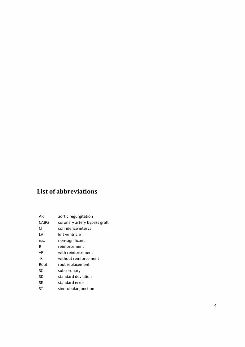
List of abbreviations
AR aortic regurgitation
CABG coronary artery bypass graft
CI confidence interval
LV left ventricle
n.s. non-significant
R reinforcement
+R with reinforcement
-R without reinforcement
Root root replacement
SC subcoronary
SD standard deviation
SE standard error
STJ sinotubular junction
Page 6
5
1. Introduction
1.1 The problem of heart valve replacement
Operations on heart valves are the second most frequent cardiac surgical
interventions. During the year 2007 more than 21.000 isolated heart valve operations were
performed in Germany with a tendency to increase with time [1]. The vast majority of these
operations involve the aortic valve (68% of all isolated heart valve procedures). The
pathophysiology of aortic valve disease is well studied, and if left untreated it results in a
lethal outcome. Despite an increasing interest in reconstructive surgery of the aortic valve
in the latest years, the majority of aortic valve procedures replace the patient’s diseased
aortic valve, with a biological or mechanical prosthesis which bears significant
disadvantages in comparison to the native human aortic valve.
1.2 The quest for the ideal heart valve replacement
The initial enthusiasm after the first implantation of mechanical valves [2] soon
weaned off due to thromboembolic complications. Thereafter, various designs were
implemented to reduce their thrombogenic potential. Bench engineering, investigations in
animals, and clinical studies emphasized the importance of hemodynamics in valve design.
Design criteria have been formulated. The materials have to be chemically inert,
compatible with human tissue, atraumatic to blood, and nonthrombogenic. They also have
to retain their structural properties over many years. Moreover it has to be technically
feasible to implant the prosthesis securely in an appropriate physiologic position. Despite
improved hemodynamics and the application of thromboresistant alloys and advanced
Page 7
6
ceramics, the goal of substituting the use of antiplatelet agents for lifelong anticoagulant
therapy remains until now elusive.
Dr. Alain Carpentier, in the late 1960s, paved the way for the development of the
“biological” valve prosthesis. Carpentier referred to this stent-mounted tissue valve as a
“bioprosthesis” [3], a hybrid of biologic and mechanical structures. Clinical investigations of
these valves confirmed that there was a low thromboembolic risk, which eliminated the
need for anticoagulation. The current consensus on the choice of prosthetic valves
recommends the use of biological prosthesis on patients older than 60-65 years old, in
which it is largely expected that the biological prosthesis will most probably outlive the
patient’s life expectance. In young patients the long term performance and durability of
biological valves remains disappointing due to the very high prevalence of mid and late
term structural valve deterioration, eventually mandating a valve re-replacement after the
second decade [4] [5].
1.3 An alternative approach for the treatment of aortic valve disease: The Ross procedure
The pulmonary autograft procedure for the treatment of aortic valve disease, first
performed by Donald Ross in 1967 [6], may be the only aortic valve replacement technique
that theoretically at least, provides all advantages of a viable, autologous, tissue valve
replacement warranting physiologic aortic valve hemodynamics and motion, as well as an
unrestricted “cross talk to surrounding structures” [7] including the aortic root, the left
ventricle and ascending aorta [8, 9]. During the Ross procedure, the patient’s native
pulmonary valve is harvested and implanted in the aortic position, while a pulmonary
homograft is implanted in the pulmonary position. The Ross procedure is being performed
in experienced centers with low operative mortality and is associated with lower incidence
of macro- and microembolism that any other mechanical or biological replacement [6, 8,
10, 11, 12] without the need for lifetime anticoagulation therapy. This makes the Ross
procedure especially appealing to young patients, whose quality of life may be affected by
Page 8
7
alternative valve substitutes, mainly due to the lifelong anticoagulation, necessary with
mechanical prostheses and the limited durability of biological valves in young patients [4,
5].
Although initially performed as a subcoronary transplant [13], the technical
complexity of the operation made the reproduction of Ross’s initial [13] and late results [6]
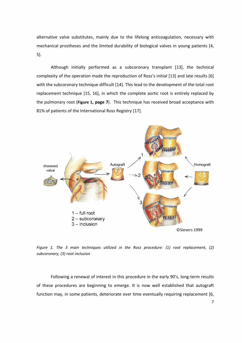
with the subcoronary technique difficult [14]. This lead to the development of the total root
replacement technique [15, 16], in which the complete aortic root is entirely replaced by
the pulmonary root (Figure 1, page 7). This technique has received broad acceptance with
81% of patients of the International Ross Registry [17].
Figure 1. The 3 main techniques utilized in the Ross procedure: (1) root replacement, (2)
subcoronary, (3) root inclusion
Following a renewal of interest in this procedure in the early 90’s, long-term results
of these procedures are beginning to emerge. It is now well established that autograft
function may, in some patients, deteriorate over time eventually requiring replacement [6,
©Sievers 1999
Page 9
8
8, 12, 18, 19, 20, 21, 22]. Concerns surfaced lately regarding the ability of the isolated,
unsupported pulmonary root to withstand the systemic circulation over time and resist
progressive dilatation, threatening valve competence [22, 23], and leading to an
unexpected increased rate of reoperation beginning around 7 to 8 years after the initial
operation[24, 25, 26]. In a recently published meta-analysis, Takkenberg et al. have
summarized the prevalence of structural and non-structural valve deterioration [27],
reoperations and other major events observed in large studies of patients operated with
the Ross procedure [25]. The incidence of autograft structural or non-structural valve
deterioration in the published large studies averages at 0.78%/year (95% CI: 0.43 – 1.40)
whereas the incidence of right sided homograft structural or non-structural valve
deterioration is approximately 0.55%/year (95% CI: 0.26 – 1.17).
1.4 Research on the modes of autograft failure of the Ross procedure
After the realization of this potential, significant research has been conducted
regarding the mode of autograft failure after the Ross procedure. Early autograft failure is
often attributed to technical errors, as was the case with the technically demanding and
difficult to reproduce subcoronary technique [14]. The introduction of the root
replacement technique [15] seems to ameliorate early autograft failure, however reports of
progressive autograft dilatation [8, 21, 28, 29] and subsequent late autograft failure have
recently emerged [18, 21].
Understanding the modes of autograft failure after the Ross procedure, many
groups have employed modified techniques or autograft reinforcement to correct
abnormalities in the aortic root area and thus prevent anatomic mismatch [28, 29], or to
stabilize parts of the aortic annulus prone to dilatation [16, 17, 22, 30]. However this long
term impact of reinforcements on the autograft function and durability remains largely
unknown.
Page 10
9
1.5 The present study
The main focus of the present study was to unveil the effect of such reinforcement
procedures on autograft function using data from the large patient population of the
German-Dutch Ross Registry. The presence of two different techniques in the Ross Registry
(subcoronary and root replacement) presents a challenge for this analysis, mainly because
the evolution of the native aortic root pathology hosting a subcoronary implant is very
different than the evolution of the freestanding pulmonary autograft root technique, and
as such, reinforcement techniques might play different roles and serve different purposes
in each of these techniques.
Page 11
10
2. Patients and Methods
2.1 Study population and operative data
The analysis was performed using data from the German-Dutch Ross Registry. The
registry includes data from 12 departments of cardiac surgery in Germany and The
Netherlands since 1988.
Follow-up data from each center were entered in the database and a systematic
prospective registry was started in January 2002 (Clinical trial ID NCT 00708409). The
employed surgical technique was according to the surgeon’s preference, with more or less
each center having adopted the one or the other technique. The operative technique
(subcoronary / root replacement) was specific for each institution and remained the same
throughout the time period of the study. The vast majority of subcoronary procedures were
performed in one center. In the root replacement technique, one center performed no
reinforcement procedures at all, whereas the incidence of prophylactically performed
reinforcement procedures increased with time in all other centers performing the root
replacement Ross procedure. Thirty patient operated with the root inclusion technique
were included in the subcoronary group, to create a group with all native root preserving
procedures.
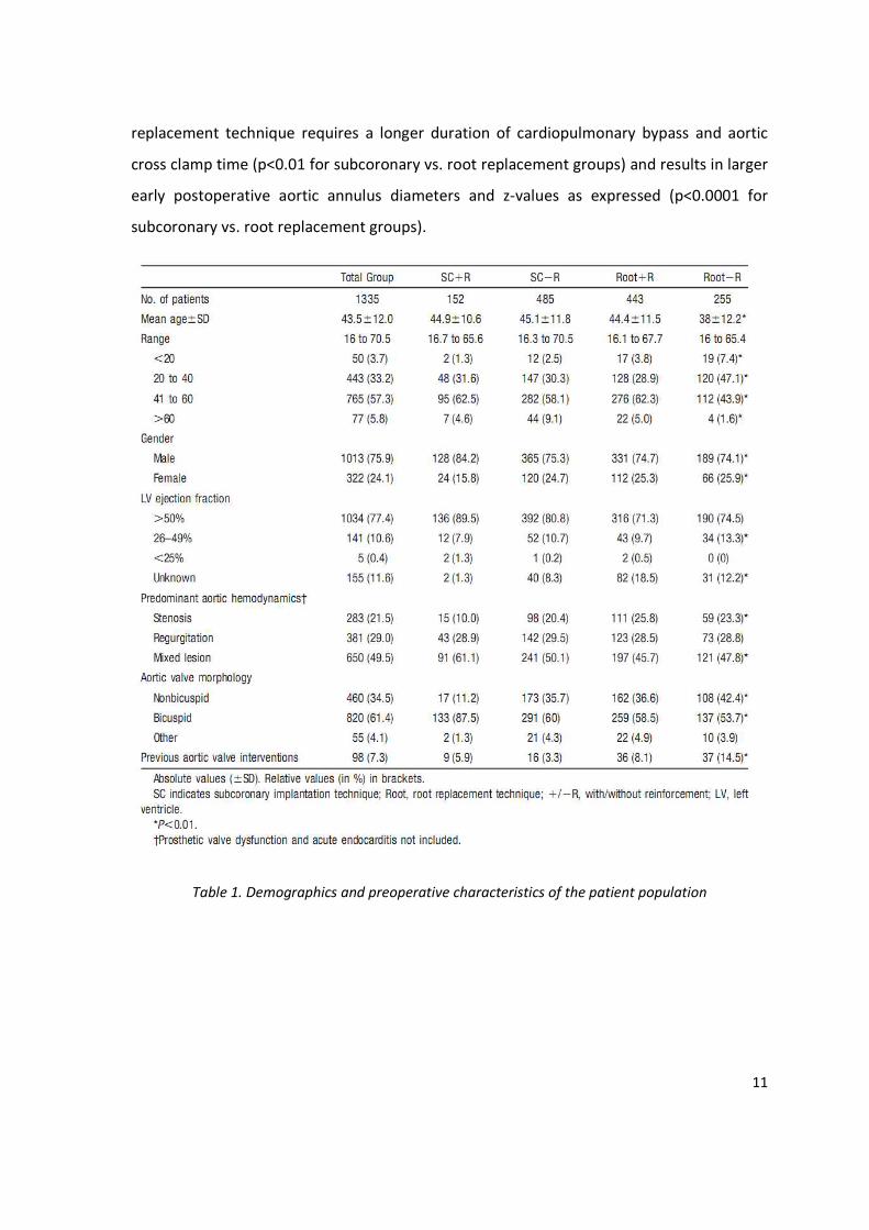
A total of 1335 patients were entered in the registry as of January 2008. The
patients’ preoperative characteristics as well as operative technique and presence or
absence of reinforcement are summarized in Table 1 page 11, and Table 2 page 12,
respectively. In brief, the patient population consists of young patients, with either normal
or slightly reduced left ventricular. It is important to note that in this study, patients with
tricuspid and bicuspid aortic valves where included, in contrast to a belief shared by some
groups in the literature that the Ross procedure in the setting of the aortic valve is not
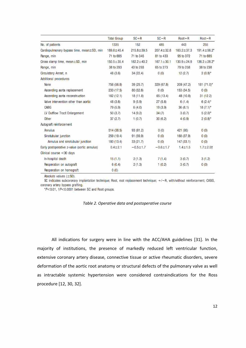
advisable. In terms of intraoperative characteristics (Table 2, page 12), the root
Page 12
11
replacement technique requires a longer duration of cardiopulmonary bypass and aortic
cross clamp time (p<0.01 for subcoronary vs. root replacement groups) and results in larger
early postoperative aortic annulus diameters and z-values as expressed (p<0.0001 for
subcoronary vs. root replacement groups).
Table 1. Demographics and preoperative characteristics of the patient population
Page 13
12
Table 2. Operative data and postoperative course
All indications for surgery were in line with the ACC/AHA guidelines [31]. In the
majority of institutions, the presence of markedly reduced left ventricular function,
extensive coronary artery disease, connective tissue or active rheumatic disorders, severe
deformation of the aortic root anatomy or structural defects of the pulmonary valve as well
as intractable systemic hypertension were considered contraindications for the Ross
procedure [12, 30, 32].
Page 14
13
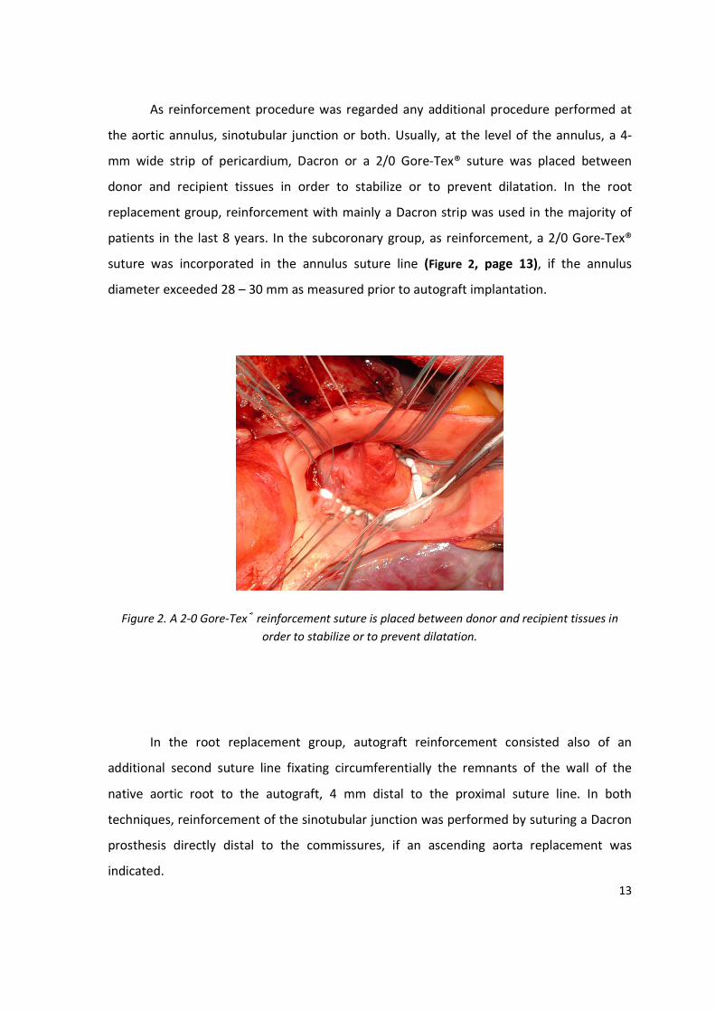
As reinforcement procedure was regarded any additional procedure performed at
the aortic annulus, sinotubular junction or both. Usually, at the level of the annulus, a 4-
mm wide strip of pericardium, Dacron or a 2/0 Gore-Tex® suture was placed between
donor and recipient tissues in order to stabilize or to prevent dilatation. In the root
replacement group, reinforcement with mainly a Dacron strip was used in the majority of
patients in the last 8 years. In the subcoronary group, as reinforcement, a 2/0 Gore-Tex®
suture was incorporated in the annulus suture line (Figure 2, page 13), if the annulus
diameter exceeded 28 – 30 mm as measured prior to autograft implantation.
Figure 2. A 2-0 Gore-Tex® reinforcement suture is placed between donor and recipient tissues in
order to stabilize or to prevent dilatation.
In the root replacement group, autograft reinforcement consisted also of an
additional second suture line fixating circumferentially the remnants of the wall of the
native aortic root to the autograft, 4 mm distal to the proximal suture line. In both
techniques, reinforcement of the sinotubular junction was performed by suturing a Dacron
prosthesis directly distal to the commissures, if an ascending aorta replacement was
indicated.
Page 15
14
Written informed consent was obtained from all patients. The study was approved
by the ethics committee of the medical faculty of the University of Lübeck (No. 08-030).
2.2 Clinical and echocardiographic follow up
Clinical and echocardiographic follow-up was performed at discharge and on a
yearly basis. The echocardiographic data acquisition protocol of the registry was
standardized in all centers. Autograft dimensions were measured at four levels (annulus,
sinus of Valsalva, sinotubular junction, proximal ascending aorta) as described by Roman et
al [33]: (1) annulus at the level of the autograft leaflet hinges, (2) sinus of Valsalva at the
largest anteroposterior diameter, (3) sinotubular junction at the distal rim of the sinuses of
Valsalva, and (4) the proximal ascending aorta 2 cm above the sinotubular junction.
The degree of aortic regurgitation was assessed by multiple techniques with the
parasternal long-axis and apical 5-chamber view. Pulsed wave Doppler and color flow
Doppler imaging were used for mapping the left ventricular outflow tract, including
determination of the ratio of jet height to left ventricular outflow tract height. Continuous
Doppler imaging was applied to measure the deceleration slope and pressure half-time of
the autograft regurgitation jet. Aortic regurgitation was graded with the use of standard
criteria in a majority of the examinations [34]. Because this is a multicenter study, the final
decision of autograft regurgitation (AR) grading was left to the decision of the responsible
echocardiographer’s preference and experience, and regurgitation severity was reported
on a scale of grade 0 to 4 [34]. Trace (trivial) aortic insufficiency defined as a very tiny
regurgitation jet in early diastole near the detection limit, was included in the analysis as
grade 0.5. Mean duration of follow-up was 6.09 ± 3.97 years (median 5.6 years; range 0.01
– 19.2 years; 8205 patient-years). Follow-up completeness was 93 %. The 7% missing follow
up visits were evenly distributed across the groups. Classification of the mode of valve
failure has been performed according to the latest guidelines for reporting outcome after
valve interventions [27]. All indications for autograft reoperations were in accordance to
Page 16
15
the ACC/AHA guidelines [31]. In two patients sub-valvular aortic aneurysms were the
primary indication for reoperation.
2.3 Statistical analysis
Frequencies are presented as absolute numbers and percentages. Continuous data
are expressed as mean ± standard deviation (SD). Patients were classified according to the
operative technique (SC, Root) and the presence (+R) or absence (-R) of R. Comparisons
between the groups were performed using the Mann-Whitney U test and the Fisher’s exact
test. Actuarial estimates of survival and freedom from autograft reoperation were
accomplished with Kaplan-Meier methods. Survival curves were compared using the log-
rank test (SPSS 11.0 for Windows, SPSS Inc., Chicago, IL). The Cox model was used to assess
the consistency of treatment effect by testing for interactions between the type of surgery
(technique and presence of autograft reinforcement) and prespecified baseline
characteristics. In order to identify predictive variables for shorter time to autograft re-
operation, we first performed a univariate analyses by using the Cox proportional hazard
regression model. Multivariable Cox proportional hazard models were used to confirm
whether differences between the operative groups persisted in the presence of pre-
operative variables. The presence of interactions and the proportionality of hazards
assumption was checked for the final model including operative group and significant pre-
operative variables. The following factors were analyzed as potential risk factors for
autograft re-operation due to structural and non-structural failure (infective endocarditis as
a re-operation indication in 8 patients was excluded): age, gender, year of surgery,
predominant aortic hemodynamics, hypertension, previous aortic valve intervention,
presence of bicuspid aortic valve, operative technique and presence of reinforcement
procedures.
Page 17
16
2.3.1 The problem of analyzing serial echocardiographic measurements: Reporting without
distorting
The statistical analysis of serial echocardiographic measurement in large patient
populations is complicated by several factors: echocardiographic information is obtained at
different time points, follow up appointments may be missed, valve function is variable
over time, there is significant inter- and intraobserver variability and the use of different
equipment may affect the measurement. The most frequently used method of analyzing
and reporting serial echocardiographic measurement is the Kaplan-Meier method.
However there are several concerns when utilizing this methodology in large patient
registries.
First, as a concept the Kaplan-Meier method considers each event as irreversible,
while the severity of valvular dysfunction or the aortic dimensions are very often variable
overtime. Therefore, by regarding each measurement as irreversible, the Kaplan-Meier
method underestimates the freedom from valvular dysfunction and inserts a significant
bias when reporting the time point at which the valvular dysfunction progressed. The
second problem with the Kaplan-Meier method is that it regards time as a continuous
variable, which this is not the case when analyzing echocardiographic measurements over
time. In the Kaplan-Meier function, every measurement is extrapolated and analyzed as if it
is to hold true or stable until the time of the next examination. This inserts a bias a
significant bias regarding the development of measurement over time in the case of missed
follow up visits or changes in follow up schedules, something that is very frequently
encountered in large registries.
Both factors mentioned above may play a minor role when analyzing small patient
groups over a short period of time, and as such the Kaplan-Meier method has been utilized
in analyses of small patient groups despite these known limitations. However in large
patient populations, biased extrapolations of measurements accumulate fast, a fact that
eventually renders the analysis and more importantly the interpretation of the results
Page 18
17
problematic. Thus in the case of the present study these factors prohibit the use of the
Kaplan-Meier method, and the analysis and reporting of our results in an accurate way
presents a major challenge.
2.3.2 Use of novel statistical methodology for the analysis and interpretation of the data
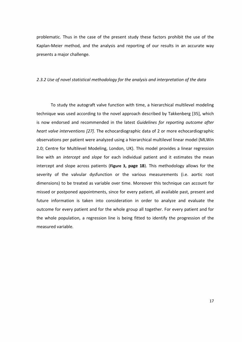
To study the autograft valve function with time, a hierarchical multilevel modeling
technique was used according to the novel approach described by Takkenberg [35], which
is now endorsed and recommended in the latest Guidelines for reporting outcome after
heart valve interventions [27]. The echocardiographic data of 2 or more echocardiographic
observations per patient were analyzed using a hierarchical multilevel linear model (MLWin
2.0; Centre for Multilevel Modeling, London, UK). This model provides a linear regression
line with an intercept and slope for each individual patient and it estimates the mean
intercept and slope across patients (Figure 3, page 18). This methodology allows for the
severity of the valvular dysfunction or the various measurements (i.e. aortic root
dimensions) to be treated as variable over time. Moreover this technique can account for
missed or postponed appointments, since for every patient, all available past, present and
future information is taken into consideration in order to analyze and evaluate the
outcome for every patient and for the whole group all together. For every patient and for
the whole population, a regression line is being fitted to identify the progression of the
measured variable.
Page 19
18
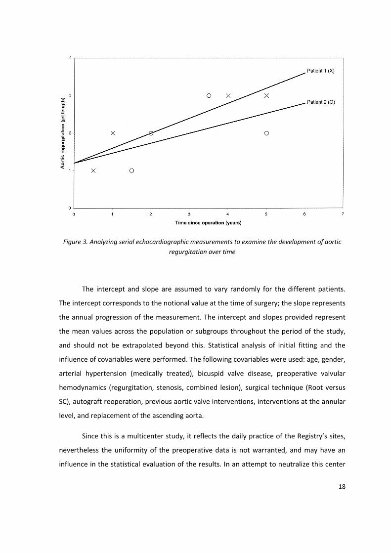
Figure 3. Analyzing serial echocardiographic measurements to examine the development of aortic
regurgitation over time
The intercept and slope are assumed to vary randomly for the different patients.
The intercept corresponds to the notional value at the time of surgery; the slope represents
the annual progression of the measurement. The intercept and slopes provided represent
the mean values across the population or subgroups throughout the period of the study,
and should not be extrapolated beyond this. Statistical analysis of initial fitting and the
influence of covariables were performed. The following covariables were used: age, gender,
arterial hypertension (medically treated), bicuspid valve disease, preoperative valvular
hemodynamics (regurgitation, stenosis, combined lesion), surgical technique (Root versus
SC), autograft reoperation, previous aortic valve interventions, interventions at the annular
level, and replacement of the ascending aorta.
Since this is a multicenter study, it reflects the daily practice of the Registry’s sites,
nevertheless the uniformity of the preoperative data is not warranted, and may have an
influence in the statistical evaluation of the results. In an attempt to neutralize this center
Page 20
19
specific influence, we integrated in all analyses presented herein a center variable, allowing
for the effect which the different centers may have to the results of this study. This model
was applied to analyze AR and aortic root dimensions over time, as well as AR as a function
of aortic root dimensions for the surgical subgroups Root and SC and subgroups with and
without R. For the small subgroup of 30 patients operated with root inclusion technique,
separate estimation of the AR development and AR dimensions over time was also
performed.
Page 21
20
3. Results
3.1 Clinical outcome and autograft reoperations
The duration of follow-up differed significantly between the groups with and
without reinforcement procedures in both techniques (subcoronary, root replacement),
with the reinforced groups having a shorter follow-up duration. This is due to the fact that
reinforcement procedures were implemented mainly in the last 6-8 years.
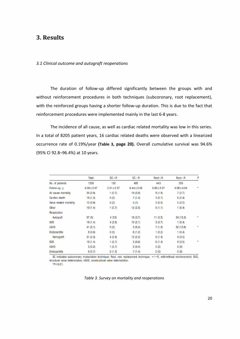
The incidence of all cause, as well as cardiac related mortality was low in this series.
In a total of 8205 patient years, 16 cardiac related deaths were observed with a linearized
occurrence rate of 0.19%/year (Table 3, page 20). Overall cumulative survival was 94.6%
(95% CI 92.8–96.4%) at 10 years.
Table 3. Survey on mortality and reoperations
Page 22
21
As a total, 67 autograft reoperations were observed (linearized occurrence rate
0.82%/year). Non-structural valve deterioration was the primary cause of failure (61.2% of
all autograft reoperations). Structural valve deterioration and endocarditis accounted for
26.9% and 11.9% of all autograft reoperations respectively.
As a first observation, the root replacement technique group had significantly more
reoperation than the subcoronary technique, and when allowing for the presence of
reinforcement procedures, the root replacement group without reinforcement accounted
for 56% of all reoperations in the study population.
When studying the modes of autograft failure in each technique significant
conclusion can be made. In the root replacement technique the main mode of failure
appears to be non structural valve deterioration (91% of all reoperations in the root
replacement group), mainly in the form of autograft dilatation. On the contrary, in the
subcoronary group the main mode of failure appears to be structural valve deterioration
(88% of all reoperation in the subcoronary group).
Freedom from autograft reoperation (with the exclusion of 8 patients operated for
infective endocarditis [27] ) was 96.8% (95% CI 95.5–99.0%) at 5 and 89.6% (95% CI 86.1–
93.0%) at 10 years. When allowing for technique and presence of R, the SC and the Root+R
revealed a significantly better freedom from reoperation at 10 years in comparison to Root-
R (94.2% (95% CI 90.4–97.9 %) and 93.2% (95% CI 88.2–98.2%) vs. 88.3% (95% CI 76.5–
90.1%) respectively, p=0.001; Figure 4, page 22).
Page 23
22
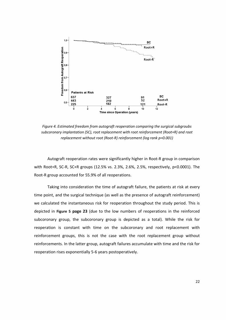
Figure 4. Estimated freedom from autograft reoperation comparing the surgical subgroubs
subcoronary implantation (SC), root replacement with root reinforcement (Root+R) and root
replacement without root (Root-R) reinforcement (log rank p=0.001)
Autograft reoperation rates were significantly higher in Root-R group in comparison
with Root+R, SC-R, SC+R groups (12.5% vs. 2.3%, 2.6%, 2.5%, respectively, p<0.0001). The
Root-R group accounted for 55.9% of all reoperations.
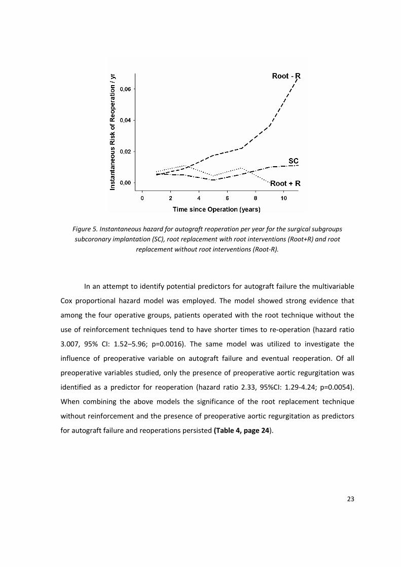
Taking into consideration the time of autograft failure, the patients at risk at every
time point, and the surgical technique (as well as the presence of autograft reinforcement)
we calculated the instantaneous risk for reoperation throughout the study period. This is
depicted in Figure 5 page 23 (due to the low numbers of reoperations in the reinforced
subcoronary group, the subcoronary group is depicted as a total). While the risk for
reoperation is constant with time on the subcoronary and root replacement with
reinforcement groups, this is not the case with the root replacement group without
reinforcements. In the latter group, autograft failures accumulate with time and the risk for
reoperation rises exponentially 5-6 years postoperatively.
Page 24
23
Figure 5. Instantaneous hazard for autograft reoperation per year for the surgical subgroups
subcoronary implantation (SC), root replacement with root interventions (Root+R) and root
replacement without root interventions (Root-R).
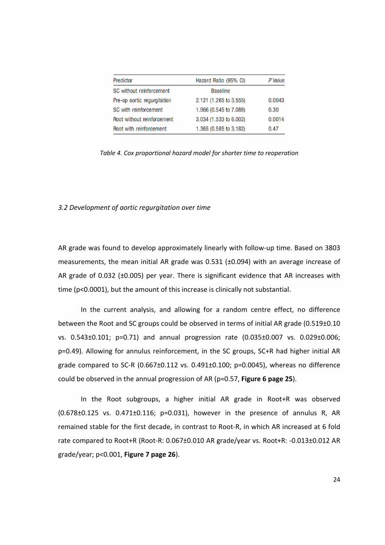
In an attempt to identify potential predictors for autograft failure the multivariable
Cox proportional hazard model was employed. The model showed strong evidence that
among the four operative groups, patients operated with the root technique without the
use of reinforcement techniques tend to have shorter times to re-operation (hazard ratio
3.007, 95% CI: 1.52–5.96; p=0.0016). The same model was utilized to investigate the
influence of preoperative variable on autograft failure and eventual reoperation. Of all
preoperative variables studied, only the presence of preoperative aortic regurgitation was
identified as a predictor for reoperation (hazard ratio 2.33, 95%CI: 1.29-4.24; p=0.0054).
When combining the above models the significance of the root replacement technique
without reinforcement and the presence of preoperative aortic regurgitation as predictors
for autograft failure and reoperations persisted (Table 4, page 24).
Page 25
24
Table 4. Cox proportional hazard model for shorter time to reoperation
3.2 Development of aortic regurgitation over time
AR grade was found to develop approximately linearly with follow-up time. Based on 3803
measurements, the mean initial AR grade was 0.531 (±0.094) with an average increase of
AR grade of 0.032 (±0.005) per year. There is significant evidence that AR increases with
time (p<0.0001), but the amount of this increase is clinically not substantial.
In the current analysis, and allowing for a random centre effect, no difference
between the Root and SC groups could be observed in terms of initial AR grade (0.519±0.10
vs. 0.543±0.101; p=0.71) and annual progression rate (0.035±0.007 vs. 0.029±0.006;
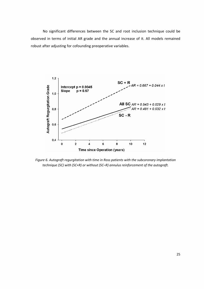
p=0.49). Allowing for annulus reinforcement, in the SC groups, SC+R had higher initial AR
grade compared to SC-R (0.667±0.112 vs. 0.491±0.100; p=0.0045), whereas no difference
could be observed in the annual progression of AR (p=0.57, Figure 6 page 25).
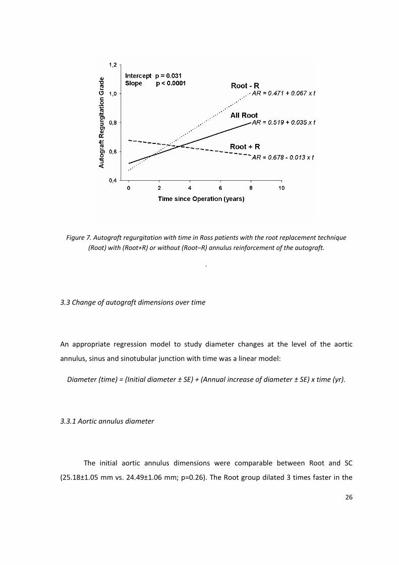
In the Root subgroups, a higher initial AR grade in Root+R was observed
(0.678±0.125 vs. 0.471±0.116; p=0.031), however in the presence of annulus R, AR
remained stable for the first decade, in contrast to Root-R, in which AR increased at 6 fold
rate compared to Root+R (Root-R: 0.067±0.010 AR grade/year vs. Root+R: -0.013±0.012 AR
grade/year; p<0.001, Figure 7 page 26).
Page 26
25
No significant differences between the SC and root inclusion technique could be
observed in terms of initial AR grade and the annual increase of it. All models remained
robust after adjusting for cofounding preoperative variables.
Figure 6. Autograft regurgitation with time in Ross patients with the subcoronary implantation
technique (SC) with (SC+R) or without (SC–R) annulus reinforcement of the autograft.
Page 27
26
Figure 7. Autograft regurgitation with time in Ross patients with the root replacement technique
(Root) with (Root+R) or without (Root–R) annulus reinforcement of the autograft.
.
3.3 Change of autograft dimensions over time
An appropriate regression model to study diameter changes at the level of the aortic
annulus, sinus and sinotubular junction with time was a linear model:
Diameter (time) = (Initial diameter ± SE) + (Annual increase of diameter ± SE) x time (yr).
3.3.1 Aortic annulus diameter
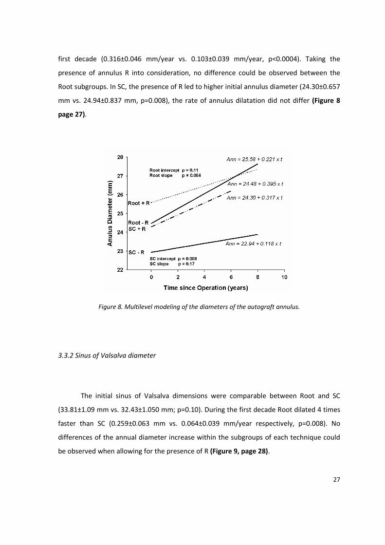
The initial aortic annulus dimensions were comparable between Root and SC
(25.18±1.05 mm vs. 24.49±1.06 mm; p=0.26). The Root group dilated 3 times faster in the
Page 28
27
first decade (0.316±0.046 mm/year vs. 0.103±0.039 mm/year, p<0.0004). Taking the
presence of annulus R into consideration, no difference could be observed between the
Root subgroups. In SC, the presence of R led to higher initial annulus diameter (24.30±0.657
mm vs. 24.94±0.837 mm, p=0.008), the rate of annulus dilatation did not differ (Figure 8
page 27).
Figure 8. Multilevel modeling of the diameters of the autograft annulus.
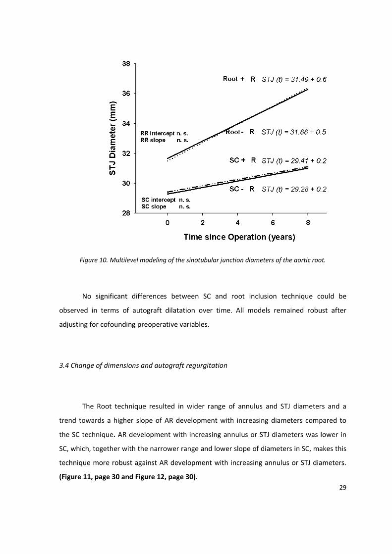
3.3.2 Sinus of Valsalva diameter
The initial sinus of Valsalva dimensions were comparable between Root and SC
(33.81±1.09 mm vs. 32.43±1.050 mm; p=0.10). During the first decade Root dilated 4 times
faster than SC (0.259±0.063 mm vs. 0.064±0.039 mm/year respectively, p=0.008). No
differences of the annual diameter increase within the subgroups of each technique could
be observed when allowing for the presence of R (Figure 9, page 28).
Page 29
28
Figure 9. Multilevel modeling of the diameters of the sinuses of Valsalva.
3.3.3 Sinotubular junction diameter
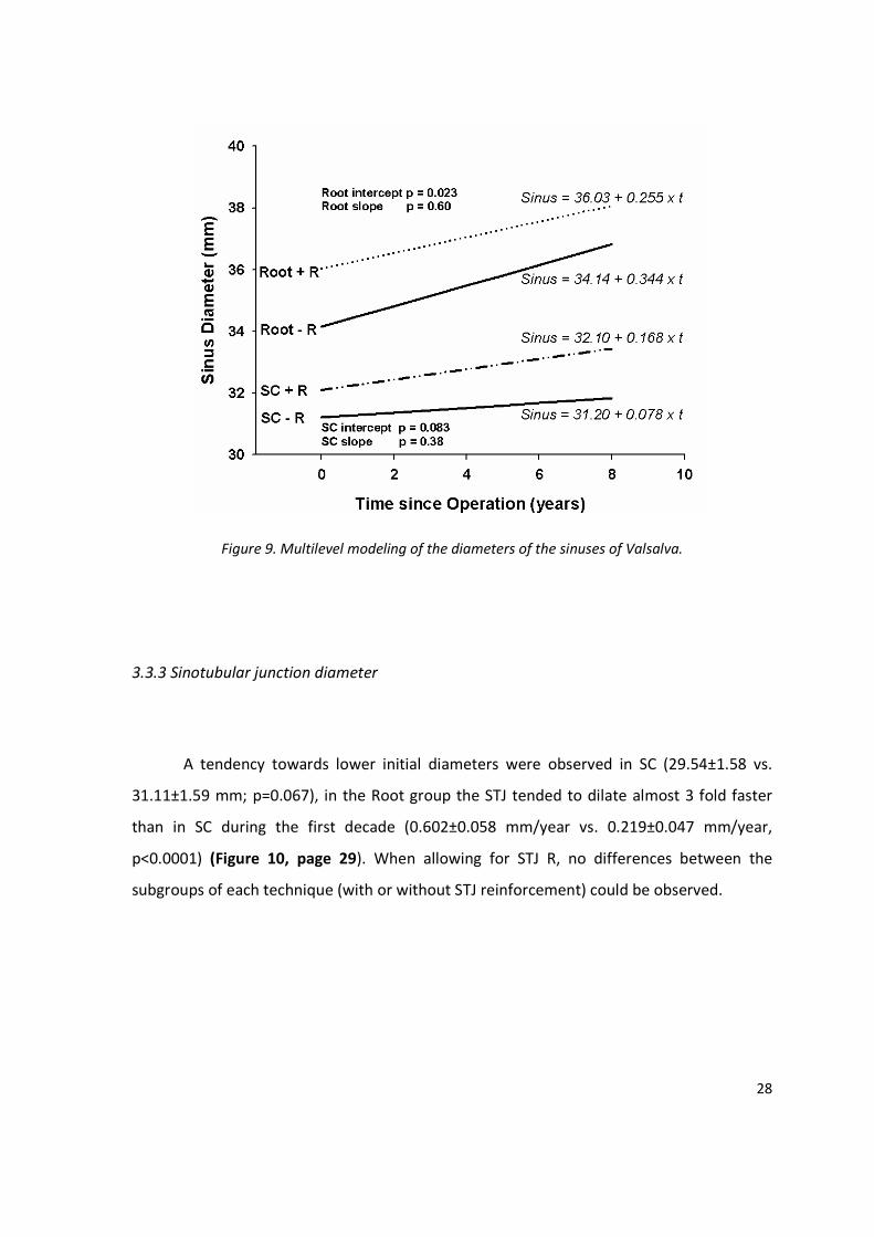
A tendency towards lower initial diameters were observed in SC (29.54±1.58 vs.
31.11±1.59 mm; p=0.067), in the Root group the STJ tended to dilate almost 3 fold faster
than in SC during the first decade (0.602±0.058 mm/year vs. 0.219±0.047 mm/year,
p<0.0001) (Figure 10, page 29). When allowing for STJ R, no differences between the
subgroups of each technique (with or without STJ reinforcement) could be observed.
Page 30
29
Figure 10. Multilevel modeling of the sinotubular junction diameters of the aortic root.
No significant differences between SC and root inclusion technique could be
observed in terms of autograft dilatation over time. All models remained robust after
adjusting for cofounding preoperative variables.
3.4 Change of dimensions and autograft regurgitation
The Root technique resulted in wider range of annulus and STJ diameters and a
trend towards a higher slope of AR development with increasing diameters compared to
the SC technique. AR development with increasing annulus or STJ diameters was lower in
SC, which, together with the narrower range and lower slope of diameters in SC, makes this
technique more robust against AR development with increasing annulus or STJ diameters.
(Figure 11, page 30 and Figure 12, page 30).
Page 31
30
Figure 11. Multilevel modeling of the autograft regurgitation grade for the various postoperative
aortic annulus diameters for the two surgical techniques.
Figure 12. Multilevel modeling of the autograft regurgitation grade for the various postoperative
sinotubular junction diameters for the two surgical techniques.
Page 32
31
4. Discussion
The Ross procedure can be performed as an attractive alternative in selected
patients, however various groups present mid-to-long term results indicating that the
autograft function may deteriorate over time with the hazard of eventually mandating a
reoperation [6, 8, 18, 20].
4.1 Previous studies
Significant research has been conducted regarding the mode of autograft failure
after the Ross procedure. Early autograft failure is often attributed to technical errors, as
was the case with the technically demanding and difficult to reproduce subcoronary
technique [14]. The introduction of the root replacement technique [15] seems to
ameliorate this early autograft failure, however reports of progressive autograft dilatation
[8, 21, 28, 29] and subsequent late autograft failure have recently emerged [18, 21, 25].
Understanding the modes of autograft failure after the Ross procedure, many
groups have employed modified techniques or R to correct abnormalities in the aortic root
area and thus prevent anatomic mismatch [28, 29], or to stabilize parts of the aortic
annulus prone to dilatation [16, 17, 22, 30]. The long term impact of R on the autograft
function and durability remains largely unknown. Thus main focus of the present study was
to unveil the effect of such R on autograft function. The presence of two different
techniques in the Ross Registry (SC and Root) presents a challenge for this analysis, mainly
because the evolution of the native aortic root pathology hosting a subcoronary implant is
very different than the evolution of the freestanding pulmonary autograft root technique,
and as such, reinforcement techniques might play different roles and serve different
purposes in each of these techniques.
Page 33
32
4.2 Present study
R in this study were performed in order to correct anatomical abnormalities or
mismatch, or prophylactically in order to stabilize the aortic root and prevent postoperative
autograft dilatation. The intention of the operating surgeon and the indication for
performing R was determined by the operating surgeon at each institution.
4.2.1 Reoperation rates
Our main observation was that a significant proportion of the Root-R subgroup
required reoperation in comparison to the three other subgroups. The Root-R group
contributed 57% of all reoperations observed in the registry. The leading cause of
reoperation in the Root group was non-structural valve deterioration, as defined in the
latest guidelines[27] (91 % of all reoperations in this group) presenting in the form of
autograft dilatation, whereas in the SC group 88% of all reoperations were attributed to
structural valve deterioration [27], mainly as cusp prolapsed (patients reoperated due to
endocarditis are excluded). The addition of R seemed to decrease reoperation rates due to
non-structural valve deterioration in the Root+R group leading to reoperation rates similar
to the SC technique. In the SC group, we could not show a significant impact of R on
reoperation rates due to either structural or non-structural valve deterioration.
Autograft failure appears after the first 6-8 years in the Root-R group with an
exponentially rising instantaneous hazard rate, while remaining stable throughout the
observational period in the SC group. For the time period studied, the Root+R subgroup had
similar reoperation risk rates as the SC group. This finding is in concordance to previous
studies [36]. From our data it could be hypothesized that the larger the preoperative
annulus dimensions, the lower the ability of the autograft to provide adequate leaflet
Page 34
33
coaptation with progressive root dilatation. In the Root group, R leads to smaller annulus
diameters, and thus they may act prophylactically. It is however unknown if in the long
term, reinforcement procedures prevent or postpone autograft function deterioration.
4.2.2 Autograft function
In this study we could not observe a significant difference with regards to AR
development over time between the SC and the Root as overall groups. This is in contrast
to a previous report of ours [8], where we found a significantly increased initial AR in the
Root patients. This difference, however, could be attributed to the additional 321 patients
(32% more than the 2006 report [8]) added to the registry in the last 2 years. Moreover,
due to the non-uniformity of the preoperative data, in this analysis we allowed for a center
effect to mitigate any systematic reporting error between the registry sites. The presence
of R effectively prevented AR development over time in the Root group, whereas in the SC
R had no effect on the AR increase, albeit for a greater initial AR. R in SC was implemented
only in large annulus observed at operation to reduce or reshape the effective annular size
to improve cusp coaptation. Here, the indication for R was not prophylactically but
therapeutic.
4.2.3 Autograft dimensions
A consistent finding in this study is the larger initial postoperative aortic root
diameters in patients undergoing R in both groups (Figure 8, page 27; Figure 9, page 28;
Figure 10, page 29), although this does not always reach the level of statistical significance.
Given however that, R are most likely to reduce the aortic root diameters, one can argue
Page 35
34
that R are often performed in order to treat underlying abnormalities, thus reducing aortic
root dimension to restore the ideal anatomical relations. Although in our series R had a
positive effect in terms of autograft durability, there are in the literature notable series of
patients operated with the Root technique without R with excellent long term outcome [6,
19]. The effects of proper patient selection bias however cannot be ruled out since an –
often intraoperative - selection of the appropriate patient-pathology is common in the
setting of the Ross procedure. It may well be that in patients with an ideal aortic root
pathology, a Root-R technique may have excellent outcome. In addition, it cannot be ruled
out that special modifications on individual patient basis in very experienced hands can
prevent postoperative dilatation without the need for R with synthetic material [19].
The SC technique was associated with significantly reduced rates of aortic root
dilatation at all levels of the aortic root. In the Root technique, R did not influence the
progressive autograft dilatation over time for the time period of the study. In this study we
could observe an increased AR in patients with increased annulus and STJ diameters.
Although a causal effect could not be established, this could be explained by the
observational nature of this study and the increased reserve of the aortic root in terms of
dilatation[37] that would lead to AR, and as such the time frame of this study could be
insufficient to show that the AR increase is solely caused by root dilatation.
4.3 Limitations
The present study is a retrospective, non randomized study. The intention of the
surgeon when performing R in SC was primarily to treat an underlying pathology, whereas
in Root, R was mainly applied routinely as a part of root replacement. Early postoperative z-
values are provided only for the aortic annulus, mainly due to the fact that large databases
of normal values in the adult population do not exist for the other counterparts of the
aortic root components. No technique to support the sinus of Valsalva was performed in
this patient population. A further post hoc subgroup analysis to identify the most
Page 36
35
appropriate type of reinforcement material and or specific operative techniques was
regarded statistically inappropriate due to the retrospective nature of this study. We
believe that this should be performed in the setting of a prospective randomized trial. A
small surgical subgroup operated with the root inclusion technique was included in the SC
group. Sub analysis of key items (AR, autograft dimensions, reoperations) did not reveal any
difference between the SC and the root inclusion technique group. A possible limitation
may be the different follow-up times of the various study groups, with R in the Root, being
mostly implemented in the last 8 years, having the shortest follow-up time. However the
differences observed in outcome and autograft function were statistically and clinically
significant for the time period studied.
4.4 Clinical implications
We can conclude that in patients undergoing the Ross procedure, autograft
reinforcement procedures performed either prophylactically in order to prevent autograft
dilatation, or therapeutically to correct an underlying a suboptimal anatomy, lead to lower
development of AR over time. Surgical autograft reinforcement is able to reduce
reoperation rates for autograft failure due to non-structural valve deterioration in the root
replacement Ross procedure for the time period of this study. These procedures appear to
be safe, present with good long term outcome and should strongly be taken into
consideration.
Page 37
36
5. Summary
Autograft reinforcement interventions (R) during the Ross procedure are intended
to preserve autograft function and improve durability. Aim of this study is to evaluate this
hypothesis.
1335 adult patients (mean age:43.5±12.0 years) underwent a Ross procedure
(subcoronary, SC, n=637;root replacement, Root, n=698). 592 patients received R of the
annulus, sinotubular junction, or both. Regular clinical and echocardiographic follow-up
was performed (mean:6.09±3.97, range:0.01–19.2 years). Longitudinal assessment of
autograft function with time was performed utilizing multilevel modeling techniques. The
Root without R (Root-R) group was associated with a 6-times increased reoperation rate
compared to Root with R (Root+R), SC with R (SC+R) and without R (SC-R) (12.9% vs. 2.3%
vs. 2.5%. vs. 2,6% respectively, p<0.001). SC and Root groups had similar rate of aortic
regurgitation (AR) development over time. Root+R patients had no progression of AR,
whereas Root-R had 6 times higher AR development compared to Root+R. In SC, R had no
remarkable effect on the annual AR progression. The SC technique was associated with
lower rates of autograft dilatation at all levels of the aortic root compared to the Root
techniques. R did not influence autograft dilatation rates in the Root group.
For the time period of the study surgical autograft stabilization techniques preserve
autograft function and result in significantly lower reoperation rates. The non-reinforced
Root was associated with significant adverse outcome. Therefore, surgical stabilization of
the autograft is advisable to preserve long term autograft function, especially in the Root
Ross procedure.
Page 38
37
6. Zusammenfassung in deutscher Sprache
6.1 Einleitung
Der Einsatz der körpereigenen Pulmonalklappe zur Behandlung einer Erkrankung
der Aortenklappe (Ross-Operation), erstmalig durchgeführt von Donald Ross im Jahre
1967[13], könnte die einzige Methode sein, welche, zumindest theoretisch, alle Vorteile
eines autologen, biologischen Klappenersatzes hat. Bei der Ross-Operation wird die native
Pulmonalklappe des Patienten entnommen und in Aortenklappenposition eingesetzt,
während ein pulmonaler Homograft (Spenderklappe) in Pulmonalposition implantiert wird.
In den frühen 90er Jahren wurde das Interesse an dieser Methode erneut geweckt,
so dass jetzt Langzeitergebnisse dieser Operationstechnik vorliegen. Es zeigt sich, dass bei
einer Reihe an Patienten Struktur und Funktion der operierten Klappe sich so verändern,
dass ein Ersatz dieser Klappe(n) erforderlich wird [6, 8, 12, 18, 19, 20, 21, 22].
Bei der Suche nach Ursachen für eine Klappendysfunktion entwickelten
verschiedene Arbeitsgruppen zusätzliche chirurgische Methoden zur Stabilisierung des
Autografts („Reinforcement“- R). Damit soll eine Korrektur der Aortenwurzelanatomie und
ihre Stabilität im Langzeitverlauf angestrebt werden [28, 29], zusätzlich war eine
Verstärkung des dilatationsanfälligen Annulus aortae beabsichtigt [16, 17, 22, 30]. Der
Langzeiteffekt dieser Maßnahmen auf Funktion und Haltbarkeit des Autograftes ist jedoch
weitgehend unbekannt.
Das Ziel der vorliegenden Arbeit ist, mit Hilfe der großen Patientenpopulation des
Deutsch-Holländischen Ross-Registers die Auswirkungen von R auf die Funktion des
Autograftes im Langzeitverlauf zu untersuchen.
6.2 Patienten und Methoden
Page 39
38
Zur Analyse wurden Daten des Deutsch-Holländischen Ross-Registers aus 12
herzchirurgischen Abteilungen seit dem Jahr 1988 herangezogen. Bis Januar 2008 wurden
1335 Patienten in das Register aufgenommen. Tabelle 1, Seite 11 und Table 2, Seite 12
enthalten die präoperativen Charakteristika der Patienten sowie die Besonderheiten der
Operationstechniken (subkoronar, SC; Wurzelersatz, Root) mit (+R) bzw. ohne (-R) R.
Klinische und echokardiographische Verlaufsuntersuchungen wurden bei Entlassung
sowie einmal jährlich durchgeführt. Die mittlere Dauer der Verlaufsbeobachtung betrug
6.09 ± 3.97 Jahre (Median 5.6 Jahre; 0.01–19.2 Jahre; 8205 Patientenjahre). Die
Vollständigkeit der Datensätze betrug 93%.
Für den Vergleich zwischen den Gruppen wurden der Mann-Whitney U-Test und der
Fisher`s Exact-Test herangezogen. Kaplan-Meier-Methoden dienten der Abschätzung der
Überlebenswahrscheinlichkeit und der Freiheit von Autograft-Reoperationen.
Überlebenskurven wurden mit Hilfe des log-rank-Tests (SPSS 11.0 for Windows, SPSS Inc.,
Chicago, IL) verglichen. Mit Hilfe des Cox-Modells wurden Interaktionen zwischen Art der
Operation (OP-Technik und der Anwendung von Autograft-R) und verschiedenen
präoperativen Charakteristika untersucht.
Um die Funktion des Autograftes in Abhängigkeit von der Zeit zu modellieren,
wurde eine hierarchische Multilevel-Analysentechnik (sog. Mehrebenenanalyse) eingesetzt,
die von den aktuellen „Guidelines for reporting outcome after heart valve interventions“
[27] gefordert wird.
6.3 Ergebnisse
Die Anzahl an Autograft-Reoperationen bei strukturell und nicht strukturell
bedingten Klappenfehlfunktionen waren in der Root-R Gruppe, verglichen mit den Gruppen
Page 40
39
Root+R, SC-R und SC+R signifikant erhöht (12,5% vs. 2,3%, 2,6% bzw. 2,5%; p<0,0001). Die
Root-R Gruppe enthält 55,9% aller Reoperationen (Tabelle 3, Seite 20 und Abbildung 5,
Seite 23). Bei SC und Root+R war der Anteil an Reoperationen nach 10 Jahren verglichen
mit der Root-R Gruppe signifikant niedriger (p=0.001).
Das multivariable Cox-Modell der vier Operationsgruppen zeigte, dass Patienten mit
präoperativer Aortenregurgitation (AR) und Patienten, welche eine Operation mit
Wurzelersatz, aber ohne Reinforcement erhalten haben, zu früheren Zeitpunkten
reoperationspflichtig werden (Tabelle 4, Seite 24).
Der Schweregrad der AR nahm annähernd linear während der
Nachbeobachtungszeit zu. Das auf 3803 Einzelmessungen basierende Gesamtmittel der
initialen AR war 0,531 (±0,094) mit einem durchschnittlichen Anstieg des AR-
Schweregrades von 0,032 (±0,005) pro Jahr. Abbildung 6, Seite 25 und Abbildung 7, Seite
26 zeigen die zeitlichen Entwicklungen der AR in den vier chirurgischen Untergruppen.
Die Entwicklung der Dimensionen des Aortenannulus über die Jahre ist in Abbildung
8, Seite 27 dargestellt. Der Annulus der Gesamtgruppe mit Wurzelersatz dilatierte im
Vergleich zur SC-Gruppe 3 mal schneller in der ersten Dekade (0,316±0,046 mm/Jahr vs.
0,103±0,039 mm/Jahr, p<0,0004). Vergleichbar nahmen die Dimensionen auf Höhe der
Sinus Valsalvae in der Gesamtgruppe mit Wurzelersatz im Vergleich zur SC-Gruppe 4 mal
schneller zu (0,259±0,063 mm/Jahr vs. 0,064±0,039 mm/Jahr, p=0.008; Abbildung 9, Seite
28), die Dimensionen des sinutubulären Übergangs (STJ) dilatierten bei größeren
Ausgangswerten 3 mal schneller in der ersten Dekade (Wurzelersatzgruppe 0,602±0,058
mm/Jahr vs. SC 0,219±0,047 mm/Jahr, p<0,0001; Abbildung 10, Seite 29).
Bei der Wurzelersatz-Technik waren größere Spannweiten der Annulus- und STJ-
Diametern auffällig. Mit zunehmendem Durchmesser war ein Trend zu einem steileren
Anstieg des AR-Schweregrades zu verzeichnen. Die SC-Gruppe zeigte ein geringe Zunahme
des AR-Schweregrades bei größeren Annulus- oder STJ-Diametern. Dies war assoziiert mit
einer kleineren Spannweite der Diameter in der Gesamtgruppe und einem geringeren
Anstieg der Diameter im zeitlichen Verlauf (Abbildungen 11, Seite 30 und 12, Seite 30).
Page 41
40
6.4 Diskussion
Der Einsatz von zusätzlichen chirurgischen Maßnahmen („reinforcement“) bei der
Ross-Operation kann zur Korrektur einer pathologischen Aortenwurzelgeometrie beitragen
oder aber als Prophylaxe dienen, um die Aortenwurzel im Langzeitverlauf zu stabilisieren
und so einer gefürchteten Dilatation des Autograftes mit Funktionsbeeinträchtigung
entgegen zu wirken.
Die wesentlichste Beobachtung der vorliegenden Arbeit war, dass Ross-Operationen
als Wurzelersatz ohne Unterstützungsmaßnahmen zu einem signifikant höheren Anteil an
Autograftfehlfunktionen führte mit der Notwendigkeit einer Reoperation (57 % aller
Reoperationen des gesamten Registers). Hauptursache waren nicht-strukturelle
Klappenveränderungen (87 % aller Reoperationen dieser Gruppe) in Form einer Autograft-
Dilatation, während in der SC-Gruppe 63% aller Reoperationen auf strukturellen
Klappenveränderungen, vorwiegend als Klappenprolaps, basierten. Die Anwendung von
chirurgischen Unterstützungsmaßnahmen in der Wurzelersatz-Gruppe senkte den Anteil an
Reoperationen, so dass Reoperationshäufigkeiten vergleichbar der SC-Gruppe erkennbar
waren.
Die vorliegenden Daten unterstützen die häufig diskutierte Hypothese, dass vornehmlich
zusätzliche Annulus-Interventionen zur Verkleinerung und Verstärkung dieser Ebene der
Aortenwurzel führen und die Geometrie und Funktion des Autografts über viele Jahre
erhalten. Bei komplettem Wurzelersatz werden die zusätzlichen Maßnahmen sowohl
prophylaktisch (zur Verhinderung einer Dilatation) wie auch therapeutisch (zur Korrektur
einer abnormen Geometrie) eingesetzt, während bei der SC-Technik Annulus-Maßnahmen
praktisch ausschließlich mit therapeutischem Ziel Eingang fanden. Damit sollte effektiv eine
zugrundeliegende Wurzelpathologie korrigiert werden, um die Ross-Operation überhaupt
zu ermöglichen. Somit sind Vergleichbarkeit dieser Intervention zwischen den
Operationsgruppen und ihre Interpretation nur bedingt möglich. So vermindern
Page 42
41
unterstützende Maßnahmen beim Wurzelersatz effektiv die Entwicklung einer AR, während
in der SC-Gruppe kein wesentlicher Einfluss auf den Anstieg des AR-Schweregrades gesehen
wurde. Offen muss auch bleiben, ob die zusätzlichen chirurgischen Maßnahmen eine
Verschlechterung der Autograftfunktion im Langzeitverlauf verhindern oder lediglich
aufschieben.
6.5 Schlussfolgerungen
Im untersuchten Zeitraum der Studie erhalten zusätzliche stabiliserende
Maßnahmen bei der Ross-Operation die Geometrie und Funktion des Autografts aufrecht
und führen zu so zu signifikant niedrigeren Reoperationsraten. Ein Wurzelersatz ohne
Annulusstabilisierung führt nach 6 – 8 Jahren zu einer Wurzeldilatation mit rapid
progredienter Autograftdysfunktion. Die chirurgische Stabilisierung des Autograftes bei der
Wurzelersatztechnik ist daher ratsam, um die Funktion des Autograftes im Langzeitverlauf
zu gewährleisten.
Page 43
42
Appendix
A. References
1 Gummert JF, Funkat A, Beckmann A, Schiller W, Hekmat K, Ernst M, Haverich A. Cardiac
surgery in Germany during 2007: a report on behalf of the German Society for Thoracic and
Cardiovascular Surgery. Thorac Cardiovasc Surg; 56: 328-336 (2008)
2 Hufnagel CA, Harvey WP, Rabil PJ, Mc DT. Surgical correction of aortic insufficiency. Surgery;
35: 673-683 (1954)
3 Carpentier A. The concept of bioprosthesis. Thoraxchir Vask Chir; 19: 379-383 (1971)
4 Grunkemeier GL, Li HH, Naftel DC, Starr A, Rahimtoola SH. Long-term performance of heart
valve prostheses. Curr Probl Cardiol; 25: 73-154 (2000)
5 Rahimtoola SH. Choice of prosthetic heart valve for adult patients. J Am Coll Cardiol; 41:
893-904 (2003)
6 Chambers JC, Somerville J, Stone S, Ross DN. Pulmonary autograft procedure for aortic
valve disease: long-term results of the pioneer series. Circulation; 96: 2206-2214 (1997)
7 Yacoub MH, Kilner PJ, Birks EJ, Misfeld M. The aortic outflow and root: a tale of dynamism
and crosstalk. Ann Thorac Surg; 68: S37-43 (1999)
8 Hanke T, Stierle U, Boehm JO, Botha CA, Matthias Bechtel JF, Erasmi A, Misfeld M, Hemmer
W, Rein JG, Robinson DR, Lange R, Horer J, Moritz A, Ozaslan F, Wahlers T, Franke UF, Hetzer R,
Hubler M, Ziemer G, Graf B, Ross DN, Sievers HH. Autograft regurgitation and aortic root
dimensions after the Ross procedure: the German Ross Registry experience. Circulation; 116: I251-
258 (2007)
9 Kouchoukos NT, Davila-Roman VG, Spray TL, Murphy SF, Perrillo JB. Replacement of the
aortic root with a pulmonary autograft in children and young adults with aortic-valve disease. N
Engl J Med; 330: 1-6 (1994)
10 Elkins RC, Lane MM, McCue C, Ward KE. Pulmonary autograft root replacement: mid-term
results. J Heart Valve Dis; 8: 499-503; discussion 503-496 (1999)
11 Paparella D, David TE, Armstrong S, Ivanov J. Mid-term results of the Ross procedure. J Card
Surg; 16: 338-343 (2001)
Page 44
43
12 Sievers HH, Hanke T, Stierle U, Bechtel MF, Graf B, Robinson DR, Ross DN. A critical
reappraisal of the Ross operation: renaissance of the subcoronary implantation technique?
Circulation; 114: I504-511 (2006)
13 Ross DN. Replacement of aortic and mitral valves with a pulmonary autograft. Lancet; 2:
956-958 (1967)
14 Jones EL, Shah VB, Shanewise JS, Martin TD, Martin RP, Coto JA, Broniec R, Shen Y. Should
the freehand allograft be abandoned as a reliable alternative for aortic valve replacement? Ann
Thorac Surg; 59: 1397-1403; discussion 1403-1394 (1995)
15 Stelzer P, Jones DJ, Elkins RC. Aortic root replacement with pulmonary autograft.
Circulation; 80: III209-213 (1989)
16 Stelzer P, Weinrauch S, Tranbaugh RF. Ten years of experience with the modified Ross
procedure. J Thorac Cardiovasc Surg; 115: 1091-1100 (1998)
17 Botha CA. The Ross operation: utilization of the patient's own pulmonary valve as a
replacement device for the diseased aortic valve. Expert Rev Cardiovasc Ther; 3: 1017-1026 (2005)
18 Elkins RC, Thompson DM, Lane MM, Elkins CC, Peyton MD. Ross operation: 16-year
experience. J Thorac Cardiovasc Surg; 136: 623-630, 630 e621-625 (2008)
19 Yacoub MH, Klieverik LM, Melina G, Edwards SE, Sarathchandra P, Bogers AJ, Squarcia U,
Sani G, van Herwerden LA, Takkenberg JJ. An evaluation of the Ross operation in adults. J Heart
Valve Dis; 15: 531-539 (2006)
20 Chiappini B, Absil B, Rubay J, Noirhomme P, Funken JC, Verhelst R, Poncelet A, El Khoury G.
The Ross procedure: clinical and echocardiographic follow-up in 219 consecutive patients. Ann
Thorac Surg; 83: 1285-1289 (2007)
21 Klieverik LM, Takkenberg JJ, Bekkers JA, Roos-Hesselink JW, Witsenburg M, Bogers AJ. The
Ross operation: a Trojan horse? Eur Heart J; 28: 1993-2000 (2007)
22 Luciani GB, Casali G, Favaro A, Prioli MA, Barozzi L, Santini F, Mazzucco A. Fate of the aortic
root late after Ross operation. Circulation; 108 Suppl 1: II61-67 (2003)
23 David TE, Omran A, Webb G, Rakowski H, Armstrong S, Sun Z. Geometric mismatch of the
aortic and pulmonary roots causes aortic insufficiency after the Ross procedure. J Thorac Cardiovasc
Surg; 112: 1231-1237; discussion 1237-1239 (1996)
24 Takkenberg JJ, Zondervan PE, van Herwerden LA. Progressive pulmonary autograft root
dilatation and failure after Ross procedure. Ann Thorac Surg; 67: 551-553; discussion 553-554
(1999)
Page 45
44
25 Takkenberg JJ, Klieverik LM, Schoof PH, van Suylen RJ, van Herwerden LA, Zondervan PE,
Roos-Hesselink JW, Eijkemans MJ, Yacoub MH, Bogers AJ. The Ross procedure: a systematic review
and meta-analysis. Circulation; 119: 222-228 (2009)
26 Charitos EI, Hanke T, Stierle U, Robinson DR, Bogers AJ, Hemmer W, Bechtel M, Misfeld M,
Gorski A, Boehm JO, Rein JG, Botha CA, Lange R, Hoerer J, Moritz A, Wahlers T, Franke UF, Breuer
M, Ferrari-Kuehne K, Hetzer R, Huebler M, Ziemer G, Takkenberg JJ, Sievers HH. Autograft
reinforcement to preserve autograft function after the ross procedure: a report from the german-
dutch ross registry. Circulation; 120: S146-154 (2009)
27 Akins CW, Miller DC, Turina MI, Kouchoukos NT, Blackstone EH, Grunkemeier GL,
Takkenberg JJ, David TE, Butchart EG, Adams DH, Shahian DM, Hagl S, Mayer JE, Lytle BW.
Guidelines for reporting mortality and morbidity after cardiac valve interventions. J Thorac
Cardiovasc Surg; 135: 732-738 (2008)
28 David TE, Omran A, Ivanov J, Armstrong S, de Sa MP, Sonnenberg B, Webb G. Dilation of the
pulmonary autograft after the Ross procedure. J Thorac Cardiovasc Surg; 119: 210-220 (2000)
29 Svensson G, Aljassim O, Svensson SE, Bech-Hanssen O, Kjellman U. Anatomical mismatch of
the pulmonary autograft in the aortic root may be the cause of early aortic insufficiency after the
Ross procedure. Eur J Cardiothorac Surg; 21: 1049-1054 (2002)
30 Bohm JO, Botha CA, Rein JG, Roser D. Technical evolution of the Ross operation: midterm
results in 186 patients. Ann Thorac Surg; 71: S340-343 (2001)
31 Bonow RO, Carabello BA, Kanu C, de Leon AC, Jr., Faxon DP, Freed MD, Gaasch WH, Lytle
BW, Nishimura RA, O'Gara PT, O'Rourke RA, Otto CM, Shah PM, Shanewise JS, Smith SC, Jr., Jacobs
AK, Adams CD, Anderson JL, Antman EM, Fuster V, Halperin JL, Hiratzka LF, Hunt SA, Nishimura R,
Page RL, Riegel B. ACC/AHA 2006 guidelines for the management of patients with valvular heart
disease: a report of the American College of Cardiology/American Heart Association Task Force on
Practice Guidelines (writing committee to revise the 1998 Guidelines for the Management of
Patients With Valvular Heart Disease): developed in collaboration with the Society of Cardiovascular
Anesthesiologists: endorsed by the Society for Cardiovascular Angiography and Interventions and
the Society of Thoracic Surgeons. Circulation; 114: e84-231 (2006)
32 Duebener LF, Stierle U, Erasmi A, Bechtel MF, Zurakowski D, Bohm JO, Botha CA, Hemmer
W, Rein JG, Sievers HH. Ross procedure and left ventricular mass regression. Circulation; 112: I415-
422 (2005)
33 Roman MJ, Devereux RB, Kramer-Fox R, O'Loughlin J. Two-dimensional echocardiographic
aortic root dimensions in normal children and adults. Am J Cardiol; 64: 507-512 (1989)
34 Perry GJ, Helmcke F, Nanda NC, Byard C, Soto B. Evaluation of aortic insufficiency by
Doppler color flow mapping. J Am Coll Cardiol; 9: 952-959 (1987)
Page 46
45
35 Takkenberg JJ, van Herwerden LA, Galema TW, Bekkers JA, Kleyburg-Linkers VE, Eijkemans
MJ, Bogers AJ. Serial echocardiographic assessment of neo-aortic regurgitation and root dimensions
after the modified Ross procedure. J Heart Valve Dis; 15: 100-106; discussion 106-107 (2006)
36 Brown JW, Ruzmetov M, Rodefeld MD, Mahomed Y, Turrentine MW. Incidence of and risk
factors for pulmonary autograft dilation after Ross aortic valve replacement. Ann Thorac Surg; 83:
1781-1787; discussion 1787-1789 (2007)
37 Notzold A, Scharfschwerdt M, Thiede L, Huppe M, Sievers HH. In-vitro study on the
relationship between progressive sinotubular junction dilatation and aortic regurgitation for several
stentless aortic valve substitutes. Eur J Cardiothorac Surg; 27: 90-93 (2005)
Page 47
46
B. Index of figures
Figure 1. The 3 main techniques utilized in the Ross procedure: (1) root replacement, (2)
subcoronary, (3) root inclusion ......................................................................................................... 7
Figure 2. A 2-0 Gore-Tex® reinforcement suture is placed between donor and recipient tissues in
order to stabilize or to prevent dilatation. ...................................................................................... 13
Figure 3. Analyzing serial echocardiographic measurements to examine the development of aortic
regurgitation over time .................................................................................................................. 18
Figure 4. Estimated freedom from autograft reoperation comparing the surgical subgroubs
subcoronary implantation (SC), root replacement with root reinforcement (Root+R) and root
replacement without root (Root-R) reinforcement (log rank p=0.001) ............................................ 22
Figure 5. Instantaneous hazard for autograft reoperation per year for the surgical subgroups
subcoronary implantation (SC), root replacement with root interventions (Root+R) and root
replacement without root interventions (Root-R). .......................................................................... 23
Figure 6. Autograft regurgitation with time in Ross patients with the subcoronary implantation
technique (SC) with (SC+R) or without (SC–R) annulus reinforcement of the autograft. .................. 25
Figure 7. Autograft regurgitation with time in Ross patients with the root replacement technique
(Root) with (Root+R) or without (Root–R) annulus reinforcement of the autograft. ........................ 26
Figure 8. Multilevel modeling of the diameters of the autograft annulus. ....................................... 27
Figure 9. Multilevel modeling of the diameters of the sinuses of Valsalva. ...................................... 28
Figure 10. Multilevel modeling of the sinotubular junction diameters of the aortic root. ................ 29
Figure 11. Multilevel modeling of the autograft regurgitation grade for the various postoperative
aortic annulus diameters for the two surgical techniques. .............................................................. 30
Figure 12. Multilevel modeling of the autograft regurgitation grade for the various postoperative
sinotubular junction diameters for the two surgical techniques. ..................................................... 30
Page 48
47
C. Index of tables
Table 1. Demographics and preoperative characteristics of the patient population ........................ 11
Table 2. Operative data and postoperative course .......................................................................... 12
Table 3. Survey on mortality and reoperations ............................................................................... 20
Table 4. Cox proportional hazard model for shorter time to reoperation ........................................ 24
Page 49
48
E. Acknowledgements
I remain thankful to Prof. Dr. med Hans-Hinrich Sievers for entrusting me this project; to
Prof. Dr. med. Ulrich Stierle for his patience and guidance throughout the course of this
study; Dr. Derek R. Robinson for his valuable statistical consultancy; Mrs. Katrin Meyer for
her excellent data management; Mrs. Petra Lingens and Mrs. Ilse Beyer for their secretarial
support; Mrs. Jana Engelmann and Mrs. Anja Paap for their documentation support at the
Department of Cardiac and Thoracic Vascular Surgery, University Clinics Schleswig-Holstein,
Campus Lübeck.
Page 50
49
F. Curriculum vitae
Personal Data
Name Efstratios I. Charitos
Place of Birth Nea Smyrni, Athens, Greece
Date of Birth 12- Dec - 1981
Family status Single
Contact Details
Address: Helmholtzstrasse 23, 23562, Lübeck, DE
Telephone (work): +49-451-500-4763
Telephone (home): +49-451-9895892
Email: [email protected]
Education
1986 – 1999 Basic Education
1999 – 2005 School of Medicine, University of Athens, Greece
Employment History
2005 – 2007 Research Fellow, Dep’t of Clinical Therapeutics, Univ. of Athens Medical School
2007 – 2009 Assistenzarzt, Klinik für Herzchirurgie, UKSH, Campus Lübeck
Page 51
50
G. Publications
Charitos EI, Hanke T, Stierle U, Robinson DR, Bogers AJ, Hemmer W, Bechtel M,Misfeld M,
Gorski A, Boehm JO, Rein JG, Botha CA, Lange R, Hoerer J, Moritz A, Wahlers T, Franke UF,
Breuer M, Ferrari-Kuehne K, Hetzer R, Huebler M, Ziemer G, Takkenberg JJ, Sievers HH;
German-Dutch Ross Registry. Autograft reinforcement to preserve autograft function after
the ross procedure: a report from the German-Dutch Ross registry. Circulation. 2009
15;120 (11 Suppl) : S146-54.
Reoperations on the autograft and homograft after the Ross procedure: A report from the
German-Dutch Ross registry
E. Charitos, W. Hemmer, J. Rein, J. Takkenberg, A. Bogers, T. Hanke, J. Boehm, C. Botha,
J.F.M. Bechtel, A. Erasmi, M. Misfeld, D. Robinson, R. Lange, J. Hörer, A. Moritz, F. Özaslan,
T. Wahlers, U. Franke, R. Hetzer, M. Hübler, G. Ziemer, B. Graf, D. Ross, U. Stierle, H.-H.
Sievers, on behalf of the German-Dutch Ross Registry (Luebeck, Stuttgart, Rotterdam,
Konstanz, Sussex, München, Frankfurt/Main, Köln, Jena, Berlin, Tübingen, Schwerin,
London), DGTHG 2009
Effect of autograft reinforcement techniques on autograft durability and function after the
Ross procedure: Results from the German-Dutch Ross Registry
T. Hanke, E. Charitos, U. Stierle, W. Hemmer, R. Lange, J. Hörer, M. Hübler, J. Takkenberg,
A.J.J.C. Bogers, H.-H. Sievers (Lübeck, Stuttgart, München, Berlin, Rotterdam), DGTHG 2009
Efstratios Charitos, Thorsten Hanke, Ulrich Stierle, Wolfgang Hemmer, Rüdiger Lange,
Jürgen Hörer, Michael Hübler, Johanna J Takkenberg, Ad J Bogers, and Hans H Sievers.
Effect of autograft reinforcement techniques on autograft durability and function after the
Ross procedure: Results from the German-Dutch Ross Registry. Circulation, Oct 2008; 118:
S_1013.