1 Lehrstuhl für Genetik Technische Universität München “Analysis of humanized antibody diversification in rabbits and mice using transgenic Immunoglobulin heavy chain (IgH) mini-loci” BASILE SIEWE Vollständiger Abdruck der von der Fakultät Wissenschaftszentrum Weihenstephan für Ernährung, Landnutzung und Umwelt der Technischen Universität München zur Erlangung des akademischen Grades eines Doktors der Naturwissenschaften genehmigten Dissertation. Vorsitzender: Univ.-Prof. Dr. S. Scherer Prüfer der Dissertation 1. apl. Prof. Dr. J. Graw 2. Univ.-Prof. Dr. A. Gierl Die Dissertation wurde am 24.01.2007 bei der Technischen Universität München eingereicht und durch die Fakultät Wissenschaftszentrum Weihenstephan für Ernährung, Landnutzung und Umwelt am 01.03.2007 angenommen.
Transcript
1
Lehrstuhl für Genetik Technische Universität München
“Analysis of humanized antibody diversification in rabbits and mice using transgenic Immunoglobulin heavy chain
(IgH) mini-loci”
BASILE SIEWE
Vollständiger Abdruck der von der Fakultät Wissenschaftszentrum Weihenstephan für Ernährung, Landnutzung und Umwelt der Technischen Universität München zur Erlangung des akademischen Grades eines
Doktors der Naturwissenschaften
genehmigten Dissertation. Vorsitzender: Univ.-Prof. Dr. S. Scherer Prüfer der Dissertation 1. apl. Prof. Dr. J. Graw
2. Univ.-Prof. Dr. A. Gierl Die Dissertation wurde am 24.01.2007 bei der Technischen Universität München eingereicht und durch die Fakultät Wissenschaftszentrum Weihenstephan für Ernährung, Landnutzung und Umwelt am 01.03.2007 angenommen.
2
To my family Especially to my son
Basile-Jordan Siewe Jr.
Table of contents
i
Contents
1 Introduction 1 1.1 The immune system 1 1.1.1 The innate or non-adaptive immune system 1 1.1.2 The adaptive or acquired immune system 2 1.2 Antibody structure and function 4 1.2.1 Antibody structure 4 1.2.2 Antibody function 5 1.3 Creation of antibody diversity 7 1.3.1 Combinatorial diversity 7 1.3.2 Junctional diversity 9 1.3.3 Generation of the light chain 10 1.3.4 Generation of the heavy chain 11 1.3.5 Somatic mutation 13 1.3.6 Somatic hypermutation versus somatic gene conversion 17 1.4 Investigating somatic mutation 19 1.5 Goal of this work 22 2 Material and Methods 23 2.1 Bacterial strains 23 2.2 Commercially acquired vectors and plasmids 23 2.3 Oligonucleotides 24 2.3.1 PCR amplification of human VH genes from genomic DNA 24 2.3.2 PCR amplification of human VH genes sub-cloned in TOPO
vectors 24
2.3.3 PCR amplification of rabbit spacer sequences from BAC template 25 2.3.4 Oligonucleotides for the modification of the pGEM linker 25 2.3.5 Oligonucleotides for the modification of the pBeloBAC II linker 25 2.3.6 Oligonucleotides for the PCR amplification of the gentamycin
selection cassette 26
2.3.7 Oligonucleotides for the PCR amplification of the kanamycin selection cassette
26
2.3.8 Oligonucleotides for the modification of BAC AY386695 via ET-cloning
26
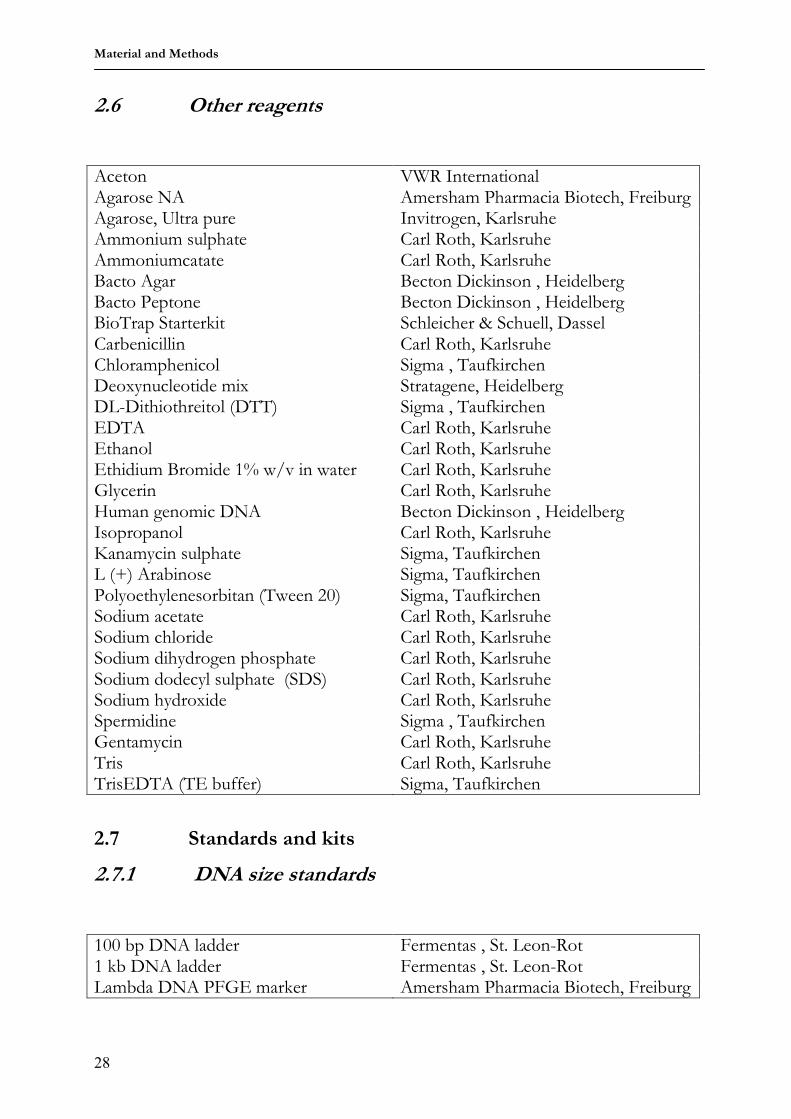
2.3.9 Oligonucleotides for genotyping 26 2.3.10 PCR Oligos for VDJ amplification 26 2.5 Restriction enzymes 27 2.5.2 DNA modifying proteins 27 2.5.3 Antibodies 27 2.5.4 Other protein reagents 27 2.6 Other reagents 2 2.7 Standards and kits 28 2.7.1 DNA size standards 28
Table of contents
ii
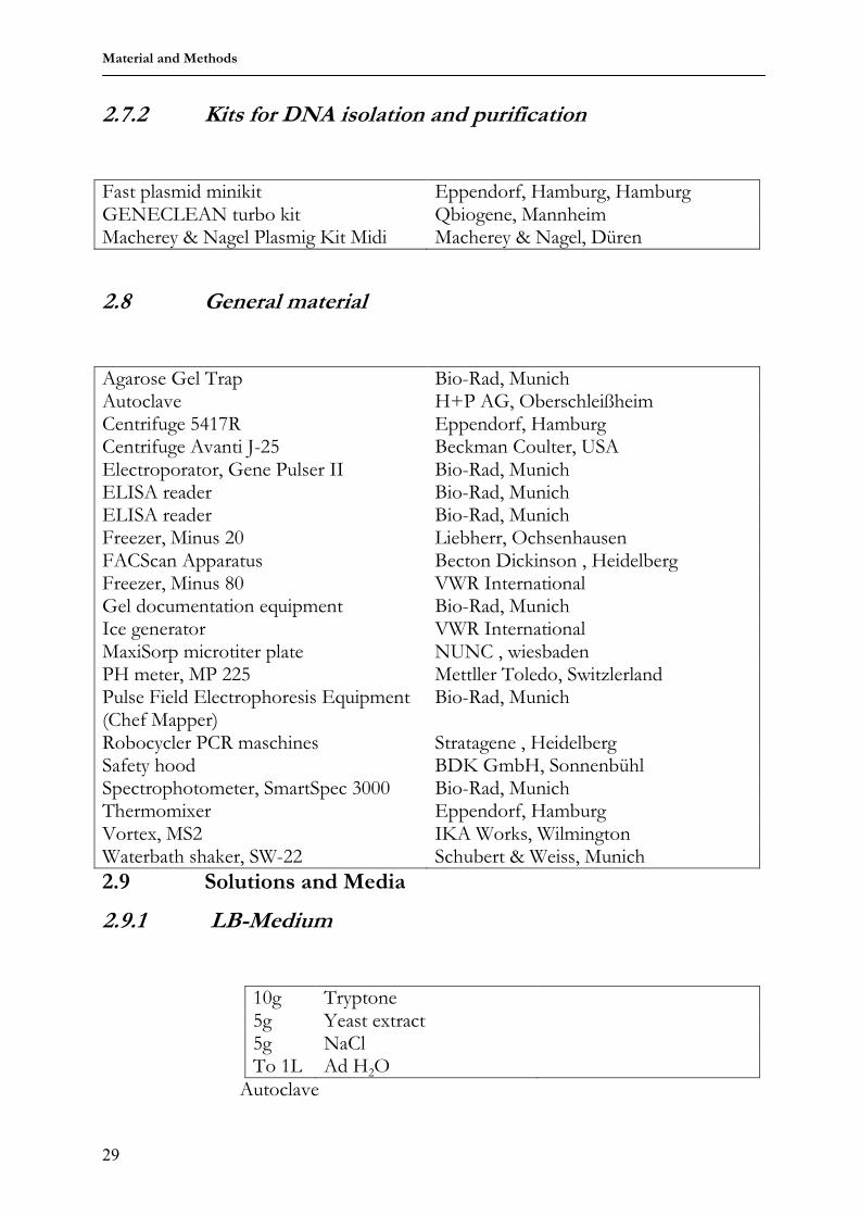
2.7.2 Kits for DNA isolation and purification 29 2.8 General material 29 2.9 Solutions and media 29 2.9.1 LB-Medium 29 2.9.2 Agar-plates 30 2.9.3 ELISA reagents 30 2.9.4 Cutting buffer 30 2.9.5 Antibiotic solutions 30 2.9.6 TAE Buffer 31 2.10 Molecular biology methods 31 2.10.1 Transformation of E.Coli by electroporation 31 2.10.2 Chemical transformation of E.Coli 31 2.10.3 Cloning PCR products 32 2.10.4 Restriction endonuclease digestion of DNA 32 2.10.5 Ethanol precipitation of DNA 33 2.10.6 Dephosphorylation of linearized plasmid DNA by CIP 33 2.10.7 Ligation of DNA fragments 33 2.10.8 Mini-prep: small scale preparation of plasmid DNA 34 2.10.9 Mini-prep: small scale preparation of plasmid DNA 34 2.10.10 Genomic DNA preparation 34 2.10.11 Quantification of DNA solutions 35 2.10.12 Agarose gel electrophoresis 35 2.10.13 Pulse field gel electrophoresis 36 2.10.14 Extraction of DNA fragments from agarose gels 36 2.10.15 Polymerase chain reaction 37 2.10.16 sequencing 37 2.10.17 BAC modification via homologous recombination in E. coli 37 2.11 Transgenic constructs 41 2.11.1 Modifying the pGEM vector 41 2.11.2 Modifying the pBeloBAC II vector 43 2.11.3 Generation of rabbit spacer and human VH plasmid libraries 44 2.11.4 Generation of a chimeric (rabbit spacer I5-6 and human VH
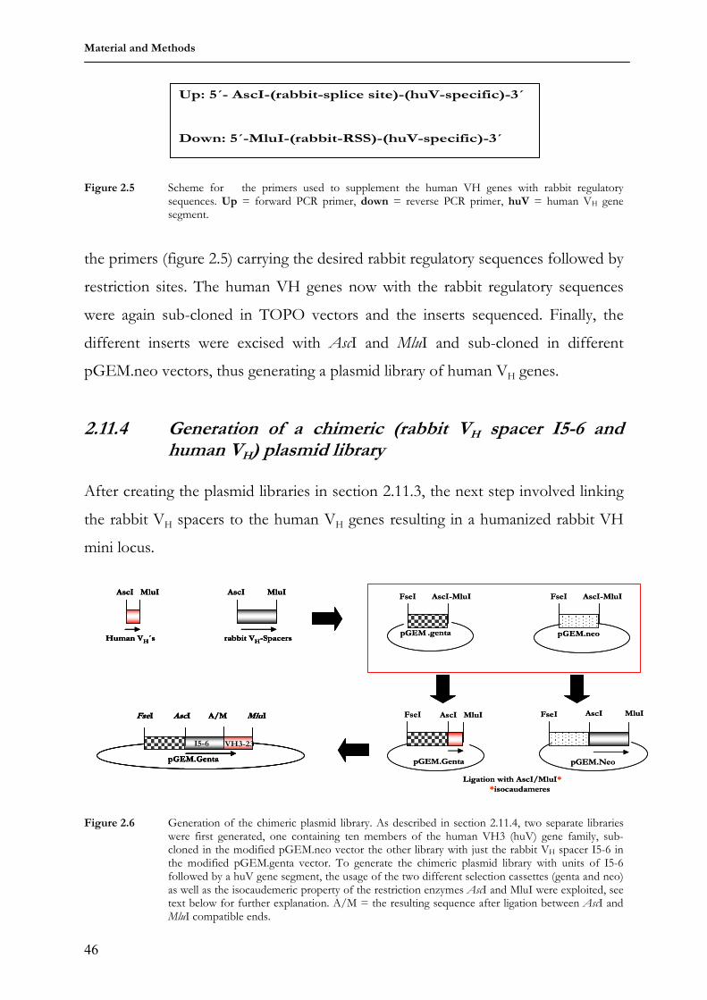
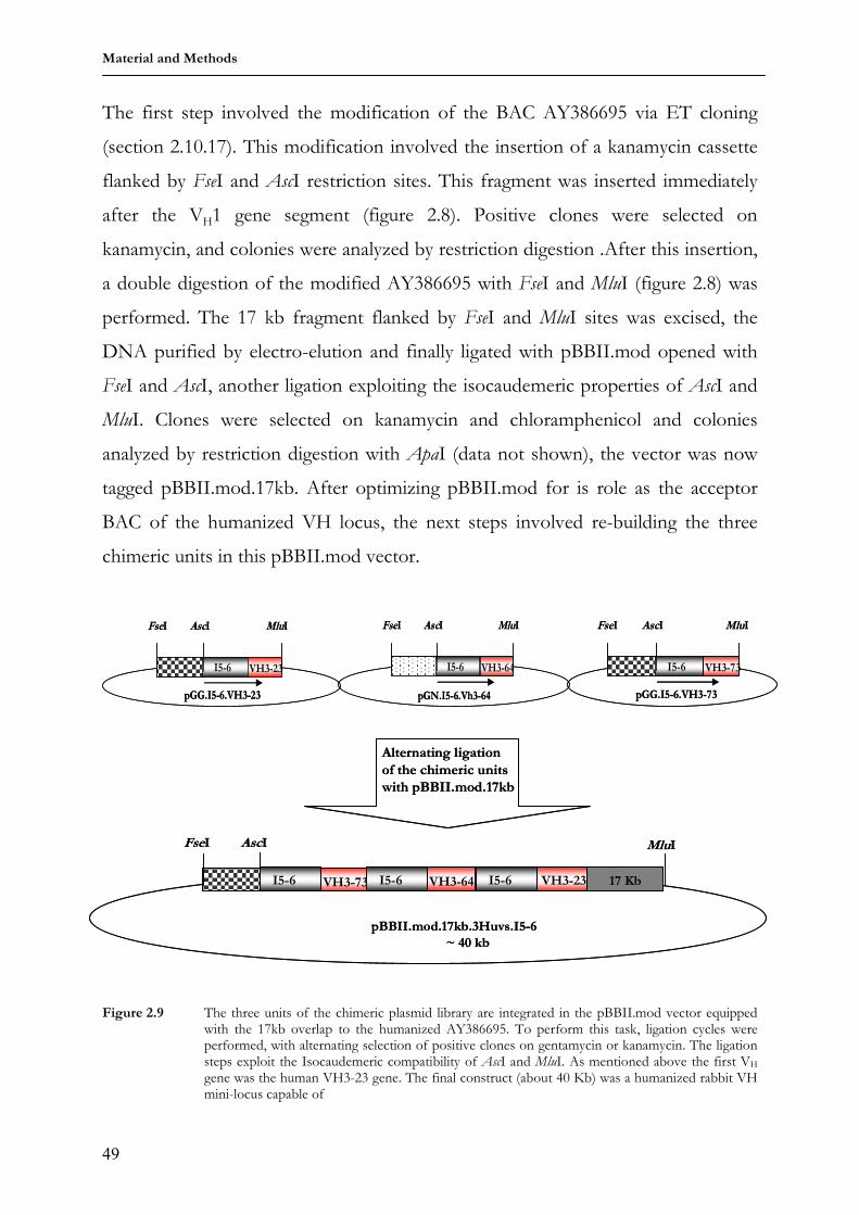
plasmid library 46
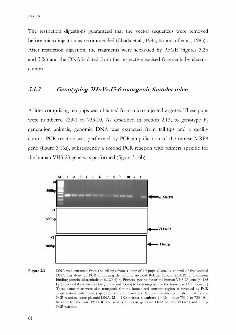
2.11.4 Rebuilding the humanized VH locs in the pBBII.mod vector 48 2.12 Generation of transgenic animals 50 2.13 Animal breeding 51 2.14 Genotyping transgenic animals 51 2.15 Immunization of transgenic animals 52 2.16 Immunohistochemical methods 53 2.16.1 Flow cytometry and cell sorting 53 2.16.2 Enzyme-Linked Immunosorbent Assay (ELISA) 55 3 Results 58 3.1 3HuVs.I5-6 transgenic mice 58 3.1.1 Purification of transgenic constructs for micro-injection 58 3.1.2 Genotyping 3HuVs.I5-6 transgenic founder mice 61
Table of contents
iii
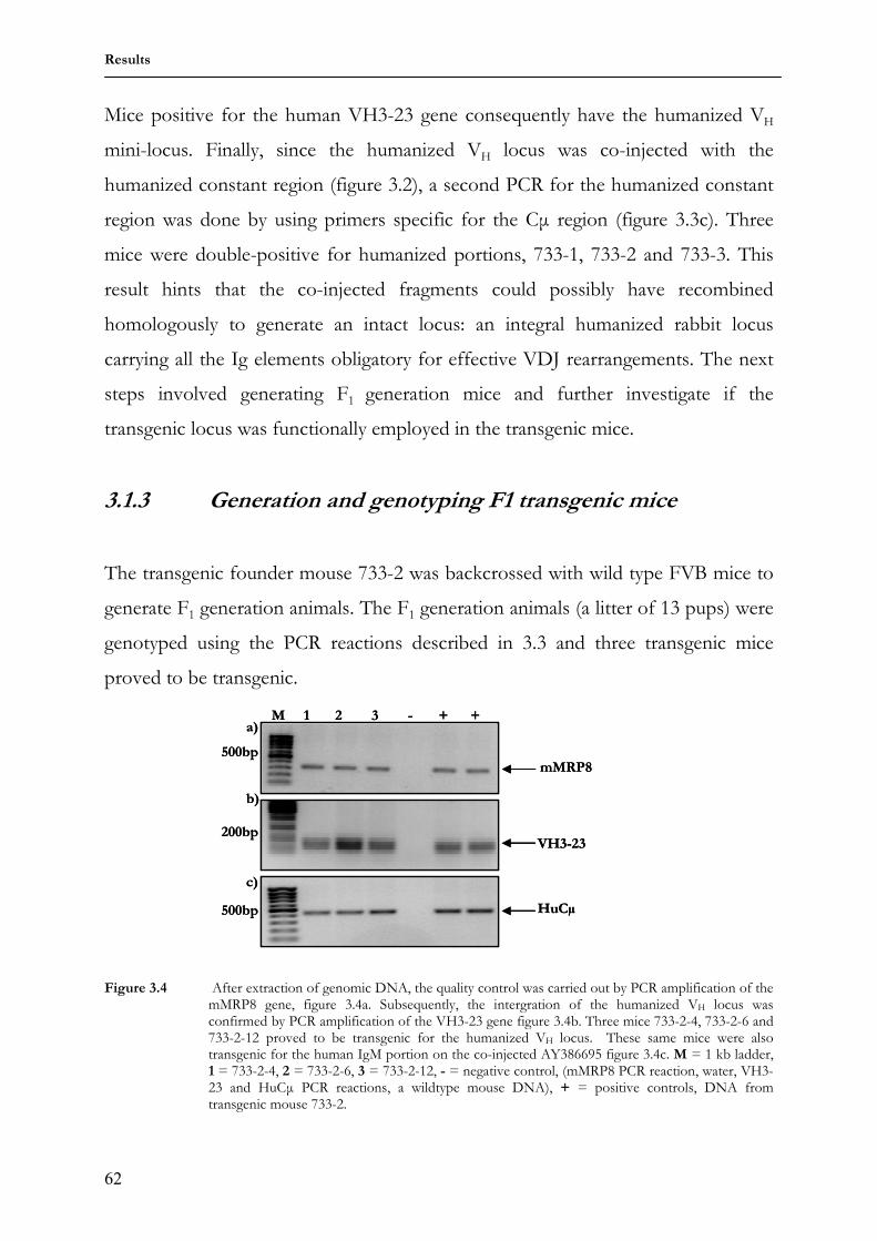
3.1.3 Generation and GenotypingF1 transgenic mice 62 3.1.4 Expression of antibodies carrying human Cµ chains in serum of
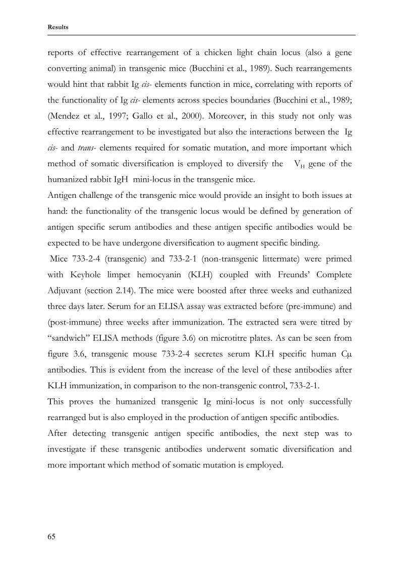
transgenic mouse 733-2-4 63
3.1.5 Expression of antibodies containing human Cµ chains on transgenic B cell surface
64
3.1.6 Transgenic antibody diversity and response to antigen 64 3.2 Comparing somatic diversification of antibodies in mice and
rabbits using a transgenic humanized mini-locus 69
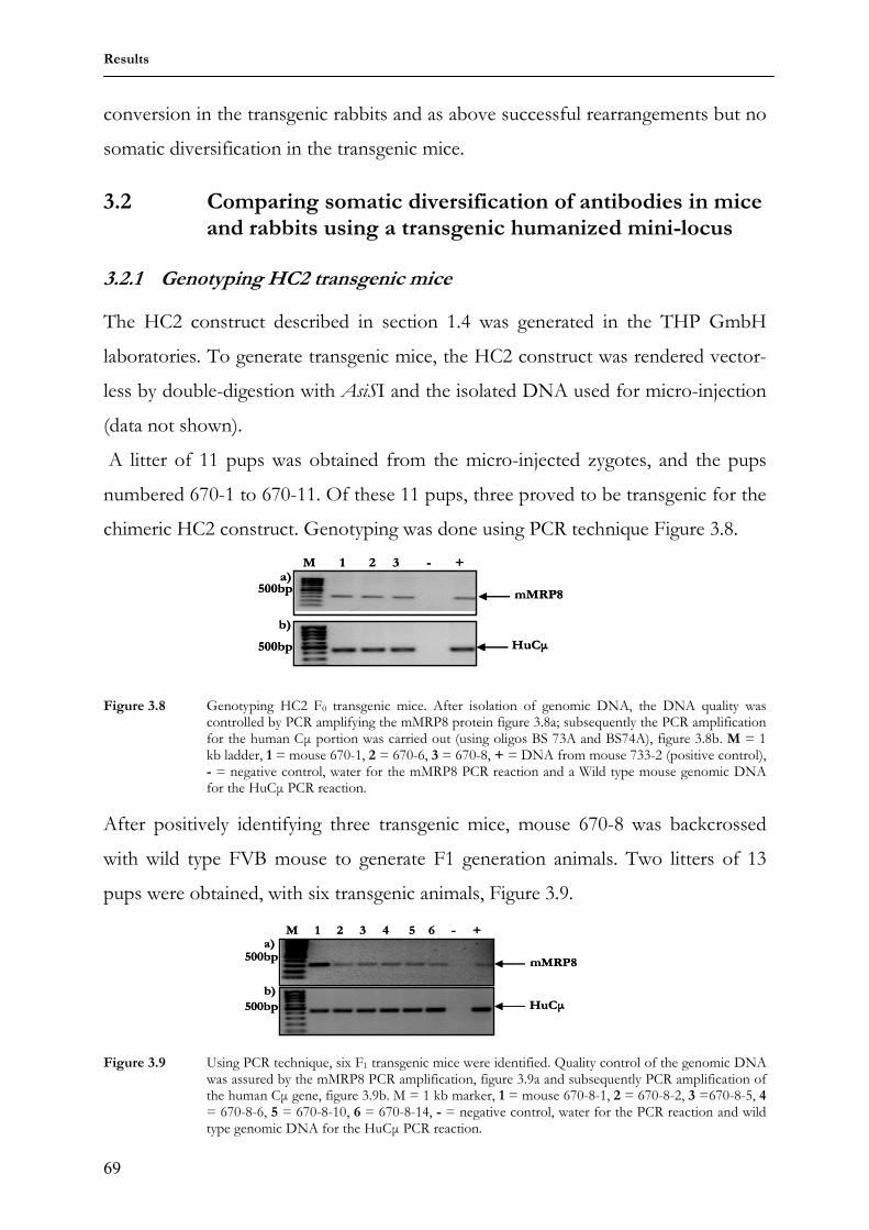
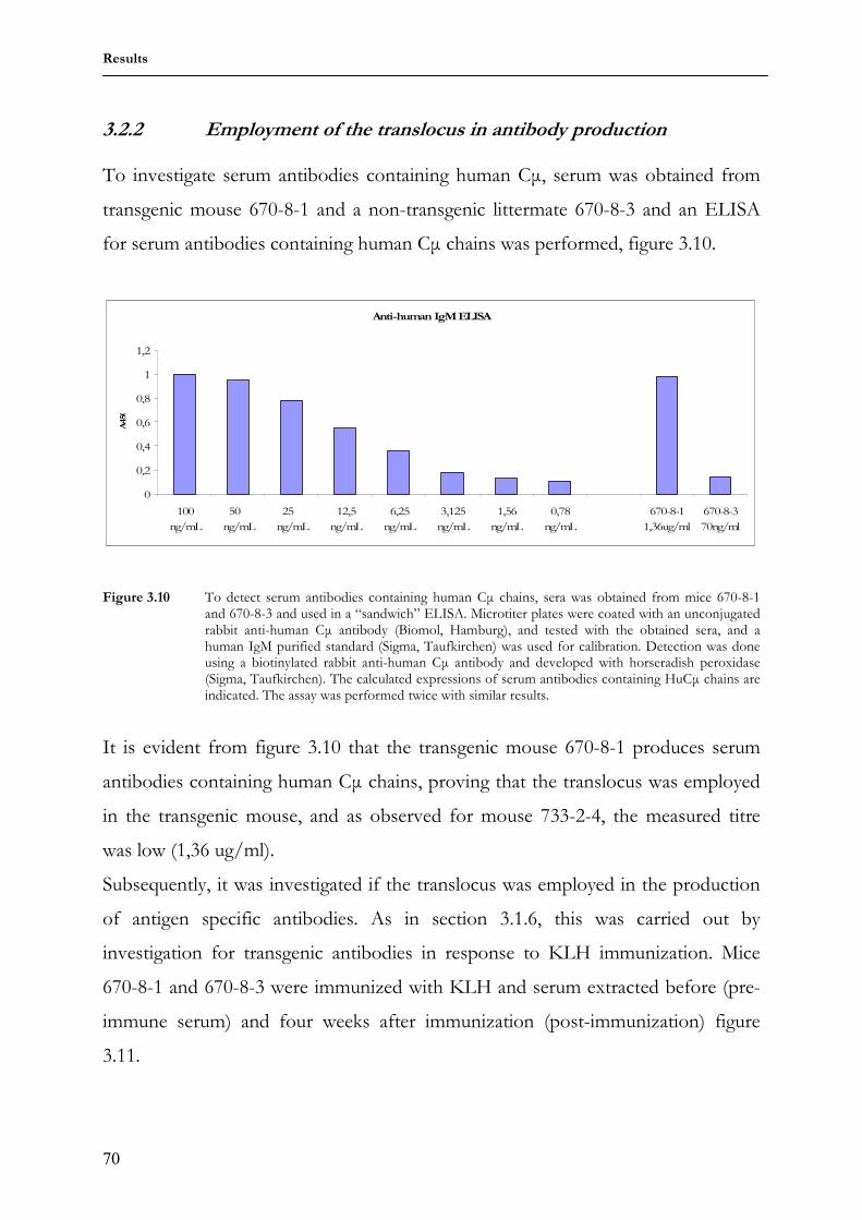
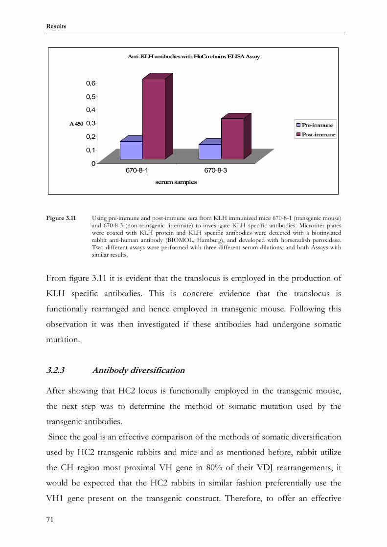
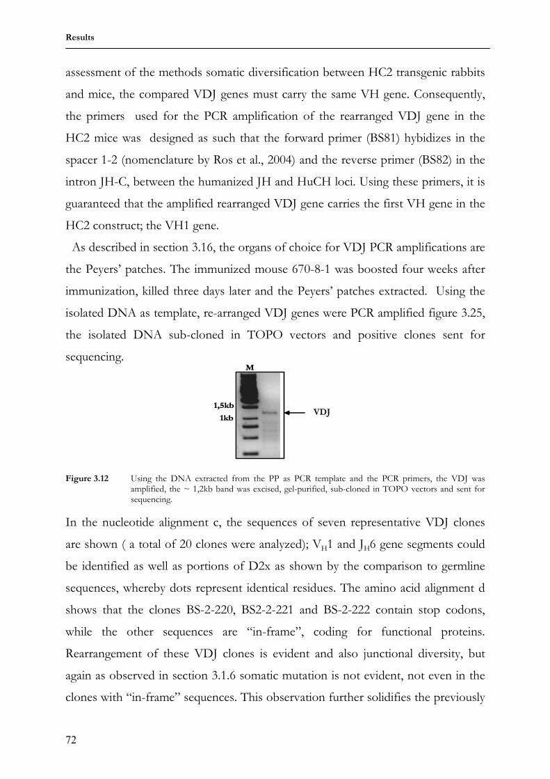
3.2.1 Genotyping HC2 transgenic mice 69 3.2.2 Employment of the translocus in antibody production 70 3.2.3 Antibody diversification 71 3.2.4 Genotyping HC2 transgenic rabbits 74 3.2.5 Employment of the translocus in antibody production 75 3.2.6 Antibody diversification 76 4 Discussion 78 4.1 3HuVs.I5-6 transgenic mice 79 4.1.1 Transgenic integration 79 4.1.2 Transgene expression 79 4.1.2 Somatic diversification of humanized antibodies 80 4.2 HC2 mice 82 4.2.1 Transgenic integration 82 4.2.2 Transgene employment and expression 82 4.2.3 Somatic diversification of the humanized antibodies 83 4.3 HC2 transgenic rabbits 84 4.3.1 Transgenic integration 84 4.3.2 Transgene employment and expression 84 4.3.3 Somatic diversification of the humanized antibodies 85 5 Sequence alignments 88 6 summary 92 7 Reference list 93 Acknowledgements 100 Curriculum vitae 101
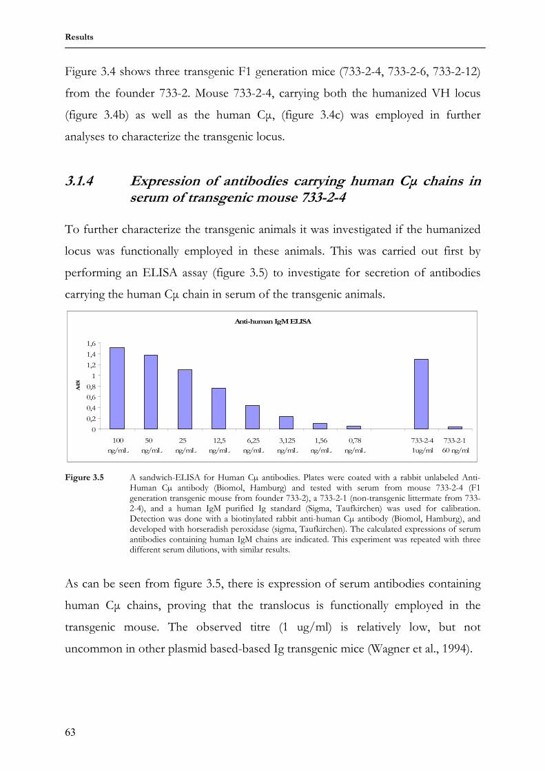
Abbreviations
iv
Abbreviations Ad add ADCC Antibody dependent cellular cytotoxicity AID Activation induced deaminase AID Activation induced deaminase BAC Bacterial artificial chromosome BCR B cell receptor Bp basepair BSA Bovine serum albumin C Constant region CDR Complementary determining region CIP Calf intestinal phosphatase D Diversity region DNA Deoxyribonucleic acid E enhancer E.coli Escherichia coli EDTA Ethylenediaminetetaacetic acid EtBr Ethidium bromide FACs Flourescence-activated cell sorter FR Framework region HuCµ Human C mu chain Ig Immunoglobulin IgH Immunoglobulin heavy chain IgL Immunoglobulin light chain IgV Immunoglobulin V region J Joining region Kb Kilobase kDA Kilo Dalton KLH Keyhole limpet hemocyanin LB Luria broth mMRP8 Murine myeloid related protein Nt nucleotide Oligo oligonucleotide PBS Phosphate buffered saline PCR Polymerase chain reaction PFGE Pulse field gel electrophoresis RAG Recombination activating gene Rpm Rounds per minute RSS Recombination signal sequence SDS Sodium dodecyl sulfate SHM Somatic hypermutation TRIS Trishydroxymethylaminomethan U units V Variable region
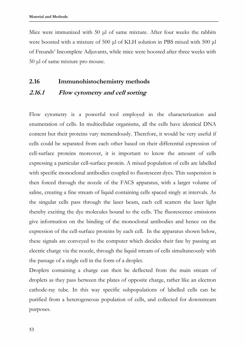
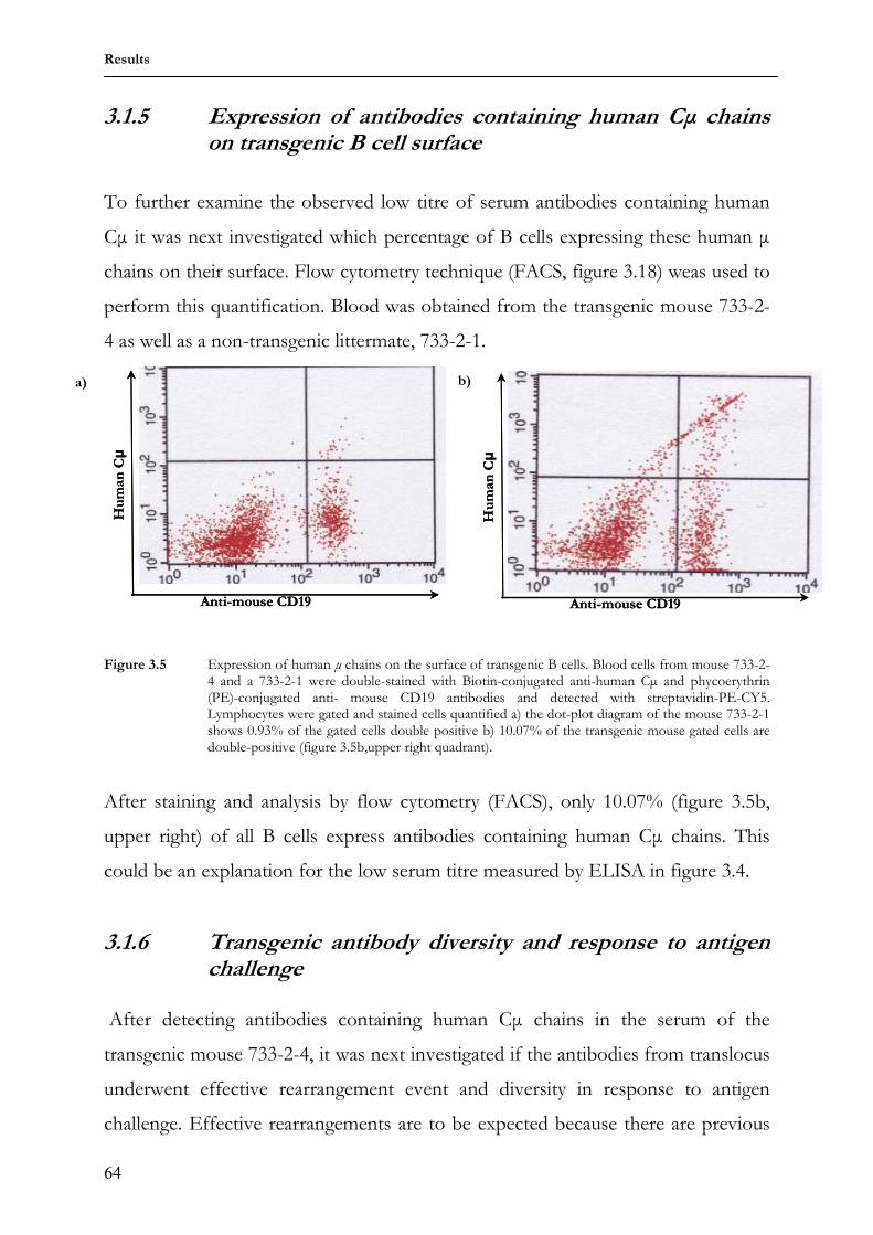
The principal function of the immune system is to protect animals from infectious
organisms and their toxic products. This system has evolved a powerful range of
mechanisms to locate foreign cells, viruses or macromolecules to neutralize these
invading molecules and to eliminate them from the body. This surveillance is
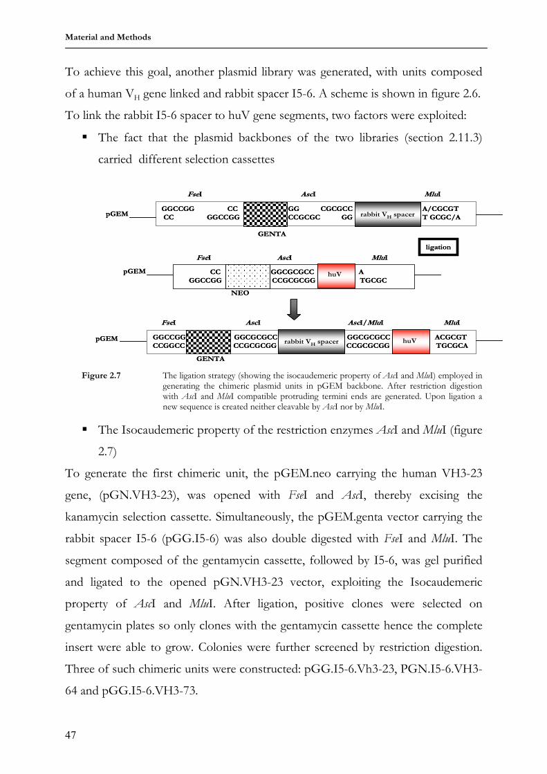
performed by a system of proteins and cells that circulate throughout the body, the
components of which can be divided into two broad categories: the non-adaptive
or innate immunity and the adaptive or acquired immunity (Janeway, Jr., 1992;
Janeway, Jr., 2001).
1.1.1 The innate or non-adaptive immune system
The innate immune system refers to immunity mediated by cells that respond non-
specifically to foreign molecules. These components of the immune system can
distinguish between foreign tissues and organisms but are unable to distinguish a
particular invader. They carry out their functions by recognizing structures which
are shared by most invading micro-organisms, but which are not features of self,
for example specific lipopolysaccharides and high-mannose containing
oligosaccharides. The components of the innate system can be divided into a
cellular and a humoral part. A complex of plasma proteins termed the complement
system makes up the main constituent of the humoral arm of the innate system.
These complement proteins bind to invaders, a process known as opsonization,
thereby marking them for destruction. The cellular arm is composed of
macrophages, whose principal function involves destruction of foreign cells by
phagocytosis. The lacrimal cells, which secrete lysozymes that destroy the outer
surface of many bacteria, are also components of the innate system. Other
Introduction
2
elements of the innate immune system include granulocytes and dendritic cells that
activate and regulate other immune elements through secretion of cytokines and
finally the natural killer cells that destroy foreign cells via cell lysis. Some members
of the innate system activate the adaptive system; dendritic cells for example are
known to act as antigen presenting cells, thereby creating a link between the innate
and the adaptive system. Non-adaptive immunity does not improve with repeated
exposure to the same foreign molecule, thus these systems can easily be evaded by
mutations. It is presumably the inadequacy of innate recognition that led to the
evolution of the adaptive immunity (Janeway, Jr., 1992).
1.1.2 The adaptive or acquired immune system
The adaptive immune system exhibits two major differences to the innate system:
first the adaptive immune system is directed against specific molecules, and second,
its actions are enhanced by re-exposure, a property termed memory, which enables
immunization. Adaptive immunity is mediated by cells called lymphocytes, which
synthesize cell-surface receptors or secrete proteins that bind specifically to foreign
molecules. These secreted proteins are known as antibodies and the specific
molecules they bind to are termed antigens. The immune system contains more
than 109 lymphocytes distributed throughout the body, enabling them to respond
rapidly at any site. Lymphocytes arise continuously from progenitor stem cells- the
hematopoietic cells- in the bone marrow. The bone marrow and thymus represent
the central or primary lymphoid organs. These are sites of lymphocyte generation
and maturation. They then circulate through the blood and lymphatic systems
resting and accumulating in specialized structures called secondary lymphoid
organs, where actual interaction with antigen occurs. These secondary lymphoid
organs include lymph nodes, spleen, tonsils, the Peyer’s patches in the small
intestines and the appendix. There are two lymphocyte sub-populations: B -and T-
cells.
Introduction
3
T-cells arise from the common lymphoid progenitor cell in the bone marrow
but mature in the thymus. The T-cells serve two major functions, first as
coordinators of other acquired immune responses, accomplished by their
production of a wide variety of cytokines and surface cell signals, and secondly in
that they mediate the primary answer to intra-cellular infections. There are two sub-
types of T-cells
• The cytotoxic T-cells or CD8+ cells
• The helper T-cells or CD4+ cells
Each T-cell has a unique surface molecule called a T-cell receptor, which
recognizes short peptides (generated from invading pathogens or viruses),
displayed on the surface of specialized cells termed antigen presenting cells (APC).
The peptide display is usually in conjunction with a membrane glycoprotein
molecule called the major histocompatibility complex (MHC). The different T-cell
sub-types recognize peptides from different cellular locations, presented in
conjunction with different types of MHC molecules.
B-cells are the mediators of humoral immunity. Every B-cell has an
Immunoglobulin (Ig) molecule on its surface, termed the B-cell Receptor (BCR).
The BCR recognizes a unique three-dimensional moiety on an antigen, termed an
epitope. B-cells mature in the bone marrow, and the millions of different B-cells
produced daily proceed to circulate throughout the bloodstream. Since each B-cell
has a different BCR, each will bind to a different substrate. Recognition and
binding of its specific target induces clonal expansion resulting in many B-cells that
recognize the same target. Recognition of a specific target leads to the
differentiation of B-cells to Memory and Plasma cells.
Introduction
4
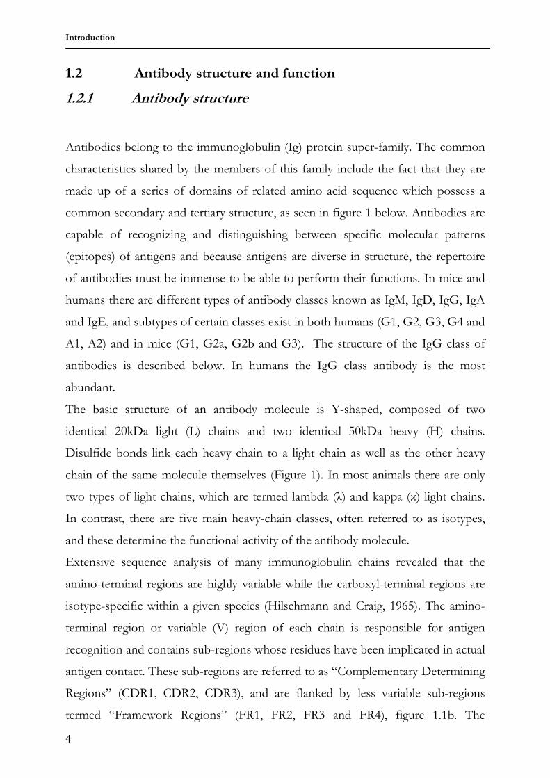
1.2 Antibody structure and function
1.2.1 Antibody structure
Antibodies belong to the immunoglobulin (Ig) protein super-family. The common
characteristics shared by the members of this family include the fact that they are
made up of a series of domains of related amino acid sequence which possess a
common secondary and tertiary structure, as seen in figure 1 below. Antibodies are
capable of recognizing and distinguishing between specific molecular patterns
(epitopes) of antigens and because antigens are diverse in structure, the repertoire
of antibodies must be immense to be able to perform their functions. In mice and
humans there are different types of antibody classes known as IgM, IgD, IgG, IgA
and IgE, and subtypes of certain classes exist in both humans (G1, G2, G3, G4 and
A1, A2) and in mice (G1, G2a, G2b and G3). The structure of the IgG class of
antibodies is described below. In humans the IgG class antibody is the most
abundant.
The basic structure of an antibody molecule is Y-shaped, composed of two
identical 20kDa light (L) chains and two identical 50kDa heavy (H) chains.
Disulfide bonds link each heavy chain to a light chain as well as the other heavy
chain of the same molecule themselves (Figure 1). In most animals there are only
two types of light chains, which are termed lambda (λ) and kappa (κ) light chains.
In contrast, there are five main heavy-chain classes, often referred to as isotypes,
and these determine the functional activity of the antibody molecule.
Extensive sequence analysis of many immunoglobulin chains revealed that the
amino-terminal regions are highly variable while the carboxyl-terminal regions are
isotype-specific within a given species (Hilschmann and Craig, 1965). The amino-
terminal region or variable (V) region of each chain is responsible for antigen
recognition and contains sub-regions whose residues have been implicated in actual
antigen contact. These sub-regions are referred to as “Complementary Determining
Regions” (CDR1, CDR2, CDR3), and are flanked by less variable sub-regions
termed “Framework Regions” (FR1, FR2, FR3 and FR4), figure 1.1b. The
Introduction
5
carboxyl-terminal region of the antibody molecule is made up of one or more
domains which are comparatively constant (C) in sequence and which perform an
array of effector functions.
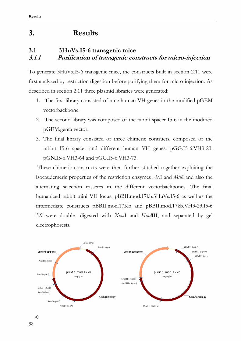
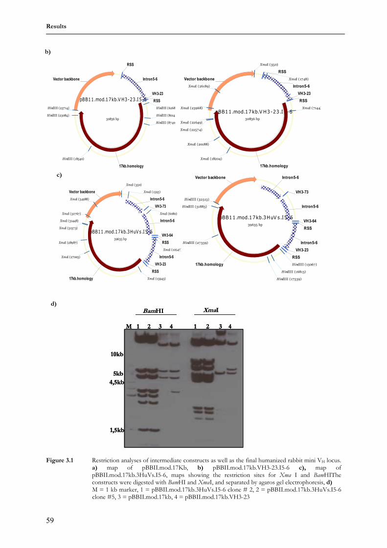
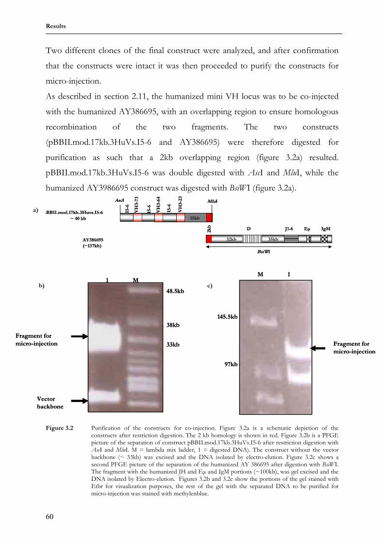
Figure 1.1 Figure 1.1a depicts an antibody molecule (IgG sub-class).Two heavy (H) and two light (L) pairs comprise a single antibody molecule. Within each chain there are two regions described as constant(C) and variable (V) regions. The genes that code for the variable regions in both chains (VH and VL genes) generate the highly diverse binding sites capable of binding almost any type of antigen. The constant and hinge regions of the antibody are encoded by CL, CH1, CH2 and CH3 genes segments. Digestion of an antibody molecule with the protease papain results in three fragments: two identical fragments carrying the antigen-binding activity, called the Fab fragments, for Fragments of antigen binding, the other fragment which carries no antigen-binding activity but crystallizes readily and is termed the Fc fragment (fragment crystallizable) Figure 1.1b depicts the frame work (indicated in red) and the complementary determining regions (indicated in yellow). (figure adapted from Paul, 2003)
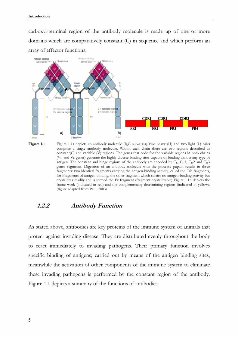
1.2.2 Antibody Function
As stated above, antibodies are key proteins of the immune system of animals that
protect against invading disease. They are distributed evenly throughout the body
to react immediately to invading pathogens. Their primary function involves
specific binding of antigens; carried out by means of the antigen binding sites,
meanwhile the activation of other components of the immune system to eliminate
these invading pathogens is performed by the constant region of the antibody.
Figure 1.1 depicts a summary of the functions of antibodies.
FR1 FR2 FR3 FR4
CDR1 CDR2 CDR3
FR1 FR2 FR3 FR4
CDR1 CDR2 CDR3
a) b)
Introduction
6
Figure 1.2 The functions of antibodies in the elimination of invading pathogens. Antibodies use a multitude
of methods to eradicate invading pathogens. Theses different eradication pathways, a, b, c and d are further elucidated below. (Red spots = bacterial toxins, blue structure = macrophages, reddish-brown structure = natural killer cells, Y-shaped structures = antibodies and the red rectangle = a component of the complement system of proteins.)
Figure 1.2 shows four pathways employed by antibodies in neutralizing antigens
and eliminating pathogens.
a. Antibodies can bind to and effectively neutralize invading bacterial
toxins
b. Also, antibodies complexed with antigens by specific binding a
process known as opsonization can in turn be bound by other
components of the immune system that possess Fc receptors. In this
example the natural killer cells are bound to opsonized bacteria,
resulting in the lysis of the bound bacteria. This is an example of
antibody dependent cellular cytotoxicity (ADCC).
c. Alternatively, opsonized bacteria can be engulfed by macrophages,
scavenger cells of the immune system. Macrophages as well possess
Fc receptors and engulf the opsonized pathogen via phagocytosis.
d. The opsonization of pathogen also leads to recruitment of the
complement system. This is a set of plasma proteins that act together
in a cascade manner to facilitate removal of coated pathogens by
phagocytes or by directly killing them. Here one of the complement
components is activated by oposonization of pathogens.
Bacterial
colony
a
b c
d
Bacterial
colony
a
b c
d neutralization
Opsonization and ADCC
Opsonization and phagocytosis
Opsonization and complement
system
Introduction
7
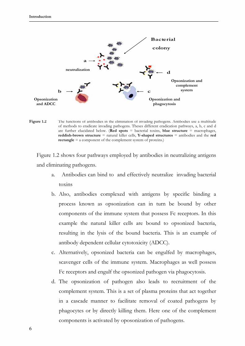
1.3 Creation of antibody diversity
1.3.1 Combinatorial diversity
The mouse was used as a model for the studies involving the creation of antibody
diversity and it was thus observed that in the mouse genome, gene segments coding
for the variable region are present in multiple copies, scattered along a
chromosome (Seidman et al., 1978). This means that during gene rearrangement,
different gene segments can be joined together to form a stretch of DNA that
codes for an entire variable region. The process of gene rearrangement is restricted
to B cells (Dreyer and Bennett, 1965; Hozumi and Tonegawa, 1976; Tonegawa et
al., 1977). Furthermore, different heavy chains can pair with different light chains
to create a functional antibody molecule. This mechanism termed combinatorial
diversity has important consequences:
• It enables a limited number of gene segments to generate an extremely
diverse set of proteins;
• Each cell assembles a different set of gene segments in the formation of
antibody molecules. This results in the expression of a unique antibody in
each cell.
Figure 1.3 Using the example of a mouse heavy chain, the processes leading to the generation of
antibody diversity are outlined. Ig gene segments are shown as red and green blocks. The depicted events are discussed in detail in the ensuing sections. (Figure adapted from Paul, 2003)
Introduction
8
The rearrangement processes involved in the formation of the V region of an
antibody is dependent on the protein products of the recombination activating
genes, RAG-1 and RAG-2 (Seidman et al., 1978; Alt and Baltimore, 1982). These
enzymes belong to a complex of enzymes termed the V (D) J recombinase that play
key roles in the rearrangement processes. Germ-line V and D DNA segments are
followed by a conserved heptamer sequence CACAGTG and a nonamer sequence
ACAAAAACC, separated by an non-conserved spacer sequence of either 12 or 23
base pairs (Lewis and Gellert, 1989; Gearhart and Bogenhagen, 1983). Likewise, all
D and J segments are immediately preceded by a consensus nonamer
GGTTTTTGT and a consensus heptamer CACAGTG. The two consensus
sequences are separated by a short non-conserved spacer sequence of either 12 or
23 base pairs.
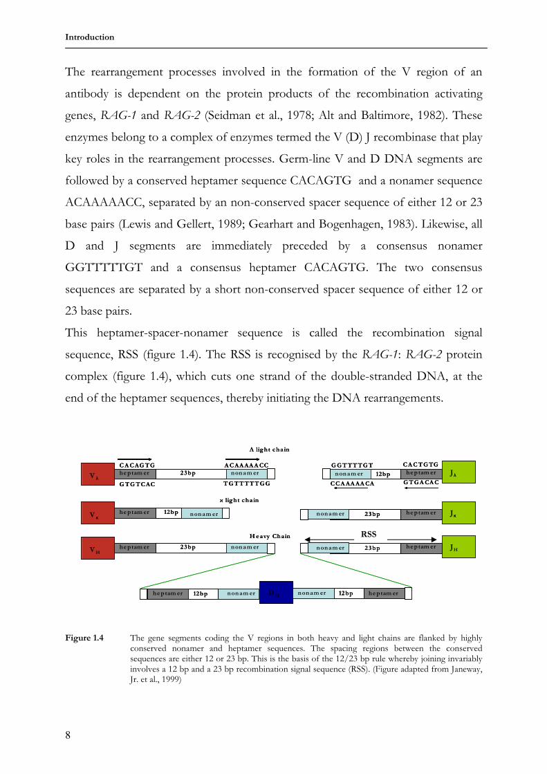
This heptamer-spacer-nonamer sequence is called the recombination signal
sequence, RSS (figure 1.4). The RSS is recognised by the RAG-1: RAG-2 protein
complex (figure 1.4), which cuts one strand of the double-stranded DNA, at the
end of the heptamer sequences, thereby initiating the DNA rearrangements.
Figure 1.4 The gene segments coding the V regions in both heavy and light chains are flanked by highly conserved nonamer and heptamer sequences. The spacing regions between the conserved sequences are either 12 or 23 bp. This is the basis of the 12/23 bp rule whereby joining invariably involves a 12 bp and a 23 bp recombination signal sequence (RSS). (Figure adapted from Janeway, Jr. et al., 1999)
heptam ernonam er 12bp
CACTGTG
GTGACAC
GGTTTTGT
CCAAAAACA
Jλ
heptam er 23bp nonam erVH
Heavy Chain
heptam er
CACAGTG
GTGTCAC
23bp nonam er
ACAAAAACC
TGTTTTTGG
V λ
Λ light cha in
heptam er 12bpV κ
κ light cha in
nonam er heptam ernonam er 23bp Jκ
heptam ernonam er 23bp JH
nonam er 12bpDHnonam er12bpheptam er heptam er
heptam ernonam er 12bp
CACTGTG
GTGACAC
GGTTTTGT
CCAAAAACA
Jλ
heptam er 23bp nonam erVH
Heavy Chain
heptam er
CACAGTG
GTGTCAC
23bp nonam er
ACAAAAACC
TGTTTTTGG
V λ
Λ light cha in
heptam er 12bpV κ
κ light cha in
nonam er heptam ernonam er 23bp Jκheptam ernonam er 23bp Jκ
heptam ernonam er 23bp JH
nonam er 12bpDHnonam er12bpheptam er heptam er
RSS
Introduction
9
Joining always involves a 12 bp spacer to 23 bp spacer, a principle known as the
“12/23 bp rule”. This rule allows joining of the regions in a precise manner, for
example in the heavy chain, two DH regions cannot join with each other nor can a
VH join directly with a JH. In the case of a light chain joining, a VL fragment must be
joined to a JL fragment. Antibody diversity is generated in three main ways, two
(combinatorial and junctional diversity, figure 1.3) of which result as a consequence
of the rearrangement processes by which a functional V region is completed. In
humans and mice, these two processes generate a diversified primary antibody
repertoire, but in birds, rabbits, cows, pigs, sheep, and horses, there is limited
diversity of the primary antibody repertoire, and as described in sections 1.3.5 and
1.3.6, rabbits further diversify their primary repertoire by a process termed gene
conversion. The third process is a mutational process which takes place after the
gene rearrangements. This mutational process which is most often antigen driven
(figure 1.3) is restricted to the rearranged V region (Gearhart and Bogenhagen,
1983) and is elucidated below. The mutational process leads to the generation of
the secondary antibody repertoire.
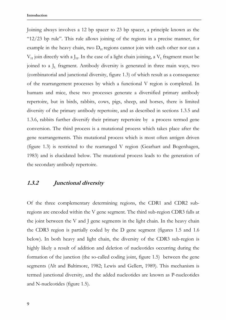
1.3.2 Junctional diversity
Of the three complementary determining regions, the CDR1 and CDR2 sub-
regions are encoded within the V gene segment. The third sub-region CDR3 falls at
the joint between the V and J gene segments in the light chain. In the heavy chain
the CDR3 region is partially coded by the D gene segment (figures 1.5 and 1.6
below). In both heavy and light chain, the diversity of the CDR3 sub-region is
highly likely a result of addition and deletion of nucleotides occurring during the
formation of the junction (the so-called coding joint, figure 1.5) between the gene
segments (Alt and Baltimore, 1982; Lewis and Gellert, 1989). This mechanism is
termed junctional diversity, and the added nucleotides are known as P-nucleotides
and N-nucleotides (figure 1.5).
Introduction
10
Figure 1.5 Generation of junctional diversity between the VH and DJH genes. The RAG protein complex
(composed of two proteins RAG-1 and RAG-2) recognise, bind and cleave the RSS motifs. The segments to be recombined are then brought by the RAG protein complex and the DNA strands the end of gene segments are cleaved by the RAG proteins to form a hairpin, while the ends of the RSS motifs are retained as a double strand break. The rest of the RSS are then joined exactly (signal joint) to form a loop of DNA which plays no further role in the ensuing processes. The double-strand break the end the end of the RSS motifs is the repaired by cell machinery and in this process, the palindromic sequences (P nucleotides) are generated. The resulting joint between the VH and the DJH genes is called the coding joint, and is further diversified by erratic addition of nucleotides (N nucleotides), a process regulated by the enzyme Terminal Deoxynucleotidyl Transferase , TDT (Kunkel et al., 1986; Snow et al., 1987).
1.3.3 Generation of the light chain
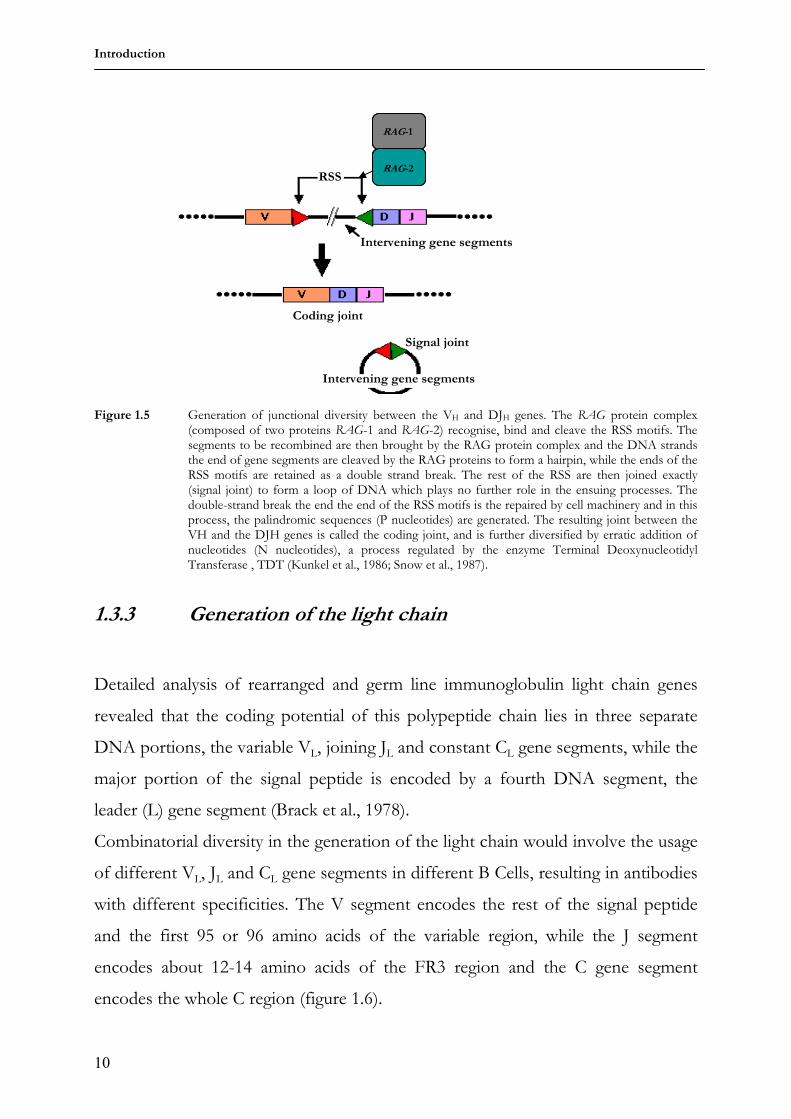
Detailed analysis of rearranged and germ line immunoglobulin light chain genes
revealed that the coding potential of this polypeptide chain lies in three separate
DNA portions, the variable VL, joining JL and constant CL gene segments, while the
major portion of the signal peptide is encoded by a fourth DNA segment, the
leader (L) gene segment (Brack et al., 1978).
Combinatorial diversity in the generation of the light chain would involve the usage
of different VL, JL and CL gene segments in different B Cells, resulting in antibodies
with different specificities. The V segment encodes the rest of the signal peptide
and the first 95 or 96 amino acids of the variable region, while the J segment
encodes about 12-14 amino acids of the FR3 region and the C gene segment
encodes the whole C region (figure 1.6).
RSS
Intervening gene segments
Coding joint
Signal joint
Intervening gene segments
RAG-2
RAG-1
RSS
Intervening gene segments
Coding joint
Signal joint
Intervening gene segments
RSS
Intervening gene segments
Coding joint
Signal joint
Intervening gene segments
RAG-2
RAG-1
RAG-2
RAG-1
Introduction
11
Figure 1.6 Rearrangement events leading to the creation of a light chain. The V segment of the light chain is composed from two segments: a variable (V) and a joining (J) region. These segments which are separated on the genomic DNA level are brought together by somatic recombination to form the functional light chain V region. The C region is encoded in a separate exon, and then is joined to the rearranged VJ segment as a result of splicing, which excises the introns between the leader (L) and the VJ and the VJ and the C segments. The leader directs the nascent protein into the cells secretory pathway. (Figure adapted from Janeway, Jr. et al., 1999)
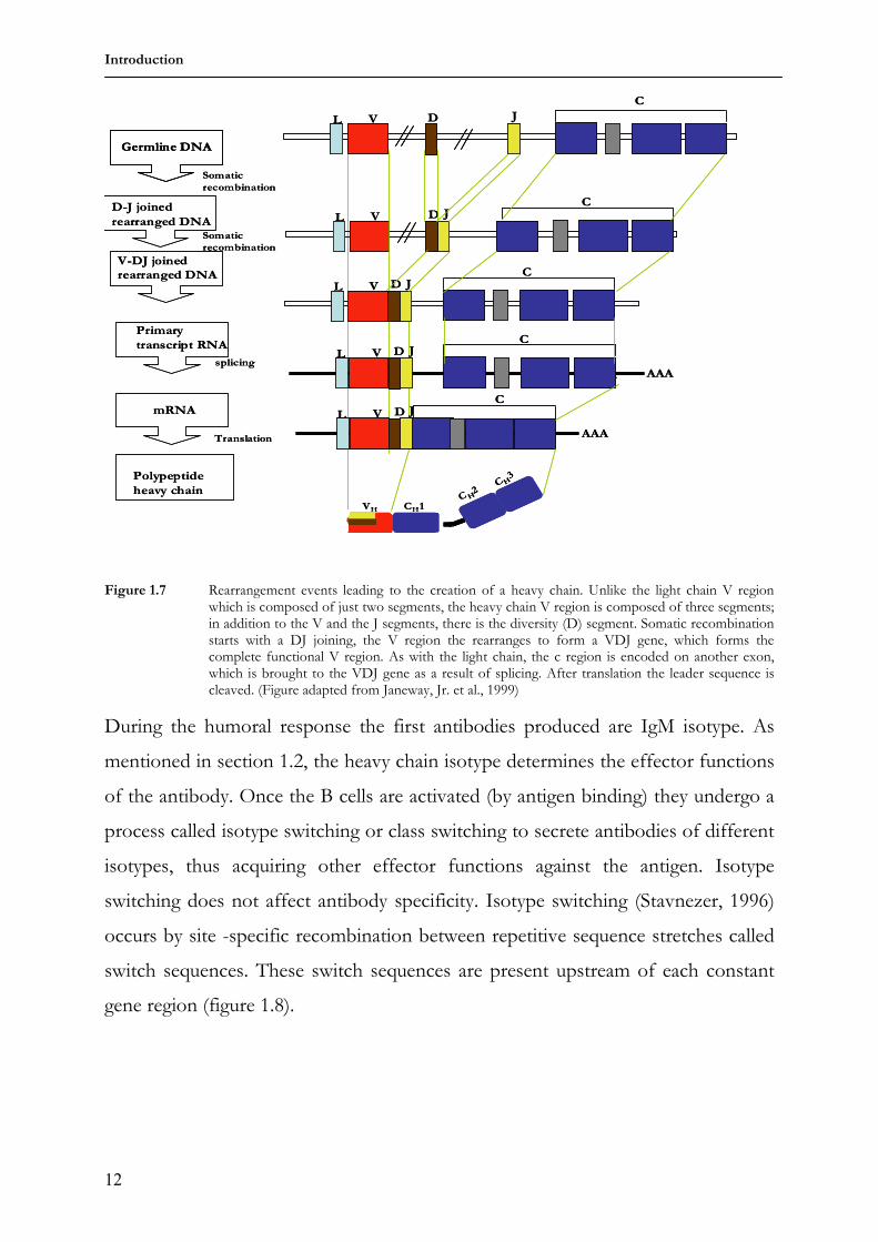
1.3.4 Generation of the heavy chain
The heavy chain on the other hand is encoded by four separate gene segments. In
addition to the VH, JH and CH segments, there is the DH segment (diversity
segment), which encodes the core portion of the CDR3. The process of
recombination producing a functional heavy chain occurs in two separate stages:
first the DH segment is joined to the JH segment, followed by recombination of a
VH segment to the DJH to complete the heavy chain variable region. Same as with
the light chain, the VDJ segment is joined to the constant-domain coding segment
by RNA splicing (figure 1.7). Combinatorial diversity is more pronounced in the
heavy chain because different D gene segments can be used in addition to the
different V and J segments.
JVL C
L V J C
CL V J
AAA
L V J
AAAC
Germline DNA
Somatic
recombination
V-J joinedrearranged DNA
Primary
transcript RNA
splicing
mRNA
Translation
Polypeptide
light chain
Transcription
JVL CJVL C
L V J CL V J C
CL V J
AAA
L V JL V J
AAA
L V JL V J
AAAC
Germline DNAGermline DNA
Somatic
recombination
V-J joinedrearranged DNA
Primary
transcript RNA
splicing
mRNA
Translation
Polypeptide
light chain
Transcription
VL CL
Introduction
12
Figure 1.7 Rearrangement events leading to the creation of a heavy chain. Unlike the light chain V region which is composed of just two segments, the heavy chain V region is composed of three segments; in addition to the V and the J segments, there is the diversity (D) segment. Somatic recombination starts with a DJ joining, the V region the rearranges to form a VDJ gene, which forms the complete functional V region. As with the light chain, the c region is encoded on another exon, which is brought to the VDJ gene as a result of splicing. After translation the leader sequence is cleaved. (Figure adapted from Janeway, Jr. et al., 1999)
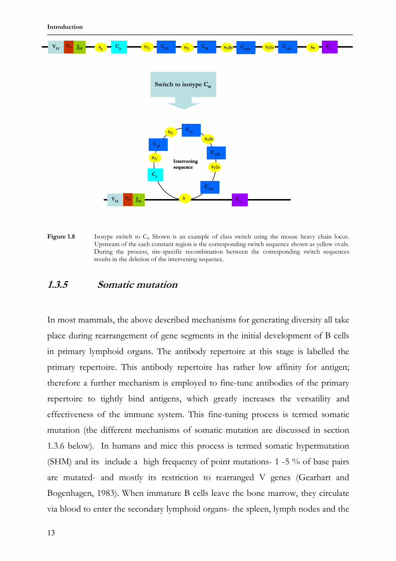
During the humoral response the first antibodies produced are IgM isotype. As
mentioned in section 1.2, the heavy chain isotype determines the effector functions
of the antibody. Once the B cells are activated (by antigen binding) they undergo a
process called isotype switching or class switching to secrete antibodies of different
isotypes, thus acquiring other effector functions against the antigen. Isotype
switching does not affect antibody specificity. Isotype switching (Stavnezer, 1996)
occurs by site -specific recombination between repetitive sequence stretches called
switch sequences. These switch sequences are present upstream of each constant
gene region (figure 1.8).
JVL
C
AAA
D
JVL
CD
JVL
CD
JVL
CD
AAA
JVL
CD
AAA
VH CH1C H
2CH
3
JVL
C
AAA
D
JVL
CD
JVL
CD
JVL
CD
AAA
JVL
CD
AAA
VH CH1C H
2CH
3
Germline DNA
Somatic
recombination
D-J joined
rearranged DNASomaticrecombination
V-DJ joinedrearranged DNA
Primary
transcript RNA
splicing
mRNA
Translation
Polypeptide
heavy chain
Germline DNAGermline DNA
Somatic
recombination
D-J joined
rearranged DNASomaticrecombination
V-DJ joinedrearranged DNA
Primary
transcript RNA
splicing
mRNA
Translation
Polypeptide
heavy chain
Introduction
13
Figure 1.8 Isotype switch to Cα. Shown is an example of class switch using the mouse heavy chain locus.
Upstream of the each constant region is the corresponding switch sequence shown as yellow ovals. During the process, site-specific recombination between the corresponding switch sequences results in the deletion of the intervening sequence.
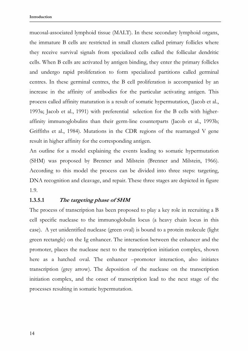
1.3.5 Somatic mutation
In most mammals, the above described mechanisms for generating diversity all take
place during rearrangement of gene segments in the initial development of B cells
in primary lymphoid organs. The antibody repertoire at this stage is labelled the
primary repertoire. This antibody repertoire has rather low affinity for antigen;
therefore a further mechanism is employed to fine-tune antibodies of the primary
repertoire to tightly bind antigens, which greatly increases the versatility and
effectiveness of the immune system. This fine-tuning process is termed somatic
mutation (the different mechanisms of somatic mutation are discussed in section
1.3.6 below). In humans and mice this process is termed somatic hypermutation
(SHM) and its include a high frequency of point mutations- 1 -5 % of base pairs
are mutated- and mostly its restriction to rearranged V genes (Gearhart and
Bogenhagen, 1983). When immature B cells leave the bone marrow, they circulate
via blood to enter the secondary lymphoid organs- the spleen, lymph nodes and the
mucosal-associated lymphoid tissue (MALT). In these secondary lymphoid organs,
the immature B cells are restricted in small clusters called primary follicles where
they receive survival signals from specialized cells called the follicular dendritic
cells. When B cells are activated by antigen binding, they enter the primary follicles
and undergo rapid proliferation to form specialized partitions called germinal
centres. In these germinal centres, the B cell proliferation is accompanied by an
increase in the affinity of antibodies for the particular activating antigen. This
process called affinity maturation is a result of somatic hypermutation, (Jacob et al.,
1993a; Jacob et al., 1991) with preferential selection for the B cells with higher-
affinity immunoglobulins than their germ-line counterparts (Jacob et al., 1993b;
Griffiths et al., 1984). Mutations in the CDR regions of the rearranged V gene
result in higher affinity for the corresponding antigen.
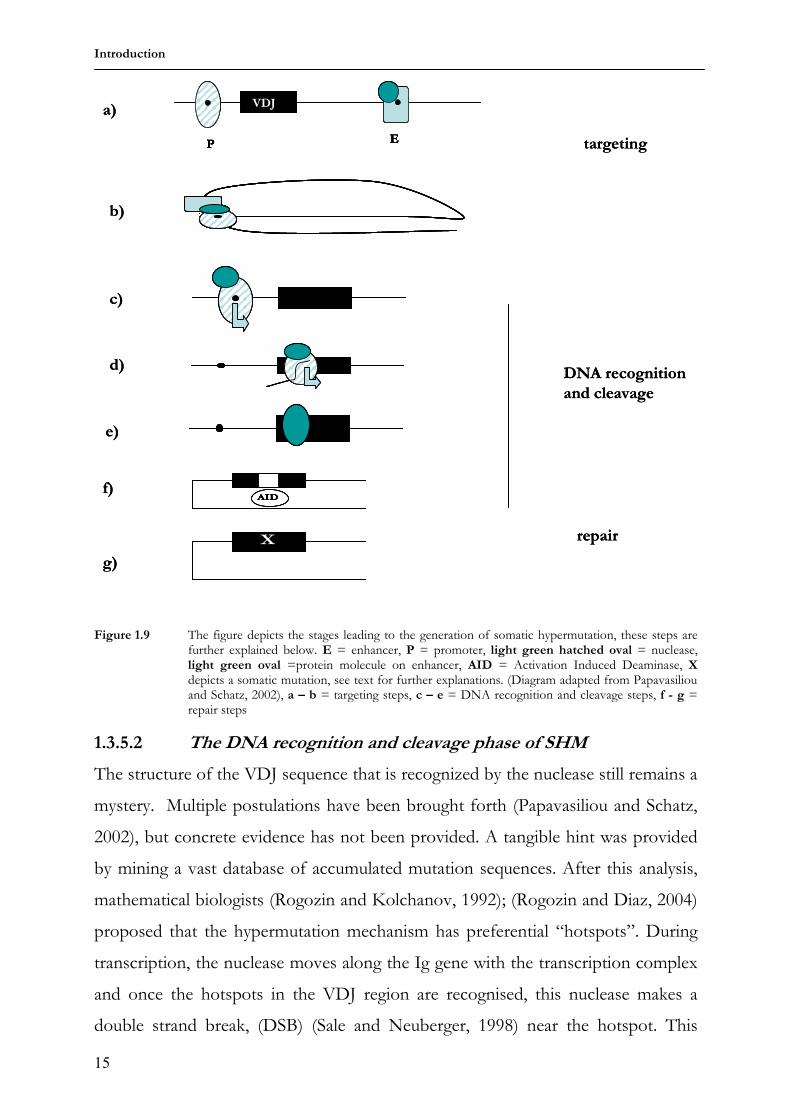
An outline for a model explaining the events leading to somatic hypermutation
(SHM) was proposed by Brenner and Milstein (Brenner and Milstein, 1966).
According to this model the process can be divided into three steps: targeting,
DNA recognition and cleavage, and repair. These three stages are depicted in figure
1.9.
1.3.5.1 The targeting phase of SHM
The process of transcription has been proposed to play a key role in recruiting a B
cell specific nuclease to the immunoglobulin locus (a heavy chain locus in this
case). A yet unidentified nuclease (green oval) is bound to a protein molecule (light
green rectangle) on the Ig enhancer. The interaction between the enhancer and the
promoter, places the nuclease next to the transcription initiation complex, shown
here as a hatched oval. The enhancer –promoter interaction, also initiates
transcription (grey arrow). The deposition of the nuclease on the transcription
initiation complex, and the onset of transcription lead to the next stage of the
processes resulting in somatic hypermutation.
Introduction
15
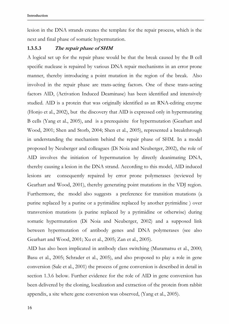
Figure 1.9 The figure depicts the stages leading to the generation of somatic hypermutation, these steps are further explained below. E = enhancer, P = promoter, light green hatched oval = nuclease, light green oval =protein molecule on enhancer, AID = Activation Induced Deaminase, X depicts a somatic mutation, see text for further explanations. (Diagram adapted from Papavasiliou and Schatz, 2002), a – b = targeting steps, c – e = DNA recognition and cleavage steps, f - g = repair steps
1.3.5.2 The DNA recognition and cleavage phase of SHM
The structure of the VDJ sequence that is recognized by the nuclease still remains a
mystery. Multiple postulations have been brought forth (Papavasiliou and Schatz,
2002), but concrete evidence has not been provided. A tangible hint was provided
by mining a vast database of accumulated mutation sequences. After this analysis,
mathematical biologists (Rogozin and Kolchanov, 1992); (Rogozin and Diaz, 2004)
proposed that the hypermutation mechanism has preferential “hotspots”. During
transcription, the nuclease moves along the Ig gene with the transcription complex
and once the hotspots in the VDJ region are recognised, this nuclease makes a
double strand break, (DSB) (Sale and Neuberger, 1998) near the hotspot. This
P
E
E
VDJ
P
EE
E
VDJ
XX repair
DNA recognition
and cleavage
targeting
a)
b)
c)
d)
e)
f)
g)
AIDAID
P
E
E
VDJ
P
EE
E
VDJ
XX repair
DNA recognition
and cleavage
targeting
a)
b)
c)
d)
e)
f)
g)
AIDAID
Introduction
16
lesion in the DNA strands creates the template for the repair process, which is the
next and final phase of somatic hypermutation.
1.3.5.3 The repair phase of SHM
A logical set up for the repair phase would be that the break caused by the B cell
specific nuclease is repaired by various DNA repair mechanisms in an error prone
manner, thereby introducing a point mutation in the region of the break. Also
involved in the repair phase are trans-acting factors. One of these trans-acting
factors AID, (Activation Induced Deaminase) has been identified and intensively
studied. AID is a protein that was originally identified as an RNA-editing enzyme
(Honjo et al., 2002), but the discovery that AID is expressed only in hypermutating
B cells (Yang et al., 2005), and is a prerequisite for hypermutation (Gearhart and
Wood, 2001; Shen and Storb, 2004; Shen et al., 2005), represented a breakthrough
in understanding the mechanism behind the repair phase of SHM. In a model
proposed by Neuberger and colleagues (Di Noia and Neuberger, 2002), the role of
AID involves the initiation of hypermutation by directly deanimating DNA,
thereby causing a lesion in the DNA strand. According to this model, AID induced
lesions are consequently repaired by error prone polymerases (reviewed by
Gearhart and Wood, 2001), thereby generating point mutations in the VDJ region.
Furthermore, the model also suggests a preference for transition mutations (a
purine replaced by a purine or a pyrimidine replaced by another pyrimidine ) over
transversion mutations (a purine replaced by a pyrimidine or otherwise) during
somatic hypermutation (Di Noia and Neuberger, 2002) and a supposed link
between hypermutation of antibody genes and DNA polymerases (see also
Gearhart and Wood, 2001; Xu et al., 2005; Zan et al., 2005).
AID has also been implicated in antibody class switching (Muramatsu et al., 2000;
Basu et al., 2005; Schrader et al., 2005), and also proposed to play a role in gene
conversion (Sale et al., 2001) the process of gene conversion is described in detail in
section 1.3.6 below. Further evidence for the role of AID in gene conversion has
been delivered by the cloning, localization and extraction of the protein from rabbit
appendix, a site where gene conversion was observed, (Yang et al., 2005).
Introduction
17
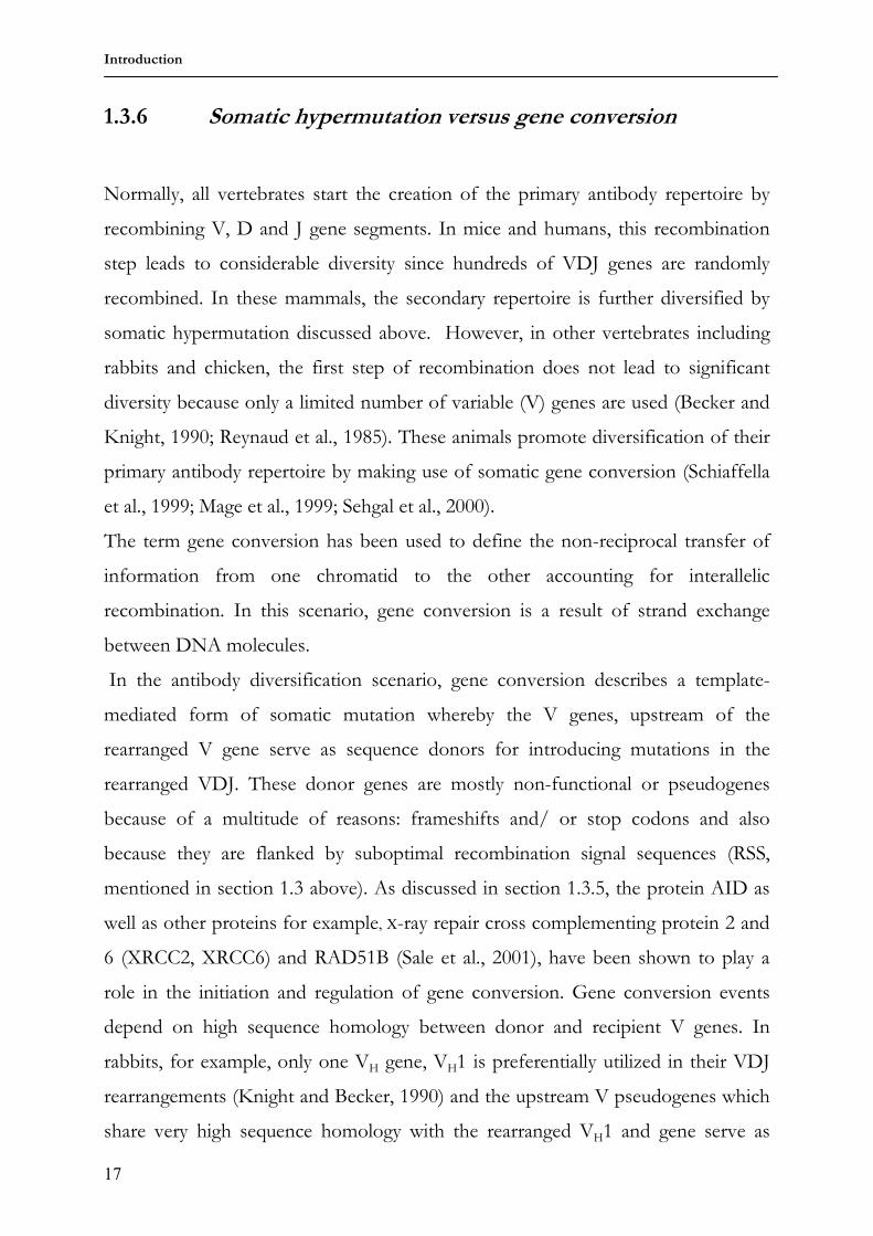
1.3.6 Somatic hypermutation versus gene conversion
Normally, all vertebrates start the creation of the primary antibody repertoire by
recombining V, D and J gene segments. In mice and humans, this recombination
step leads to considerable diversity since hundreds of VDJ genes are randomly
recombined. In these mammals, the secondary repertoire is further diversified by
somatic hypermutation discussed above. However, in other vertebrates including
rabbits and chicken, the first step of recombination does not lead to significant
diversity because only a limited number of variable (V) genes are used (Becker and
Knight, 1990; Reynaud et al., 1985). These animals promote diversification of their
primary antibody repertoire by making use of somatic gene conversion (Schiaffella
et al., 1999; Mage et al., 1999; Sehgal et al., 2000).
The term gene conversion has been used to define the non-reciprocal transfer of
information from one chromatid to the other accounting for interallelic
recombination. In this scenario, gene conversion is a result of strand exchange
between DNA molecules.
In the antibody diversification scenario, gene conversion describes a template-
mediated form of somatic mutation whereby the V genes, upstream of the
rearranged V gene serve as sequence donors for introducing mutations in the
rearranged VDJ. These donor genes are mostly non-functional or pseudogenes
because of a multitude of reasons: frameshifts and/ or stop codons and also
because they are flanked by suboptimal recombination signal sequences (RSS,
mentioned in section 1.3 above). As discussed in section 1.3.5, the protein AID as
well as other proteins for example, X-ray repair cross complementing protein 2 and
6 (XRCC2, XRCC6) and RAD51B (Sale et al., 2001), have been shown to play a
role in the initiation and regulation of gene conversion. Gene conversion events
depend on high sequence homology between donor and recipient V genes. In
rabbits, for example, only one VH gene, VH1 is preferentially utilized in their VDJ
rearrangements (Knight and Becker, 1990) and the upstream V pseudogenes which
share very high sequence homology with the rearranged VH1 and gene serve as
Introduction
18
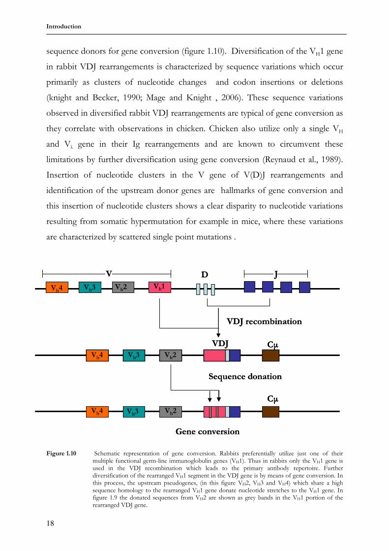
sequence donors for gene conversion (figure 1.10). Diversification of the VH1 gene
in rabbit VDJ rearrangements is characterized by sequence variations which occur
primarily as clusters of nucleotide changes and codon insertions or deletions
(knight and Becker, 1990; Mage and Knight , 2006). These sequence variations
observed in diversified rabbit VDJ rearrangements are typical of gene conversion as
they correlate with observations in chicken. Chicken also utilize only a single VH
and Vλ gene in their Ig rearrangements and are known to circumvent these
limitations by further diversification using gene conversion (Reynaud et al., 1989).
Insertion of nucleotide clusters in the V gene of V(D)J rearrangements and
identification of the upstream donor genes are hallmarks of gene conversion and
this insertion of nucleotide clusters shows a clear disparity to nucleotide variations
resulting from somatic hypermutation for example in mice, where these variations
are characterized by scattered single point mutations .
Figure 1.10 Schematic representation of gene conversion. Rabbits preferentially utilize just one of their
multiple functional germ-line immunoglobulin genes (VH1). Thus in rabbits only the VH1 gene is used in the VDJ recombination which leads to the primary antibody repertoire. Further diversification of the rearranged VH1 segment in the VDJ gene is by means of gene conversion. In this process, the upstream pseudogenes, (in this figure VH2, VH3 and VH4) which share a high sequence homology to the rearranged VH1 gene donate nucleotide stretches to the VH1 gene. In figure 1.9 the donated sequences from VH2 are shown as grey bands in the VH1 portion of the rearranged VDJ gene.
Vh1Vh2Vh3Vh4
V D J
VDJ CµVh2Vh3Vh4
Gene conversion
Cµ
Vh4 Vh3 Vh2
VDJ recombination
Sequence donation
Vh1Vh2Vh3Vh4
V D J
VDJ CµVh2Vh3Vh4
Gene conversion
Cµ
Vh4 Vh3 Vh2
VDJ recombination
Sequence donation
Introduction
19
In addition to irrevocable evidence of the employment of gene conversion in the
diversification of the V gene in V (D) J rearrangements, mutations in the D, J and
also in the region immediately 3’ of the JH region have been observed. These
sequence variations were attributed to somatic hypermutation because the D and J
sequences lack potential sequence donors (Crane et al., 1996; Sehgal et al., 2000).
The observation of diversification in these regions makes it likely that somatic
hypermutation also occurs in the rearranged VH gene segments, (Winstead et al.,
1999; Lanning and Knight, 1997). It can thus be concluded that rabbits employ
both gene conversion and hypermutation in the diversification of V(D)J genes.
1.4 Investigating somatic mutation
Intense research is ongoing to elucidate the mechanisms underlying somatic
mutation and considerable progress has been made as some of the cis- and trans-
elements regulating Ig rearrangement and somatic mutation have been identified.
The cis-elements include transcriptional enhancers and promoters while the trans-
elements include the proteins AID, XRCC2, XRCC6 and RAD51B (reviewed in
Maizels, 2005; Jung et al., 2006).
To carry out such investigations, Ig transgenes are extensively used and it has been
reported that some of these cis-elements function across species boundaries; a
chicken light chain was successfully rearranged and expressed in transgenic mice
(Bucchini et al., 1987; Lauster et al., 1993; Bulfone-Paus et al., 1995) and human
Ig loci have been shown to rearrange and be expressed in transgenic mice (Taylor
et al., 1992; Bruggemann et al., 1989; Bruggemann et al., 1991; Popov et al., 1999;
Magadan et al., 2002; Jakobovits, 1995; Gallo et al., 2000). Somatic diversification
of the transgenic chicken light chain locus was not investigated, but hypermutation
of the human Ig loci in the transgenic mice was observed.
As mentioned above in section 1.3.6 somatic hypermutation and gene conversion
have been attributed to specific species: mice and humans employ somatic
hypermutation meanwhile chicken and rabbits employ gene diversification.
Introduction
20
Nonetheless reports of gene conversion in mice (a bona fide hypermutator) have
been made (Xu and Selsing, 1994; Tsai et al., 2002), although irrefutable proof is
still lacking. Postulations have been made stating that somatic hypermutation and
gene conversion constitute alternative pathways of resolving an AID-induced lesion
in the V gene of a rearranged V(D)J segment (Maizels, 2005; Neuberger, 2002), but
the interactions between the cis- and trans- elements involved in somatic
diversification need further elucidation. An immense contribution to the
understanding of Ig diversification in rabbits was made by the construction of a
rabbit BAC library, of which three partially overlapping BACs, (Ros et al., 2004)
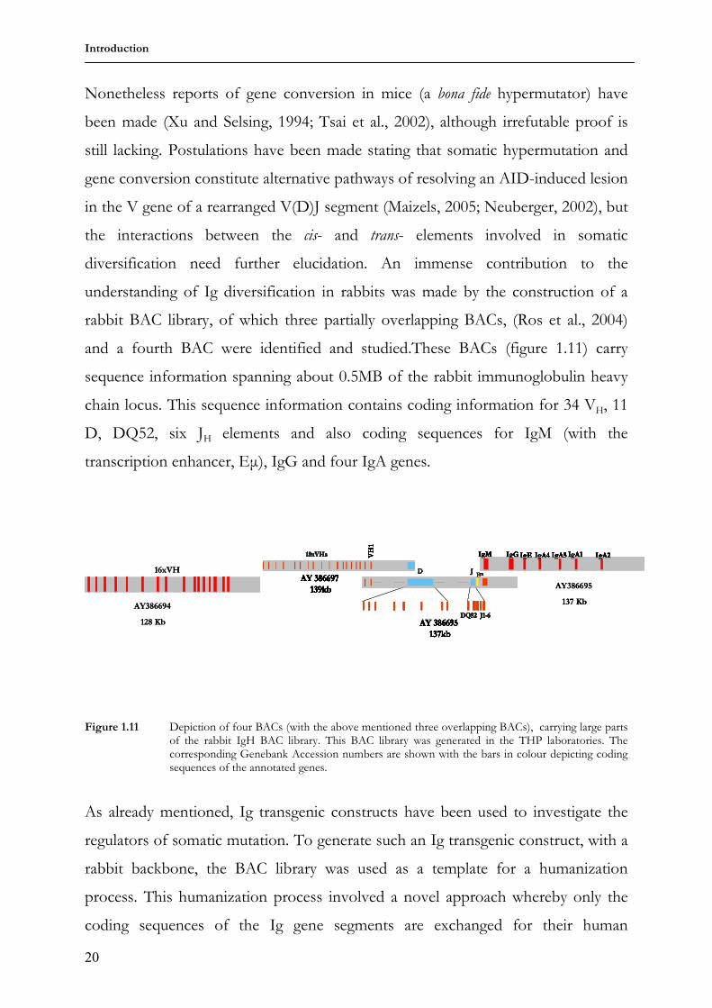
and a fourth BAC were identified and studied.These BACs (figure 1.11) carry
sequence information spanning about 0.5MB of the rabbit immunoglobulin heavy
chain locus. This sequence information contains coding information for 34 VH, 11
D, DQ52, six JH elements and also coding sequences for IgM (with the
transcription enhancer, Eµ), IgG and four IgA genes.
Figure 1.11 Depiction of four BACs (with the above mentioned three overlapping BACs), carrying large parts
of the rabbit IgH BAC library. This BAC library was generated in the THP laboratories. The corresponding Genebank Accession numbers are shown with the bars in colour depicting coding sequences of the annotated genes.
As already mentioned, Ig transgenic constructs have been used to investigate the
regulators of somatic mutation. To generate such an Ig transgenic construct, with a
rabbit backbone, the BAC library was used as a template for a humanization
process. This humanization process involved a novel approach whereby only the
coding sequences of the Ig gene segments are exchanged for their human
counterparts. In so doing the cis elements regulating Ig rearrangements (RSS,
promoters) and somatic mutation (transcription enhancers and promoters) are left
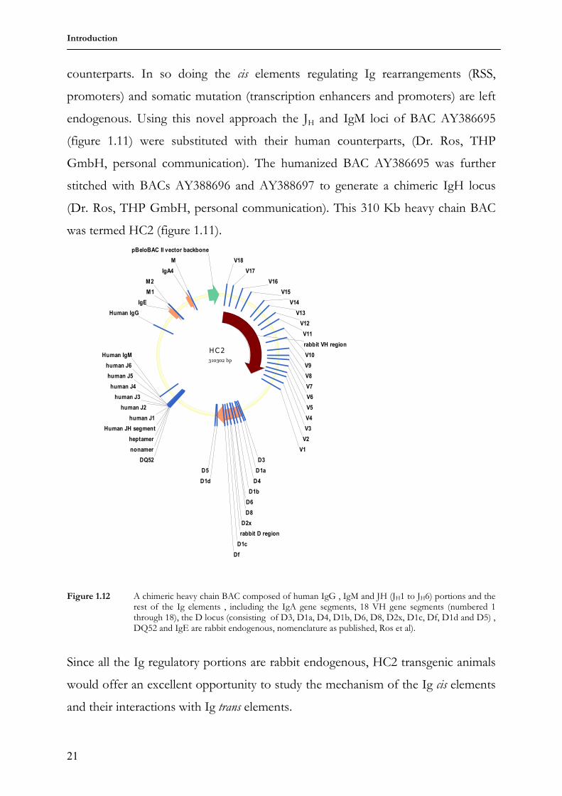
endogenous. Using this novel approach the JH and IgM loci of BAC AY386695
(figure 1.11) were substituted with their human counterparts, (Dr. Ros, THP
GmbH, personal communication). The humanized BAC AY386695 was further
stitched with BACs AY388696 and AY388697 to generate a chimeric IgH locus
(Dr. Ros, THP GmbH, personal communication). This 310 Kb heavy chain BAC
was termed HC2 (figure 1.11).
HC2
310302 bp
rabbit D region
Human JH segment
Human IgM
IgA4
IgE
Human IgG
V16
heptamer
nonamer
D3
D1a
D4
D6
D1b
D8
D2x
Df
D1c
D5
D1d
DQ52
human J1
human J2
human J3
human J4
human J5
human J6
rabbit VH region
M
M1
M2
pBeloBAC II vector backbone
V1
V2
V3
V18
V17
V15
V14
V13
V12
V11
V10
V9
V8
V7
V6
V5
V4
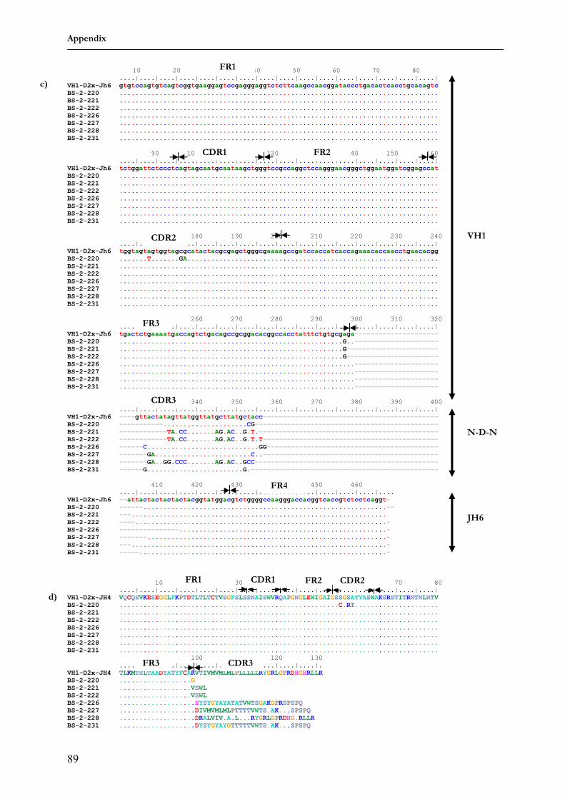
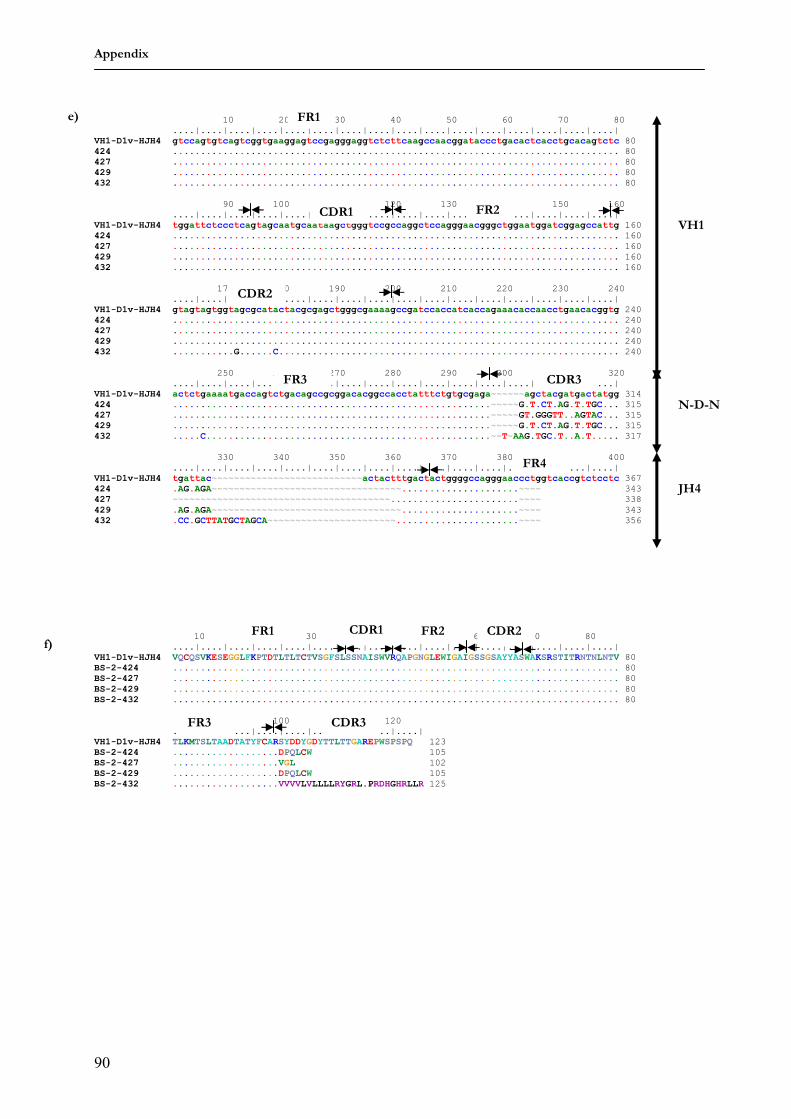
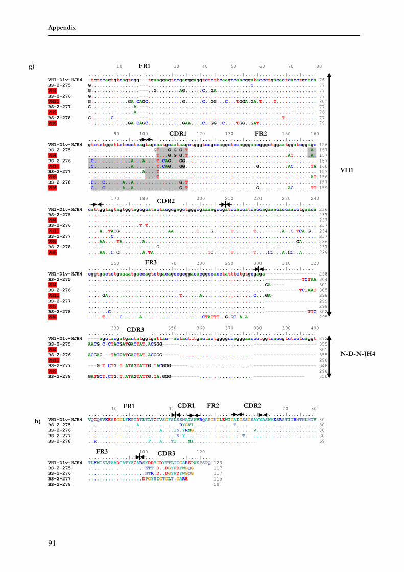
Figure 1.12 A chimeric heavy chain BAC composed of human IgG , IgM and JH (JH1 to JH6) portions and the rest of the Ig elements , including the IgA gene segments, 18 VH gene segments (numbered 1 through 18), the D locus (consisting of D3, D1a, D4, D1b, D6, D8, D2x, D1c, Df, D1d and D5) , DQ52 and IgE are rabbit endogenous, nomenclature as published, Ros et al).
Since all the Ig regulatory portions are rabbit endogenous, HC2 transgenic animals
would offer an excellent opportunity to study the mechanism of the Ig cis elements
and their interactions with Ig trans elements.
Introduction
22
Additionally, more insight to the mechanisms underlying Ig rearrangements and
somatic mutation can be obtained by the generation of transgenic animals using a
fully human transgenic IgH locus. In a further context, production of functional
fully human antibodies by transgenic animals could be of important therapeutic
use. It has been reported that three constructs carrying overlapping genomic
fragments were able to recombine in a homologous manner to reconstitute a core
human IgH locus in transgenic mice (Wagner et al., 1996). In this respect, the
generation of a humanized rabbit VH locus (using BAC AY3886697 as a template)
and co-injection with the humanized BAC AY386695 construct would result in the
constitution of a core rabbit humanized IgH locus in transgenic animals: since co-
injection of two linear constructs have been shown to integrate at the same site.
1.5 Goal of this work
In the present work, the generation of a humanized rabbit VH locus, using BAC
AY3886697 as template is described. This humanized VH locus was co-injected
with the humanized AY386695 construct to generate transgenic mice. These
transgenic mice analyzed to give an insight to following questions:
Will the rabbit Ig regulatory elements function across species boundaries?
More importantly which method of somatic diversification would result
from the interaction between the rabbit Ig cis elements (a gene converter)
and mouse Ig trans elements (a hypermutator)? Gene conversion or
hypermutation?
Furthermore, to proffer a more efficient assessment of the different methods of
somatic diversification, the HC2 chimeric construct was used to generate both
transgenic mice and rabbits and the transgenic animals analysed with following
question in mind:
Will the same IgH transgenic construct hypermutate in mice and gene convert
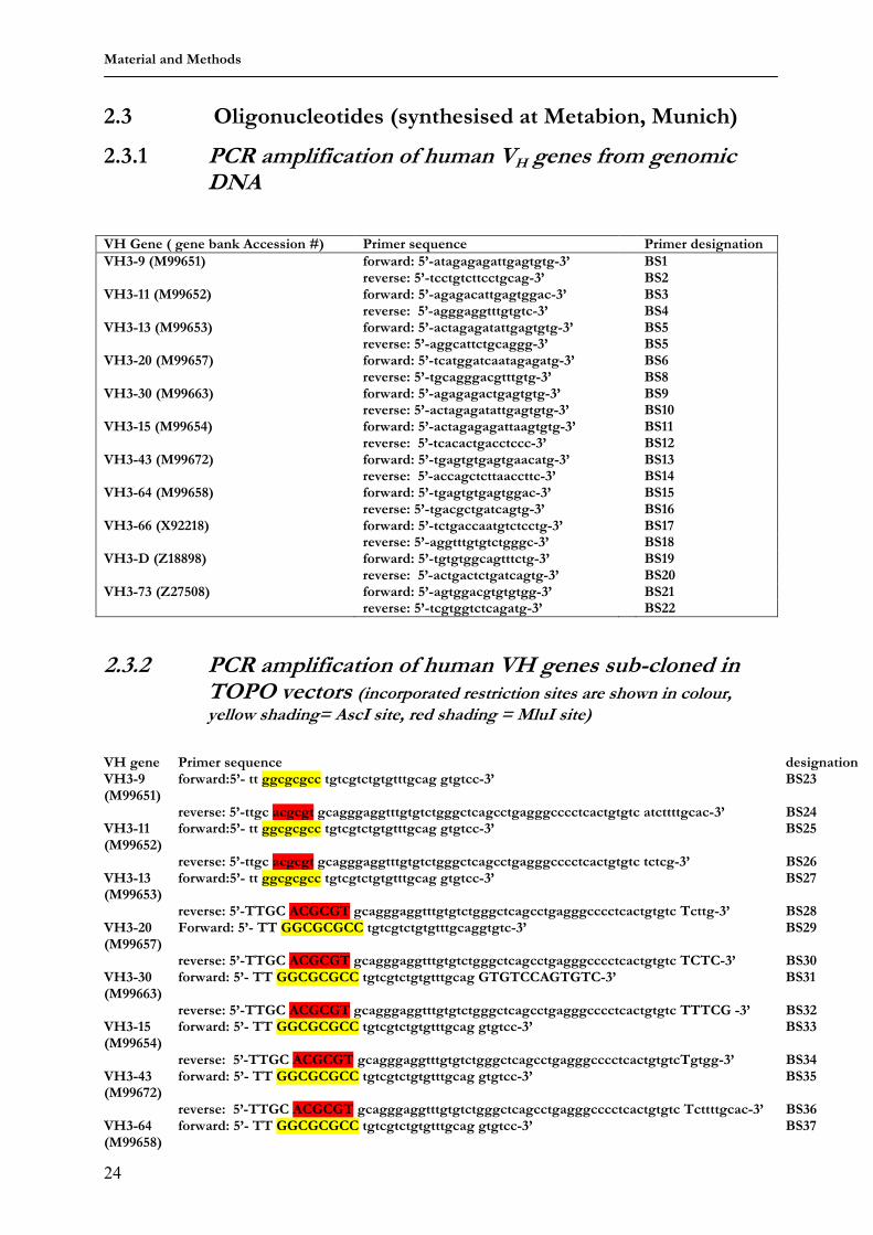
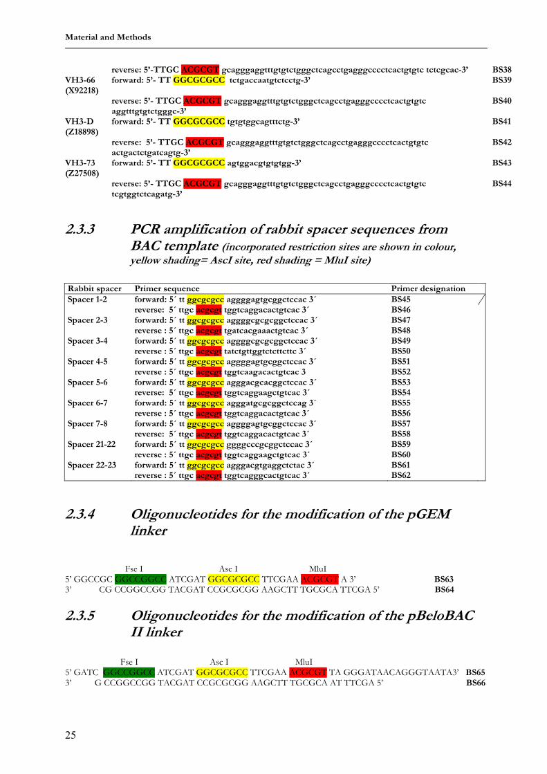
2.3.2 PCR amplification of human VH genes sub-cloned in TOPO vectors (incorporated restriction sites are shown in colour, yellow shading= AscI site, red shading = MluI site)
VH gene Primer sequence designation VH3-9 (M99651)
2.3.4 Oligonucleotides for the modification of the pGEM linker
Fse I Asc I MluI 5’ GGCCGC GGCCGGCC ATCGAT GGCGCGCC TTCGAA ACGCGT A 3’ BS63 3’ CG CCGGCCGG TACGAT CCGCGCGG AAGCTT TGCGCA TTCGA 5’ BS64
2.3.5 Oligonucleotides for the modification of the pBeloBAC II linker
Fse I Asc I MluI 5’ GATC GGCCGGCC ATCGAT GGCGCGCC TTCGAA ACGCGT TA GGGATAACAGGGTAATA3’ BS65 3’ G CCGGCCGG TACGAT CCGCGCGG AAGCTT TGCGCA AT TTCGA 5’ BS66
Material and Methods
26
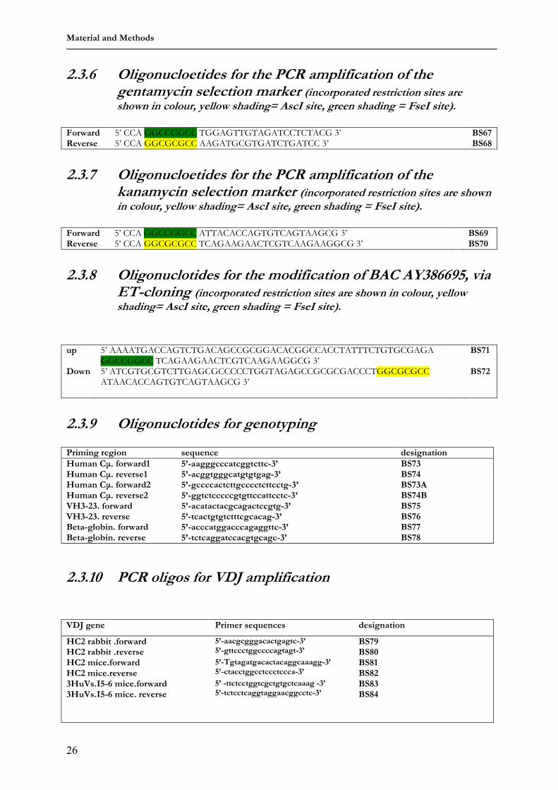
2.3.6 Oligonucloetides for the PCR amplification of the gentamycin selection marker (incorporated restriction sites are shown in colour, yellow shading= AscI site, green shading = FseI site).
Forward 5’ CCA GGCCGGCC TGGAGTTGTAGATCCTCTACG 3’ BS67 Reverse 5’ CCA GGCGCGCC AAGATGCGTGATCTGATCC 3’ BS68
2.3.7 Oligonucloetides for the PCR amplification of the
kanamycin selection marker (incorporated restriction sites are shown in colour, yellow shading= AscI site, green shading = FseI site).
Forward 5’ CCA GGCCGGCC ATTACACCAGTGTCAGTAAGCG 3’ BS69 Reverse 5’ CCA GGCGCGCC TCAGAAGAACTCGTCAAGAAGGCG 3’ BS70
2.3.8 Oligonuclotides for the modification of BAC AY386695, via
ET-cloning (incorporated restriction sites are shown in colour, yellow shading= AscI site, green shading = FseI site).
up 5’ AAAATGACCAGTCTGACAGCCGCGGACACGGCCACCTATTTCTGTGCGAGA
GGCCGGCC TCAGAAGAACTCGTCAAGAAGGCG 3’ BS71
Down 5’ ATCGTGCGTCTTGAGCGCCCCCTGGTAGAGCCGCGCGACCCTGGCGCGCC ATAACACCAGTGTCAGTAAGCG 3’
BS72
2.3.9 Oligonuclotides for genotyping Priming region sequence designation
Human Cµ. forward1 5’-aagggcccatcggtcttc-3’ BS73 Human Cµ. reverse1 5’-acggtgggcatgtgtgag-3’ BS74 Human Cµ. forward2 5’-gccccactcttgcccctcttcctg-3’ BS73A Human Cµ. reverse2 5’-ggtctcccccgtgttccattcctc-3’ BS74B VH3-23. forward 5’-acatactacgcagactccgtg-3’ BS75 VH3-23. reverse 5’-tcactgtgtctttcgcacag-3’ BS76 Beta-globin. forward 5’-acccatggacccagaggttc-3’ BS77 Beta-globin. reverse 5’-tctcaggatccacgtgcagc-3’ BS78
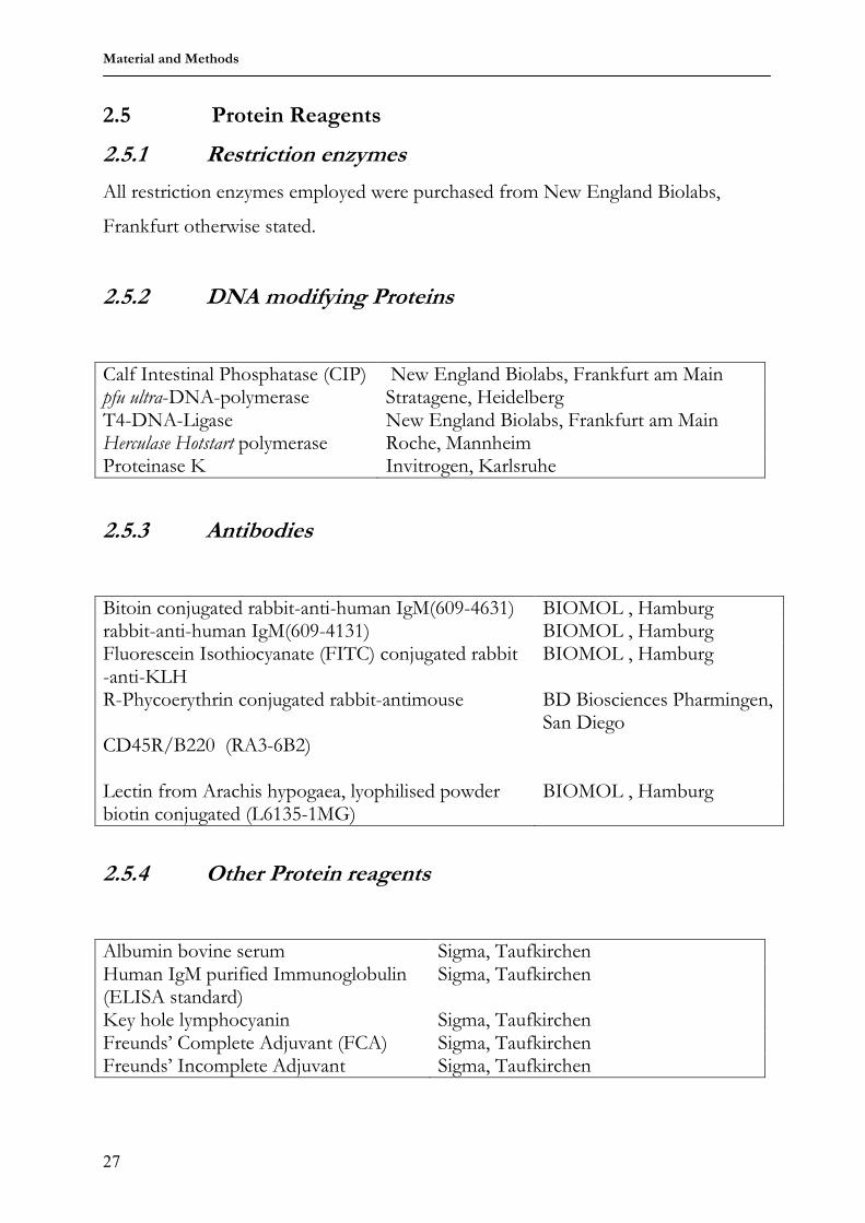
All restriction enzymes employed were purchased from New England Biolabs,
Frankfurt otherwise stated.
2.5.2 DNA modifying Proteins
Calf Intestinal Phosphatase (CIP) New England Biolabs, Frankfurt am Main pfu ultra-DNA-polymerase Stratagene, Heidelberg T4-DNA-Ligase New England Biolabs, Frankfurt am Main Herculase Hotstart polymerase Roche, Mannheim Proteinase K Invitrogen, Karlsruhe
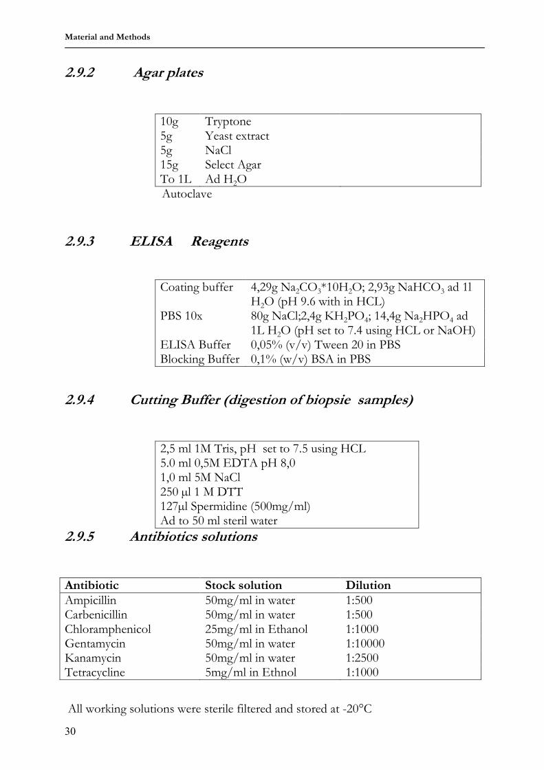
10g Tryptone 5g Yeast extract 5g NaCl To 1L Ad H2O
Autoclave
Material and Methods
30
2.9.2 Agar plates
10g Tryptone 5g Yeast extract 5g NaCl 15g Select Agar To 1L Ad H2O Autoclave
2.9.3 ELISA Reagents
Coating buffer 4,29g Na2CO3*10H2O; 2,93g NaHCO3 ad 1l H2O (pH 9.6 with in HCL)
PBS 10x 80g NaCl;2,4g KH2PO4; 14,4g Na2HPO4 ad 1L H2O (pH set to 7.4 using HCL or NaOH)
ELISA Buffer 0,05% (v/v) Tween 20 in PBS Blocking Buffer 0,1% (w/v) BSA in PBS
2.9.4 Cutting Buffer (digestion of biopsie samples)
2,5 ml 1M Tris, pH set to 7.5 using HCL 5.0 ml 0,5M EDTA pH 8,0 1,0 ml 5M NaCl 250 µl 1 M DTT 127µl Spermidine (500mg/ml) Ad to 50 ml steril water
2.9.5 Antibiotics solutions
Antibiotic Stock solution Dilution Ampicillin 50mg/ml in water 1:500 Carbenicillin 50mg/ml in water 1:500 Chloramphenicol 25mg/ml in Ethanol 1:1000 Gentamycin 50mg/ml in water 1:10000 Kanamycin 50mg/ml in water 1:2500 Tetracycline 5mg/ml in Ethnol 1:1000
All working solutions were sterile filtered and stored at -20°C
Material and Methods
31
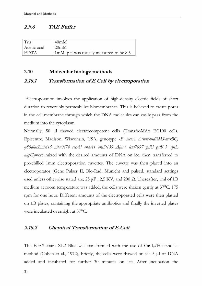
2.9.6 TAE Buffer
2.10 Molecular biology methods
2.10.1 Transformation of E.Coli by electroporation
Electroporation involves the application of high-density electric fields of short
duration to reversibly permeabilize biomembranes. This is believed to create pores
in the cell membrane through which the DNA molecules can easily pass from the
nupG)were mixed with the desired amounts of DNA on ice, then transferred to
pre-chilled 1mm electroporation cuvettes. The cuvette was then placed into an
electroporator (Gene Pulser II, Bio-Rad, Munich) and pulsed, standard settings
used unless otherwise stated are; 25 µF , 2,5 KV, and 200 Ω. Thereafter, 1ml of LB
medium at room temperature was added, the cells were shaken gently at 37°C, 175
rpm for one hour. Different amounts of the electroporated cells were then platted
on LB plates, containing the appropriate antibiotics and finally the inverted plates
were incubated overnight at 37°C.
2.10.2 Chemical Transformation of E.Coli
The E.coli strain XL2 Blue was transformed with the use of CaCl2/Heatshock-
method (Cohen et al., 1972), briefly, the cells were thawed on ice 5 µl of DNA
added and incubated for further 30 minutes on ice. After incubation the
Tris 40mM Acetic acid 20mM EDTA 1mM pH was usually measured to be 8.5
Material and Methods
32
cells/DNA mixture was pulsed for 30 seconds at 42°C placed briefly on ice and
supplemented with 250 µl of LB medium. The bacteria were then incubated at
37°C and 175 rpm for one hour and plated on LB plates containing the appropriate
selective antibiotics and finally the inverted plates were incubated overnight at
37°C.
2.10.3 Cloning PCR Products
PCR products were cloned using a rapid and selective method. This was achieved
by means of commercially purchased kits. Invitrogen, The Netherlands, offers
different kits for cloning varying PCR products. The TOPO Zero Blunt Kit was
used to clone blunt end PCR products, obtained by amplification with proof
reading poymerase meanwhile TOPO TA kit was used to create PCR products with
A overhang, finally to clone large PCR products, >3kb, the TOPO XL kit was
used.
2.10.4 Restriction endonuclease digestion of DNA
Restriction endonucleases were used to digest double-stranded DNA for analytical
or preparative purposes. These restriction enzymes were purchased from New
England Biolabs, unless otherwise stated, and used according to the manufacturers’
instructions. A typical reaction consisted of a 20 µl volume containing about 1µg of
DNA, 1x reaction buffer (from supplier), and if required 100 µg/ml BSA. The
reaction was incubated at the recommended temperature (usually 37°C) for times
from 30 minutes to overnight. Complete digestion was confirmed by agarose gel
electrophoresis.
Material and Methods
33
2.10.5 Ethanol precipitation of DNA
DNA was precipitated from aqueous solutions using salt and alcohol. A 1/10
volume of 3.3 M sodium acetate, pH 5.2 and a 2.5 volume of ice-cold absolute
ethanol were added to the DNA containing aqueous solution. After overnight
incubation at -20°C, the solution was centrifuged at 14000 rpm, (Eppendorf
5417R) 4°C for 30 minutes. The resulting pellet was washed with ice-cold 70%
ethanol, dried and dissolved in water.
2.10.6 Dephosphorylation of linearized plasmid DNA by CIP
To prevent the re-ligation of linearized vector and to promote the insertion of the
desired DNA fragment (insert), calf intestinal phosphatase (CIP) was used to
dephosphorylate the 5’ ends of the linearized vector. Dephosphorylation was
carried out directly following plasmid linearization. 1Unit CIP (New England
Biolabs) was added to the digestion mixture. After one hour incubation at 37°C,
the mixture was loaded to agarose gel in which the CIP and restriction enzymes
were inactivated. The dephosphorylated plasmid was gel-extracted and precipitated
with ethanol.
2.10.7 Ligation of DNA fragments
DNA fragments bearing either sticky ends, meaning both vector and insert have
compatible overhangs or blunt ends, meaning the ends of the vector and insert
have no overhangs can be ligated in vitro with T4 DNA ligase.
Linearized, dephosphorylated vector was mixed with insert DNA, usually in vector:
insert molar ratios of 1:5 or 1:10. Total DNA was at 200 ng in a total volume of 20
µl, containing 1x ligase buffer and 1U T4 DNA ligase. The ligation mixture was
incubated at 16°C overnight and was subsequently used to transform bacteria.
Material and Methods
34
2.10.8 Mini-prep: small-scale preparation of plasmid DNA
Following amplification of plasmid DNA in E.coli, DNA was extracted using a
rapid method (‘mini-prep’) to screen for clones with the correct insert. Single
colonies were picked from agar plates, and used to inoculate 3 ml of LB medium
containing the suitable selective antibiotic and cultured overnight at 37°C, 225 rpm.
For medium and high copy plasmids up to a size of about 7 kb, mini-prep DNA
extraction was done using the Eppendorf, (Hamburg) “fast-plasmid kit”. For larger
plasmid and single copy BAC vector plasmids, the Qiagen (Qiagen, Hilden,
Germany) “plasmid purification kit”. In both cases, the extraction was performed
following the manufacturers’ instructions and reagents supplied in the kit. The
extracted DNA was analyzed by restriction digestion.
2.10.9 Midi-prep: large-scale preparation of plasmid DNA
For preparations of large amounts of plasmid DNA, the Macherey & Nagel
Nucleobond BAC 100 purification kit was used. About 20 µl of a bacterial culture
from a clone known to contain the desired insert was used to inoculate 500 ml LB
medium containing the appropriate antibiotic and grown overnight. In the case of
medium and high copy plasmids, 100 ml of LB was used. Extraction was carried
following the manufacturers instructions with the reagents supplied in the kit.
2.10.10 Genomic DNA preparation
Genomic DNA was obtained from mouse tail-tip or rabbit tear-tip biopsies. .
Tissue samples were incubated overnight in a lysis buffer consisting of 375 µl
cutting buffer, 20 µl 20%SDS and 5 µl Proteinase K. The debris was gotten rid off
by centrifuging at 14000 rpm for 2 minutes in a table centrifuge (Ependorf 5417R).
The supernatant was transferred in fresh tubes, the DNA was precipitated by
Material and Methods
35
adding 400µl Isopropanol and again centrifuged for two minutes at 14000 rpm
(Ependorf 5417R).The pellet was washed with 400 µl 70% Ethanol, centrifuged
again for two minutes at 14000 rpm (Ependorf 5417R) and the pellet washed again
with 400 µl 100% Ethanol, dried and re-dissolved in 200 µl 0.1 TE buffer. 2 µl were
used for PCR reactions.
2.10.11 Quantification of DNA solutions
The concentration of DNA was determined by spectrophotometry. The ultraviolet
(UV) absorption was measured at a wavelength of 260 nm (OD260) using a quartz
cuvette. For double-stranded DNA one OD260 corresponds to approximately 50
µg/ml DNA. In addition, the OD260 was measured to estimate the purity of the
nucleic acid sample. A ratio A260/A280 significantly less than 1.8-2.0 would indicate
phenol or protein contamination.
2.10.12 Agarose gel electrophoresis
Conventional gel electrophoresis is commonly used to separate and analyze nuleic
acids. Separation of DNA molecules from 100 bp to 20 kb was usually performed
on agarose gels (Ultra pure Agarose, Invitrogen) in 1x TAE buffer; the agarose
percentage was used according to the DNA sizes to be separated. Gel run was
performed with variable time and volt conditions according to separation range and
agarose percentage using an electrophoresis power supply (Bio-Rad, Munich). For
size comparison, a DNA molecular ladder was loaded on the gel next to the
samples. After gel run, DNA was stained with the intercalating fluorescent reagent
ethidium bromide (EtBr), which was added either, to the gel before solidification or
in TAE buffer for after-run staining at the concentration of 0,5 µg/ml. Stained
DNA was visualized on a UV-transilluminator at a wavelength of 254 nm and
photographed with a gel documentation apparatus (Bio-Rad, Munich).
Material and Methods
36
2.10.13 Pulse field gel electrophoresis (PFGE)
Conventional gel electrophoresis has an upper limit to the sizes that can be
resolved. Pulse field gel electrophoresis overcomes the problems of resolution of
large DNA fragments by using two sets of electrodes that are fixed at angles to
each other. The current is alternated between these sets of electrodes at defined
intervals. Separation is based upon the potential of smaller DNA molecules to
reorient faster than larger DNA molecules. PFGE was used for the resolution of
high weight molecular DNA (20-200 kb), derived from the restriction digestion of
BAC DNA. Pulse Field Agarose was used at a concentration of 1% in 0.5% TBE.
Run was carried out in 0.5% TBE using the Chef-Mapper apparatus (Bio-Rad,
Munich). λ mix DNA standard was used for size comparison. The temperature was
maintained at 14°C, by means of a cooling system. The run time and settings were
generated automatically according to input of separation range. This was done by
software delivered with the apparatus. Gel was stained with EtBr after the run and
visualized as described above.
2.10.14 Extraction of DNA fragments from agarose gels
After gel electrophoresis, the desired DNA bands were excised with a scalpel under
irradiation with low-energy long wavelength UV light (320 nm) to minimize
damaging the DNA. The DNA fragments were eluted from the gel pieces by
means of the GENECLEAN kit (Qbiogene) according to the provided protocol.
In case of preparation of fragments to inject into fertilized oocytes for the
generation of transgenic mice, as well as after PCR amplification of Human VH
genes and antibiotic selection cassettes, contact with EtBr and UV irradiation was
absolutely avoided. After EtBr-free gel run, the gel was stained with 0.5%
Material and Methods
37
Methyleneblue , de-stained by washing with water and the desired bands were the
excised after visualization on a transilluminator.
In case of high molecular weight fragments, the DNA was extracted by means of
electroelution, using the BioTrap apparatus according to the provided protocol.
The DNA was thereafter precipitated with EtOH.
2.10.15 Polymerase chain reaction (PCR)
This method of DNA amplification had different applications, for example
production of DNA fragments to sub-clone and genotyping of transgenic animals.
According to the specific purpose, PCR amplifications were realized from various
templates, genomic DNA, cloned DNA like plasmids and BACs. Furthermore
different types of DNA polymerases (sources stated below) were chosen and the
reaction settings were according to the manufacturers’ instructions.
In general, otherwise stated, PCR reaction mixes were set up with 1x PCR buffer,
4mM of a deoxyribonucleotide triphosphate mixture (dNTP), 4 picomoles of each
pimers, 1Unit of DNA polymerase and the amount of DNA template was
specifically determined in each case.PCR reactions were performed in Strategene
Robocylcler PCR machines.
2.10.16 Sequencing
Sequencing was performed by the company AGOWA GmbH in Berlin. For all
fragments sub-cloned in the TOPO vectors, the sequencing primer was the T7
primer, present on the commercially acquired vector.
Material and Methods
38
2.10.17 BAC modification via homologous recombination in E.coli
BAC modification using homologous recombination allows amendments of large
DNA constructs like BACs, PACs and Cosmids, where the normal cloning
techniques based on the use of restriction enzymes and ligation cannot be applied.
Using this targeted homologous recombination in E.coli insertions, deletions and
point mutations can be introduced in such large DNA constructs. Normally used
E.coli strains are deficient in the recombination machinery; an exogenous
recombination has to be imported. For this purpose several approaches have been
established(Yang et al., 1997; Muyrers et al., 1999; Yu et al., 2000; Swaminathan et
al., 2001). In this work the method proposed by Stewart and co-workers (Zhang et
al., 1998) was used for BAC modification.
Briefly, the exogenous plasmid expressing the proteins which mediate homologous
recombination is transformed into competent cells carrying the target BAC. In this
case the plasmid used was the pSC101αβγ-tetra plasmid. The plasmid expresses the
phage recombinase proteins Redα, Redβ and Redγ under the control of an
arabinose inducible promoter, and a temperature sensitive origin; propagation is
permissible at 30°C.
To make competent cells for the transformation, overnight cultures of the cells
carrying the target BAC are generated by using glycerol stocks to inoculate 1.0 ml
LB medium containing the selective antibiotics. This is carried out in a 2 ml
Eppendorf tube with a punctured lid and shaken overnight at 37°C, 1050 rpm in a
heating block (Eppendorf, Hamburg).
The next day 20 µl of the overnight culture is used to inoculate three 2 ml tubes,
each with 1,4 ml LB medium and the selective antibiotic. The cultures are grown
for two to three hours till OD600=0.2-0.25, shaking at 37°C, 1050 rpm. One of the
three inoculated tubes is used for the measurement. Once the desired OD is
attained the two remaining tubes are spun for 30 seconds at 11,000 rpm in a pre-
cooled (between -5°C and +2°C, Ependorf 5417R) centrifuge. The supernatant is
quickly discarded and the pellet washed by re-suspending in 1 ml ice-cold 10%
Material and Methods
39
glycerol, the wash step is repeated twice. After the final wash step the supernatant
is discarded leaving 20 to 30 µl cell suspension in the tube. Keeping the tube on ice
1µl (0.2 to 0.5 µg) of the pSC101αβγ-tetra plasmid is added to the one tube of the
freshly made competent cells and as a negative control 1µl water is added to the
second tube. The mixture is then transferred to an ice-cold 1mm electroporation
cuvette and electroporation carried out at 1350 V, 10 µF, 600 Ω using the Bio-Rad
Gene Pulser II. After electroporation the cells were re-suspended in 1ml sterile LB
medium and incubated at 30°C, 1050rpm for 70 minutes. Varying amounts from
10µl-100µl are platted on LB plates with the selective antibiotics. The pSC101αβγ-
tetra plasmid has a tetracycline selection cassette. The day after, about six colonies
from the plates were used to inoculate 3ml LB medium with the selective
antibiotics and grown at 30°C, the selective temperature for the propagation of the
pSC101αβγ-tetra plasmid. After DNA isolation as described above, the presence of
the transformed plasmid was confirmed by restriction digestion.
About 20 µl bacterial cultures of positive clones were used to inoculate 3 ml LB
medium again with the selective antibiotics, this time the cultures were incubated at
37°C. After DNA isolation the intactness of the target BAC was confirmed by
restriction digestion. Now that the homologous recombination mediating plasmid
has been successfully transformed into cells carrying the target BAC, the next step
is to create a targeting construct carrying the desired modification to be introduced
in the BAC. This targeting construct is created by PCR amplifying a selection
cassette, with oligonucleotides carrying a 50 bp homology to the target BAC and
preceding these 50 bp homologies are the desired mutations to be introduced
(figure 2.1). The PCR product is gel analysed under EtBr free conditions; the gel is
stained with Methylenblue as described above and purified using the
GENECLEAN kit (QBIOGENE).
Next the bacterial cells carrying both the target BAC and the pSC101αβγ-tetra
plasmid are made competent for the transformation of the targeting construct.
Six Eppendorf (Hamburg) tubes are set each with 1.4ml LB medium containing the
selective antibiotic –in this case neo-, and inoculated with 15-20µl of fresh
Material and Methods
40
overnight culture of the cells carrying the target BAC and the pSC101αβγ-tetra
plasmid. The cells are propagated at 30°C, 1050 rpm till OD600= 0.15-0.18 is
reached. One or two of the six tubes are used for OD measurements. As soon as
the optimal OD600 is attained 20 % L (+) Arabinose is added to a final
concentration of 0.1-0.2% thereby inducing expression of the recombinase
proteins, one tube is left un-induced as a negative control.
Figure 2.1 A schematic view of BAC modification by homologous recombination as described by
(Zhang et al., 1998).
The tubes are then incubated at 37°C, 1050 rpm for 50 minutes, until an
OD600=0.35-0.4 is attained. Three tubes two induced and the un-induced control
are then spun down at 11000 rpm, for 30 seconds in a pre-cooled Eppendorf
(Hamburg) centrifuge, and the pellet re-suspended in 1ml ice-cold 10% glycerol.
The wash step is repeated twice and after the third time the supernatant is
discarded leaving 20 to 30 µl cell suspension is left in the tubes. Still keeping the
tubes on ice, 1-2 µl about 0.3-0.5µg of the targeting construct is added to one of
the induced tubes and to the non-induced negative control.
RpsL NeoNeo
RpsL NeoNeo
Target region
RpsL NeoNeoRpsL NeoNeoRpsL NeoNeo
Homologous
recombination
50bp homology to
target region
50bp homology to
target region
The purified PCR product is transformed into freshly made competent bacteria carrying, the target BAC as well as the pSC101αβγ-tetra plasmid, which mediates homologous recombination.
The pSC101αβγ-tetra plasmid is induced using L-Arabinose to produce the proteins which regulate and direct homologous recombination between the 50 bp arms of the PCR product and tht corresponding region in the target BAC. Transformants are selected because of the presence of the neo cassette and the intactness of the insert is confirmed by colony analysis
A selection cassette is PCR amplified with primers carrying desired sequences as well as 50 bp homolgies (shown here in grey and black) to the target region
Material and Methods
41
The same volume of sterile water is added to the second induced tube as a second
negative control. After mixing briefly by pipetting very gently up and down, the
mixture is transferred to an ice-old 1mm electroporation cuvette and electroporated
at 1350 V, 10 µF, and 600 Ωs. The cells are then re-suspended in 1ml LB medium
and incubated for 70 minutes at 37°C shaking at 1050rpm. 3x 330 µl of the sample
probe and 1x 330 µl of the negative controls are plated on LB plates with selective
antibiotics and incubated overnight at 37°C. Colony analysis is then carried out and
positives confirmed by restriction digestion and sequencing.
2.11 Transgenic constructs The first step in the generation of the transgenic constructs was the customisation
of two commercially acquired plasmid vectors, (pGEM and pbeloBAC II) to
accommodate the employed cloning strategies.
2.11.1 Modifying the pGEM vector
The initial step involved the modification of the linker region to provide desired
restriction sites: FseI, AscI and MluI. To modify the pGEM linker, the synthesized
lyophilized oligonucleotides, BS63 and BS64, were dissolved in distilled H2O to a
final concentration of 10 ng/µl. Annealing reactions were set up using varying
concentrations of the two oligonucleotides. Three different set ups with each 2.6
ng, 7 ng and 13 ng of each oligonucleotide in a total volume of 20 µl were
supplemented with 2 µl 10x Roche T4-ligase buffer. The reactions were heated in a
water bath at 97°C for 2 minutes and cooled to room temperature. The pGEM
vector was opened by double restriction digestion with NotI and HindIII, and the
annealed linker was then ligated to the opened vector (figure 2.2a).
Varying ratios of opened vector: annealed linker from 1:2, 1:5 and 1:10 were set up
in a total volume of 10 µl, supplemented with 1 µl 10X T4-ligase Buffer (Roche,
Mannheim) and 1µl T4-Ligase (Roche) and incubated overnight at 16°C. A
Material and Methods
42
negative control reaction with no vector DNA was also set up. The following day,
XL2-Blue cells were chemically transformed with 5 µl of the ligation reactions, and
the insertion of the desired sites was confirmed by restriction digestion.
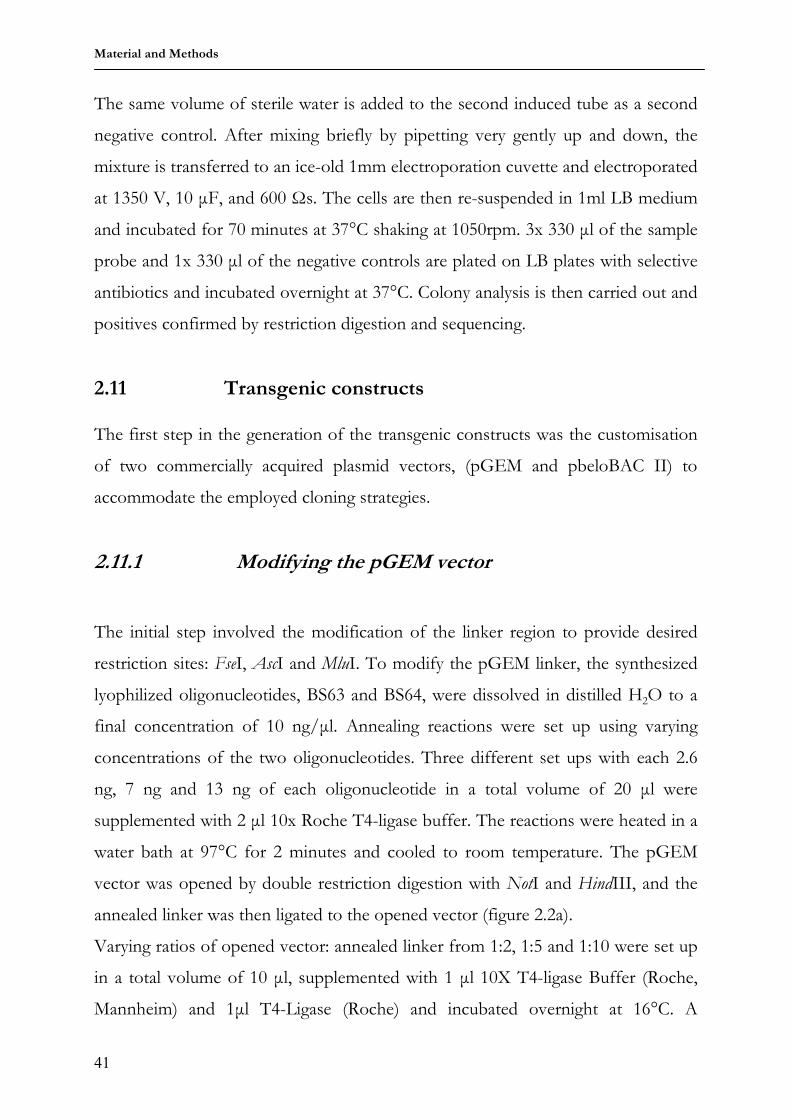
Figure 2.2 Generation of customized pGEM.genta and pGEM.neo vectors. After the linker modification of a
commercially acquired pGEM vector (inserting the sites FseI-AscI-MluI, in that order) two different selection cassettes, gentamycin (figure 2.2 b) and kanamycin (figure 2.2c) were ligated into the FseI and AscI sites.
After this confirmation, using the pRep-Genta (Gene Bridges, Dresden) plasmid as
Template, the gentamycin selection cassette was amplified by primers BS67 and
BS68. The PCR product was then double digested with FseI and AscI (intergrated
in the primers) and the digested gel-purified product was ligated with the modified
pGEM vector also opened with FseI and AscI. The resulting vector was tagged
pGEM.Genta (figure 2.2b).
A second pGEM vector tagged pGEM.neo (figure 2.2c), was also generated
following the same scheme but or this vector a kanamycin selection cassette was
PCR amplified with oligos BS69 and BS70, double digested with FseI and AscI sites
and ligated with the modified pGEM opened with the same restriction enzymes.
c)
pGEM
3178 bp
T7
SP6
MCS
Amp
f1 Ori
HindIII (39)
NotI (13)
pGEM.genta
4115 bp
gentamycin selection cassette
pGEM-backbone
AscI (964)
FseI (25)
MluI (977)
pGEM.Neo
4563 bp
kanamycin selection cassette
pGEM-backbone
AscI ( 1412)
FseI (25)
MluI (1425)
Ligation with the
gentamycin
selection cassette
Ligation with the
kanamycin
selection cassette
pGEM
3178 bp
T7
SP6
MCS
Amp
f1 Ori
HindIII (39)
NotI (13)
pGEM.genta
4115 bp
gentamycin selection cassette
pGEM-backbone
AscI (964)
FseI (25)
MluI (977)
pGEM.Neo
4563 bp
kanamycin selection cassette
pGEM-backbone
AscI ( 1412)
FseI (25)
MluI (1425)
Ligation with the
gentamycin
selection cassette
Ligation with the
gentamycin
selection cassette
Ligation with the
kanamycin
selection cassette
a) b)
Material and Methods
43
2.11.2 Modifying the pBeloBAC II (pBBII) vector
A commercially acquired pBBII vector was modified and used as the backbone for
building the final transgenic construct. The pBBII vector is the “state of the art”
vector used in building large constructs; inserts of up to 500 kb can be readily
accommodated by the vector (Woo et al., 1994). This BAC vector system is based
on the E.coli F factor, and in bacteria such plasmids carrying the F factor are
maintained in low copy number (Shizuya et al., 1992). This low copy maintenance
in bacteria effectively reduces the potential for recombination between DNA
fragments (inserts) carried by the plasmid.
Figure 2.3 The pBBII vector was opened with BamHI and HindIII and the linker modified using oligos BS65
and BS66 to carry restriction sites for FseI, AscI and MluI. The reactions set up were same as described for the pGEM vector in section 2.11.1. The insertion of these sites was confirmed by restriction digestion (data not shown).
This property is of paramount importance for the humanization procedure of the
rabbit VH locus, because this locus is composed of highly homologous segments
(Ros et al., 2004). Other advantages offered by the pBBII vector includes: easy
transfection of such large plasmids into E.coli by electroporation and also the
genetic stability of the cloned DNA (Shizuya et al., 1992). The commercially
acquired pBBII vector was double digested with BamHI and HindIII and the linker
modified as described in section 2.4 using oligonucleotides BS65 and BS66. The
modified linker had additional restriction sites for FseI, AscI and MluI. Theis
modified pBBII vector was named pBBII.mod (figure 2.3).
pBB11
7507 bp
ori
T7 promoter
Sp6 promoter
sopA
sopB
sopC
chloramphenicol cassette
Bam HI (355)
HindIII (385)
Linker
mofdification,
insertion of
FseI, AscI and
MluI sites
pBB11
7507 bp
ori
T7 promoter
Sp6 promoter
sopA
sopB
sopC
chloramphenicol cassette
Bam HI (355)
HindIII (385)
Linker
mofdification,
insertion of
FseI, AscI and
MluI sites
Linker
mofdification,
insertion of
FseI, AscI and
MluI sites
Material and Methods
44
2.11.3 Generation of rabbit VH spacer and human VH plasmid libraries
As mentioned in section 1.4 the humanization process in this work employs a novel
approach whereby only the coding sequences of the rabbit VH locus are exchanged
for their human counterparts. In this process the regulatory elements are left rabbit
endogenous. BAC AY3886697 was used as the template for this humanization
process. The rabbit IgH locus fulfils two prerequisites vital for gene conversion:
A functional VH C most proximal gene (since rabbits preferentially use
this gene in Ig rearrangements, section 1.3.6)
The upstream genes share great sequence homology to this VH C most
proximal gene, enabling them to serve as sequence donors during gene
conversion.
In the humanization process these properties had to be maintained to generate a
functional transgenic rabbit VH mini-locus. Consequently the human VH genes
used for the humanization process all belonged to the same family, the VH3 family
of genes (Brezinschek et al., 1995), thus ensuring sequence homology and the VH C
most proximal gene was the human VH3-23 gene which is the most abundantly
used human VH gene (Brezinschek et al., 1995).
The starting point of the humanization process was the creation of two small
plasmid libraries:
One composed of rabbit VH spacer sequences carrying the regulatory
elements
And the second composed of the coding sequences of the human VH3 gene
family.
To generate the rabbit spacer library, BAC AY386697 was digested with BstbI (data
not shown) and the digested DNA separated using PFGE, and the DNA extracted
by electro-elution. This was to generate small templates to be used in PCR
amplification of the rabbit VH spacer sequences. The desired amplicon is shown in
figure 2.4
Material and Methods
45
Figure 2.4 The rabbit spacer sequences were PCR amplified with primers BS45-BS62, shown here as arrows.
The forward primer carries an AscI site while the revers primer carries a MluI site. The obtained amplicon is a rabbit spacer sequence, with the promoter and leader sequences flanked by AscI and MluI restriction sites
Following this scheme, spacer sequences I1-2, I2-3, I3-4, I4-5 and I5-6 were
successfully amplified (nomenclature by Ros et al., 2004). Subsequently it was
attempted to sub-clone the different PCR products in TOPO vectors, but only
spacer I5-6 could successfully be sub-cloned. It was thereafter excised by digestion
with AscI and MluI and sub-cloned into the customized pGEM.genta vector.
VH gene Genebank number VH gene Genebank number
VH3-23 M99660 VH3-48 M99675
VH3-66 X92218 VH3-21 M99658
VH3-11 M99652 VH3-64 M99682
VH3-30 M99663 VH3-D Z18898
VH3-9 M99651 VH3-74 Z17392
VH3-13 M99653 VH3-33 M99665
VH3-72 Z27508 VH3-7 M99649
VH3-73 Z27508 VH3-43 M99672
VH3-15 M99654 VH3-49 M99676
Table 2.1 the 18 members of the human VH3 gene family. The members in bold could be successfully
amplified and sub-cloned in the TOPO vectors.
To generate the human VH gene plasmid library, human genomic DNA was used
as template in PCR reactions with primers BS1-BS22. The PCR products were then
sub-cloned in TOPO vectors and the inserts sequenced. Table 2.1 shows the
human VH3 gene family members.
To furnish the humanized VH locus with the functionality of a rabbit endogenous
locus capable of undergoing Ig rearrangements, it was of paramount importance to
supplement these sub-cloned human VH coding sequences with rabbit regulatory
portions consisting of the splicing signal and the recombination signal sequence. To
achieve this goal, the sub-cloned human VH genes were again PCR amplified, with
RabbitV
H
genewithRSS
Rabbit
VH spacer promoter
leader
RabbitV
H
genewithRSS
Rabbit
VH spacer promoter
leader
Material and Methods
46
Figure 2.5 Scheme for the primers used to supplement the human VH genes with rabbit regulatory
sequences. Up = forward PCR primer, down = reverse PCR primer, huV = human VH gene segment.
the primers (figure 2.5) carrying the desired rabbit regulatory sequences followed by
restriction sites. The human VH genes now with the rabbit regulatory sequences
were again sub-cloned in TOPO vectors and the inserts sequenced. Finally, the
different inserts were excised with AscI and MluI and sub-cloned in different
pGEM.neo vectors, thus generating a plasmid library of human VH genes.
2.11.4 Generation of a chimeric (rabbit VH spacer I5-6 and human VH) plasmid library
After creating the plasmid libraries in section 2.11.3, the next step involved linking
the rabbit VH spacers to the human VH genes resulting in a humanized rabbit VH
mini locus.
Figure 2.6 Generation of the chimeric plasmid library. As described in section 2.11.4, two separate libraries
were first generated, one containing ten members of the human VH3 (huV) gene family, sub-cloned in the modified pGEM.neo vector the other library with just the rabbit VH spacer I5-6 in the modified pGEM.genta vector. To generate the chimeric plasmid library with units of I5-6 followed by a huV gene segment, the usage of the two different selection cassettes (genta and neo) as well as the isocaudemeric property of the restriction enzymes AscI and MluI were exploited, see text below for further explanation. A/M = the resulting sequence after ligation between AscI and MluI compatible ends.
To achieve this goal, another plasmid library was generated, with units composed
of a human VH gene linked and rabbit spacer I5-6. A scheme is shown in figure 2.6.
To link the rabbit I5-6 spacer to huV gene segments, two factors were exploited:
The fact that the plasmid backbones of the two libraries (section 2.11.3)
carried different selection cassettes
Figure 2.7 The ligation strategy (showing the isocaudemeric property of AscI and MluI) employed in
generating the chimeric plasmid units in pGEM backbone. After restriction digestion with AscI and MluI compatible protruding termini ends are generated. Upon ligation a new sequence is created neither cleavable by AscI nor by MluI.
The Isocaudemeric property of the restriction enzymes AscI and MluI (figure
2.7)
To generate the first chimeric unit, the pGEM.neo carrying the human VH3-23
gene, (pGN.VH3-23), was opened with FseI and AscI, thereby excising the
kanamycin selection cassette. Simultaneously, the pGEM.genta vector carrying the
rabbit spacer I5-6 (pGG.I5-6) was also double digested with FseI and MluI. The
segment composed of the gentamycin cassette, followed by I5-6, was gel purified
and ligated to the opened pGN.VH3-23 vector, exploiting the Isocaudemeric
property of AscI and MluI. After ligation, positive clones were selected on
gentamycin plates so only clones with the gentamycin cassette hence the complete
insert were able to grow. Colonies were further screened by restriction digestion.
Three of such chimeric units were constructed: pGG.I5-6.Vh3-23, PGN.I5-6.VH3-
After generating the last plasmid library with the chimeric units, the final step
involved re-building the humanized mini VH locus in the pBBII.mod vector.
2.11.5 Re-building the humanized VH locus in the pBBII.mod
vector As mentioned in section 1.4, the co-injection of overlapping fragments leads to
homologous recombination and ultimately the reconstitution of a core locus.
Following this principle, the construction of the humanized VH locus with an
overlapping region to the humanized BAC AY386695, would lead to the
reconstitution of a core humanize rabbit IgH locus upon co-injection of the two
fragments.
To create such an overlapping region, the pBBII.mod vector was first
complemented with a 17kb region homologous to construct AY386695 (figure 2.8).
Figure 2.8 Sub-cloning of a 17kb homologous region in the pBBII.mod vector. BACs AY386697 and
AY386695 share a large homologous region, a 17kb region, shown here in grey, was sub-cloned in the pBBII.mod vector. After modification of BAC AY386695, a double digestion with FseI and MluI ensued and the resulting 17kb flanked by FseI and MluI restriction sites. pBBII.mod was opened with FseI and AscI and the 17kb fragment sub-cloned. The pBBII.mod vector was now named pBBII.mod.17kb
FseI-Neo-AscI
2 1
VH D J1-6 Eµ IgM
AY386695
(~137kb)
MluI
excision with FseI/MluI
FseI-Neo-AscI MluI
pBB11.mod
17kb
18 2 1
MluIVH
AY386697
(~139kb)
32kb 35kb17kb
17kb
FseI-Neo-AscI
2 1
VH D J1-6 Eµ IgM
AY386695
(~137kb)
MluI
excision with FseI/MluIexcision with FseI/MluI
FseI-Neo-AscI MluI
pBB11.mod
17kb
18 2 1
MluIVH
AY386697
(~139kb)
32kb 35kb17kb
17kb
Material and Methods
49
The first step involved the modification of the BAC AY386695 via ET cloning
(section 2.10.17). This modification involved the insertion of a kanamycin cassette
flanked by FseI and AscI restriction sites. This fragment was inserted immediately
after the VH1 gene segment (figure 2.8). Positive clones were selected on
kanamycin, and colonies were analyzed by restriction digestion .After this insertion,
a double digestion of the modified AY386695 with FseI and MluI (figure 2.8) was
performed. The 17 kb fragment flanked by FseI and MluI sites was excised, the
DNA purified by electro-elution and finally ligated with pBBII.mod opened with
FseI and AscI, another ligation exploiting the isocaudemeric properties of AscI and
MluI. Clones were selected on kanamycin and chloramphenicol and colonies
analyzed by restriction digestion with ApaI (data not shown), the vector was now
tagged pBBII.mod.17kb. After optimizing pBBII.mod for is role as the acceptor
BAC of the humanized VH locus, the next steps involved re-building the three
chimeric units in this pBBII.mod vector.
Figure 2.9 The three units of the chimeric plasmid library are integrated in the pBBII.mod vector equipped
with the 17kb overlap to the humanized AY386695. To perform this task, ligation cycles were performed, with alternating selection of positive clones on gentamycin or kanamycin. The ligation steps exploit the Isocaudemeric compatibility of AscI and MluI. As mentioned above the first VH gene was the human VH3-23 gene. The final construct (about 40 Kb) was a humanized rabbit VH mini-locus capable of
pGG.I5-6.VH3-23
FseI AscI MluI
VH3-23I5-6
pGG.I5-6.VH3-23
FseI AscI MluI
VH3-23I5-6
FseI AscI MluI
pGN.I5-6.Vh3-64
VH3-64I5-6
FseI AscI MluI
pGN.I5-6.Vh3-64
VH3-64I5-6
pGG.I5-6.VH3-73
FseI AscI MluI
VH3-73I5-6
pGG.I5-6.VH3-73
FseI AscI MluI
VH3-73I5-6
MluI
pBBII.mod.17kb.3Huvs.I5-6
~ 40 kb
VH3-23 17 KbI5-6VH3-73 VH3-64I5-6I5-6
FseI AscI MluI
pBBII.mod.17kb.3Huvs.I5-6
~ 40 kb
VH3-23 17 KbI5-6I5-6VH3-73 VH3-64I5-6I5-6I5-6I5-6
FseI AscI
Alternating ligation
of the chimeric units
with pBBII.mod.17kb
Alternating ligation
of the chimeric units
with pBBII.mod.17kb
Material and Methods
50
The pBB11.mod BAC vector was opened with FseI and AscI, thereby excising the
neomycin selection cassette. The first chimeric unit, pGG.I5-6.VH3-23 (figure 2.9)
was also digested with FseI and MluI excising the piece composed of the
gentamycin selection cassette, spacer I5-6 and VH3-23. This insert was then ligated
with the opened pBB11.mod BAC vector and colonies selected on gentamycin.
The selection on gentamycin ensures that only clones with the desired insert can
survive. This construct was subsequently prepared for the ligation of the second
chimeric unit pGN.I5-6.VH3-64 by excising the neomycin cassette with FseI and
AscI. Again, the second unit was also digested with FseI and MluI but this time the
insert carried a neomycin selection cassette. After ligation the colonies were
selected on neomycin, again only clones with the desired insert can survive the
selection. This ligation cycle was repeated with the last chimeric unit pGG.I5-
6.VH3-73 and the final construct is a humanized VH locus (figure 2.9), with human
VH elements VH3-23, Vh3-64 and VH3-73 interspersed by the rabbit spacer I5-6
and other rabbit regulatory sequences required for successful rearrangement of the
VH locus in vivo. An in addition it also carries the 17kb overlap to the humanized
construct AY 388695, thus enabling homologous recombination after co-injection
of both BAC constructs.
2.12 Generation of transgenic animals
The dominating technique leading to random integration of a transgenic construct
in the mice genome involves the micro-injection of DNA into the pro-nucleus of a
developing zygote. Transgenesis was performed by pro-nuclear injection as
described by Gordon et al (Gordon et al., 1980). After fertilization of a mouse egg,
the male and female pro-nuclei stay separated for a few hours before they fuse to
create the zygotic nucleus allowing a 3-4 hour window ideal for DNA micro-
injection (Auerbach, 2004). During this time span, these independent pro-nuclei
can be visualized under a microscope and DNA can be injected into the larger male
pro-nucleus. The nuclear membranes then break down and the pro-nuclei fuse to
Material and Methods
51
give the zygotic nucleus. The eggs which survived the injections are then
transferred to the oviducts of pseudopregnant mice (Auerbach, 2004). In this work
micro-injection experiments were performed by Dr. Imgard Thorey in the facilities
of the Munich Gene Centre under supervision of Dr.Vet. Ingrid Renner-Mueller.
Generation of transgenic rabbits was out-sourced to the company BioProtein
Technologies, Paris. The pubs were then bred in the facilities of THP GmbH.
2.13 Animal breeding
FVB/N strain mice were bred in the facilities of the Gene Centre Munich under
the supervision of Dr. Ingrid Renner-Müller. A2 allotype rabbits (Mage et al., 1984;
Esteves et al., 2004) were bred in the THP facilities after pro-nuclei injections were
carried out at BioProtein Technologies, Paris.
2.14 Genotyping transgenic animals
Transgenic mice and rabbit founders were genotyped by using isolated genomic
DNA, in PCR reactions for the transgenic constructs. Genomic DNA was
obtained from mice tail tip and from rabbit ear tip biopsies. The PCR settings
included an initial denaturation step was at 94°C for 8 mins, followed by a
denaturation for 45 secs at 45°C, the annealing temperature was primer dependent
while the elongation time was amplicon-size dependent. The 35 cycles were
followed by a final extension step at 72°C for 5 mins. Summarized below (Table
2.2) are the different conditions for the respective PCR reactions. The herculase
hotstart polymerase (ROCHE, Mannheim) was employed in all reactions.
Genotyping PCR reactions Amplicon Step Temp. time Cycle # Murine myeloid related protein (mMRP) 8; used for murine genomic DNA quality control (326 nt PCR product)
35 Rabbit β-globin gene, used for rabbit genomic DNA quality control (200 nt PCR product)
Annealing Elongation
58°C 72°C
45 secs 30 secs
Human Cµ fragment, used to detect transgenic rabbits ( 672 nt PCR product using primers 73 and 74, and 450 nt using 73A and 73B)
Annealing Elongation
63°C 72°C
50 secs 50 secs
Human Vh3-23 gene fragment, used to detect transgenic mice ( 185 nt PCR product)
Annealing Elongation
60°C 72°C
40 secs 20 secs
Final extension 72°C 5 mins 1
Table 2.2 The PCR settings used in genotyping the transgenic rabbits and mice
After each PCR reaction the obtained product was analysed by gel electrophoresis.
Founders were again crossed with wild type animals to generate F1 progeny. The F1
progeny were again genotyped using the PCR reactions and settings in Table 2.2.
2.15 Immunization of transgenic animals
To investigate the employment of the translocus in the production of antigen
specific antibodies, transgenic animals were immunized with keyhole limpet
hemocyanin (KLH). Hemocyanins are copper-containing oxygen-transporting
proteins found in many arthropods and mollusc species. KLH with a molecular
weight of about 400 kDa, is isolated from the Californian giant keyhole limpet
megathura crenulata, a marine gastropod. KLH has been recognized as a potent
immunogenic antigen, a property accepted to be related to its sugar constituents.
Based on this immunostimulatory property, KLH is being used not only as an
immunoactivator but also in medical research and clinical studies (Kurokawa et al.,
2002). In this study immunizations were done via the subcutaneous route with
injections at four different sites. Before immunizations the animals were bled (pre
bleed, an average of about 150 ul blood was obtained).
To immunize rabbits, 500 µl of keyhole limpet hemocyanin (KLH) solution in PBS
was mixed with 500 µl of Freunds’ Complete Adjuvants and used pro rabbit.
Material and Methods
53
Mice were immunized with 50 µl of same mixture. After four weeks the rabbits
were boosted with a mixture of 500 µl of KLH solution in PBS mixed with 500 µl
of Freunds’ Incomplete Adjuvants, while mice were boosted after three weeks with
50 µl of same mixture pro mouse.
2.16 Immunohistochemistry methods
2.16.1 Flow cytometry and cell sorting
Flow cytometry is a powerful tool employed in the characterization and
enumeration of cells. In multicellular organisms, all the cells have identical DNA
content but their proteins vary tremendously. Therefore, it would be very useful if
cells could be separated from each other based on their differential expression of
cell-surface proteins moreover, it is important to know the amount of cells
expressing a particular cell-surface protein. A mixed population of cells are labelled
with specific monoclonal antibodies coupled to fluorescent dyes. This suspension is
then forced through the nozzle of the FACS apparatus, with a larger volume of
saline, creating a fine stream of liquid containing cells spaced singly at intervals. As
the singular cells pass through the laser beam, each cell scatters the laser light
thereby exciting the dye molecules bound to the cells. The fluorescence emissions
give information on the binding of the monoclonal antibodies and hence on the
expression of the cell-surface proteins by each cell. In the apparatus shown below,
these signals are conveyed to the computer which decides their fate by passing an
electric charge via the nozzle, through the liquid stream of cells simultaneously with
the passage of a single cell in the form of a droplet.
Droplets containing a charge can then be deflected from the main stream of
droplets as they pass between the plates of opposite charge, rather like an electron
cathode-ray tube. In this way specific subpopulations of labelled cells can be
purified from a heterogeneous population of cells, and collected for downstream
purposes.
Material and Methods
54
Figure 2.10 A depiction of the Fluorescence-Activated Cell Sorter (FACS) principle. The process begins by
forcing the stained cells through the nozzle thereby generating a thin liquid film of cells with the cells passing through the laser beam one cell at a time. These cells are then scanned by the laser. The laser beam is scattered by the passing cell, depending on the size and granularity of the cell. During the scattering process, the laser light excites the fluorescent dye tagged to the antibody which in turn emits a colour of light detected by the photomultiplier tube. Cells stained with a particular dye can be sorted into a tube for further analysis. (Picture taken from www.zellsorter.de )
Data from the flow cytometer can be displayed in different forms using the
software from the supplier, for example as a histogram or in the form of a two-
dimensional scatter diagram. Furthermore by examining large numbers of cells,
flow cytometry can deliver quantitative data on the percentage of cells bearing
different molecules.
To stain B cells from peripheral blood, 100 µl of blood was placed in a 15 ml
falcon tube. The red blood cells are lysed by addition of FACS lysing solution (BD
Biosciences Pharmingen, Mannheim) and incubating 10 minutes on ice. The cells
are then centrifuged for 5 minutes at 250x g, 4°C (Eppendorf centrifuge
5417R).The cell pellet is resuspended in 500µl CellWash solution (BD Biosciences
Pharmingen, Mannheim) and the primary antibodies are added in the appropriate
working solutions. Following incubation on ice for 15 minutes, the cells are again
Cell suspension
Staining of cells
with fluorescent
antibodies for
specific cell-
surface antigens
nozzel
Laser beam
Flow
cytometer/sort
electronics
Cell sorting
Analysis
Material and Methods
55
pelleted by centrifugation at 250x g, 4°C (Eppendorf centrifuge 5417R) and
resuspended in 2 ml cell wash. The detection antibodies were then added, the cells
incubated for 15 minutes on ice and analysed using the flow cytometer (FACScan,
BD Biosciences Pharmingen, Mannheim). When preparing cells for sorting, the
steps with cell wash were done with PBS supplemented with 2% FCS. Before
sorting the suspension was pressed through a sieve. Sorting was performed in the
laboratory of Dr. Ellwart, GSF institute of molecular immunology. The sorted cells
were collected in PBS supplemented with 20% FCS and genomic DNA was
extracted as described above.
2.16.2 Enzyme-Linked Immunosorbent Assay (ELISA)
ELISA is an immunohistochemical technique used to detect proteins subsequent to
their reaction with specific antibodies. In a standard ELISA a specific antigen is
immobilized on the polystyrol-surface of a 96-well-microtier plastic plate. After
addition of an antibody preparation containing antibodies against the antigen the
interaction can be detected and evaluated photometrically. Alternatively, an
antibody (the “capture” antibody) can be coated on the microtiter plate and a
protein preparation can be tested for the presence of reacting antigens, in this case
a second labelled antibody (the “detecting” antibody) is used for detection. This
alternative form is termed sandwich ELISA (figure 2.11).
To detect the production of KLH specific antibodies after immunization of
transgenic animals with KLH, the standard ELISA was used. 100µl of the KLH
protein (10µg/ml) was pipetted in the wells of the MaxiSorp microtiter plate
(NUNC). 100µl BSA (1µg/ml) was also pipetted in the uncoated blank wells and
the plate was incubated overnight at 4°C.
Material and Methods
56
Figure 2.11 Principle of the sandwich ELISA (further described in text), (adapted from www.uni-hamburg.de)
Unbound protein was discarded by washing the plate 2x (wash steps were
performed using the Wellwash machine, Thermo Labsytems) Then 200 µl of
blocking buffer was pipetted in the wells and to block residual sticky sites on the
plate. After incubating for 1h at room temperature, the plate was again washed 2x
with 200 µl washing buffer and 100 µl of the diluted serum preparation (varying
dilutions 1:10,1:20,1:40, in blocking buffer) were then pipetted in the wells and the
plate incubated for 3h at room temperature.
After incubation, the plate was again washed 6x and 100µl of 1:8 (diluted in
blocking buffer) anti-human IgM (609-4631, Biomol, Hamburg) antibody was
pipetted into the wells. 100 µl of blocking buffer was pipetted in the blank control
wells. The plate was then incubated for 1h at room temperature and the wash step
repeated after incubation. 100 µl of a 1:8000 dilution of peroxidase-streptavidin-
conjugate (Sigma, Taufkirchen) was pipetted into the wells and incubated for 30
minutes at room temperature. After the incubation, the plate was again washed and
100 µl of a substrate solution (3,3’5,5’-tetramethylbenzidine base TMB, Sigma,
Taufkirchen) was pippetted into the wells. The plate was further incubated until the
substrate, a colourless substrate (Chromogen, figure 2.3) is converted blue by the
peroxidase. The reaction was stopped by addition of 100 µl 1N H2SO4 whereby the
solution turned yellow.
Detection antibody
Target ptotein
Capture antibody
Microtiter plate
Inactive substrate
active substrateDetection antibody
Target ptotein
Capture antibody
Microtiter plate
Inactive substrate
active substrate
Material and Methods
57
The absorption was then measured at λ= 450nm in an ELISA reader (Bio-Rad,
Munich). The obtained absorption values were used to calculate the different
antigen concentration, in comparison to the blank control. The calculated data were
then represented as a histogram.
To quantify the amount of serum antibodies containing human µ chains used in the
transgenic animals after KLH immunization, the sandwich ELISA method was
used. Plates were coated with 100µl of a 1:2500 dilution of the anti-human IgM
antibody (609-4131, BIOZOL, Hamburg), the “capture” primary antibody in this
case and tested with serum from the transgenic animals. The secondary “detection”
antibody was the biotin conjugated anti-human IgM (609-4631, Biomol, Hamburg).
A 1:200 dilution was used.
Finally, detection was done using the peroxidase-streptavidin-conjugate, as
described above.
After absorption readings (ELISA reader, BioRad München), the different levels
of antibodies containing chains were calculated by interpolation on calibration
curves generated using a human reference serum (I-8260, human IgM reagent
grade, Sigma, Taufkirchen).
Results
58
3. Results 3.1 3HuVs.I5-6 transgenic mice 3.1.1 Purification of transgenic constructs for micro-injection To generate 3HuVs.I5-6 transgenic mice, the constructs built in section 2.11 were
first analyzed by restriction digestion before purifying them for micro-injection. As
described in section 2.11 three plasmid libraries were generated:
1. The first library consisted of nine human VH genes in the modified pGEM
vectorbackbone
2. The second library was composed of the rabbit spacer I5-6 in the modified
pGEM.genta vector.
3. The final library consisted of three chimeric contructs, composed of the
rabbit I5-6 spacer and different human VH genes: pGG.I5-6.VH3-23,
pGN.I5-6.VH3-64 and pGG.I5-6.VH3-73.
These chimeric constructs were then further stitched together exploiting the
isocaudemeric properties of the restriction enzymes AscI and MluI and also the
alternating selection cassetes in the different vectorbackbones. The final
humanized rabbit mini VH locus, pBBII.mod.17kb.3HuVs.I5-6 as well as the
intermediate constructs pBBII.mod.17Kb and pBBII.mod.17kb.VH3-23.I5-6
3.9 were double- digested with XmaI and HindIII, and separated by gel
electrophoresis.
pBB11.mod.17kb
26329 bp
17kb.homology
Vector backbone
XmaI (350)
XmaI (2637)
XmaI (13697)
XmaI (15681)
XmaI (18067)
XmaI (18142)
XmaI (19461)
XmaI (21682)
pBB11.mod.17kb
26329 bp
17kb.homology
Vector backbone
HindIII (1761)
HindIII (3507)
HindIII (4233)
HindIII (14033)
HindIII (18577)
HindIII (19207)
a)
Results
59
pBB11.mod.17kb.VH3-23.I5-6
30836 bp
Intron5-6
RSS
VH3-23
RSS
17kb.homology
Vector backbone
HindIII (6268)
HindIII (8014)
HindIII (8740)
HindIII (18540)
HindIII (23084)
HindIII (23714)
pBB11.mod.17kb.VH3-23.I5-6
30836 bp
Intron5-6
RSS
VH3-23
RSS
17kb.homology
Vector backbone
XmaI (350)
XmaI (1748)
XmaI (7144)
XmaI (18204)
XmaI (20188)
XmaI (22574)
XmaI (22649)
XmaI (23968)
XmaI (26189)
pBB11.mod.17kb.3HuVs.I5-6
39635 bp
Intron5-6
VH3-73
VH3-64
Intron5-6
RSS
VH3-23
RSS
Intron5-6
17kb.homology
Vector backbone
XmaI (350)
XmaI (1593)
XmaI (6080)
XmaI (10547)
XmaI (15943)
XmaI (27003)
XmaI (28987)
XmaI (31373)
XmaI (31448)
XmaI (32767)
XmaI (34988)
pBB11.mod.17kb.3HuV s.I5-6
39635 bp
Intron5-6
VH3-73
VH3-64
Intron5-6
RSS
VH3-23
RSS
Intron5-6
17kb.homology
Vector backbone
HindIII (15067)
HindIII (16813)
HindIII (17539)
HindIII (27339)
HindIII (31883)
HindIII (32513)
Figure 3.1 Restriction analyses of intermediate constructs as well as the final humanized rabbit mini VH locus. a) map of pBBII.mod.17Kb, b) pBBII.mod.17kb.VH3-23.I5-6 c), map of pBBII.mod.17kb.3HuVs.I5-6, maps showing the restriction sites for Xma I and BamHIThe constructs were digested with BamHI and XmaI, and separated by agaros gel electrophoresis, d) M = 1 kb marker, 1 = pBBII.mod.17kb.3HuVs.I5-6 clone # 2, 2 = pBBII.mod.17kb.3HuVs.I5-6 clone #5, 3 = pBBII.mod.17kb, 4 = pBBII.mod.17kb.VH3-23
M 1 2 3 4 1 2 3 4
10kb
5kb
4,5kb
1,5kb
BamHI XmaI
M 1 2 3 4 1 2 3 4
10kb
5kb
4,5kb
1,5kb
M 1 2 3 4 1 2 3 4
10kb
5kb
4,5kb
1,5kb
BamHI XmaI
b)
c)
d)
Results
60
Two different clones of the final construct were analyzed, and after confirmation
that the constructs were intact it was then proceeded to purify the constructs for
micro-injection.
As described in section 2.11, the humanized mini VH locus was to be co-injected
with the humanized AY386695, with an overlapping region to ensure homologous
recombination of the two fragments. The two constructs
(pBBII.mod.17kb.3HuVs.I5-6 and AY386695) were therefore digested for
purification as such that a 2kb overlapping region (figure 3.2a) resulted.
pBBII.mod.17kb.3HuVs.I5-6 was double digested with AscI and MluI, while the
humanized AY3986695 construct was digested with BsiWI (figure 3.2a).
Figure 3.2 Purification of the constructs for co-injection. Figure 3.2a is a schematic depiction of the
constructs after restriction digestion. The 2 kb homology is shown in red. Figure 3.2b is a PFGE picture of the separation of construct pBBII.mod.17kb.3HuVs.I5-6 after restriction digestion with AscI and MluI. M = lambda mix ladder, 1 = digested DNA). The construct without the vector backbone (~ 33kb) was excised and the DNA isolated by electro-elution. Figure 3.2c shows a second PFGE picture of the separation of the humanized AY 386695 after digestion with BsiWI. The fragment with the humanized JH and Eµ and IgM portions (~100kb), was gel excised and the DNA isolated by Electro-elution. Figures 3.2b and 3.2c show the portions of the gel stained with Etbr for visualization purposes, the rest of the gel with the separated DNA to be purified for micro-injection was stained with methylenblue.
D J1-6 Eµ IgM
AY386695
(~137kb)
32kb 35kb
2kb
pBBII.mod.17kb.3Huvs.I5-6
~ 40 kb
VH
3-2
3
I5-6
I5-6
I5-6
VH
3-6
4
VH
3-7
3
15kb
AscI MluI
BsiWI
D J1-6 Eµ IgM
AY386695
(~137kb)
32kb 35kb
2kb
pBBII.mod.17kb.3Huvs.I5-6
~ 40 kb
VH
3-2
3
I5-6
I5-6
I5-6
VH
3-6
4
VH
3-7
3
15kb
AscI MluI
BsiWI
1 M
48.5kb
38kb
33kb
Fragment for
micro-injection
Vector
backbone
1 M
48.5kb
38kb
33kb
Fragment for
micro-injection
Vector
backbone
a)
b) c)
145.5kb
97kb
M 1
Fragment for
micro-injection
145.5kb
97kb
M 1
Fragment for
micro-injection
Results
61
The restriction digestions guaranteed that the vector sequences were removed
before micro-injection as recommended (Chada et al., 1985; Krumlauf et al., 1985) .
After restriction digestion, the fragments were separated by PFGE (figures 3.2b
and 3.2c) and the DNA isolated from the respective excised fragments by electro-
A litter comprising ten pups was obtained from micro-injected zygotes. These pups
were numbered 733-1 to 733-10. As described in section 2.13, to genotype F0
generation animals, genomic DNA was extracted from tail-tips and a quality
control PCR reaction was performed by PCR amplification of the mouse MRP8
gene (figure 3.16a), subsequently a second PCR reaction with primers specific for
the human VH3-23 gene was performed (figure 3.16b).
Figure 3.3 DNA was extracted from the tail-tips from a litter of 10 pups a) quality control of the isolated DNA was done by PCR amplifying the murine myeloid Related Protein (mMRP)8, a calcium binding protein (Marenholz et al., 2004) b) Primers specific for of the human VH3-23 gene (~ 185 bp ) revealed three mice (733-1, 733-2 and 733-3) to be transgenic for the humanized VH locus. C) These same mice were also transgenic for the humanized constant region as revealed by PCR amplification with primers specific for the human Cµ (~675bp). Positive controls (+) of for the PCR reactions were plasmid DNA. M = 1Kb marker, numbers 1 – 10 = mice 733-1 to 733-10, - = water for the mMRP8 PCR, and wild type mouse genomic DNA for the VH3-23 and HuCµ PCR reaction.
M 1 2 3 4 5 6 7 8 9 10 - +
500bp
mMRP8
VH3-23
HuCµ
500bp
500bp
a)
b)
c)
M 1 2 3 4 5 6 7 8 9 10 - +
500bp
mMRP8
VH3-23
HuCµ
500bp
500bp
a)
b)
c)
Results
62
Mice positive for the human VH3-23 gene consequently have the humanized VH
mini-locus. Finally, since the humanized VH locus was co-injected with the
humanized constant region (figure 3.2), a second PCR for the humanized constant
region was done by using primers specific for the Cµ region (figure 3.3c). Three
mice were double-positive for humanized portions, 733-1, 733-2 and 733-3. This
result hints that the co-injected fragments could possibly have recombined
homologously to generate an intact locus: an integral humanized rabbit locus
carrying all the Ig elements obligatory for effective VDJ rearrangements. The next
steps involved generating F1 generation mice and further investigate if the
transgenic locus was functionally employed in the transgenic mice.
3.1.3 Generation and genotyping F1 transgenic mice
The transgenic founder mouse 733-2 was backcrossed with wild type FVB mice to
generate F1 generation animals. The F1 generation animals (a litter of 13 pups) were
genotyped using the PCR reactions described in 3.3 and three transgenic mice
proved to be transgenic.
Figure 3.4 After extraction of genomic DNA, the quality control was carried out by PCR amplification of the mMRP8 gene, figure 3.4a. Subsequently, the intergration of the humanized VH locus was confirmed by PCR amplification of the VH3-23 gene figure 3.4b. Three mice 733-2-4, 733-2-6 and 733-2-12 proved to be transgenic for the humanized VH locus. These same mice were also transgenic for the human IgM portion on the co-injected AY386695 figure 3.4c. M = 1 kb ladder, 1 = 733-2-4, 2 = 733-2-6, 3 = 733-2-12, - = negative control, (mMRP8 PCR reaction, water, VH3-23 and HuCµ PCR reactions, a wildtype mouse DNA), + = positive controls, DNA from transgenic mouse 733-2.
M 1 2 3 - + +
500bpmMRP8
500bp HuCµ
VH3-23
a)
b)
c)
200bp
M 1 2 3 - + +
500bpmMRP8
500bp HuCµ
VH3-23
a)
b)
c)
200bp
Results
63
Figure 3.4 shows three transgenic F1 generation mice (733-2-4, 733-2-6, 733-2-12)
from the founder 733-2. Mouse 733-2-4, carrying both the humanized VH locus
(figure 3.4b) as well as the human Cµ, (figure 3.4c) was employed in further
analyses to characterize the transgenic locus.
3.1.4 Expression of antibodies carrying human Cµ chains in serum of transgenic mouse 733-2-4
To further characterize the transgenic animals it was investigated if the humanized
locus was functionally employed in these animals. This was carried out first by
performing an ELISA assay (figure 3.5) to investigate for secretion of antibodies
carrying the human Cµ chain in serum of the transgenic animals.
Anti-human IgM ELISA
0
0,2
0,4
0,6
0,8
1
1,2
1,4
1,6
100
ng/mL
50
ng/mL
25
ng/mL
12,5
ng/mL
6,25
ng/mL
3,125
ng/mL
1,56
ng/mL
0,78
ng/mL
733-2-4
1ug/ml
733-2-1
60 ng/ml
A450
Figure 3.5 A sandwich-ELISA for Human Cµ antibodies. Plates were coated with a rabbit unlabeled Anti-
Human Cµ antibody (Biomol, Hamburg) and tested with serum from mouse 733-2-4 (F1 generation transgenic mouse from founder 733-2), a 733-2-1 (non-transgenic littermate from 733-2-4), and a human IgM purified Ig standard (Sigma, Taufkirchen) was used for calibration. Detection was done with a biotinylated rabbit anti-human Cµ antibody (Biomol, Hamburg), and developed with horseradish peroxidase (sigma, Taufkirchen). The calculated expressions of serum antibodies containing human IgM chains are indicated. This experiment was repeated with three different serum dilutions, with similar results.
As can be seen from figure 3.5, there is expression of serum antibodies containing
human Cµ chains, proving that the translocus is functionally employed in the
transgenic mouse. The observed titre (1 ug/ml) is relatively low, but not
uncommon in other plasmid based-based Ig transgenic mice (Wagner et al., 1994).
Results
64
3.1.5 Expression of antibodies containing human Cµ chains on transgenic B cell surface
To further examine the observed low titre of serum antibodies containing human
Cµ it was next investigated which percentage of B cells expressing these human µ
chains on their surface. Flow cytometry technique (FACS, figure 3.18) weas used to
perform this quantification. Blood was obtained from the transgenic mouse 733-2-
4 as well as a non-transgenic littermate, 733-2-1.
Figure 3.5 Expression of human µ chains on the surface of transgenic B cells. Blood cells from mouse 733-2-
4 and a 733-2-1 were double-stained with Biotin-conjugated anti-human Cµ and phycoerythrin (PE)-conjugated anti- mouse CD19 antibodies and detected with streptavidin-PE-CY5. Lymphocytes were gated and stained cells quantified a) the dot-plot diagram of the mouse 733-2-1 shows 0.93% of the gated cells double positive b) 10.07% of the transgenic mouse gated cells are double-positive (figure 3.5b,upper right quadrant).
After staining and analysis by flow cytometry (FACS), only 10.07% (figure 3.5b,
upper right) of all B cells express antibodies containing human Cµ chains. This
could be an explanation for the low serum titre measured by ELISA in figure 3.4.
3.1.6 Transgenic antibody diversity and response to antigen challenge
After detecting antibodies containing human Cµ chains in the serum of the
transgenic mouse 733-2-4, it was next investigated if the antibodies from translocus
underwent effective rearrangement event and diversity in response to antigen
challenge. Effective rearrangements are to be expected because there are previous
a) b)
Hum
an Cµ
Anti-mouse CD19
Hum
an Cµ
Anti-mouse CD19
Hum
an Cµ
Anti-mouse CD19
Hum
an Cµ
Anti-mouse CD19
Results
65
reports of effective rearrangement of a chicken light chain locus (also a gene
converting animal) in transgenic mice (Bucchini et al., 1989). Such rearrangements
would hint that rabbit Ig cis- elements function in mice, correlating with reports of
the functionality of Ig cis- elements across species boundaries (Bucchini et al., 1989;
(Mendez et al., 1997; Gallo et al., 2000). Moreover, in this study not only was
effective rearrangement to be investigated but also the interactions between the Ig
cis- and trans- elements required for somatic mutation, and more important which
method of somatic diversification is employed to diversify the VH gene of the
humanized rabbit IgH mini-locus in the transgenic mice.
Antigen challenge of the transgenic mice would provide an insight to both issues at
hand: the functionality of the transgenic locus would be defined by generation of
antigen specific serum antibodies and these antigen specific antibodies would be
expected to be have undergone diversification to augment specific binding.
Mice 733-2-4 (transgenic) and 733-2-1 (non-transgenic littermate) were primed
with Keyhole limpet hemocyanin (KLH) coupled with Freunds’ Complete
Adjuvant (section 2.14). The mice were boosted after three weeks and euthanized
three days later. Serum for an ELISA assay was extracted before (pre-immune) and
(post-immune) three weeks after immunization. The extracted sera were titred by
“sandwich” ELISA methods (figure 3.6) on microtitre plates. As can be seen from
figure 3.6, transgenic mouse 733-2-4 secretes serum KLH specific human Cµ
antibodies. This is evident from the increase of the level of these antibodies after
KLH immunization, in comparison to the non-transgenic control, 733-2-1.
This proves the humanized transgenic Ig mini-locus is not only successfully
rearranged but is also employed in the production of antigen specific antibodies.
After detecting transgenic antigen specific antibodies, the next step was to
investigate if these transgenic antibodies underwent somatic diversification and
more important which method of somatic mutation is employed.
Results
66
0
0,02
0,04
0,06
0,08
0,1
0,12
0,14
A 450
Pre-immune Post-immune
Anti-KLH antibodies with Human Cu chains ELISA
733-2-4
733-2-1
Figure 3.6 Plates were coated with KLH protein and KLH specific antibodies carrying human Cµ antibodies in the sera were detected with a biotinylated rabbit anti-human IgM antibody (BIOMOL, Hamburg) and developed with horseradish peroxidase (Sigma, Taufkirchen). Data are presented as mean +SD. Error bars indicate standard deviation. Two different assays were performed with three different serum dilutions, and both Assays with similar results.
Classically, somatic diversification is investigated by cloning the expressed V genes
from the germinal centers in lymphoid organs and comparing the obtained V
sequences to their germline counterparts (Klix et al., 1998). To clone these
expressed V regions the appropriate lymphoid organ is of significance, since the B
cells from the organ of choice must be in germinal centers, where B cell maturation
occurs. It has been shown in sheep (Reynaud et al., 1991) and mice (Gonzalez-
Fernandez and Milstein, 1993), that the peyers patches are the optimal organs for
acquiring diversified V(D)J sequences. In this work, the peyers patches were also
the organ of choice for the PCR amplification of transgenic VDJ sequences. To
PCR amplify the expressed VDJ gene from the transgenic IgH mini-locus, specific
PCR primers were designed (figure 3.7a). The forward primer was designed such
that it had a hybridizing region common to all three human VH genes of the
transgenic IgH mini-locus. To achieve this, a nucleotide alignment the leader
sequences of the three human genes VH3-23, VH3-64 and VH3-73 was done
(figure 3.7b). A sequence stretch consensus to all three leaders (figure 3.7b,
highlighted in red) was used as the forward PCR primer (BS83). Similarly, since the
human JH sequence used in the expressed VDJ gene is unknown, the reverse PCR
Results
67
primer (BS84) was designed to carry a sequence common to all the human JH
sequences (figure 3.7 c, highlighted in red) on the transgenic construct and also a
sequence hybridizing to the JH-C intron (the intron between the JH and C loci).
Consensus (1) AC ACTACTTCGACCTCTGGGGCCAGGG ACCCTGGTC
IntronJH6-C
HuJH6 (51) ACCGTCTCCTCAGGTAGGAACGGCCTC
HuJH3 (38) ACCGTCTCTTCAGGT
HuJH5 (39) ACCGTCTCCTCAGGT
HuJH4 (36) ACCGTCTCCTCAGGT
HuJH1 (40) ACCGTCTCCTCAGGT
HuJH2 (41) ACTGTCTCCTCAGGT
Figure 3.7 PCR amplification of VDJ genes, a) primers were designed such that the forward primer
(highlighted in red), hybridizes to the leader sequence common to all three VH genes in the transgenic construct and b) the reverse primer (also highlighted in red) hybridizes partly to the JH6 and to the Intron JH6-Cµ sequences c) Using these primers two PCR bands were obtained, shown is 1 µl of the purified products. The different sizes are due to different JH sequences used in the VDJ gene. M = 1 kb marker, 1 = VH1DJH4 PCR product, 2 = VH1DJH6 PCR product.
Using these primers, VDJ genes of two different sizes were obtained (figure 3.7d).
The obtained PCR products of different sizes can be explained by the JH usage in
the expressed VDJ genes. The JH4 and JH6 genes are preferentially used in humans
and in transgenic mice with a human Ig locus, the same preferential usage was
observed (Gallo et al., 2000). The obtained PCR sequences were then sub-cloned in
TOPO vectors and sent for sequencing. A total of 20 sequences were analyzed but
for convenience purposes, a nucleotide alignment of just four representative clones
is shown, alignment 5a, (appendix). As is evident from the nucleotide alignment
D J1-6 Eµ IgMVH
3-23
VH
3-64
VH
3-73
32kb 35kb
Leader primer IntronJH6-C
primer
D J1-6 Eµ IgMVH
3-23
VH
3-64
VH
3-73
32kb 35kb
Leader primer IntronJH6-C
primer
a)
b)
c)
d)
M 1 2
1kb
2kb
M 1 2
1kb
2kb
Results
68
(dots represent identical nucleotides to the germline sequence) the humanized
rabbit IgH mini-locus undergoes effective re-arrangements in the transgenic mice.
Not only can V, D and J genes can be attributed also junctional diversity (N-D-N)
is apparent. All the four clones shown in the nucleotide alignment use the human
VH3-23 and JH6 genes (CDR and FR regions are indicated, annotations according
to the International Immunogenetics Information Systems (IMGT), Montpellier,
France) and even portions of the D2x sequence are identified. The usage of the
human VH3-23 and JH6 genes, coincides with the observation of preferential usage
of human Ig genes in transgenic mice (Gallo et al., 2000). As described above,
diversification is analyzed by comparing the obtained VDJ sequence to the
germline counterpart, in this case the germline counterpart being human VH3-23
gene. In the nucleotide alignment, dots represent identical bases and as is evident
no somatic mutation is observed.
Alignment 5b shows an amino acid alignment of the four representative clones, of
these four only clones BS-2-44 and BS-2-45 have sequences in frame and code for
functional proteins. This shows that the obtained sequences code for functional
proteins albeit no somatic mutation is evident.
The above results give a strong hint that the transgenic locus undergoes effective
re-arrangements and gives rise to specific antibodies in response to antigen
challenge. Again this demonstrates the observation that Ig cis elements are
conserved amongst species, in this case rabbit Ig cis sequences are functional in
mice. Furthermore these results also give the first hint that interaction between the
Ig cis and trans elements determine the method of somatic diversification employed.
As seen in the alignment from alignment 5a, the Ig locus of a gene converting
animal (rabbit) does not undergo somatic diversification in mice, a hypermutator.
To further examine this hypothesis a more effective comparison is required; a
comparison of the somatic diversification methods of both rabbits and mice
transgenic for the same Ig locus. This comparison was performed with rabbits and
mice transgenic for the HC2 construct (figure 1.11). If the hypothesis is to be
confirmed, it would be expected that this IgH mini-locus undergoes gene
Results
69
conversion in the transgenic rabbits and as above successful rearrangements but no
somatic diversification in the transgenic mice.
3.2 Comparing somatic diversification of antibodies in mice and rabbits using a transgenic humanized mini-locus
3.2.1 Genotyping HC2 transgenic mice
The HC2 construct described in section 1.4 was generated in the THP GmbH
laboratories. To generate transgenic mice, the HC2 construct was rendered vector-
less by double-digestion with AsiSI and the isolated DNA used for micro-injection
(data not shown).
A litter of 11 pups was obtained from the micro-injected zygotes, and the pups
numbered 670-1 to 670-11. Of these 11 pups, three proved to be transgenic for the
chimeric HC2 construct. Genotyping was done using PCR technique Figure 3.8.
Figure 3.8 Genotyping HC2 F0 transgenic mice. After isolation of genomic DNA, the DNA quality was controlled by PCR amplifying the mMRP8 protein figure 3.8a; subsequently the PCR amplification for the human Cµ portion was carried out (using oligos BS 73A and BS74A), figure 3.8b. M = 1 kb ladder, 1 = mouse 670-1, 2 = 670-6, 3 = 670-8, + = DNA from mouse 733-2 (positive control), - = negative control, water for the mMRP8 PCR reaction and a Wild type mouse genomic DNA for the HuCµ PCR reaction.
After positively identifying three transgenic mice, mouse 670-8 was backcrossed
with wild type FVB mouse to generate F1 generation animals. Two litters of 13
pups were obtained, with six transgenic animals, Figure 3.9.
Figure 3.9 Using PCR technique, six F1 transgenic mice were identified. Quality control of the genomic DNA was assured by the mMRP8 PCR amplification, figure 3.9a and subsequently PCR amplification of the human Cµ gene, figure 3.9b. M = 1 kb marker, 1 = mouse 670-8-1, 2 = 670-8-2, 3 =670-8-5, 4 = 670-8-6, 5 = 670-8-10, 6 = 670-8-14, - = negative control, water for the PCR reaction and wild type genomic DNA for the HuCµ PCR reaction.
a)
b)
500bp
M 1 2 3 - +
500bp
mMRP8
HuCµ
a)
b)
500bp
M 1 2 3 - +
500bp
mMRP8
HuCµ
M 1 2 3 4 5 6 - +a)
b)
mMRP8
HuCµ
500bp
500bp
M 1 2 3 4 5 6 - +a)
b)
mMRP8
HuCµ
500bp
500bp
Results
70
3.2.2 Employment of the translocus in antibody production To investigate serum antibodies containing human Cµ, serum was obtained from
transgenic mouse 670-8-1 and a non-transgenic littermate 670-8-3 and an ELISA
for serum antibodies containing human Cµ chains was performed, figure 3.10.
Anti-human IgM ELISA
0
0,2
0,4
0,6
0,8
1
1,2
100
ng/mL
50
ng/mL
25
ng/mL
12,5
ng/mL
6,25
ng/mL
3,125
ng/mL
1,56
ng/mL
0,78
ng/mL
670-8-1
1,36ug/ml
670-8-3
70ng/ml
A450
Figure 3.10 To detect serum antibodies containing human Cµ chains, sera was obtained from mice 670-8-1 and 670-8-3 and used in a “sandwich” ELISA. Microtiter plates were coated with an unconjugated rabbit anti-human Cµ antibody (Biomol, Hamburg), and tested with the obtained sera, and a human IgM purified standard (Sigma, Taufkirchen) was used for calibration. Detection was done using a biotinylated rabbit anti-human Cµ antibody and developed with horseradish peroxidase (Sigma, Taufkirchen). The calculated expressions of serum antibodies containing HuCµ chains are indicated. The assay was performed twice with similar results.
It is evident from figure 3.10 that the transgenic mouse 670-8-1 produces serum
antibodies containing human Cµ chains, proving that the translocus was employed
in the transgenic mouse, and as observed for mouse 733-2-4, the measured titre
was low (1,36 ug/ml).
Subsequently, it was investigated if the translocus was employed in the production
of antigen specific antibodies. As in section 3.1.6, this was carried out by
investigation for transgenic antibodies in response to KLH immunization. Mice
670-8-1 and 670-8-3 were immunized with KLH and serum extracted before (pre-
immune serum) and four weeks after immunization (post-immunization) figure
3.11.
Results
71
0
0,1
0,2
0,3
0,4
0,5
0,6
A 450
670-8-1 670-8-3
serum samples
Anti-KLH antibodies with HuCu chains ELISA Assay
Pre-immune
Post-immune
Figure 3.11 Using pre-immune and post-immune sera from KLH immunized mice 670-8-1 (transgenic mouse) and 670-8-3 (non-transgenic littermate) to investigate KLH specific antibodies. Microtiter plates were coated with KLH protein and KLH specific antibodies were detected with a biotinylated rabbit anti-human antibody (BIOMOL, Hamburg), and developed with horseradish peroxidase. Two different assays were performed with three different serum dilutions, and both Assays with similar results.
From figure 3.11 it is evident that the translocus is employed in the production of
KLH specific antibodies. This is concrete evidence that the translocus is
functionally rearranged and hence employed in transgenic mouse. Following this
observation it was then investigated if these antibodies had undergone somatic
mutation.
3.2.3 Antibody diversification After showing that HC2 locus is functionally employed in the transgenic mouse,
the next step was to determine the method of somatic mutation used by the
transgenic antibodies.
Since the goal is an effective comparison of the methods of somatic diversification
used by HC2 transgenic rabbits and mice and as mentioned before, rabbit utilize
the CH region most proximal VH gene in 80% of their VDJ rearrangements, it
would be expected that the HC2 rabbits in similar fashion preferentially use the
VH1 gene present on the transgenic construct. Therefore, to offer an effective
Results
72
assessment of the methods somatic diversification between HC2 transgenic rabbits
and mice, the compared VDJ genes must carry the same VH gene. Consequently,
the primers used for the PCR amplification of the rearranged VDJ gene in the
HC2 mice was designed as such that the forward primer (BS81) hybidizes in the
spacer 1-2 (nomenclature by Ros et al., 2004) and the reverse primer (BS82) in the
intron JH-C, between the humanized JH and HuCH loci. Using these primers, it is
guaranteed that the amplified rearranged VDJ gene carries the first VH gene in the
HC2 construct; the VH1 gene.
As described in section 3.16, the organs of choice for VDJ PCR amplifications are
the Peyers’ patches. The immunized mouse 670-8-1 was boosted four weeks after
immunization, killed three days later and the Peyers’ patches extracted. Using the
isolated DNA as template, re-arranged VDJ genes were PCR amplified figure 3.25,
the isolated DNA sub-cloned in TOPO vectors and positive clones sent for
sequencing.
Figure 3.12 Using the DNA extracted from the PP as PCR template and the PCR primers, the VDJ was
amplified, the ~ 1,2kb band was excised, gel-purified, sub-cloned in TOPO vectors and sent for sequencing.
In the nucleotide alignment c, the sequences of seven representative VDJ clones
are shown ( a total of 20 clones were analyzed); VH1 and JH6 gene segments could
be identified as well as portions of D2x as shown by the comparison to germline
sequences, whereby dots represent identical residues. The amino acid alignment d
shows that the clones BS-2-220, BS2-2-221 and BS-2-222 contain stop codons,
while the other sequences are “in-frame”, coding for functional proteins.
Rearrangement of these VDJ clones is evident and also junctional diversity, but
again as observed in section 3.1.6 somatic mutation is not evident, not even in the
clones with “in-frame” sequences. This observation further solidifies the previously
1,5kb
1kb
M
1,5kb
1kb
M
VDJ
Results
73
mentioned hypothesis that the Ig cis elements of a gene converting animal, cannot
interact with the Ig trans elements of a somatic hypermutating animal to result in
somatic mutation.
It has been reported that, though unfractionated populations of B lymphocytes
from PP transgenic mice have already undergone hypermutation, B cell populations
in the germinal centres show the most extensive somatic mutation (Gonzalez-
Fernandez and Milstein, 1993).To exploit this possibility, the FACS technique was
used to sort for PP B cells in germinal centres Figure 3.13, and DNA isolated from
the enriched cells used as PCR template for a second VDJ PCR amplification.
Figure 3.13 Cells from the Peyers patches were double-stained with Biotin-conjugated anti-peanut agglutinin,
PNA (this is a plant lectin which is used as a marker for germinal center B cells (Butcher et al., 2005; Butcher et al., 1982) and a PE-conjugated B220 (a murine B cell marker). The double-stained cells PNA+B220+ were sorted (R2, gated cells) and genomic DNA extracted and a VDJ PCR reaction repeated.
The PCR product (data not shown) was sub-cloned in TOPO vectors and clones
sent for sequencing. A nucleotide alignment of (alignment e) shows the sequence of
four representative VDJ clones (a total of 15 clones were analyzed). Again, effective
rearrangement is evident, as well as junctional diversity (N-D-N). Usage of the
VH1, JH6 as well as portions of the D1v gene segments is evident. In the
alignment, dots represent identical nucleotides to the germline sequences. Though
these clones are obtained from PNA+B220+ B cells from the PP of a immunized
animal, no somatic hypermutation is manifested.
B220 stained cells
PN
A sta
ined
cells
B220 stained cells
PN
A sta
ined
cells
B220 stained cells
PN
A sta
ined
cells
Results
74
One can assume that in the HC2 transgenic mice, the complete machinery of Ig cis
and trans elements, and especially the necessary interactions necessary for somatic
mutation is lacking.
The results from the HC2 transgenic mice consolidate the theory postulated in
section 3.1, saying that a rabbit IgH locus can effectively rearrange in mice but the
Ig trans regulatory elements required for somatic mutation are not present, hence
no somatic mutation is occurs. The concluding corroboration for this hypothesis
would be gotten by investigation of somatic mutation in HC2 transgenic rabbits.
3.2.4 Genotyping HC2 transgenic rabbits
A litter of 8 pups was obtained from the micro-injected zygotes, and the pups
numbered 2001-2003, genomic DNA was obtained from ear-tip biopsies and
transgenic integration investigated by PCR technique, figure 3.14.
Figure 3.14 After isolation of genomic DNA from ear-tip biopsies, the quality of the DNA was controlled by PCR amplification of the rabbit ubiquitous betaglobin gene, figure 3.14a. Subsequently using oligos BS73A and BS74A, the 475bp portion of the human Cµ gene was also PCR amplified, (figure 3.14b) confirming integration of the HC2 construct. M = 1 kb ladder, 1 = rabbit 2002, 2 = 2003, 3 = 2004, - = negative control (water for the betaglobin PCR, and for the HuCµ PCR, WT DNA).
Figure 3.26 shows the three F0 generation HC2 rabbits. Rabbit 2003 was
backcrossed with wild type rabbits to generate F1 generation animals. From a litter
5 pups, one transgenic F1 generation rabbit was identified, figure 3.15.
M 1 2 3 - +
500bp
500bp
a)
b)
betaglobin
HuCµ
M 1 2 3 - +
500bp
500bp
a)
b)
betaglobin
HuCµ
Results
75
Figure 3.15 Genomic DNA was obtained as usual from ear tip biopsies and the quality control betaglobin PCR reaction performed, figure 3.15a. Subsequently, the PCR amplification of the HuCµ was performed (figure 3.15b) with one F1 rabbit transgenic for the HC2 locus. M = 1kb marker, 1 = 2014, 2 = 2015, 3 = 2016, 4 = 2015, 5 = 2017, - = negative control (water for the betaglobin PCR, and for the HuCµ PCR, WT DNA).
HC2 rabbit 2017 was then further analysed for expression and employment of the
translocus.
3.2.5 Employment of the translocus in antibody production An ELISA assay was performed to detect serum antibodies with HuCµ chains,
figure 3.16.
Anti-human IgM ELISA
0
0,5
1
1,5
2
2,5
100
ng/mL
50
ng/mL
25
ng/mL
12,5
ng/mL
6,25
ng/mL
3,125
ng/mL
1,56
ng/mL
0,78
ng/mL
2017
546ng/ml
2015
8,48ng/ml
A450
Figure 3.16 Microtiter plates were coated with an unconjugated rabbit anti-human Cµ antibody (Biomol,
Hamburg), tested with the obtained sera from rabbits 2017 and non-transgenic littermate 2015, and a human IgM purified standard (Sigma, Taufkirchen) was used for calibration. Detection was done using a biotinylated rabbit anti-human Cµ antibody and developed with horseradish peroxidase (Sigma, Taufkirchen). The calculated expressions of serum antibodies containing HuCµ chains are indicated. The assay was performed twice with similar results.
Following detection of serum antibodies containing HuCµ chains, it was next
investigated if these antibodies also participated in the production of antigen
specific antibodies. As above for the transgenic mice, rabbits 2017 and 2012 were
+ - 1 2 3 4 5 M
betaglobin
HuCµ
500bp
500bp
a)
b)
+ - 1 2 3 4 5 M
betaglobin
HuCµ
500bp
500bp
+ - 1 2 3 4 5 M
betaglobin
HuCµ
500bp
500bp
a)
b)
Results
76
primed with KLH and boosted four weeks later. Pre – and Post - immune sera
from both animals were extracted and an ELISA assay (figure 3.17) for KLH
specific antibodies with HuCµ chains carried out.
0
0,05
0,1
0,15
0,2
0,25
0,3
0,35
0,4
0,45
A 450
2017 2012
serum samples
Anti-KLH antibodies with human Cu chains ELISA assay
Pre-immune
Post-immune
Figure 3.17 Using pre-immune and post-immune sera from KLH immunized rabbits 2017 (transgenic) and (non-transgenic littermate) 2012 to detect for KLH specific antibodies. Microtiter plates were coated with KLH protein and KLH specific antibodies were detected with a biotinylated rabbit anti-human antibody (BIOMOL, Hamburg), and developed with horseradish peroxidase. Two different assays were performed with three different serum dilutions, and both Assays with similar results.
As is evident from figure 3.17, rabbit 2017 does have serum antibodies with HuCµ
chains specific for KLH.
The next step was therefore to investigate if these antigen specific antibodies had
undergone somatic mutation.
3.2.6 Antibody diversification
Four day after the boost, rabbit 2017 was euthanized and the Peyers’ patches
extracted. Genomic DNA was isolated from the PP and used as a template for the
PCR amplification of VDJ genes (figure 3.18). As mentioned above, the primers
(BS79 and BS80) were designed to exclusively amplify VDJ rearrangements
containing the VH1 gene of the HC2 construct.
Results
77
Figure 3.18 Using a forward primer specific for the VH1 gene and the reverse primer specific for JH4, the VDJ gene (~500bp PCR product) was amplified, sub-cloned in TOPO vectors and sent for sequencing.
Alignment 5g (Appendix) depicts the nucleotide sequences of four representative
clones (a total of 22 clones were analyzed). This alignment shows usage of the VH1
and human JH4 gene segments, with some portions of the D1v identified. The dots
represent identities to germline sequences. As is evident from the alignment these
clones have undergone somatic mutation in the form of gene conversion and also
some hypermutation. The VH genes on the HC2 construct which have served as
sequence donors are highlighted in red beneath the corresponding clones and the
donated sequence is highlighted in grey. For example, the VH4 gene served as
donor to clone BS-2-275 and the donated sequence starts from nucleotide 109 to
115 (shaded grey).
The amino acid alignment h shows that all the sequences except for BS-2-278 code
for functional proteins. This means that the sequenced clones represent KLH
specific transgenic antibody genes which have undergone affinity maturation
contrary to the sequences obtained from the transgenic mice above.
This proves that the rabbit Ig cis elements require species specific trans elements to
regulate somatic mutation.
1,5kb
500bp
M
VDJ
1,5kb
500bp
M
VDJ
Discussion
78
4. Discussion Following successful gene rearrangement, immunoglobulin V genes are further
diversified to amplify their function of binding a multitude of foreign antigen, a
process called affinity maturation. In humans and mice affinity maturation is
attained by somatic hypermutation (Tonegawa, 1983), a non-templated mutational
process during which single nucleotide substitutions are introduced in and around
the rearranged V region. On the other hand, chicken and rabbits employ a
templated mutational process, gene conversion (Reynaud et al., 1987; Becker and
Knight, 1990); although they have been reports of gene conversion in mice (Xu and
Selsing, 1994; Tsai et al., 2002).
Using Ig transgenic animals, the mechanisms underlying the gene rearrangement
events and the subsequent somatic mutation are still under investigation.
Consequently, some Ig cis and trans- acting regulators have been identified (Jung et
al., 2006), and it has also been reported that some of these Ig cis regulators function
across species boundaries (Mendez et al., 1997; Bucchini et al., 1987; Bruggemann
et al., 1991).
In this study, the generation of transgenic animals carrying a rabbit humanized IgH
mini-locus is described, with groundbreaking work in the generation of Ig
transgenic rabbits. The humanization process described involves a novel approach
where only the coding sequences of the rabbit IgH locus are substituted for their
human counterparts. The resulting humanized IgH mini-locus contained the
required regulators of V gene rearrangement: RSS and splice site, as well as Ig cis
acting elements including the VH promoter and enhancer regions; all these
regulatory elements remaining rabbit endogenous.
It was shown that the rabbit humanized IgH mini-loci successfully rearranged and
were employed in transgenic mice and rabbits, but no gene conversion was
observed in the transgenic mice, as opposed to transgenic rabbits, where gene
conversion of the translocus was observed.
Discussion
79
4.1 3HuVs.I5-6 transgenic mice 4.1.1 Transgene integration Initial genotyping of transgenic founders and F1 generation mice was done with
PCR technique using genomic DNA from tail tip biopsies. Three different PCR
reactions were performed: PCR amplification of the mouse ubiquitous MRP8 gene,
the human VH3-23 gene on the 3HuVs.I5-6 construct and finally amplification of a
portion of the human Cµ portion of the humanized AY386695. Transgenic mice
were positive for both the VH3-23 and the HuCµ portions hinting that the co-
injection of the two fragments with a 2 kb overlapping region could have resulted
in homologous recombination and the formation of an intact humanized IgH
locus.
Since the study was focused on the functionality of the translocus, confirmation of
transgene integration using PCR methods sufficed; though by using this rather
simple technique, neither the number of integrated sites nor the exact number of
integrated copies can be determined.
4.1.2 Transgene expression
Using the F1 transgenic mouse 733-2-4, serum antibodies containing human Cµ
chains could be identified by “sandwich “ELISA method. Though the measured
titre was rather low, it was not uncommon for other plasmid-based generated
transgenic mice (Wagner et al., 1996). Furthermore, by flow cytometry it could be
confirmed that only about 10% of the B cells express the transgenic human Cµ
chains on their cell surface. This low percentage is a feasible explanation for the
measured titre of serum antibodies containing human Cµ chains. These
observations give the first concrete hints the translocus was functionally employed
in the transgenic mouse. To consolidate this preliminary observation, the
functionality of the translocus was further investigated by detecting antigen specific
serum antibodies.
Discussion
80
This was done by immunizing mice with KLH, a potent immunogenic antigen.
After immunization, antigen specific antibodies were detected using a “sandwich
ELISA”. This assay confirmed the presence of serum KLH specific antibodies
carrying human Cµ chains.
These experiments reveal that the humanized rabbit IgH min-locus successfully
rearranged and was functional in the transgenic mice. The functionality of the mini-
locus was confirmed by the detection of serum KLH specific antibodies carrying
human Cµ chains. Furthermore, these results confirm the functionality of rabbit
IgH cis regulators in mice, correlating with the observation of functional
rearrangement of a rabbit IgL locus in transgenic mice (Bucchini et al., 1987).
4.1.3 Somatic diversification of humanized antibodies
After showing that the humanized mini-locus rearranged and was functionally
employed in the transgenic mice, it was next investigated if the antibodies carrying
human Cµ chains underwent somatic diversification.
This was done by PCR amplification of rearranged VDJ genes. The organ of
choice for the amplification is of paramount importance, since only B cells in
germinal centres have undergone affinity maturation; therefore the organ of choice
for the VDJ amplification must contain B cells in germinal centres. The mice
Peyers’ patches (PP) have been reported as optimal organs for PCR amplification
of diversified Ig genes (Gonzalez-Fernandez and Milstein, 1993). In this study the
PPs of the transgenic mice were therefore also used as the organ of choice for the
PCR reactions. Using PCR primers specific for the translocus, two different VDJ
PCR products were obtained. These two different VDJ PCR products are
explained by prefential JH usage in the transgenic mice. In humans the JH6 and the
JH4 genes are preferentially used in rearranged VDJ genes, (Brezinschek et al.,
1995). This preferential usage has also been reported in mice transgenic for the
human IgH locus (Gallo et al., 2000). The VDJH6 PCR product was sub-cloned and
sequenced.
Discussion
81
For simplicity reasons, the sequences of just four representative of 20 clones are
shown. Upon sequence analysis, done by comparing the obtained sequences to
their germ-line counterparts, the usage of the human VH3-23 and JH6 gene
segments could be identified. It was difficult to identify D gene segment usage,
because most often only portions of the D gene segment are incorporated in the
rearranged VDJ gene (Gallo et al., 2000), furthermore, the D segment is the core of
the CDR3 region which contribute highly to junctional diversity, whereby
nucleotides are erratically added and deleted. The observed junctional diversity of
the four representative clones proved that the obtained sequences constitute
diverse repertoire and not a mono- or oligo expansion of a limited set of
rearrangements. Of the four sequences two have an open reading frame; with the
clone BS-2-44 containing a CDR3 region of about 10 amino acids. A length similar
to the average CDR3 length reported in adult human sequences (Sanz, 1991).
Although combinatorial (different JH usage) and junctional diversity were observed,
the obtained VDJ clones showed no point mutations; the hallmarks of somatic
hypermutation.
The observed junctional diversity is a viable explanation for the detection of serum
KLH specific antibodies in the transgenic mice. This would correlate with other
reports where antigen binding in transgenic mice was mostly dictated by CDR3
diversity (Suarez et al., 2005). This means that the obtained sequences coding for
the humanized antibodies are comparable to human counterparts.
It can be concluded that the rabbit Ig cis regulatory elements do permit successful
rearrangements of V gene in the transgenic mice, but the Ig trans regulatory
elements required for the interactions with the cis elements to promote somatic
mutation are missing.
To ratify this hypothesis, it was necessary to perform a more comprehensive
comparison of the methods of somatic mutation between rabbits and mice. This
comparison would be more significant by employing rabbits and mice transgenic
for the same Ig locus. Thus, transgenic rabbits and mice were generated using the
HC2 construct; a humanized rabbit IgH mini-locus with 18 VH, 11D, DQ52, IgE
Discussion
82
and IgA4 rabbit gene segments and six JH, IgM and IgG human gene segments.
As with the 3HuVs.I5-6 construct all the described Ig cis regulatory elements
(Maizels, 2005; Jung et al., 2006; Perlot et al., 2005) remained rabbit endogenous.
4.2 HC2 mice
4.2.1 Transgene intergration
Founder animals where genotyped by using genomic DNA obtained from ear-tip
biopsies as PCR template for the PCR amplification of a portion of the human Cµ
gene segment present on the HC2 translocus. Transgenic founder, 670-8 was then
back-crossed with WT mice to generate F1 generation transgenic animals. F1
animals were again genotyped by PCR technique and for simplicity reasons only
mouse 670-8-1 was used in further experiments.
4.2.2 Transgene employment and expression
Using the F1 transgenic mouse 670-8-1, serum antibodies containing human Cµ
chains could be identified by “sandwich “ELISA method. Again low serum titres
were observed. To further characterize the employment of the translocus, it was
investigated antigen specific antibodies containing HuCµ chains were produced
upon antigen challenge.
After KLH immunization, sera from mouse 670-8-1 and a non-transgenic
littermate 670-8-3 were investigated for KLH specific antibodies carrying HuCµ
chains. Using the ELISA technique the production of KLH specific antibodies with
HuCµ chains was confirmed in transgenic mouse 670-8-1 in contrary to the non-
transgenic mouse 670-8-3 where the production of KLH specific antibodies was
not observed.
Discussion
83
These results correlate with the observations for the 3HuVs.I5-6 above, proving
that the HC2 transgenic locus was successfully rearranged and functional in the
transgenic mouse.
4.2.3 Somatic diversification of the humanized antibodies
Following detection of antigen specific antibodies with HuCµ chains, indicating
successful rearrangement and functionality of the HC2 translocus in the transgenic
mouse, it was next investigated if these antibodies underwent somatic
diversification.
Using genomic DNA isolated from the PPs of transgenic mouse 670-8-1, VDJ
sequences were PCR amplified, sub-cloned in TOPO vectors and sequenced. For
simplicity reasons, only the sequences from six representative of 20 clones are
shown. Sequence analysis done by comparing obtained sequences to the germ-line
counterparts confirmed usage of the rabbit VH1, D2x (portions), as well as human
JH6 gene segments was confirmed. Furthermore, extensive junctional diversity is
evident, proving the obtained sequences constitute a diverse repertoire of
rearranged VDJ genes.
As is evident from the amino acid alignment of the obtained sequences, sequences
from clones BS-2-226,227,228 and 231 code for functional humanized antibodies,
whereas sequences from clones BS-2-220 and BS-2-221 contain stop codons.
Since these antibodies have been shown to be KLH specific, it would be expected
that the obtained sequences show high somatic mutation. In contrary no somatic
mutation is evident; the point mutations in clone BS-2-220 (position 168, A to T
mutation, position 176, C to G mutation, and position 177, G to A) most probably
represent Taq polymerase error (Keohavong and Thilly, 1989), in addition this
clone contains a stop codon.
Normally, a very high percentage of the VDJ sequences obtained from the PPs of
transgenic mice have undergone somatic mutation (Gonzalez-Fernandez and
Milstein, 1993; Delbos et al., 2005). Furthermore VDJ sequences from B cells in
Discussion
84
germinal centres show the highest mutation rates (Gonzalez-Fernandez and
Milstein, 1993). In this study, B220+PNA+ B cells were sorted from PPs of
transgenic mouse 670-8-1 and VDJ sequences PCR amplified. Successful
rearrangement is evident (15 clones were analayzed) with the usage of rabbit VH1,
D1v (portions) as well as human JH4 gene segments. Junctional diversity is also
evident but again no somatic mutation events in the rearranged V region are
apparent.
The results from the HC2 mice go a long way to buttress the already proposed
hypothesis of the lack of necessary Ig trans elements to interact with the rabbit Ig cis
elements to regulate somatic mutation.
Investigation of the HC2 rabbits gave the final substantiation to the hypothesis.
4.3 HC2 transgenic rabbits
4.3.1 Transgene integration
Founder rabbits were genotyped by PCR amplification for the HuCµ gene of the
HC2 construct. Following this confirmation, F1 generation rabbits were generated
by back crossing transgenic founder TPO3 with WT animals. The F1 transgenic
rabbits were again genotyped by PCR amplification for the HuCµ gene. The F1
HC2 transgenic rabbit 2017 was thereafter used in the investigations. The
generation of rabbits transgenic for a humanized IgH mini-locus represent
groundbreaking work in rabbit transgenesis.
4.3.2 Transgene employment and expression
Using the ELISA technique, transgenic rabbit 2017 was shown to have serum titres
of antibodies containing HuCµ chains Upon immunization with KLH, these
antibodies containing HuCµ chains were shown using ELISA technique to be
employed in the production of KLH specific antibodies.
Discussion
85
4.4.3 Somatic diversification of humanized antibodies
A chicken IgL locus, (a gene converting animal) has been shown to successfully
rearrange in transgenic mice (Bucchini et al., 1987; Lauster et al., 1993). Though
rearrangement was observed, neither expression nor somatic mutation was
investigated. In the present study, expression of the humanized rabbit IgH locus is
shown and to investigate re-arrangement and somatic diversification, genomic
DNA from the PPs of the KLH immunized rabbit 2017 was isolated and used in
the PCR amplification of VDJ genes. Primers were specific for VDJ genes carrying
VH1 and human JH4. The pre-selection for VDJ genes with the VH1 was to ease
comparison with the obtained VDJ sequences from the transgenic HC2 mice with
rearranged VH1 gene. And furthermore, rabbits preferentially utilize the VH1 gene
in their VDJ genes (Becker et al., 1990; Becker and Knight, 1990).
The nucleotide sequence alignment obtained from four representative of 22 clones
show usage of the rabbit VH1, portions of D1v and human JH4 gene segments.
Successful re-arrangement is also evident as well as high junctional diversity.
Somatic mutation is also obvious. The hallmarks of gene conversion are apparent:
stretches of nucleotides in the re-arranged VDJ clone “donated” by other upstream
VH genes (Becker and Knight, 1990). In the nucleotide alignment g, the donor VH
genes (shaded in red) are aligned under the rearranged VDJ clones with the
donated sequences (grey shadings). All the identified donor sequences are present
on the HC2 construct and furthermore, the gene conversion events are also
concentrated in the CDR regions, leading to amino acid substitutions as is evident
in the amino acid alignment h. All the obtained sequences from the clones, code
for functional proteins except, clone BS-2-278, which contains a stop codon. This
proves that the sequences code for functional antibodies which have undergone
affinity maturation to bind KLH.
In addition to gene conversion, rabbits use hypermutation to diversify the
rearranged VDJ genes (Schiaffella et al., 1999). In the obtained sequences, point
mutations typical of point mutations are also evident. These point mutations are
Discussion
86
summarized in the table below. Since the nucleotide analysis is based on PCR
amplifications, Taq polymerase error must be taken into account, but as mentioned
above, these PCR errors are composed mainly of A*T to G*C transitions. As is
evident from the table below, there is a bias for transitions over transversions (ratio
6:4) a ratio comparable to other published data (Lanning and Knight, 1997).
Table 4.1 Dissection of the somatic hypermutation events from HC2 rabbit
Mutation Number Clone Position Region A N 1 BS-2-275 58 CDR1
G N 3 BS-2-277 17, 99,169, FR1, FR1, FR2,
C N 4 BS-2-278 BS-2-276
69, 185 179, 181
FR1, CDR2, CDR2, CDR2
T N 2 BS-2-278 9, 248 FR1, FR3 Total Mutations 10 Transitions G A 1 T C 2
C T 3 Total Transitions 6 Transversions A T 1 G C 2
C G 1 Total Transversions 4
From these data it is apparent that, the humanized antibodies from the HC2 mini-
locus undergo somatic gene conversion and hypermutation; the mechanisms of
affinity maturation described for rabbit Ig genes. Finally, the hypothesis that rabbit
Ig cis elements require, species specific Ig trans elements to regulate somatic
mutation is thus consolidated.
In the investigations of the mechanisms underlying somatic mutation, it has
previously been postulated that Ig trans elements, play a decisive role in the method
of somatic mutation employed in the diversification of rearranged IgV genes,
(Maizels, 2005). The postulations describe somatic hypermutation and gene
conversion to be two methods which could be used to repair a lesion caused by the
protein AID in the rearranged IgV gene, figure 4.1. Using chicken bursa lines
capable of undergoing gene conversion constitutively it has been shown that in the
deficiency of species specific Ig trans factors, for example the genes implicated in
Discussion
87
homologous recombination, there is a shift from gene conversion to hypermutation
(Neuberger, 2002; Sale et al., 2001).
In this study, using a novel transgenic approach a genetic system was used to
established that these Ig trans acting factors function in a species specific, since the
humanized rabbit IgH mini-loci successfully rearranged in transgenic mice but no
somatic mutation was evident in contrary, the HC2 locus rearranged and
underwent somatic mutation in transgenic rabbits.
Somatic hypermutation gene conversion
Figure 4.1 Hypermutation and gene conversion represent alternative methods used to repair an AID induced lesion in the rearranged IgV gene. In the absence of trans acting factors for example the genes implicated in homologous recombination (XRCC2, XRCC3 and RAD51B), there is a shift in the method of repair from gene conversion to hypermutation, (figure adapted from Neuberger, 2002).
6. Summary Somatic mutation of rearranged immunoglobulin genes (IgH and IgL) is a key process because it leads to the diversification of the antibody chains, a vital progression in the affinity maturation process whereby antibody-antigen binding is enhanced. Two different mechanisms of somatic mutation have been described and attributed to different species: somatic hypermutaion (in man and mouse) occurs by fixation of individual non-templated nucleotide substitutions, whereas gene conversion (in rabbits, sheep, birds and cattle) occurs by templated substitutions with sequences donated by upstream pseudogenes to the rearranged IgV gene segment. These two processes have been described to be alternative methods employed in revamping a lesion caused by the protein Activation Induced Deanimase (AID) in the rearranged IgV gene segment. The decision which method is thereby used depends on interplay of cis (Ig promoter and enhancer) and trans (genes involved in homologous recombination) elements. In this work a transgenic vector is created using a novel approach, whereby the coding sequences of a rabbit IgH locus are substituted with their human counterparts. In the first part of the study, this transgenic vector is used to generated transgenic mice, and somatic diversification of the humanized antibodies investigated; the question posed being what would be the result of the interaction between rabbit ( a gene converting animal) cis regulatory elements (on the transgenic vector) and the trans elements of the mouse host ( a hypermutating animal)? The results advocate a species specific activity of the Ig cis and trans elements as no somatic hypermutation was observed, albeit successful rearrangement and employment of the translocus. In the second part of the work, a second humanized rabbit transgenic vector was used to generate transgenic rabbits and mice and the somatic diversification of the humanized antibodies investigated. Mice and rabbits transgenic for the same locus proffer a much comprehensible comparison of somatic hypermutation in these animals. The question to be answered in this second part was would the humanized antibodies undergo somatic hypermutation in the transgenic mice and gene conversion in the transgenic rabbits? The results give credit to the hypothesis from the first part: the humanized antibodies from the transgenic mice showed no somatic hypermutation, while those from the transgenic rabbits did undergo effective gene conversion. In total these results argue for a species specific interaction of Ig cis and trans regulatory elements in determining the method of somatic mutation employed.
References
93
7. Reference List
Alt,F.W. and Baltimore,D. (1982). Joining of immunoglobulin heavy chain gene segments: implications from a chromosome with evidence of three D-JH fusions. Proc. Natl. Acad. Sci. U. S. A 79, 4118-4122.
Auerbach,A.B. (2004). Production of functional transgenic mice by DNA pronuclear microinjection. Acta Biochim. Pol. 51, 9-31.
Basu,U., Chaudhuri,J., Alpert,C., Dutt,S., Ranganath,S., Li,G., Schrum,J.P., Manis,J.P., and Alt,F.W. (2005). The AID antibody diversification enzyme is regulated by protein kinase A phosphorylation. Nature 438, 508-511.
Becker,R.S. and Knight,K.L. (1990). Somatic diversification of immunoglobulin heavy chain VDJ genes: evidence for somatic gene conversion in rabbits. Cell 63, 987-997.
Becker,R.S., Suter,M., and Knight,K.L. (1990). Restricted utilization of VH and DH genes in leukemic rabbit B cells. Eur. J. Immunol. 20, 397-402.
Brack,C., Hirama,M., Lenhard-Schuller,R., and Tonegawa,S. (1978). A complete immunoglobulin gene is created by somatic recombination. Cell 15, 1-14.
Brenner,S. and Milstein,C. (1966). Origin of antibody variation. Nature 211, 242-243.
Brezinschek,H.P., Brezinschek,R.I., and Lipsky,P.E. (1995). Analysis of the heavy chain repertoire of human peripheral B cells using single-cell polymerase chain reaction. J. Immunol. 155, 190-202.
Bruggemann,M., Caskey,H.M., Teale,C., Waldmann,H., Williams,G.T., Surani,M.A., and Neuberger,M.S. (1989). A repertoire of monoclonal antibodies with human heavy chains from transgenic mice. Proc. Natl. Acad. Sci. U. S. A 86, 6709-6713.
Bruggemann,M., Spicer,C., Buluwela,L., Rosewell,I., Barton,S., Surani,M.A., and Rabbitts,T.H. (1991). Human antibody production in transgenic mice: expression from 100 kb of the human IgH locus. Eur. J. Immunol. 21, 1323-1326.
Bucchini,D., Reynaud,C.A., Ripoche,M.A., Grimal,H., Jami,J., and Weill,J.C. (1987). Rearrangement of a chicken immunoglobulin gene occurs in the lymphoid lineage of transgenic mice. Nature 326, 409-411.
Bulfone-Paus,S., Reiners-Schramm,L., and Lauster,R. (1995). The chicken immunoglobulin lambda light chain gene is transcriptionally controlled by a modularly organized enhancer and an octamer-dependent silencer. Nucleic Acids Res. 23, 1997-2005.
Butcher,E.C., Rouse,R.V., Coffman,R.L., Nottenburg,C.N., Hardy,R.R., and Weissman,I.L. (1982). Surface phenotype of Peyer's patch germinal center cells: implications for the role of germinal centers in B cell differentiation. J. Immunol. 129, 2698-2707.
Butcher,E.C., Rouse,R.V., Coffman,R.L., Nottenburg,C.N., Hardy,R.R., and Weissman,I.L. (2005). Surface phenotype of Peyer's patch germinal center cells: implications for the role of germinal centers in B cell differentiation. 1982. J. Immunol. 175, 1363-1372.
References
94
Chada,K., Magram,J., Raphael,K., Radice,G., Lacy,E., and Costantini,F. (1985). Specific expression of a foreign beta-globin gene in erythroid cells of transgenic mice. Nature 314, 377-380.
Cohen,S.N., Chang,A.C., and Hsu,L. (1972). Nonchromosomal antibiotic resistance in bacteria: genetic transformation of Escherichia coli by R-factor DNA. Proc. Natl. Acad. Sci U. S. A 69, 2110-2114.
Crane,M.A., Kingzette,M., and Knight,K.L. (1996). Evidence for limited B-lymphopoiesis in adult rabbits. J. Exp. Med. 183, 2119-2121.
Delbos,F., De Smet,A., Faili,A., Aoufouchi,S., Weill,J.C., and Reynaud,C.A. (2005). Contribution of DNA polymerase eta to immunoglobulin gene hypermutation in the mouse. J. Exp. Med. 201, 1191-1196.
Di Noia,J. and Neuberger,M.S. (2002). Altering the pathway of immunoglobulin hypermutation by inhibiting uracil-DNA glycosylase. Nature 419, 43-48.
Dreyer,W.J. and Bennett,J.C. (1965). The molecular basis of antibody formation: a paradox. Proc. Natl. Acad. Sci. U. S. A 54, 864-869.
Esteves,P.J., Lanning,D., Ferrand,N., Knight,K.L., Zhai,S.K., and Van Der,L.W. (2004). Allelic variation at the VHa locus in natural populations of rabbit (Oryctolagus cuniculus, L.). J. Immunol. 172, 1044-1053.
Gallo,M.L., Ivanov,V.E., Jakobovits,A., and Davis,C.G. (2000). The human immunoglobulin loci introduced into mice: V (D) and J gene segment usage similar to that of adult humans. Eur. J. Immunol. 30, 534-540.
Gearhart,P.J. and Bogenhagen,D.F. (1983). Clusters of point mutations are found exclusively around rearranged antibody variable genes. Proc. Natl. Acad. Sci. U. S. A 80, 3439-3443.
Gearhart,P.J. and Wood,R.D. (2001). Emerging links between hypermutation of antibody genes and DNA polymerases. Nature Rev. Immunol. 1, 187-192.
Gonzalez-Fernandez,A. and Milstein,C. (1993). Analysis of somatic hypermutation in mouse Peyer's patches using immunoglobulin kappa light-chain transgenes. Proc. Natl. Acad. Sci. U. S. A 90, 9862-9866.
Gordon,J.W., Scangos,G.A., Plotkin,D.J., Barbosa,J.A., and Ruddle,F.H. (1980). Genetic transformation of mouse embryos by microinjection of purified DNA. Proc. Natl. Acad. Sci. U. S. A 77, 7380-7384.
Griffiths,G.M., Berek,C., Kaartinen,M., and Milstein,C. (1984). Somatic mutation and the maturation of immune response to 2-phenyl oxazolone. Nature 312, 271-275.
Hilschmann,N. and Craig,L.C. (1965). Amino acid sequence studies with Bence-Jones proteins. Proc. Natl. Acad. Sci. U. S. A 53, 1403-1409.
Honjo,T., Kinoshita,K., and Muramatsu,M. (2002). Molecular mechanism of class switch recombination: linkage with somatic hypermutation. Annu. Rev. Immunol. 20, 165-196.
Hozumi,N. and Tonegawa,S. (1976). Evidence for somatic rearrangement of immunoglobulin genes coding for variable and constant regions. Proc. Natl. Acad. Sci. U. S. A 73, 3628-3632.
References
95
Jacob,J., Kelsoe,G., Rajewsky,K., and Weiss,U. (1991). Intraclonal generation of antibody mutants in germinal centres. Nature 354, 389-392.
Jacob,J., Przylepa,J., Miller,C., and Kelsoe,G. (1993b). In situ studies of the primary immune response to (4-hydroxy-3-nitrophenyl)acetyl. III. The kinetics of V region mutation and selection in germinal center B cells. J. Exp. Med. 178, 1293-1307.
Jakobovits,A. (1995). Production of fully human antibodies by transgenic mice. Curr. Opin. Biotechnol. 6, 561-566.
Janeway,C.A., Jr. (1992). The immune system evolved to discriminate infectious nonself from noninfectious self. Immunol. Today 13, 11-16.
Janeway,C.A., Jr. (2001). How the immune system works to protect the host from infection: a personal view. Proc. Natl. Acad. Sci U. S. A 98, 7461-7468.
Janeway,C.A., Jr., Paul Travers, Mark Walport, and Donald Capra (1999). Immunobiology.
Jung,D., Giallourakis,C., Mostoslavsky,R., and Alt,F.W. (2006). Mechanism and control of V(D)J recombination at the immunoglobulin heavy chain locus. Annu. Rev. Immunol. 24, 541-570.
Keohavong,P. and Thilly,W.G. (1989). Fidelity of DNA polymerases in DNA amplification. Proc. Natl. Acad. Sci U. S. A 86, 9253-9257.
Klix,N., Jolly,C.J., Davies,S.L., Bruggemann,M., Williams,G.T., and Neuberger,M.S. (1998). Multiple sequences from downstream of the J kappa cluster can combine to recruit somatic hypermutation to a heterologous, upstream mutation domain. Eur. J. Immunol. 28, 317-326.
Knight,K.L. and Becker,R.S. (1990). Molecular basis of the allelic inheritance of rabbit immunoglobulin VH allotypes: implications for the generation of antibody diversity. Cell 60, 963-970.
Krumlauf,R., Hammer,R.E., Brinster,R., Chapman,V.M., and Tilghman,S.M. (1985). Regulated expression of alpha-fetoprotein genes in transgenic mice. Cold Spring Harb. Symp. Quant. Biol. 50, 371-378.
Kunkel,T.A., Gopinathan,K.P., Dube,D.K., Snow,E.T., and Loeb,L.A. (1986). Rearrangements of DNA mediated by terminal transferase. Proc. Natl. Acad. Sci. U. S. A 83, 1867-1871.
Kurokawa,T., Wuhrer,M., Lochnit,G., Geyer,H., Markl,J., and Geyer,R. (2002). Hemocyanin from the keyhole limpet Megathura crenulata (KLH) carries a novel type of N-glycans with Gal(beta1-6)Man-motifs. Eur. J. Biochem. 269, 5459-5473.
Lanning,D.K. and Knight,K.L. (1997). Somatic hypermutation: mutations 3' of rabbit VDJ H-chain genes. J. Immunol. 159, 4403-4407.
Lauster,R., Reynaud,C.A., Martensson,I.L., Peter,A., Bucchini,D., Jami,J., and Weill,J.C. (1993). Promoter, enhancer and silencer elements regulate rearrangement of an immunoglobulin transgene. EMBO J. 12, 4615-4623.
Lewis,S. and Gellert,M. (1989). The mechanism of antigen receptor gene assembly. Cell 59, 585-588.
Fernandez,A. (2002). Production of antigen-specific human monoclonal antibodies: comparison of mice carrying IgH/kappa or IgH/kappa/lambda transloci. Biotechniques 33, 680, 682, 684.
Mage,R.G., Bernstein,K.E., McCartney-Francis,N., Alexander,C.B., Young-Cooper,G.O., Padlan,E.A., and Cohen,G.H. (1984). The structural and genetic basis for expression of normal and latent VHa allotypes of the rabbit. Mol. Immunol. 21, 1067-1081.
Mage,R.G., Sehgal,D., Schiaffella,E., and Anderson,A.O. (1999). Gene-conversion in rabbit B-cell ontogeny and during immune responses in splenic germinal centers. Vet. Immunol. Immunopathol. 72, 7-15.
Marenholz,I., Heizmann,C.W., and Fritz,G. (2004). S100 proteins in mouse and man: from evolution to function and pathology (including an update of the nomenclature). Biochem. Biophys. Res. Commun. 322, 1111-1122.
Mendez,M.J., Green,L.L., Corvalan,J.R., Jia,X.C., Maynard-Currie,C.E., Yang,X.D., Gallo,M.L., Louie,D.M., Lee,D.V., Erickson,K.L., Luna,J., Roy,C.M., Abderrahim,H., Kirschenbaum,F., Noguchi,M., Smith,D.H., Fukushima,A., Hales,J.F., Klapholz,S., Finer,M.H., Davis,C.G., Zsebo,K.M., and Jakobovits,A. (1997). Functional transplant of megabase human immunoglobulin loci recapitulates human antibody response in mice. Nat. Genet. 15, 146-156.
Muramatsu,M., Kinoshita,K., Fagarasan,S., Yamada,S., Shinkai,Y., and Honjo,T. (2000). Class switch recombination and hypermutation require activation-induced cytidine deaminase (AID), a potential RNA editing enzyme. Cell 102, 553-563.
Muyrers,J.P., Zhang,Y., Testa,G., and Stewart,A.F. (1999). Rapid modification of bacterial artificial chromosomes by ET- recombination. Nucleic Acids Res. 27, 1555-1557.
Neuberger,M.S. (2002). Antibodies: a paradigm for the evolution of molecular recognition. Biochem. Soc. Trans. 30, 341-350.
Papavasiliou,F.N. and Schatz,D.G. (2002). Somatic hypermutation of immunoglobulin genes. Merging mechanisms for genetic diversity. Cell 109 Suppl, S35-S44.
Paul,W.E. (2003). Fundamental Immunology. Lippincott Williams & Wilkins).
Perlot,T., Alt,F.W., Bassing,C.H., Suh,H., and Pinaud,E. (2005). Elucidation of IgH intronic enhancer functions via germ-line deletion. Proc. Natl. Acad. Sci U. S. A 102, 14362-14367.
Popov,A.V., Zou,X., Xian,J., Nicholson,I.C., and Bruggemann,M. (1999). A human immunoglobulin lambda locus is similarly well expressed in mice and humans. J. Exp. Med. 189, 1611-1620.
Reynaud,C.A., Anquez,V., Dahan,A., and Weill,J.C. (1985). A single rearrangement event generates most of the chicken immunoglobulin light chain diversity. Cell 40, 283-291.
Reynaud,C.A., Anquez,V., Grimal,H., and Weill,J.C. (1987). A hyperconversion mechanism generates the chicken light chain preimmune repertoire. Cell 48, 379-388.
Reynaud,C.A., Mackay,C.R., Muller,R.G., and Weill,J.C. (1991). Somatic generation of diversity in a mammalian primary lymphoid organ: the sheep ileal Peyer's patches. Cell 64, 995-1005.
References
97
Rogozin,I.B. and Diaz,M. (2004). Cutting edge: DGYW/WRCH is a better predictor of mutability at G:C bases in Ig hypermutation than the widely accepted RGYW/WRCY motif and probably reflects a two-step activation-induced cytidine deaminase-triggered process. J. Immunol. 172, 3382-3384.
Rogozin,I.B. and Kolchanov,N.A. (1992). Somatic hypermutagenesis in immunoglobulin genes. II. Influence of neighbouring base sequences on mutagenesis. Biochim. Biophys. Acta 1171, 11-18.
Ros,F., Puels,J., Reichenberger,N., van Schooten,W., Buelow,R., and Platzer,J. (2004). Sequence analysis of 0.5 Mb of the rabbit germline immunoglobulin heavy chain locus. Gene 330, 49-59.
Sale,J.E., Calandrini,D.M., Takata,M., Takeda,S., and Neuberger,M.S. (2001). Ablation of XRCC2/3 transforms immunoglobulin V gene conversion into somatic hypermutation. Nature 412, 921-926.
Sale,J.E. and Neuberger,M.S. (1998). TdT-accessible breaks are scattered over the immunoglobulin V domain in a constitutively hypermutating B cell line. Immunity. 9, 859-869.
Sanz,I. (1991). Multiple mechanisms participate in the generation of diversity of human H chain CDR3 regions. J. Immunol. 147, 1720-1729.
Schiaffella,E., Sehgal,D., Anderson,A.O., and Mage,R.G. (1999). Gene conversion and hypermutation during diversification of VH sequences in developing splenic germinal centers of immunized rabbits. J. Immunol. 162, 3984-3995.
Schrader,C.E., Linehan,E.K., Mochegova,S.N., Woodland,R.T., and Stavnezer,J. (2005). Inducible DNA breaks in Ig S regions are dependent on AID and UNG. J. Exp. Med. 202, 561-568.
Sehgal,D., Schiaffella,E., Anderson,A.O., and Mage,R.G. (2000). Generation of heterogeneous rabbit anti-DNP antibodies by gene conversion and hypermutation of rearranged VL and VH genes during clonal expansion of B cells in splenic germinal centers. Eur. J. Immunol. 30, 3634-3644.
Seidman,J.G., Leder,A., Edgell,M.H., Polsky,F., Tilghman,S.M., Tiemeier,D.C., and Leder,P. (1978). Multiple related immunoglobulin variable-region genes identified by cloning and sequence analysis. Proc. Natl. Acad. Sci. U. S. A 75, 3881-3885.
Shen,H.M., Ratnam,S., and Storb,U. (2005). Targeting of the activation-induced cytosine deaminase is strongly influenced by the sequence and structure of the targeted DNA. Mol. Cell Biol. 25, 10815-10821.
Shen,H.M. and Storb,U. (2004). Activation-induced cytidine deaminase (AID) can target both DNA strands when the DNA is supercoiled. Proc. Natl. Acad. Sci U. S. A 101, 12997-13002.
Shizuya,H., Birren,B., Kim,U.J., Mancino,V., Slepak,T., Tachiiri,Y., and Simon,M. (1992). Cloning and stable maintenance of 300-kilobase-pair fragments of human DNA in Escherichia coli using an F-factor-based vector. Proc. Natl. Acad. Sci. U. S. A 89, 8794-8797.
Snow,E.T., Kunkel,T.A., and Loeb,L.A. (1987). Base substitution mutagenesis by terminal transferase: its role in somatic mutagenesis. Mutat. Res. 180, 137-146.
Stavnezer,J. (1996). Immunoglobulin class switching. Curr. Opin. Immunol. 8, 199-205.
References
98
Suarez,E., Magadan,S., Sanjuan,I., Valladares,M., Molina,A., Gambon,F., Diaz-Espada,F., and Gonzalez-Fernandez,A. (2005). Rearrangement of only one human IGHV gene is sufficient to generate a wide repertoire of antigen specific antibody responses in transgenic mice. Mol. Immunol.
Swaminathan,S., Ellis,H.M., Waters,L.S., Yu,D., Lee,E.C., Court,D.L., and Sharan,S.K. (2001). Rapid engineering of bacterial artificial chromosomes using oligonucleotides. Genesis. 29, 14-21.
Taylor,L.D., Carmack,C.E., Schramm,S.R., Mashayekh,R., Higgins,K.M., Kuo,C.C., Woodhouse,C., Kay,R.M., and Lonberg,N. (1992). A transgenic mouse that expresses a diversity of human sequence heavy and light chain immunoglobulins. Nucleic Acids Res. 20, 6287-6295.
Tonegawa,S. (1983). Somatic generation of antibody diversity. Nature 302, 575-581.
Tonegawa,S., Brack,C., Hozumi,N., and Schuller,R. (1977). Cloning of an immunoglobulin variable region gene from mouse embryo. Proc. Natl. Acad. Sci. U. S. A 74, 3518-3522.
Tsai,H.F., D'Avirro,N., and Selsing,E. (2002). Gene conversion-like sequence transfers in a mouse antibody transgene: antigen selection allows sensitive detection of V region interactions based on homology. Int. Immunol. 14, 55-64.
Wagner,S.D., Gross,G., Cook,G.P., Davies,S.L., and Neuberger,M.S. (1996). Antibody expression from the core region of the human IgH locus reconstructed in transgenic mice using bacteriophage P1 clones. Genomics 35, 405-414.
Wagner,S.D., Williams,G.T., Larson,T., Neuberger,M.S., Kitamura,D., Rajewsky,K., Xian,J., and Bruggemann,M. (1994). Antibodies generated from human immunoglobulin miniloci in transgenic mice. Nucleic Acids Res. 22, 1389-1393.
Winstead,C.R., Zhai,S.K., Sethupathi,P., and Knight,K.L. (1999). Antigen-induced somatic diversification of rabbit IgH genes: gene conversion and point mutation. J. Immunol. 162, 6602-6612.
Woo,S.S., Jiang,J., Gill,B.S., Paterson,A.H., and Wing,R.A. (1994). Construction and characterization of a bacterial artificial chromosome library of Sorghum bicolor. Nucleic Acids Res. 22, 4922-4931.
Xu,B. and Selsing,E. (1994). Analysis of sequence transfers resembling gene conversion in a mouse antibody transgene. Science 265, 1590-1593.
Xu,Z., Fulop,Z., Zhong,Y., Evinger,A.J., III, Zan,H., and Casali,P. (2005). DNA lesions and repair in immunoglobulin class switch recombination and somatic hypermutation. Ann. N. Y. Acad. Sci 1050, 146-162.
Yang,G., Obiakor,H., Sinha,R.K., Newman,B.A., Hood,B.L., Conrads,T.P., Veenstra,T.D., and Mage,R.G. (2005). Activation-induced deaminase cloning, localization, and protein extraction from young VH-mutant rabbit appendix. Proc. Natl. Acad. Sci U. S. A 102, 17083-17088.
Yang,X.W., Model,P., and Heintz,N. (1997). Homologous recombination based modification in Escherichia coli and germline transmission in transgenic mice of a bacterial artificial chromosome. Nat. Biotechnol. 15, 859-865.
Yu,D., Ellis,H.M., Lee,E.C., Jenkins,N.A., Copeland,N.G., and Court,D.L. (2000). An efficient recombination system for chromosome engineering in Escherichia coli. Proc. Natl. Acad. Sci. U. S. A 97, 5978-5983.
References
99
Zan,H., Shima,N., Xu,Z., Al Qahtani,A., Evinger Iii,A.J., Zhong,Y., Schimenti,J.C., and Casali,P. (2005). The translesion DNA polymerase theta plays a dominant role in immunoglobulin gene somatic hypermutation. EMBO J. 24, 3757-3769.
Zhang,Y., Buchholz,F., Muyrers,J.P., and Stewart,A.F. (1998). A new logic for DNA engineering using recombination in Escherichia coli. Nat. Genet. 20, 123-128.
Acknowledgements
100
Acknowldgements This work was carried out at the facilities of the company “Therapeutic Human Polyclonals GmbH”, Am Neuland 3, Bernried. I would like to thank the management: Dr. Roland Bülow and Dr. Win van Schooten for giving me this excellent opportunity. My indepth gratitude also goes to Prof. Dr. Jochen Graw, GSF; Neuherberg for accepting to supervise and represent this work at the Technical University of Munich, without him this would have not been possible. To the THP GmbH Team: especially Dr. Ros Francesca, who was always there for numerous discussions and for being so patient with me (mille grazie), Dominique Ostler, for valuable advice particularly in animal handling and to the rest of the team for keeping up with my consistent bugging: Jürgen, Nicole I, Nicole II, Tajana, Imke, Caro, Brigitte, Christoph, Daniela, Thomas and Irmgard. Not forgetting Dr. Renner-Müller Ingrid, for her excellent work in handling the transgenic mice. Thanks to Dr. Josef Platzer for his contribution to this work and Dr. Sonja Offner for painstakingly proof-reading the manuscript. To all my friends especially, Pais and Nick for keeping up with my workhaholic habits, not forgetting Lisa, for always being there for me, through thick and thin, without whose support I would not have achieved so much.
Curriculum Vitae
101
Curriculum Vitae Personal data Family name Siewe First name Basile Date of Birth September, 18th 1976 Place of birth Bamunka, Kamerun
Academic education and scientific experience 1997-1999 Undergraute Studies of Biology at the Friedrich-Alexander
University of Erlangen-Nuremberg, Germany 1999-2002 Graduate studies at the Technical University of Munich,
faculty of Bisosciences 02.2002 – 11.2002 Diploma thesis at Max plank Institute of biochemistry, topic
“The roles played by the mRNA binding proteins, HuR and PTB in the p27kip translational regulation” Tutor: Dr Ludger Hengst
12.2002-11.2006 PhD work at “Therapeutic human polyclonals GmbH”, Bernried, Germany, while employed as “Research associate”. Topic “ Analysis of humanized antibody diversification in rabbits and mice using transgenic Immunoglobulin heavy chain (IgH) mini-loci” Tutors: Dr. Platzer Josef and Prof. Jochen Graw