Institut für Adaptabilität des mo Anpassung Kumulative Dissertation humanarum (Dr. rer. hum g Da Da r Sportwissenschaft, Universität Rostock otorischen Systems – Akute un gen der neuromuskulären Funk zur Erlangung des akademischen Grades m.) der Medizinischen Fakultät der Unive vorgelegt von Martin Behrens, geboren am 25.03.1982 in Berlin atum der Einreichung: 30.10.2013 atum der Verteidigung: 27.05.2014 nd chronische ktion s Doctor rerum ersität Rostock
Transcript
Institut für Sportwissenschaft,
Adaptabilität des motorischen Systems Anpassung
Kumulative Dissertationhumanarum (Dr. rer. hum.) der
geboren am 25.03.1982
Datum der Einreichung:
Datum der Verteidigung: 27.05.2014
Institut für Sportwissenschaft, Universität Rostock
Adaptabilität des motorischen Systems – Akute und chronische Anpassungen der neuromuskulären Funktion
zur Erlangung des akademischen Grades Doctor rerum (Dr. rer. hum.) der Medizinischen Fakultät der Universität Rostock
vorgelegt von Martin Behrens,
geboren am 25.03.1982 in Berlin
Datum der Einreichung: 30.10.2013
Datum der Verteidigung: 27.05.2014
Akute und chronische der neuromuskulären Funktion
zur Erlangung des akademischen Grades Doctor rerum Medizinischen Fakultät der Universität Rostock
zef007
Schreibmaschinentext
urn:nbn:de:gbv:28-diss2014-0162-6
zef007
Schreibmaschinentext
zef007
Schreibmaschinentext
zef007
Schreibmaschinentext
zef007
Schreibmaschinentext
zef007
Schreibmaschinentext
1. Gutachter: Prof. Dr. phil. Sven Bruhn, Institut für Sportwissenschaft,
Universität Rostock
2. Gutachter: Prof. Dr. med. Dipl.-Ing. Rainer Bader, Orthopädische Klinik und
Poliklinik, Universitätsmedizin Rostock
3. Gutachter: Prof. Dr. med. Dr. rer. nat. Andree Niklas, Sportmedizin,
Universitätsmedizin Göttingen
“[…] to move things is all that mankind can do, and that for such the sole executant is muscle, whether in whispering a syllable or in felling a
forest.”
- Sir Charles Sherrington -
ABSTRACT
Körperliche Aktivität steigert die Lebensqualität, sie wirkt im Hinblick auf diverse Leiden
präventiv und fördert bei vielen Pathologien Therapie als auch Rehabilitation. Bedingt durch
die hohe Adaptabilität des motorischen Systems provoziert physische Aktivität spezifische
Anpassungen, die in Abhängigkeit von der Art, Intensität und Dauer der jeweiligen
Bewegungsintervention differieren. Zudem finden die Adaptationen auf verschiedenen
Ebenen innerhalb des neuromuskulären Systems statt.
Generell können die Anpassungen im motorischen System in zwei Subkategorien
differenziert werden. Dazu gehören (I) die akuten Anpassungen, die die Auswirkungen von
einmaligen Bewegungsinterventionen auf das neuromuskuläre System umfassen. Des
Weiteren können sich (II) chronische Anpassungen im neuromuskulären System einstellen,
die z. B. durch die Applikation repetitiver Bewegungsreize über einen längeren Zeitraum oder
das Ausbleiben von physischer Aktivität induziert werden.
Die vorliegende kumulative Dissertationsschrift befasst sich mit Teilaspekten der
Adaptabilität des motorischen Systems. Die zugrundeliegende Leitfrage war dabei, welche
Modulationen spezifische Bewegungsinterventionen und Alterungsprozesse im motorischen
System provozieren. Innerhalb von vier Studien wurden akute und chronische Anpassungen
der neuromuskulären Funktion analysiert. Im Hinblick auf die akuten Anpassungen wurden
die Effekte von Ermüdung sowie kontraktionsinduzierten Muskelverletzungen und -schmerz
auf die neuromuskuläre Funktion untersucht. Bei den chronischen Adaptationen stand der
Einfluss von Training und Alterungsprozessen auf die neuromuskuläre Funktion im Fokus.
Die Experimente wurden an den Oberschenkelmuskeln durchgeführt. Aufgrund ihrer
Bedeutung für die Kniestabilität sowie für alltägliche und sportliche Aktivitäten, wie z. B. das
Gehen, Laufen und Springen, ist es von hoher Relevanz akute und chronische Adaptationen
der neuromuskulären Funktion der Oberschenkelmuskeln zu untersuchen.
sowie Ligamentrupturen im Bereich des Sprung- und Kniegelenks gehören zu den häufigsten
Verletzungen im Sport [5, 6, 41, 84, 108, 196, 198].
Es konnte nachgewiesen werden, dass aktivitätsinduzierte Ermüdung nicht nur eine reduzierte
Muskelkraft, eine verringerte Reflexamplitude und längere Reflexlaufzeiten implizieren kann [59,
155, 176], sondern ebenfalls eine Minderung der propriozeptiven Funktion möglich ist [204].
Dabei wird die Propriozeption vermutlich durch die Erhöhung der Schwelle für
Muskelspindelentladungen und die Modulation der �-�-Koaktivierung beeinflusst [19, 131, 227].
Die Modulation des afferenten Inputs in Richtung der �-Motoneuronen kann zu einer
9
inadäquaten Funktion der gelenkumspannenden Muskulatur führen. Dadurch werden die
protektive reflektorische Aktivierung der Muskulatur und damit auch die aktive Gelenkstabilität
reduziert. Dies führt zu einer nachteiligen Beeinflussung der neuromuskulären Kontrolle des
Gelenks, die additional durch eine veränderte volitive Aktivierung verschlechtert wird. Das
daraus resultierende Aktivierungsdefizit mindert die Fähigkeit der Muskulatur schnell auf
Perturbationen reagieren zu können, die die anatomischen Limitierungen überschreiten und zu
Verletzungen führen [186]. Es wird deutlich, dass ein ermüdungsbedingtes reflektorisches und
volitives Aktivierungsdefizit die protektiven Ressourcen für das betreffende Gelenk minimiert
und somit ein erhöhtes Verletzungsrisiko bestehen kann.
Studien weisen darauf hin, dass aktivitätsinduzierte Ermüdung als Risikofaktor für non-kontakt
Verletzungen des vorderen Kreuzbandes (ACL-Verletzungen) angesehen werden kann [45, 67].
Die Experimente, die ACL-Verletzungsmechanismen im Zusammenhang mit Ermüdung
analysiert haben, reflektierten zumeist auf kinetische und kinematische Daten [30, 45, 46, 67,
79, 142, 143, 165, 217, 218, 220].
Es existieren lediglich wenige Studien, die die Reflexantworten der knieumspannenden
Muskulatur in den Fokus stellten [145, 232]. Wojtys et al. [232] haben die anteriore
Tibiatranslation und die elektromyographischen Antworten der relaxierten knieumspannenden
Muskulatur vor und nach einem Ermüdungsprotokoll, das mithilfe eines isokinetischen
Dynamometers durchgeführt wurde, gemessen. Sie stellten eine erhöhte anteriore
Tibiatranslation sowie verzögerte Muskelreflexe fest und schlussfolgerten, dass
aktivitätsinduzierte Ermüdung eine Rolle bei der Pathomechanik von Knieverletzungen spielen
kann. In einer Studie von Melnyk und Gollhofer [145] konnte gezeigt werden, dass die anteriore
Tibiaverschiebung nach einem isokinetischen Ermüdungsprotokoll der ischiocruralen
Muskulatur erhöht war. Sie führten die veränderte Kniestabilität auf die reduzierte Reflexaktivität
des M. biceps femoris und des M. semitendinosus/semimembranosus zurück. In den beiden
genannten Studien wurde jedoch keine Geschlechterdifferenzierung vorgenommen.
Demzufolge wurde die hypothetische geschlechtsspezifische Modulation der anterioren
Tibiatranslation und Reflexantworten im Zusammenhang mit aktivitätsinduzierter Ermüdung
noch nicht betrachtet. Das ist vor dem Hintergrund einer beträchtlich höheren Inzidenz von
ACL-Rupturen bei Frauen relevant, deren Ursachen noch nicht allumfassend aufgedeckt
wurden [97]. Als primäre Einflussfaktoren werden Unterschiede in der passiven und aktiven
Kniegelenkstabilität diskutiert. Die passive Kniegelenkstabilität hängt stark von der Laxizität der
Bänder und der Geometrie der artikulären Oberflächen ab, während sich die aktive Stabilität u.
a. stark auf Muskelaktivierungsmuster und -reaktionszeiten sowie die Muskelsteifigkeit stützt
[99].
Die im Rahmen der Dissertation durchgeführte Studie sollte die Auswirkungen einer
spezifischen Bewegungsintervention, welche Ermüdung induzierte, auf die Kniegelenkstabilität
bei Männern sowie Frauen analysieren.
EXPERIMENT I: Behrens, M., Mau-Moeller, A., Wassermann, F., Bruhn, S. (2013). Effect of
fatigue on hamstring reflex responses and posterior-anterior tibial translation in men and
women. Plos One, 8 (2), e56988.
10
2.1.2 Fragestellung
Innerhalb dieses Experiments wurde der akute Funktionsverlust der ischiocruralen Muskulatur
aufgrund von ermüdender physischer Aktivität analysiert. Demzufolge sollten die Auswirkungen
eines spezifischen Ermüdungsprotokolls auf die anteriore Tibiatranslation und Reflexantworten
des M. biceps femoris und des M. semitendinosus/semimembranosus bei Männern und Frauen
eruiert werden. Es wurde angenommen, dass die aktivitätsinduzierte Ermüdung die
Reflexantworten der Hamstrings reduziert und die anteriore Tibiatranslation vergrößert. Zudem
wurde geprüft, ob es einen Zusammenhang zwischen verschiedenen Hamstring/Quadriceps-
Drehmoment-Ratios und der anterioren Tibiatranslation gibt.
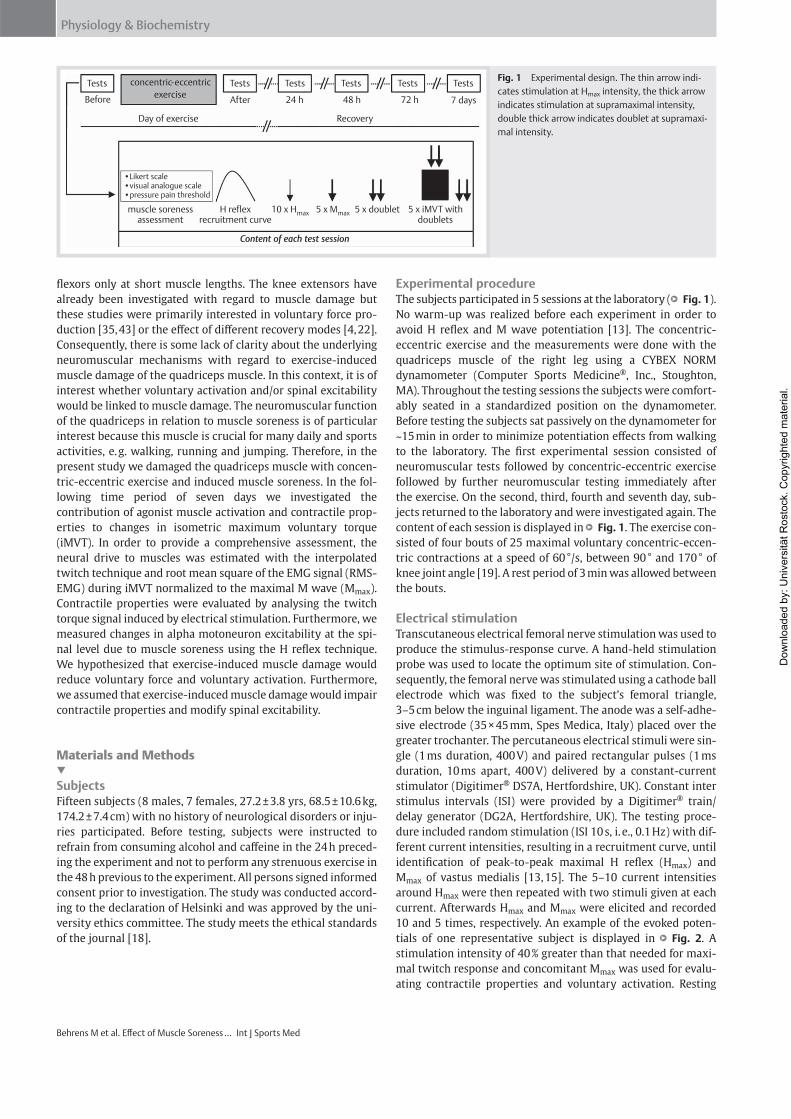
2.1.3 Methoden
Im Folgenden wird der methodische Rahmen für das durchgeführte Experiment dargestellt.
Dabei wird der methodische Ansatz global skizziert. Eine detaillierte Beschreibung der
Personenstichprobe, experimentellen Prozedur, Datenaufnahme und -analyse sowie
statistischen Analyse für das Experiment ist in der Publikation zu finden.
Für die Messung des akuten Funktionsverlustes der ischiocruralen Muskulatur aufgrund von
ermüdender physischer Aktivität wurden die Probanden vor und nach einer
Ermüdungsintervention, die aus repetitiven Sprüngen bis zur Erschöpfung bestand, untersucht.
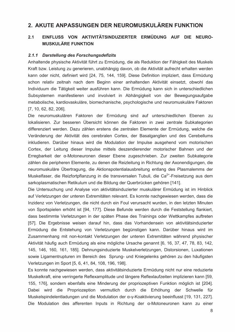
Die Messungen beinhalteten die Erhebung der anterioren Tibiatranslation mittels eines
Kniearthrometers (Abbildung 3) und der Reflexantworten des M. biceps femoris und M.
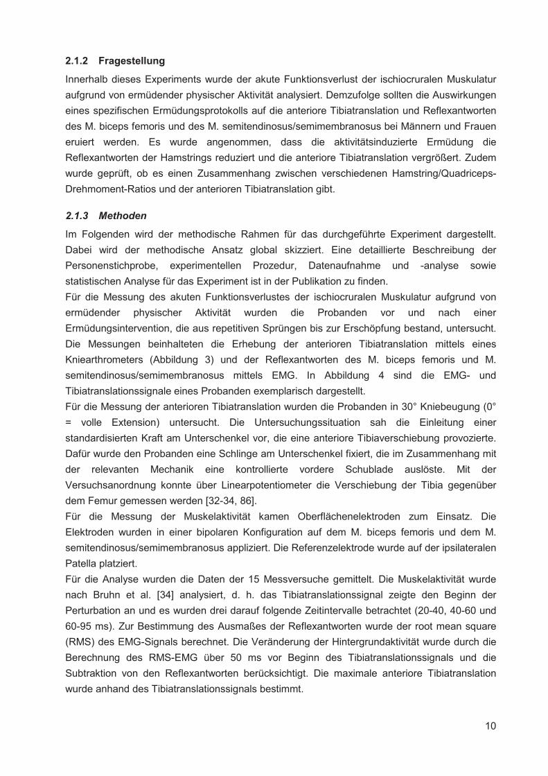
semitendinosus/semimembranosus mittels EMG. In Abbildung 4 sind die EMG- und
Tibiatranslationssignale eines Probanden exemplarisch dargestellt.
Für die Messung der anterioren Tibiatranslation wurden die Probanden in 30° Kniebeugung (0°
= volle Extension) untersucht. Die Untersuchungssituation sah die Einleitung einer
standardisierten Kraft am Unterschenkel vor, die eine anteriore Tibiaverschiebung provozierte.
Dafür wurde den Probanden eine Schlinge am Unterschenkel fixiert, die im Zusammenhang mit
der relevanten Mechanik eine kontrollierte vordere Schublade auslöste. Mit der
Versuchsanordnung konnte über Linearpotentiometer die Verschiebung der Tibia gegenüber
dem Femur gemessen werden [32-34, 86].
Für die Messung der Muskelaktivität kamen Oberflächenelektroden zum Einsatz. Die
Elektroden wurden in einer bipolaren Konfiguration auf dem M. biceps femoris und dem M.
semitendinosus/semimembranosus appliziert. Die Referenzelektrode wurde auf der ipsilateralen
Patella platziert.
Für die Analyse wurden die Daten der 15 Messversuche gemittelt. Die Muskelaktivität wurde
nach Bruhn et al. [34] analysiert, d. h. das Tibiatranslationssignal zeigte den Beginn der
Perturbation an und es wurden drei darauf folgende Zeitintervalle betrachtet (20-40, 40-60 und
60-95 ms). Zur Bestimmung des Ausmaßes der Reflexantworten wurde der root mean square
(RMS) des EMG-Signals berechnet. Die Veränderung der Hintergrundaktivität wurde durch die
Berechnung des RMS-EMG über 50 ms vor Beginn des Tibiatranslationssignals und die
Subtraktion von den Reflexantworten berücksichtigt. Die maximale anteriore Tibiatranslation
wurde anhand des Tibiatranslationssignals bestimmt.
11
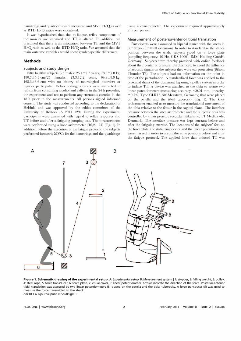
Abb. 3 Darstellung des Versuchsaufbaus zur Messung der funktionellen Kniestabilität. A Experimentelles Setup, B Kniearthrometer | 1: Stopper, 2: Gewicht, 3: Seilzug, 4: Stahlseil, 5: Kraftaufnehmer, 6: Kraftmessplatte, 7: Sichtschutz, 8: Linearpotentiometer. Die Pfeile zeigen die Richtung der wirkenden Kraft an. Die anteriore Tibiatranslation wurde von den beiden Linearpotentiometern erfasst, die auf der Patella und Tuberositas tibiae platziert waren. Die an der Wade applizierte Kraft wurde mittels eines Kraftsensors (5) aufgezeichnet.
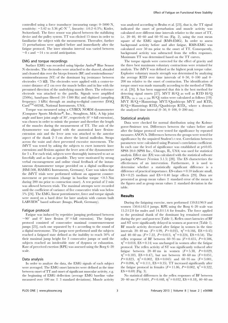
Abb. 4 EMG- und Tibiatranslationssignale eines Probanden. In dieser Abbildung wurde das EMG-Signal gleichgerichtet, um die unterschiedlichen Anteile des Dehnungsreflexes zu visualisieren. Die dicke gestrichelte Linie markiert den Beginn der anterioren Tibiatranslation. Das EMG-Signal wurde in drei Zeitfenstern betrachtet (20-40, 40-60 und 60-95 ms).
12
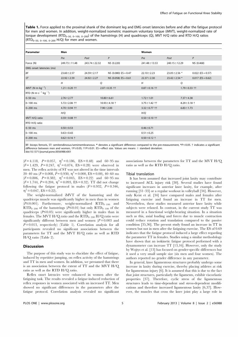
2.1.4 Ergebnisse und Diskussion
Innerhalb dieses Kapitels werden die wichtigsten Ergebnisse des Experiments vorgestellt und
anschließend kurz diskutiert. Die komplette Darstellung der Resultate der Studie ist in der
angehängten Publikation zu finden.
Die durchgeführte Studie sollte den akuten Funktionsverlust der ischiocruralen Muskulatur
aufgrund von ermüdender physischer Aktivität auf die anteriore Tibiatranslation und die
Reflexantworten des M. biceps femoris und des M. semitendinosus/semimembranosus bei
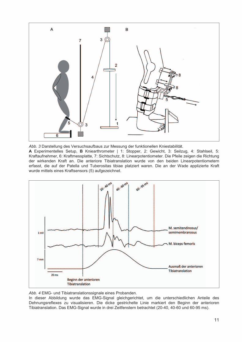
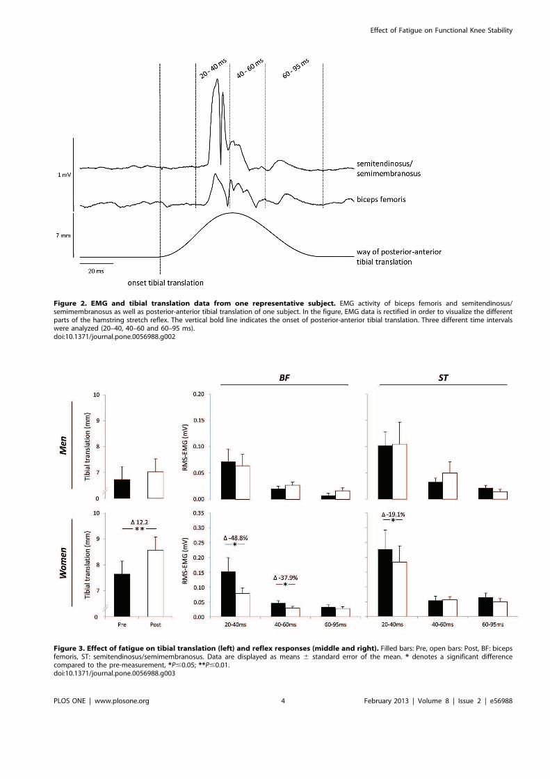
Männern und Frauen eruieren. Es wurde festgestellt, dass die anteriore Tibiatranslation bei den
Frauen signifikant erhöht war. Die Veränderung in der Mechanik ging mit einer signifikanten
Reduktion der Reflexantwort des M. biceps femoris in den Zeitfenstern 20-40 und 40-60 ms
sowie des M. semitendinosus/semimembranosus im Zeitfenster 20-40 ms einher. Bei den
Männern konnten keine statistisch signifikanten Veränderungen von Pre zu Post nachgewiesen
werden (Abbildung 5).
Abb. 5 Einfluss von Ermüdung auf die anteriore Tibiatranslation (links) und die Reflexantworten des M. biceps femoris (BF) und des M. semitendinosus/semimembranosus (ST) (Mitte und rechts). Schwarze Balken: Pre, weiße Balken: Post. Die Daten sind als Mittelwerte mit Standardfehler dargestellt. * zeigt eine signifikante Veränderung zum Pre-Wert an (* P � 0,05; ** P � 0,01).
Es wird angenommen, dass eine erhöhte Kniegelenklaxizität zur Pathomechanik von ACL-
Rupturen beitragen kann [178]. Zudem konnte gezeigt werden, dass sportliche Aktivitäten, wie
z. B. das Laufen [105, 115, 158] oder ein Volleyballtraining [125], eine akute Zunahme der
Gelenklaxizität bewirken können. Innerhalb der genannten Experimente wurde die
Kniegelenklaxizität jedoch in einer Untersuchungssituation analysiert, in der die Muskeln inaktiv
waren und somit ihrer gelenkstabilisierenden Funktion nicht nachkommen konnten. Im Verlauf
der hier vorliegenden Studie wurde die anteriore Tibiatranslation während einer
Gewichtsbelastung gemessen. In dieser Untersuchungssituation wirkten Muskel- und axiale
13
Kräfte. Die durch Muskeln und das Körpergewicht generierten Kräfte können zu einer Reduktion
der Translation und Rotation der Tibia beitragen [135, 224]. Dabei wird vor allem der
ischiocruralen Muskulatur eine große Bedeutung zugeschrieben. Sie fungiert als Antagonist zur
anterioren Tibiatranslation und schützt das ACL bei Bewegungen der Tibia relativ zum Femur
[26, 106, 152]. Im Hinblick darauf wird der Muskelaktivierung durch Dehnungsreflexe eine
potentielle Bedeutung für die Kniegelenkstabilität eingeräumt [73, 74]. Friemert et al. [74]
fanden heraus, dass vor allem die Höhe der Muskelaktivität der „short latency response“ der
Reflexantwort mit dem Ausmaß der anterioren Tibiatranslation korrespondiert. Dieser Phase der
Reflexantwort trug innerhalb der durchgeführten Studie das Analysezeitfenster von 20-40 ms
Rechnung. Das vorliegende Experiment zeigte diesen Zusammenhang bei den Frauen auf,
während bei den Männern keine signifikante Veränderung durch die Ermüdungsintervention
induziert wurde. Demnach war eine Reduktion der Reflexaktivität des M. biceps femoris in den
Analysezeiträumen 20-40 und 40-60 ms sowie des M. semitendinosus/semimembranosus im
Zeitfenster 20-40 ms bei den Frauen zu verzeichnen. Diese führte zu einer korrespondierenden
signifikanten Zunahme der anterioren Tibiatranslation. Die Ergebnisse können mit einer
unterschiedlichen Beanspruchung des neuromuskulären Systems bei Frauen und Männern
begründet werden. Dabei scheint die neuromuskuläre Kontrolle in Abhängigkeit von der
Bewegungsaufgabe moduliert zu werden. Rozzi et al. [187] haben herausgefunden, dass
Frauen im Vergleich zu Männern eine erhöhte Aktivierung der ischiocruralen Muskulatur bei der
Landung von einem Sprung aufweisen. Darüber hinaus konnten die Autoren eine erhöhte
Kniegelenklaxizität und eine reduzierte propriozeptive Funktion bei Frauen feststellen. Sie
kamen zu dem Schluss, dass die erhöhte Muskelaktivierung vermutlich ein Versuch des
neuromuskulären Systems ist, diese Defizite zu kompensieren. Wenn dieser kompensatorische
Mechanismus durch Ermüdung gestört wird, besteht eine erhöhte Verletzungsgefahr [187].
Demnach ist es vorstellbar, dass das verwendete Ermüdungsprotokoll eine stärkere Ermüdung
in der ischiocruralen Muskulatur der Frauen bewirkte, die zu den reduzierten Reflexantworten
beitrug. Dabei können folgende durch Ermüdung ausgelösten Prozesse eine Rolle spielen: (I)
Veränderungen der intrafusalen Eigenschaften [29, 239], (II) Modulation der präsynaptischen
Inhibition der Ia Afferenzen [58, 59, 184], (III) Modifikation der intrinsischen Eigenschaften von
Motoneuronen [113, 176].
14
2.2 EINFLUSS VON KONTRAKTIONSINDUZIERTEN MUSKELVERLETZUNGEN AUF
DIE NEUROMUSKULÄRE FUNKTION
2.2.1 Darstellung des Forschungsdefizits
Die kontraktionsinduzierte Muskelverletzung ist ein Phänomen, welches infolge ungewohnter
physischer und ermüdender Aktivität auftritt. Je nach Ausmaß der Muskelverletzung beinhalten
die mit ihr einhergehenden Symptome (I) die partielle Ruptur intrazellulärer Muskelstrukturen,
des Sarkolemms und der extrazellulären Matrix [71, 72, 210], (II) die Beeinträchtigung der
neuromuskulären Funktion [140, 172], (III) die Entstehung von Schwellungen und den
temporären Verlust des vollen Bewegungsausmaßes sowie (IV) das Auftreten von
Muskelschmerzen [51]. Die Muskelschmerzen, die mit Verzögerung auftreten und ihren
Höhepunkt nach zwei bis drei Tagen erreichen [51], werden in der internationalen Literatur als
„Delayed Onset Muscle Soreness“ und im Volksmund als „Muskelkater“ bezeichnet.
Es konnte gezeigt werden, dass exzentrische Muskelaktionen einen stärkeren Muskelschaden
induzieren als konzentrische oder isometrische [38]. Exzentrische Muskelaktionen, gefolgt von
konzentrischen, sind Bestandteil des Dehnungs-Verkürzungs-Zyklus (DVZ) und kommen bei
alltäglichen und sportlichen Bewegungen, wie dem Gehen, Laufen und Springen, vor. Die mit
exzentrischen Muskelaktionen einhergehenden Kräfte können zum Auftreten von
kontraktionsinduzierten Muskelverletzungen führen. Dies ist bei langandauernden und/oder
intensiven alltäglichen und sportlichen Aktivitäten, wie z. B. dem Laufen, repetitiven Springen
und Krafttraining, der Fall. Demnach kommt die kontraktionsinduzierte Muskelverletzung häufig
bei Freizeit- und Leistungssportlern sowie in der Rehabilitation nach Verletzungen und
operativen Eingriffen vor. Dabei tritt sie vor allem bei Überbelastung und Überbeanspruchung
auf [65].
Für den Menschen ist dabei der mit der Muskelverletzung einhergehende Funktionsverlust der
Muskeln von Bedeutung, weil dieser zu Leistungseinbußen führt. Darüber hinaus kann eine
inadäquate Muskelfunktion zu einem erhöhten Verletzungsrisiko beitragen. So kann eine
verminderte neuromuskuläre Funktion der gelenkumspannenden Muskulatur z. B. die
funktionelle Gelenkstabilität reduzieren [186] und somit das Verletzungsrisiko erhöhen.
Andererseits ist bekannt, dass die kontraktionsinduzierten Muskelschäden reversibel sind,
soweit die Schädigung ein gewisses Ausmaß nicht überschreitet und den betroffenen
Strukturen Gelegenheit zur Regeneration gegeben wird. Die Auswirkungen von
kontraktionsinduzierten Muskelverletzungen wurden in diversen Studien analysiert [17, 35, 50,
95, 172, 173, 175, 195, 205, 223, 225, 234]. Der Fokus der Studien lag dabei zum einen auf
den Veränderungen von Prozessen auf Muskelebene bedingt durch den strukturellen
Muskelschaden, d. h. Modulation der elektromechanischen Kopplung [101], Redistribution der
Sarkomerlängen [153], selektive Zerstörung von Muskelfasern [72] und Beeinträchtigung der
muskulären Glykogenresynthese [12]. Zum anderen wurde die Muskelfunktion in Bezug auf die
Ausdauerperformance [81] und neuromuskuläre Funktion [56] untersucht. Die Ergebnisse der
15
zitierten Studien zeigen, dass kontraktionsinduzierte Muskelverletzungen eine Abnahme der
Muskelkraft und damit auch der Performance nach sich ziehen.
Es gibt jedoch nur wenige Studien, die zwischen dem Beitrag der Beeinträchtigungen auf
Muskelebene und der Modulationen im Zentralnervensystem zur reduzierten Muskelkraft
differenzieren konnten [171, 172, 175]. Dabei ist im Hinblick auf die reduzierte Muskelkraft die
neuromuskuläre Funktion von Bedeutung. Diese beinhaltet erstens die periphere Ebene, d. h.
die muskuläre Funktion, und zweitens die zentrale Ebene, d. h. die neuronale Funktion. Der
Beitrag der beiden Ebenen zu der reduzierten Muskelkraft kann unter Zuhilfenahme von
geeigneten Methoden, wie z. B. Stimulationsprotokollen, aufgeklärt werden.
Die Beeinträchtigungen auf der peripheren Ebene infolge von kontraktionsinduzierten
Muskelverletzungen beinhalten u. a. die Störung der elektromechanischen Kopplung [101] und
die Ruptur von Strukturen auf Sarkomerebene [174]. Die Modulation auf der zentralen Ebene
beinhaltet u. a. die inadäquate willkürliche Aktivierung von Muskeln [171, 175]. Es wird
angenommen, dass die reduzierte neuronale Ansteuerung ein Schutzmechanismus ist, der das
tendomuskuläre System vor weiterem Schaden bewahrt [175]. Der mit der Muskelverletzung
einhergehende Schmerz aktiviert vermutlich Gruppe III und IV Afferenzen [162], die das
Potential besitzen Modulationen auf kortikaler [139] und spinaler Ebene [14] zu induzieren.
Diese wiederum sollen zur reduzierten willkürlichen Aktivierung nach kontraktionsinduziertem
Muskelschaden beitragen [175].
Es existieren nur wenige Studien, in denen der Beitrag der willkürlichen Aktivierung zur
reduzierten Kraft mit adäquaten Methoden untersucht wurde [140, 171, 172, 175]. Die zitierten
Studien konzentrierten sich auf die Armflexoren [171, 172], Plantarflexoren [175] und
Knieextensoren [140]. In den genannten Untersuchungen konnte eine Beeinträchtigung der
kontraktilen Eigenschaften der jeweiligen Muskulatur nachgewiesen werden. Im Hinblick auf die
willkürliche Aktivierung wurden gegensätzliche Ergebnisse gefunden. Racinais et al. [175]
konnten eine reduzierte willkürliche Aktivierung der Plantarflexoren bis 48 h nach der
Intervention, die eine Muskelverletzung induzierte, nachweisen, während die Ergebnisse der
anderen Studien keine Beeinträchtigungen für die Armflexoren und Knieextensoren zeigten
[140, 172].
Neben der willkürlichen Aktivierung von Muskeln sind Modulationen innerhalb des
Reflexbogens von Interesse, d. h. Änderungen in der Exzitabilität der �-Motoneuronen und/oder
präsynaptischen Inhibition [238]. Für die Evaluation dieser Modulationen bietet sich die H-
Reflexmethode an, die Anfang des 20. Jahrhunderts durch den deutschen Physiologen Paul
Hoffmann etabliert wurde [100]. Avela et al. [14] konnten einen reduzierten H-Reflex nach
kontraktionsinduziertem Muskelschaden nachweisen, während Racinais et al. [175] keine
Veränderung des H-Reflexes dokumentierten.
Aufgrund der widersprüchlichen Ergebnisse und Unklarheiten sollte die folgende eigene Studie,
im Hinblick auf den M. quadriceps femoris, zur Erweiterung des Wissens beitragen.
EXPERIMENT II: Behrens, M., Mau-Moeller, A., Bruhn, S. (2012). Effect of exercise-induced
muscle damage on neuromuscular function of the quadriceps muscle. International Journal of
Sports Medicine, 33 (8), 600-606.
16
2.2.2 Fragestellung
Innerhalb dieser Studie wurde die Regeneration der neuromuskulären Funktion des M.
quadriceps femoris nach einer Ermüdungsintervention, die zur Überbelastung und somit zu
mikrostrukturellen Muskelverletzungen führte, untersucht. Es wurden die Auswirkungen von
kontraktionsinduzierten Muskelverletzungen auf das isometrische maximale willkürliche
Drehmoment (iMVT), die willkürliche Aktivierung, die kontraktilen Eigenschaften und den
Schmerz des M. quadriceps femoris in einem Zeitraum von sieben Tagen analysiert. Zudem
wurde der H-Reflex des M. vastus medialis abgeschätzt. Es wurde angenommen, dass das
iMVT infolge der kontraktionsinduzierten Muskelverletzungen sinkt und die willkürliche
Aktivierung, die kontraktilen Eigenschaften sowie der H-Reflex moduliert werden.
2.2.3 Methoden
Im Folgenden wird der methodische Rahmen für das durchgeführte Experiment dargestellt.
Dabei wird der methodische Ansatz global skizziert. Eine detaillierte Beschreibung der
Personenstichprobe, experimentellen Prozedur, Datenaufnahme und -analyse sowie
statistischen Analyse für das Experiment ist in der Publikation zu finden.
Für die Untersuchung der Modulationen innerhalb des neuromuskulären Systems, ausgelöst
durch kontraktionsinduzierte Muskelverletzungen, wurden Kraftmessungen und
neurophysiologische Techniken kombiniert.
Zu den Messzeitpunkten wurden die Probanden auf einem Dynamometer fixiert, um die
Drehmomente zu messen, die durch den M. quadriceps femoris produziert wurden.
Für die Messung der Muskelaktivität kamen Oberflächenelektroden zum Einsatz. Die
Elektroden wurden in einer bipolaren Konfiguration auf dem M. rectus femoris, dem M. vastus
medialis und dem M. vastus lateralis appliziert. Die Referenzelektrode wurde auf der
ipsilateralen Patella platziert.
Die Analyse der Modulationen auf den unterschiedlichen Ebenen des neuromuskulären
Systems wurde mithilfe der transkutanen elektrischen Stimulation des N. femoralis realisiert.
Durch die Applikation von elektrischen Stimuli in Ruhebedingung und während der MVCs
konnten unterschiedliche Ebenen des neuromuskulären Systems und deren Modulation näher
untersucht werden. Dazu gehörte die Abschätzung der kontraktilen Eigenschaften des M.
quadriceps femoris in Ruhebedingung, der Erregbarkeit der spinalen �-Motoneuronen des M.
vastus medialis mittels der Exzitation der Ia Afferenzen und der willkürlichen Aktivierung des M.
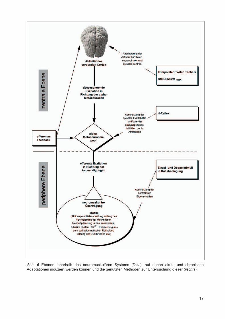
quadriceps femoris während MVC. In Abbildung 6 sind die Ebenen innerhalb des
neuromuskulären Systems, auf denen akute und chronische Adaptationen induziert werden
können und die genutzten Methoden zur Untersuchung dieser dargestellt.
17
Abb. 6 Ebenen innerhalb des neuromuskulären Systems (links), auf denen akute und chronische Adaptationen induziert werden können und die genutzten Methoden zur Untersuchung dieser (rechts).
18
Abschätzung der kontraktilen Eigenschaften
Die Abschätzung der kontraktilen Eigenschaften des M. quadriceps femoris beinhaltete die
Aufzeichnung der maximalen Muskelantworten im EMG (maximale M-Welle [Mmax]) und der
mechanischen Antworten der relaxierten Muskulatur auf supramaximale elektrische Einzel- und
Doppelstimuli, die am N. femoralis appliziert wurden [87, 124, 222]. Die Stimulationsintensität
betrug dabei 140 % der Intensität, mit der Mmax im EMG und die korrespondierende maximale
mechanische Antwort provoziert wurde. Mmax repräsentiert das volle Ausmaß der
Muskelaktivierung, induziert durch die direkte Depolarisation aller motorischen Axone. Es wird
davon ausgegangen, dass diese maximale muskuläre Antwort mit der Entladung des gesamten
Motoneuronenpools gleichgesetzt werden kann [163, 167]. Demnach stellt die supramaximale
Stimulation sicher, dass alle Muskelfasern innerviert werden. Diese Stimulationsmethode
erlaubt eine Abschätzung der kontraktilen Eigenschaften der betreffenden Muskulatur [80, 87,
124, 128]. Der involvierte Prozess umfasst die elektromechanische Kopplung inklusive der
intrazellulären Ca2+-Kinetik. Durch den Pre-Post-Vergleich der Amplitude von Mmax und der
korrespondierenden mechanischen Antwort kann auf differente Anpassungen des
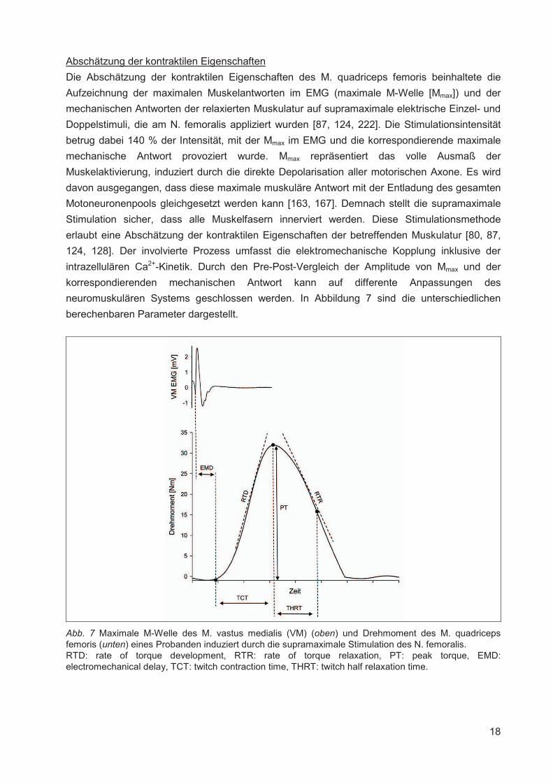
neuromuskulären Systems geschlossen werden. In Abbildung 7 sind die unterschiedlichen
berechenbaren Parameter dargestellt.
Abb. 7 Maximale M-Welle des M. vastus medialis (VM) (oben) und Drehmoment des M. quadriceps femoris (unten) eines Probanden induziert durch die supramaximale Stimulation des N. femoralis. RTD: rate of torque development, RTR: rate of torque relaxation, PT: peak torque, EMD: electromechanical delay, TCT: twitch contraction time, THRT: twitch half relaxation time.
19
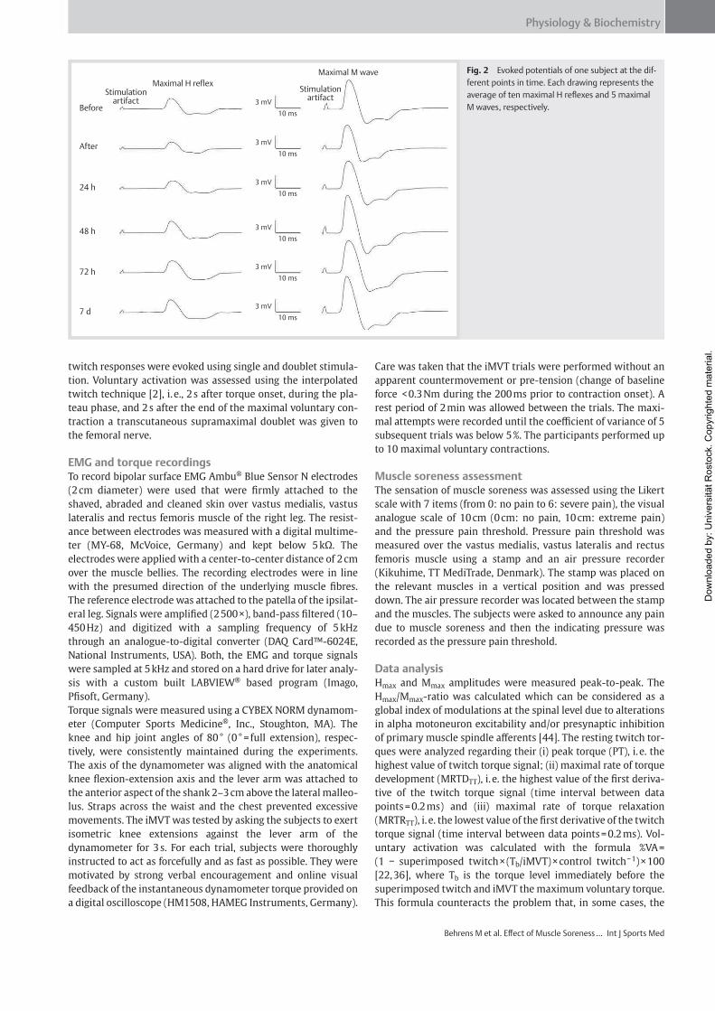
Abschätzung der Exzitabilität der spinalen �-Motoneuronen
Mit der H-Reflextechnik kann die Modulation der Exzitabilität der spinalen �-Motoneuronen
und/oder präsynaptischen Inhibition der Ia Afferenzen infolge von kurz- oder langfristigen
Interventionen abgeschätzt werden [2, 167, 197, 238]. Die Auslösung des H-Reflexes basiert
auf der kurzen elektrischen Stimulation eines gemischten peripheren Nervens, der sowohl
afferente als auch efferente Axone beinhaltet [166, 238]. Aufgrund der elektrischen Reizung mit
einer geringen Stromstärke werden die Ia-Spindelafferenzen depolarisiert und das
Aktionspotential breitet sich entsprechend dem Alles-oder-Nichts-Gesetz in Richtung der
homonymen �-Motoneuronen aus. Im Rückenmark erfolgt eine Umschaltung der Erregung auf
die �-Motoneuronen, deren Depolarisation Aktionspotentiale generiert, die sich über den
efferenten Schenkel des Reflexbogens bis zum Muskel ausbreiten. Die durch die
Elektrostimulation induzierte Reflexantwort des Muskels wird mittels Oberflächenelektroden
elektromyografisch erfasst. Die Reflexantwort variiert in Abhängigkeit von der applizierten
Stromstärke. Demnach erscheint bei geringen Stromstärken zunächst der H-Reflex. Mit
zunehmender Stimulationsintensität erhöht sich der H-Reflex, bis er sein Maximum erreicht, um
sich anschließend wieder zu reduzieren. Eine Erhöhung der Reizstärke impliziert außerdem die
Generierung eines weiteren Potentials, das als M-Welle bezeichnet wird und durch die
Stimulation der motorischen Axone entsteht. Diese direkte Muskelantwort wird durch eine
efferente Reizleitung in Richtung des Muskels ausgelöst und erreicht bei einer bestimmten
Stimulationsintensität ihr Maximum, welches als Mmax bezeichnet wird [163, 167, 238]. Die
maximale Amplitude des H-Reflexes im Verhältnis zur maximalen M-Welle (Hmax/Mmax-Ratio)
ermöglicht die Abschätzung der spinalen Exzitabilität und/oder präsynaptischen Inhibition der Ia
Afferenzen für einen gegebenen Zustand [238]. Veränderungen des Verhältnisses beider
Amplituden durch Interventionen dokumentieren Adaptationen auf der Ebene des spinalen
sensomotorischen Systems. Im Hinblick auf die durchgeführten Experimente wurde die
elektrische Stimulation am N. femoralis appliziert und die elektromyographische Erfassung der
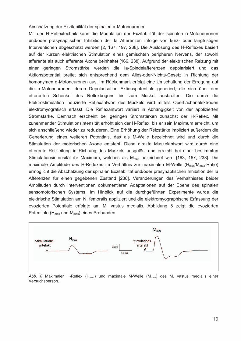
evozierten Potentiale erfolgte am M. vastus medialis. Abbildung 8 zeigt die evozierten
Potentiale (Hmax und Mmax) eines Probanden.
Abb. 8 Maximaler H-Reflex (Hmax) und maximale M-Welle (Mmax) des M. vastus medialis einer Versuchsperson.
20
Abschätzung der willkürlichen Aktivierung
Die Abschätzung der willkürlichen Aktivierung des M. quadriceps femoris erfolgte zum einen
über die Berechnung des normalisierten RMS-EMG (RMS-EMG/Mmax) und zum anderen mittels
der Interpolated Twitch Technik.
Der Vorteil der normalisierten EMG-Daten gegenüber den „einfachen“ EMG-Daten liegt in der
Reduktion von potentiellen Fehlerquellen. Dazu gehören eine veränderte Elektrodenposition bei
Wiederholungsmessungen sowie die Inter-Session-Variabilität der Hautimpedanz, des
subkutanen Fettgewebes und der Faszien [219]. Der RMS-EMG/Mmax wurde herangezogen, um
die Aktivierung des M. quadriceps femoris während der initialen Phase des
Drehmomentanstieges (RMS-EMGRTD/Mmax) und des Drehmomentmaximums (RMS-
EMGiMVT/Mmax) zu analysieren.
Als zweites Verfahren wurde die Interpolated Twitch Technik verwendet. Mit dieser Methode ist
es möglich, die volitive Aktivierung eines Muskels zu messen [8, 77, 122, 149]. Anders gesagt,
kann somit das Ausmaß des Aktivierungsdefizits für den relevanten Muskel abgeschätzt
werden. Dieses wird über die supramaximale Stimulation eines peripheren Nervens während
einer statischen MVC und der Messung des korrespondierenden mechanischen Outputs eruiert.
Es wird angenommen, dass die supramaximale Aktivierung der motorischen Axone alle
Muskelfasern rekrutiert [163], demnach auch solche, die bei der willkürlichen Anstrengung nicht
in den Kontraktionsprozess einbezogen werden können. Wenn die elektrische Stimulation
keinen additionalen mechanischen Output produziert, ist der Muskel durch den
deszendierenden Input von kortikalen, supraspinalen und spinalen Zentren voll aktiviert. Wird
das generierte Drehmoment durch die Stimuli jedoch erhöht, weist das auf eine submaximale
volitive Aktivierung hin [117].
Die Quantifizierung der volitiven Aktivierung (VA) erfolgt über die Formel:
% VA = (1 – (superimposed twitch * (Tb * iMVT-1) * control twitch-1)) * 100 [140, 212]
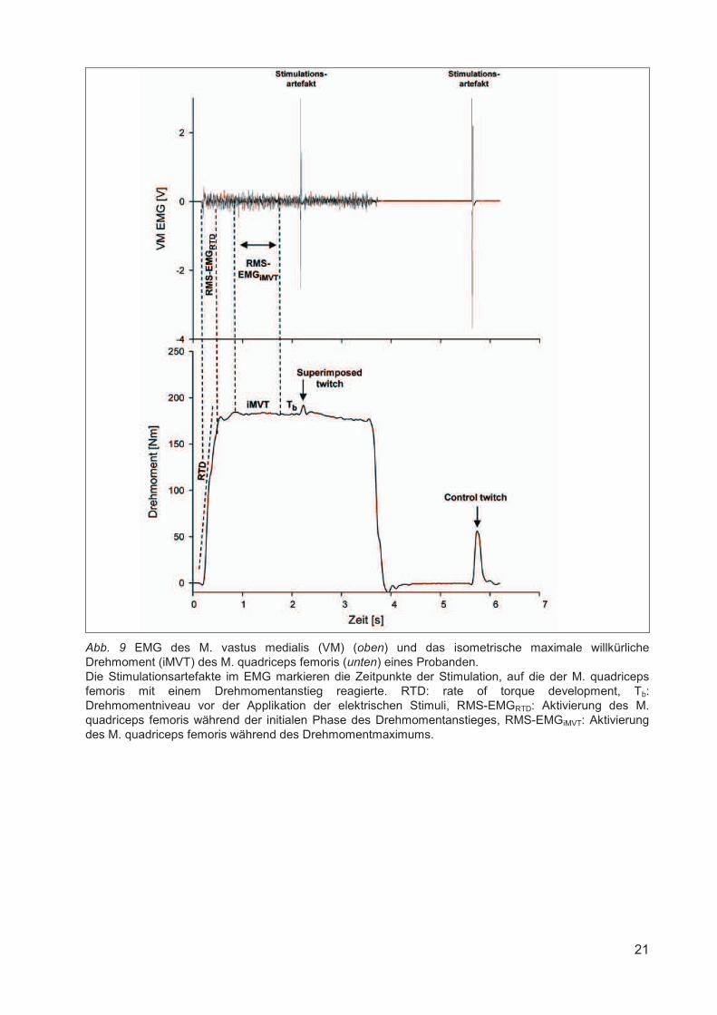
Tb bezeichnet das Drehmomentniveau vor der Applikation der elektrischen Stimuli, welches
aufgrund der Fluktuation der Drehmoment-Zeit-Kurve unter dem iMVT liegen kann. Die
beschriebene Formel wirkt dieser Fehlerquelle entgegen. In Abbildung 9 kann die Methode
anhand von Rohdaten eines Probanden nachvollzogen werden. Die Versuchsperson hatte die
Aufgabe eine isometrische MVC durchzuführen. Die elektrische Stimulation des N. femoralis
erfolgte 2 s nach dem Beginn der Kontraktion und generierte den „superimposed twitch“. Das
Ende der Kontraktion löste eine weitere 2 s später erfolgende elektrische Stimulation aus, die
den „control twitch“ produzierte. Mithilfe der in der Formel aufgeführten Parameter kann auf die
willkürliche Aktivierung in Prozent geschlossen werden. Die Abbildung zeigt zudem weitere
Parameter, die anhand der Rohdaten berechnet werden können und Aufschluss über die
neuromuskuläre Funktion geben.
21
Abb. 9 EMG des M. vastus medialis (VM) (oben) und das isometrische maximale willkürliche Drehmoment (iMVT) des M. quadriceps femoris (unten) eines Probanden. Die Stimulationsartefakte im EMG markieren die Zeitpunkte der Stimulation, auf die der M. quadriceps femoris mit einem Drehmomentanstieg reagierte. RTD: rate of torque development, Tb: Drehmomentniveau vor der Applikation der elektrischen Stimuli, RMS-EMGRTD: Aktivierung des M. quadriceps femoris während der initialen Phase des Drehmomentanstieges, RMS-EMGiMVT: Aktivierung des M. quadriceps femoris während des Drehmomentmaximums.
22
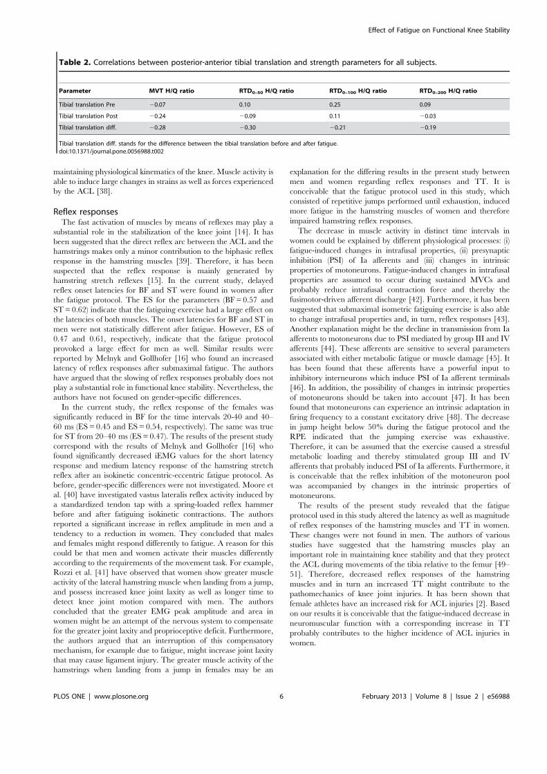
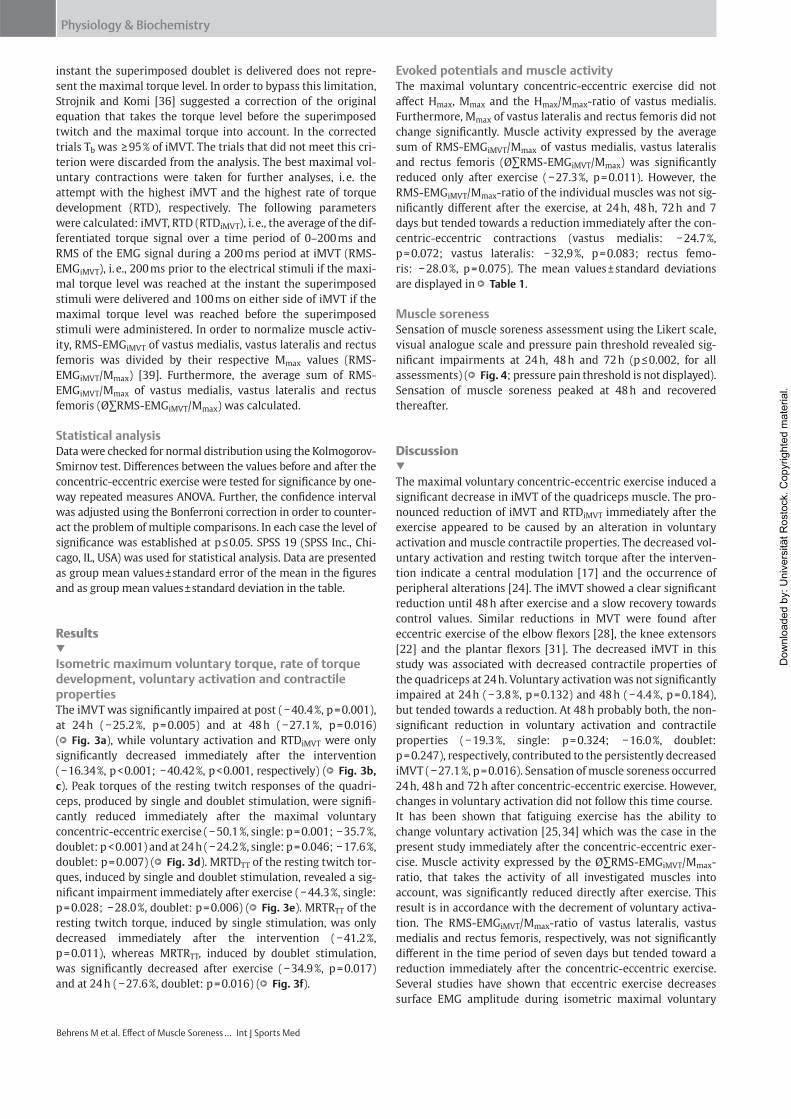
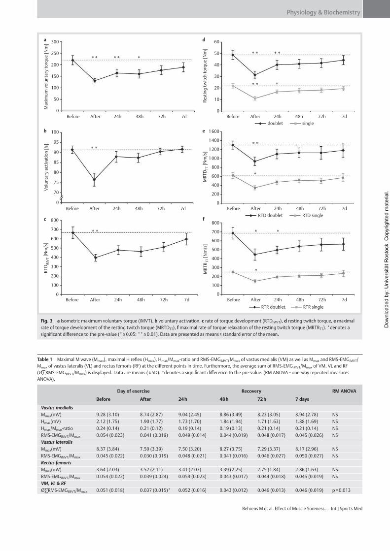
2.2.4 Ergebnisse und Diskussion
Die durchgeführte Studie untersuchte die Regeneration der neuromuskulären Funktion des M.
quadriceps femoris nach einer Ermüdungsintervention, welche zur Überbelastung und somit zu
mikrostrukturellen Muskelverletzungen führte. Dafür wurden die Auswirkungen von
kontraktionsinduzierten Muskelverletzungen auf das iMVT, die willkürliche Aktivierung, die
kontraktilen Eigenschaften und den Schmerz des M. quadriceps femoris in einem Zeitraum von
sieben Tagen (Post, 24 h, 48 h, 72 h, 7 d) analysiert. Zudem wurde die Exzitabilität des �-
Motoneuronenpools des M. vastus medialis über Ia Afferenzen zu den Messzeitpunkten
abgeschätzt. Die kontraktionsinduzierten Muskelverletzungen wurden mithilfe eines
Dynamometers durch vier Sätze mit jeweils 25 maximalen willkürlichen konzentrisch-
exzentrischen Kontraktionen bei einer Geschwindigkeit von 60°/s induziert.
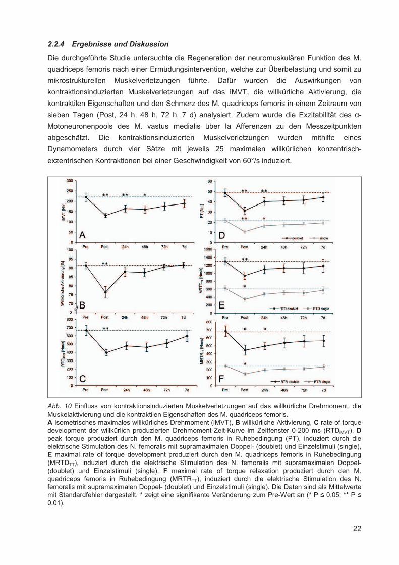
Abb. 10 Einfluss von kontraktionsinduzierten Muskelverletzungen auf das willkürliche Drehmoment, die Muskelaktivierung und die kontraktilen Eigenschaften des M. quadriceps femoris. A Isometrisches maximales willkürliches Drehmoment (iMVT), B willkürliche Aktivierung, C rate of torque development der willkürlich produzierten Drehmoment-Zeit-Kurve im Zeitfenster 0-200 ms (RTDiMVT), Dpeak torque produziert durch den M. quadriceps femoris in Ruhebedingung (PT), induziert durch die elektrische Stimulation des N. femoralis mit supramaximalen Doppel- (doublet) und Einzelstimuli (single), E maximal rate of torque development produziert durch den M. quadriceps femoris in Ruhebedingung (MRTDTT), induziert durch die elektrische Stimulation des N. femoralis mit supramaximalen Doppel- (doublet) und Einzelstimuli (single), F maximal rate of torque relaxation produziert durch den M. quadriceps femoris in Ruhebedingung (MRTRTT), induziert durch die elektrische Stimulation des N. femoralis mit supramaximalen Doppel- (doublet) und Einzelstimuli (single). Die Daten sind als Mittelwerte mit Standardfehler dargestellt. * zeigt eine signifikante Veränderung zum Pre-Wert an (* P � 0,05; ** P �0,01).
23
Unmittelbar nach der Intervention (Post) war das iMVT signifikant reduziert. Dafür kann eine
signifikante Abnahme der willkürlichen Aktivierung und der kontraktilen Eigenschaften des M.
quadriceps femoris verantwortlich gemacht werden. Die Verminderung der Muskelaktivierung
und -funktion zum Zeitpunkt Post weist auf interventionsbedingte zentrale [75] und periphere
Modulationen hin [148]. Dabei ist die deutliche Abnahme der Parameter vermutlich
überwiegend auf die akuten Effekte der Ermüdung zurückzuführen [150, 202]. Die
Folgemessungen ergaben eine signifikante Reduktion des iMVT bis 48 h nach der Intervention,
die primär auf die Verschlechterung der kontraktilen Funktion zurückzuführen war. Die
willkürliche Aktivierung, abgeschätzt mittels Interpolated Twitch Technik, zeigte zwar keine
signifikante Änderung, war aber nach 24 h um 3,8 % und nach 48 h um 4,4 % vermindert.
Abbildung 10 zeigt die Zusammenfassung der wichtigsten Ergebnisse. Demnach scheinen
akute Adaptationen in der Peripherie eine größere Rolle für die Abnahme des Drehmoments
nach kontraktionsinduzierten Muskelverletzungen zu spielen, wobei die Modulation der
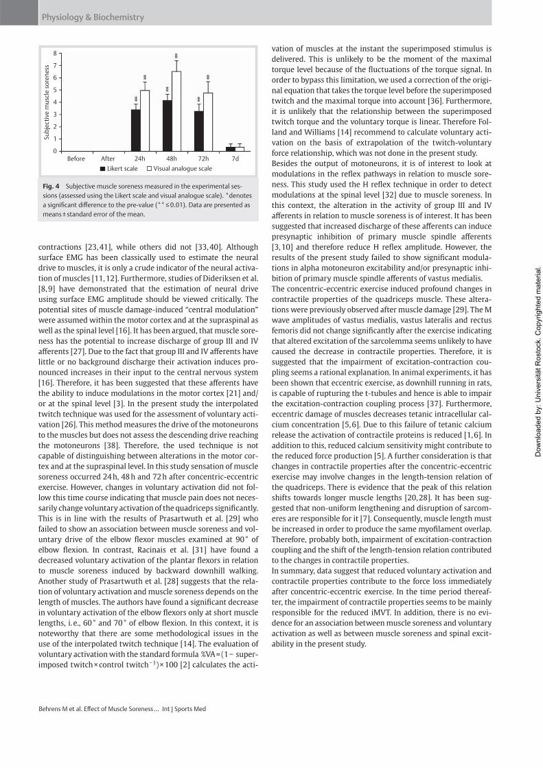
Muskelaktivierung nicht ausgeschlossen werden kann. Die Messung des Muskelschmerzes
ergab eine signifikante Zunahme zu den Messzeitpunkten 24 h, 48 h und 72 h. Die neuronalen
Parameter wiesen jedoch keine Korrespondenz mit der Schmerzentwicklung auf.
Die Reduktion des iMVT durch kontraktionsinduzierte Muskelverletzungen konnte in
unterschiedlichen Studien nachgewiesen werden [140, 172, 175]. Im Hinblick auf den Beitrag
der Muskelinaktivierung zur Abnahme des iMVT konnten Racinais et al. [175] eine reduzierte
willkürliche Aktivierung der Plantarflexoren bis 48 h nach der Intervention, die eine
Muskelverletzung induziert hat, nachweisen. Im Widerspruch dazu stehen die Ergebnisse von
Martin et al. [140] und Prasartwuth et al. [172], die keine Veränderung dieses Parameters für
die Knieextensoren und Armflexoren finden konnten. Durch den kombinierten Ansatz der
vorliegenden Studie, der die Abschätzung der willkürlichen Aktivierung über die Interpolated
Twitch Technik und den normalisierten RMS-EMG umfasste, konnte keine Modulation der
Muskelaktivierung festgestellt werden. Zudem zeigte die Exzitabilität der �-Motoneuronen des
M. vastus medialis, abgeschätzt mittels der H-Reflextechnik, ebenfalls keine Veränderung.
Demnach scheint der mit kontraktionsinduzierten Muskelverletzungen einhergehende
Muskelschmerz keine Modulation dieser neuronalen Parameter zu provozieren.
Die Verschlechterung der kontraktilen Funktion nach kontraktionsinduzierten
Muskelverletzungen wurde schon zuvor beobachtet [172]. Die Abnahme der Muskelfunktion
wird dabei primär durch die Beeinträchtigung der elektromechanischen Kopplung verursacht.
Dabei spielen vermutlich die partielle Ruptur transversaler Tubuli [213], die Abnahme der
intrazellulären Ca2+-Konzentration und eine reduzierte Ca2+-Sensitivität eine Rolle [20, 21].
Darüber hinaus wird eine Rechtsverschiebung der Längen-Spannungs-Relation durch den
strukturellen Muskelschaden diskutiert [109, 171], die durch eine ungleichmäßige Dehnung und
Ruptur von Sarkomeren zustande kommt [38]. Demzufolge muss der Muskel länger sein, damit
die Myofilamente optimal interagieren können.
24
3. CHRONISCHE ANPASSUNGEN DER NEUROMUSKULÄREN FUNKTION
3.1 EINFLUSS EINES PLYOMETRISCHEN TRAININGS AUF DIE NEUROMUSKULÄRE
FUNKTION
3.1.1 Darstellung des Forschungsdefizits
Regelmäßige physische Aktivität, z. B. Training, führt zur Steigerung der Leistungsfähigkeit des
neuromuskulären Systems. Im Hinblick auf eine Erhöhung der Muskelkraft bietet sich
Krafttraining an, welches neuronale und muskuläre Adaptationen provoziert. Dabei wird der
Kraftzuwachs in der initialen Phase des Trainings primär neuronalen Anpassungen
zugeschrieben, denen muskuläre Adaptationen folgen [66].
Zu den neuronalen Adaptationen infolge von Krafttraining gehört eine zunehmende
Verbesserung der intermuskulären Koordination, vor allem bei Kraftleistungen, die das
Zusammenspiel mehrerer Muskeln erfordern [188]. Des Weiteren kann es zur Erhöhung der
Muskelaktivierung des Agonisten kommen, die mit einer erhöhten Rekrutierung und
Feuerfrequenz von motorischen Einheiten begründet wird [60, 192]. Ein Anstieg der
agonistischen Muskelaktivität infolge eines Krafttrainings wurde in diversen Studien belegt, die
zur Detektion der trainingsbedingten Veränderungen die Elektromyographie (EMG) [88-90, 118,
157] oder Stimulationstechniken [2, 55, 61, 69, 179] verwendeten. Es gibt Hinweise darauf,
dass die durch Krafttraining induzierten Anpassungen primär auf spinaler Ebene lokalisiert sind
und kortikale Adaptationen eine untergeordnete Rolle spielen [44, 60, 104]. Zudem kann
Krafttraining die Koaktivierung des Antagonisten reduzieren [43], was wiederum zur Abnahme
der zu überwindenden Kraft für den Agonisten führt. Außerdem kann eine verminderte
Koaktivierung zur Erhöhung der agonistischen Muskelaktivität führen, indem die reziproke
Inhibition des Agonisten abnimmt [70].
Die primäre Anpassung auf Muskelebene infolge von Krafttraining ist die Zunahme des
Muskelquerschnitts, die als Muskelhypertrophie bezeichnet wird [70]. Nach der Initiierung des
ersten Trainingsstimulus werden diverse zelluläre und hormonelle Signalwege aktiviert, die zur
Hypertrophie der beanspruchten Muskulatur führen [22, 28, 52]. Die beiden dabei involvierten
Hauptprozesse sind (I) die Erhöhung der Muskelproteinsynthese [123] und (II) die Proliferation
von Satellitenzellen im Muskel [110]. Trotz des zeitnahen Einsetzens der genannten Prozesse
ist eine messbare Zunahme des Muskeldickenwachstums bei zuvor untrainierten Personen erst
nach ca. vier bis acht Wochen nachweisbar [4, 11, 76, 98]. Eine neuere Untersuchung zeigte
jedoch, dass eine signifikante Muskelquerschnittzunahme bereits nach drei Wochen eintreten
kann [200]. Die beiden Prozesse, die das Muskelwachstum induzieren, setzen zwar zeitnah
nach der Applikation des ersten Trainingsstimulus ein, tragen aber in unterschiedlichem
Ausmaß zur Hypertrophie der beanspruchten Muskulatur bei. Demnach ist die Erhöhung der
Muskelproteinsynthese der primäre Faktor für das Muskeldickenwachstum und die Aktivierung
von Satellitenzellen steht diesem nach [11]. Dabei sind die Typ II Muskelfasern stärker von der
Hypertrophie betroffen als die Typ I Muskelfasern [40, 209, 215].
Neben dem Dickenwachstum des Muskelgewebes durch Krafttraining kommt es zu weiteren
Adaptationen. Diese beinhalten Anpassungen innerhalb der Muskelfasertypen, die durch eine
Abnahme der Typ IIX und eine Zunahme der Typ IIA Muskelfasern gekennzeichnet sind [40, 90,
25
92]. Darüber hinaus kann es zur Veränderung der Sehne und des Bindegewebes [70, 119]
sowie der Muskelarchitektur kommen [1, 112].
Krafttraining kann mittels unterschiedlicher Trainingsregimes durchgeführt werden, die
wiederum spezifische Anpassungen provozieren. Dazu gehören u. a. das Training an Geräten,
Training mit freien Gewichten, Elektromyostimulationstraining und plyometrisches Training.
Der Begriff plyometrisches Training bezieht sich auf sportliche Übungen, die den DVZ
involvieren [137]. Der DVZ ist durch eine exzentrische Muskelaktion, auf die eine konzentrische
folgt, gekennzeichnet und kommt bei alltäglichen und sportlichen Bewegungen, wie dem
Gehen, Laufen und Springen, vor. Plyometrisches Training kommt bei gesunden Menschen zur
Anwendung, es wird aber auch für spezielle Patientenpopulationen, z. B. Menschen mit
Osteoporose, empfohlen [137].
Die Auswirkungen eines plyometrischen Trainings auf den menschlichen Organismus wurden in
[9, 111, 121, 231, 233] untersucht. Obwohl diverse Studien eine Kraftsteigerung nach
plyometrischem Training dokumentieren konnten [91, 189, 230], existieren nur wenige Studien,
die die zugrundeliegenden neuromuskulären Adaptationen analysiert haben. Kyrolainen et al.
[127] untersuchten neuronale Anpassungen mittels EMG und muskuläre Adaptationen mithilfe
von Muskelbiopsien. Nach dem Training stellten die Autoren eine erhöhte maximale Kraft der
Plantarflexoren und einen Anstieg der korrespondierenden EMG-Aktivität fest, jedoch keine
Änderungen auf muskulärer Ebene. Kubo et al. [121] konnten ebenfalls eine Steigerung der
Kraft der Plantarflexoren nach plyometrischem Training nachweisen. Mithilfe der Interpolated
Twitch Technik konnten die Autoren zeigen, dass eine erhöhte willkürliche Aktivierung zur
Kraftsteigerung beigetragen hat. Im Hinblick auf die Knieextensoren konnten Kyrolainen et al.
[127] keine Steigerung der maximalen Kraft, aber der Kraftanstiegssteilheit, nach einem
plyometrischen Training feststellen. Malisoux et al. [133] untersuchten muskuläre Adaptationen
des M. vastus medialis infolge eines plyometrischen Trainings mittels Muskelbiopsien und
stellten eine Erhöhung des Querschnitts für Typ-I, -IIA und -IIA/IIX Muskelfasern fest. Die
Autoren konnten außerdem eine Steigerung der Kraft und Sprunghöhe dokumentieren.
Aufgrund der übersichtlichen Anzahl an Studien, die sich mit neuronalen sowie muskulären
Adaptationen und ihrem Beitrag zu einer gesteigerten maximalen Kraft nach plyometrischem
Training beschäftigten, kommen Markovic und Mikulic [137] in ihrem Review-Artikel über
plyometrisches Training auf S. 869 zu dem Fazit: „[…] our current knowledge about plyometric
training-induced changes in neural function is limited“. Hier wird der Forschungsbedarf deutlich.
Es existieren nur wenige Studien, die die neuromuskuläre Funktion des M. quadriceps femoris
vor und nach plyometrischem Training mit geeigneten Methoden analysiert haben. Die folgende
durchgeführte Studie sollte dazu beitragen das Wissen in dieser Hinsicht zu erweitern.
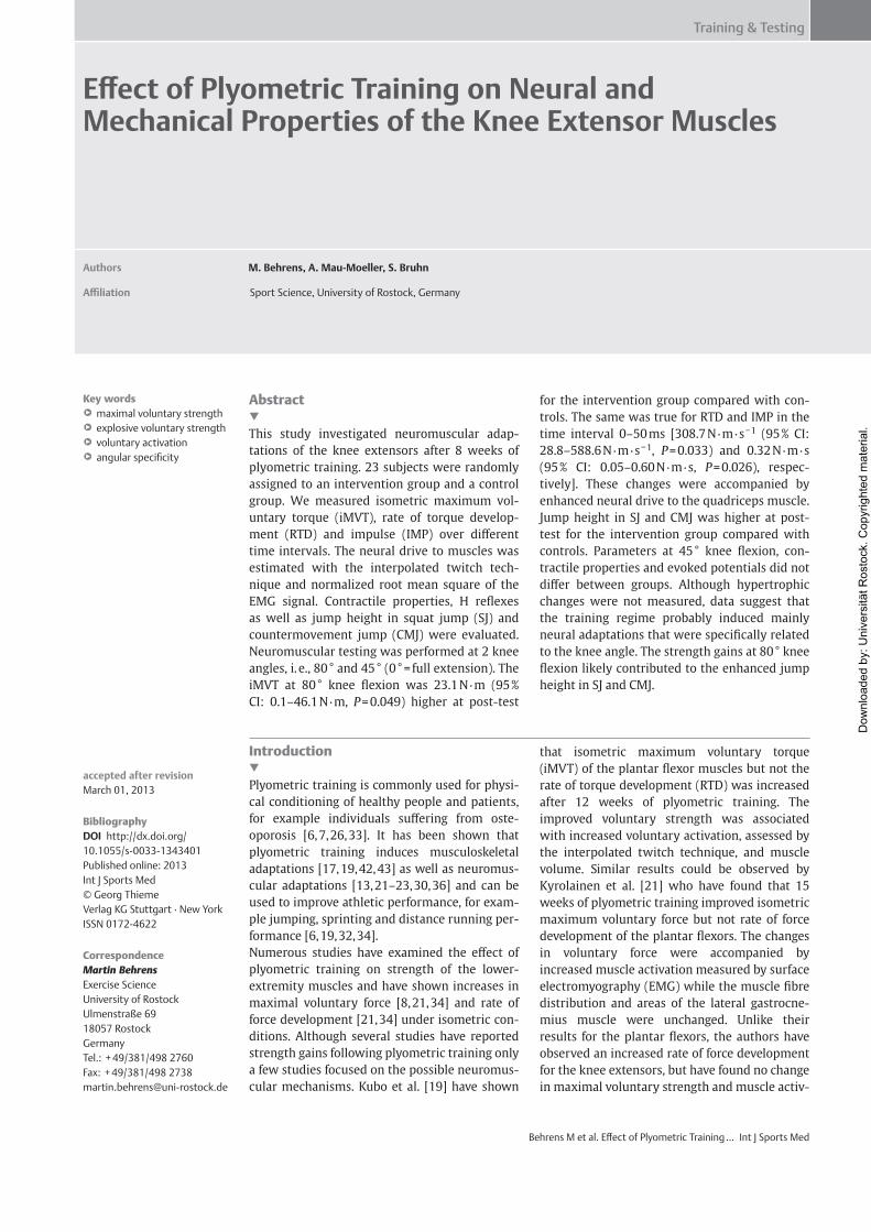
EXPERIMENT III: Behrens, M., Mau-Moeller, A., Bruhn, S. (2013). Effect of plyometric training
on neural and mechanical properties of the knee extensor muscles. International Journal of
Sports Medicine (in press).
26
3.1.2 Fragestellung
Innerhalb dieses Experiments wurde die Modulation der Leistungsfähigkeit des
neuromuskulären Systems durch Training untersucht. Es wurden die Auswirkungen eines
achtwöchigen plyometrischen Trainings auf das iMVT, die willkürliche Aktivierung, die
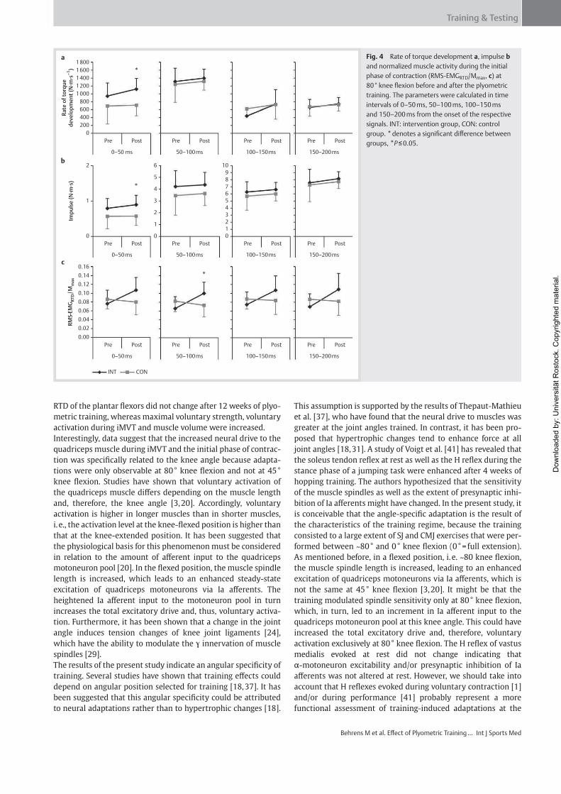
Anstiegssteilheit der Drehmoment-Zeit-Kurve (rate of torque development [RTD]), die
kontraktilen Eigenschaften des M. quadriceps femoris, den H-Reflex des M. vastus medialis und
die Sprungperformance analysiert. Es wurde davon ausgegangen, dass die
Trainingsintervention zur Erhöhung der genannten Parameter führt.
3.1.3 Methoden
Für die Untersuchung der Modulationen innerhalb des neuromuskulären Systems, ausgelöst
durch ein plyometrisches Training, wurde auf das gleiche Methodenrepertoire zurückgegriffen,
das in Kapitel 2.2.3 beschrieben ist. Dieses beinhaltete jeweils Kraftmessungen und
neurophysiologische Techniken. Eine detaillierte Beschreibung der Personenstichprobe,
experimentellen Prozedur, Datenaufnahme und -analyse sowie statistischen Analyse für das
Experiment ist in der Publikation zu finden.
Zu den Messzeitpunkten wurden die Probanden auf einem Dynamometer fixiert, um die
Drehmomente zu messen, die durch den M. quadriceps femoris produziert wurden.
Für die Messung der Muskelaktivität kamen Oberflächenelektroden zum Einsatz. Die
Elektroden wurden in einer bipolaren Konfiguration auf dem M. rectus femoris, dem M. vastus
medialis und dem M. vastus lateralis appliziert. Die Referenzelektrode wurde auf der
ipsilateralen Patella platziert.
Die Analyse der Modulationen auf den unterschiedlichen Ebenen des neuromuskulären
Systems wurde mithilfe der transkutanen elektrischen Stimulation des N. femoralis realisiert.
Durch die Applikation von elektrischen Stimuli in Ruhebedingung und während der MVCs
konnten unterschiedliche Ebenen des neuromuskulären Systems und deren Modulation näher
untersucht werden. Dazu gehörte die Abschätzung der kontraktilen Eigenschaften des M.
quadriceps femoris in Ruhebedingung, der Erregbarkeit der spinalen �-Motoneuronen des M.
vastus medialis mittels der Exzitation der Ia Afferenzen und der willkürlichen Aktivierung des M.
quadriceps femoris während MVC.
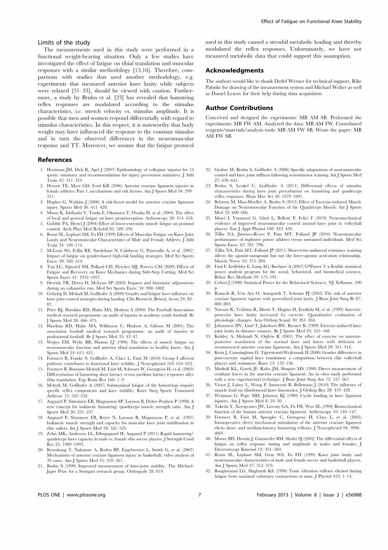
3.1.4 Ergebnisse und Diskussion
In diesem Kapitel werden die wichtigsten Ergebnisse des Experiments vorgestellt und
anschließend kurz diskutiert. Die komplette Darstellung der Resultate der Studie ist in der
angehängten Publikation zu finden.
Innerhalb dieses Experiments wurde die Modulation der Leistungsfähigkeit des
neuromuskulären Systems durch Training untersucht. Die durchgeführte Studie analysierte die
Auswirkungen eines achtwöchigen plyometrischen Trainings auf das iMVT, die willkürliche
Aktivierung, die RTD und die kontraktilen Eigenschaften des M. quadriceps femoris sowie die
Sprungperformance. Zudem wurde die Exzitabilität des �-Motoneuronenpools des M. vastus
medialis über Ia Afferenzen zu den Messzeitpunkten abgeschätzt. Die Messung der
neuromuskulären Funktion erfolgte in zwei Kniewinkeln, d. h. 80° und 45° Knieflexion (0° = volle
27
Extension), um potentielle winkelspezifische Trainingsanpassungen aufzudecken. Das Training
erfolgte zwei Mal pro Woche und beinhaltete Countermovement Jumps, Squat Jumps und Drop
Jumps.
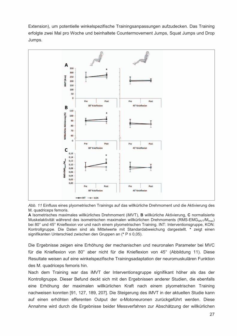
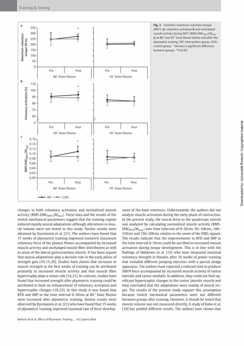
Abb. 11 Einfluss eines plyometrischen Trainings auf das willkürliche Drehmoment und die Aktivierung des M. quadriceps femoris. A Isometrisches maximales willkürliches Drehmoment (iMVT), B willkürliche Aktivierung, C normalisierte Muskelaktivität während des isometrischen maximalen willkürlichen Drehmoments (RMS-EMGiMVT/Mmax) bei 80° und 45° Knieflexion vor und nach einem plyometrischen Training. INT: Interventionsgruppe, KON: Kontrollgruppe. Die Daten sind als Mittelwerte mit Standardabweichung dargestellt. * zeigt einen signifikanten Unterschied zwischen den Gruppen an (* P � 0,05).
Die Ergebnisse zeigen eine Erhöhung der mechanischen und neuronalen Parameter bei MVC
für die Knieflexion von 80° aber nicht für die Knieflexion von 45° (Abbildung 11). Diese
Resultate weisen auf eine winkelspezifische Trainingsadaptation der neuromuskulären Funktion
des M. quadriceps femoris hin.
Nach dem Training war das iMVT der Interventionsgruppe signifikant höher als das der
Kontrollgruppe. Dieser Befund deckt sich mit den Ergebnissen anderer Studien, die ebenfalls
eine Erhöhung der maximalen willkürlichen Kraft nach einem plyometrischen Training
nachweisen konnten [91, 127, 189, 207]. Die Steigerung des iMVT in der aktuellen Studie kann
auf einen erhöhten efferenten Output der �-Motoneuronen zurückgeführt werden. Diese
Annahme wird durch die Ergebnisse beider Messverfahren zur Abschätzung der willkürlichen
28
Aktivierung gestützt. Die kontraktilen Eigenschaften zeigten keinen trainingsbedingten
Gruppenunterschied. Auf Grundlage dieser Resultate kann davon ausgegangen werden, dass
die applizierte Trainingsmaßnahme primär neuronale Anpassungen provoziert hat. Zu ähnlichen
Ergebnissen kamen Kyrolainen et al. [127], die eine Erhöhung der Muskelaktivierung der
Plantarflexoren infolge eines plyometrischen Trainings nachweisen konnten, während keine
muskulären Veränderungen detektiert wurden. Darüber hinaus existieren Studien, die
neuronale und hypertrophische Veränderungen nach einem Sprungtraining zeigen konnten
[121, 133]. Generell wird allerdings angenommen, dass primär neuronale Anpassungen für den
Anstieg der willkürlichen maximalen Kraft in den ersten Wochen eines Krafttrainings
verantwortlich sind [60, 91, 154]. Aufgrund der Winkelspezifik der Anpassungen in der
vorliegenden Studie kann primär von neuronalen Adaptationen ausgegangen werden [116]. Die
Ergebnisse einer Studie von Thepaut-Mathieu et al. [216] weisen darauf hin, dass neuronale
Anpassungen lediglich die Gelenkwinkel und damit Muskellängen betreffen, die im Training
Verwendung fanden. Damit geht einher, dass eine Kraftsteigerung ebenfalls nur in den
spezifischen Gelenkwinkeln zu beobachten ist. Dagegen scheinen sich hypertrophische
Adaptationen auf die maximale willkürliche Kraft in allen Gelenkwinkeln auszuwirken [116, 191].
Voigt et al. [226] fanden bei ihren Probanden erhöhte Dehnungsreflexe und H-Reflexe infolge
eines vierwöchigen plyometrischen Trainings. Die Autoren gingen davon aus, dass eine erhöhte
Muskelspindelsensitivität sowie eine reduzierte präsynaptische Inhibition der Ia Afferenzen zu
den Ergebnissen beitrugen. In der aktuellen Studie bestand das Training größtenteils aus
Sprungübungen, die zwischen ~80° und 0° Knieflexion (0° = volle Extension) durchgeführt
wurden. Dabei entsprachen 80° Knieflexion dem Umkehrpunkt beim Countermovement Jump
und der Startposition beim Squat Jump. Demnach waren die Gewichtsbelastung und damit der
Anspruch an die Kraftleistung im besagten Kniewinkel am höchsten. Zudem sind die Anteile des
M. quadriceps femoris und damit auch die Muskelspindeln bei 80° Knieflexion länger. Das kann
zu einer gesteigerten Erregung des M. quadriceps femoris Motoneuronenpools über Ia
Afferenzen führen, die bei einer Knieflexion von 45° nicht erfolgt [27, 122]. Demnach ist es
möglich, dass das plyometrische Training die Spindelsensitivität ausschließlich bei 80°
Knieflexion erhöht hat und damit zur Steigerung des Ia-afferenten Inputs beitrug. Dieser könnte
wiederum zur Steigerung des gesamten exzitatorischen Outputs im Sinne einer gesteigerten
willkürlichen Aktivierung während MVC beigetragen haben.
29
3.2 EINFLUSS DES ALTERNS AUF DIE NEUROMUSKULÄRE FUNKTION
3.2.1 Darstellung des Forschungsdefizits
Im Verlauf des Alterungsprozesses kommt es zu einer Reduktion der Leistungsfähigkeit des
neuromuskulären Systems. Die Abnahme der neuromuskulären Funktion des gesunden
alternden Menschen ist durch strukturelle und funktionelle Veränderungen bedingt.
Die strukturellen Veränderungen beinhalten u. a. eine Abnahme des Volumens von Neuronen
im Lobus frontalis [93, 190] und in der Substantia alba des Cerebrums [138]. Darüber hinaus
spielen Modulationen im peripheren Nervensystem eine Rolle. Dazu gehören die Reduktion der
Anzahl und des Durchmessers myelinisierter Axone [151] und die Abnahme der axonalen
Leitungsgeschwindigkeit [147] aufgrund verminderter Myelinisierung und veränderter
Internodiumlängen [3]. Es kommt des Weiteren zu einem zunehmenden Verlust von �-
Motoneuronen, induziert durch Apoptose, die Reduktion der Aktivität von insulinähnlichen
Wachstumsfaktoren (insulin-like growth factor 1, IGF-1), die erhöhte Konzentration spezifischer
Außerdem kann eine altersbedingte Abnahme der Muskelfaseranzahl und Atrophie von
Muskelfasern konstatiert werden. Dabei sind die Typ II Muskelfasern stärker betroffen als die
Typ I Muskelfasern [68, 117, 129]. Als primäre Ursache dafür wird eine reduzierte myofibrilläre
Proteinsynthese im Alter diskutiert [18]. Zudem weisen ältere Individuen eine sogenannte
anabole Resistenz auf [180], die durch eine reduzierte Fähigkeit zur Erhöhung der
Muskelproteinsynthese charakterisiert ist. Demnach reagieren jüngere und ältere Menschen
unterschiedlich auf anabole Stimuli, wie z. B. Nahrungsaufnahme und physische Aktivität [53,
180, 229]. Des Weiteren wurde die Reduktion der kontraktilen Kinetik [54] sowie die
Modifikation der Muskelarchitektur [157] beim älteren Menschen nachgewiesen. Im Hinblick auf
die Veränderung der Muskelarchitektur sind primär zwei Faktoren relevant: (I) die Reduktion der
Muskelfaszikellänge und (II) die Abnahme des Fiederungswinkels der Muskulatur [120, 157]
bedingt durch die Abnahme der Sarkomere in Serie. Dadurch werden die Längen-Spannungs-
Relation, Kraft-Geschwindigkeits-Relation und Leistungs-Geschwindigkeits-Relation des
relevanten Muskels negativ beeinflusst [157].
Die altersbedingten funktionellen Veränderungen inkludieren u. a. die Modulation der Aktivität
des zerebralen Cortexes während differenter Aufgaben, die mittels funktioneller
Magnetresonanztomographie [96, 181] und transkranieller Magnetstimulation [193]
nachgewiesen wurden. Zudem konnte bei älteren Menschen eine erhöhte Koaktivierung
antagonistischer Muskeln festgestellt werden [23, 36], die zur Reduktion der Kraftproduktion der
agonistischen Muskeln beiträgt. Die Ursache liegt in der Erhöhung der zu überwindenden Kraft
für den Agonisten. Außerdem kann eine erhöhte Koaktivierung zur Abnahme der agonistischen
Muskelaktivität führen, indem die reziproke Inhibition des Agonisten zunimmt. Des Weiteren
deuten H-Reflex Studien darauf hin, dass die spinalen �-Motoneuronen im Altersgang eine
reduzierte Erregbarkeit aufweisen [64, 221] und es zu einer Modifikation von
Hemmungsmechanismen, wie z. B. der präsynaptischen Inhibition von Ia Afferenzen [63] und
der reziproken Inhibition [114], bei spezifischen Bewegungsaufgaben kommt.
30
Die genannten und weitere Faktoren sind mitverantwortlich für die Abnahme der Muskelkraft mit
zunehmendem Alter, die in der internationalen Literatur als „Dynapenia“ charakterisiert wird [49,
134]. Die verminderte mechanische Muskelperformance, die eine reduzierte willkürliche
maximale und explosive Kraft sowie Leistung im höheren Alter beinhaltet, impliziert eine
Verschlechterung der funktionellen Kapazität während der Ausübung von Alltagstätigkeiten. Zu
diesen Tätigkeiten gehören z. B. das Gehen, Treppensteigen und Aufstehen von einem Stuhl
[3]. Demnach ist die altersbedingte Abnahme der maximalen und explosiven Kraftproduktion
[203] für die Sturzinzidenz und Mobilität im Alltag von enormer Relevanz. Es konnte gezeigt
werden, dass die Kraft der unteren Extremitäten mit der Häufigkeit von Sturzereignissen und
der Fähigkeit auf Perturbationen der posturalen Kontrolle adäquat reagieren zu können im
Zusammenhang steht [168, 169]. Darüber hinaus wurden signifikante Korrelationen zwischen
der Explosivkraft eines Individuums und der Ganggeschwindigkeit sowie der Zeit zum
Absolvieren einer definierten Anzahl von Treppenstufen gefunden [25]. Izquierdo et al. [102]
wiesen nach, dass die Fähigkeit der Beinextensoren zur schnellen Kraftproduktion mit der
posturalen Kontrolle zusammen hängt.
Aufgrund der Relevanz der maximalen und explosiven Kraft für die Mobilität im Alter ist die
Aufdeckung der Ursachen für die Abnahme dieser Kraftfähigkeiten von Bedeutung. Obwohl die
altersbedingte Abnahme der Maximal- und Explosivkraft des M. quadriceps femoris mehrfach
nachgewiesen wurde [102, 103, 170], sind die zugrundeliegenden neuromuskulären
Mechanismen ungenügend untersucht. Darüber hinaus sind die Ergebnisse der existierenden
Studien zu dieser Thematik partiell widersprüchlich. So konnten Roos et al. [183] und Wilder et
al. [228] keine altersbedingte Veränderung der willkürlichen Aktivierung des M. quadriceps
femoris während isometrischer maximaler willkürlicher Kontraktionen (maximum voluntary
contraction [MVC]) feststellen, während Stevens et al. [211] und Stackhouse et al. [208] ein
altersbedingtes Aktivierungsdefizit der Knieextensoren während isometrischer MVC bei
gesunden Männern und Frauen aufdecken konnten.
Die unterschiedlichen Ergebnisse sind zum Teil auf methodische Defizite zurückzuführen. Die
folgende durchgeführte Studie sollte in dieser Hinsicht zur Erweiterung des Wissens beitragen.
EXPERIMENT IV: Mau-Moeller, A.*, Behrens, M.*, Lindner, T., Bader, R., Bruhn, S. (2013).
Age-related changes in neuromuscular function of the quadriceps muscle in physically active
adults. Journal of Electromyography and Kinesiology, 23 (3), 640-648. (* authors contributed
equally to this work)
31
3.2.2 Fragestellung
Diese Studie untersuchte den Abfall der Leistungsfähigkeit des neuromuskulären Systems
infolge des Alterungsprozesses. Dazu wurde der Beitrag altersbedingter neuronaler und
muskulärer Veränderungen zur Reduktion des iMVT und der RTD analysiert. Zudem wurde der
H-Reflex des M. vastus medialis zwischen älteren und jüngeren Probanden verglichen. Es
wurde angenommen, dass die Reduktion neuronaler sowie muskulärer Parameter für die
Abnahme der maximalen willkürlichen Kraft im Altersgang verantwortlich ist.
3.2.3 Methoden
Für die Untersuchung der Modulationen innerhalb des neuromuskulären Systems, ausgelöst
durch den Alterungsprozess, wurde auf das gleiche Methodenrepertoire zurückgegriffen, das in
Kapitel 2.2.3 beschrieben ist. Dieses beinhaltete jeweils Kraftmessungen und
neurophysiologische Techniken. Eine detaillierte Beschreibung der Personenstichprobe,
experimentellen Prozedur, Datenaufnahme und -analyse sowie statistischen Analyse für das
Experiment ist in der Publikation zu finden.
Zu den Messzeitpunkten wurden die Probanden auf einem Dynamometer fixiert, um die
Drehmomente zu messen, die durch den M. quadriceps femoris produziert wurden.
Für die Messung der Muskelaktivität kamen Oberflächenelektroden zum Einsatz. Die
Elektroden wurden in einer bipolaren Konfiguration auf dem M. rectus femoris, dem M. vastus
medialis und dem M. vastus lateralis appliziert. Die Referenzelektrode wurde auf der
ipsilateralen Patella platziert.
Die Analyse der Modulationen auf den unterschiedlichen Ebenen des neuromuskulären
Systems wurde mithilfe der transkutanen elektrischen Stimulation des N. femoralis realisiert.
Durch die Applikation von elektrischen Stimuli in Ruhebedingung und während der MVCs
konnten unterschiedliche Ebenen des neuromuskulären Systems und deren Modulation näher
untersucht werden. Dazu gehörte die Abschätzung der kontraktilen Eigenschaften des M.
quadriceps femoris in Ruhebedingung, der Erregbarkeit der spinalen �-Motoneuronen des M.
vastus medialis mittels der Exzitation der Ia Afferenzen und der willkürlichen Aktivierung des M.
quadriceps femoris während MVC.
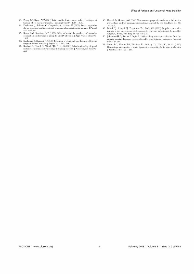
3.2.4 Ergebnisse und Diskussion
In diesem Kapitel werden die wichtigsten Ergebnisse des Experiments vorgestellt und
anschließend kurz diskutiert. Die komplette Darstellung der Resultate der Studie ist in der
angehängten Publikation zu finden.
Das durchgeführte Experiment untersuchte den Abfall der Leistungsfähigkeit des
neuromuskulären Systems infolge des Alterungsprozesses. Innerhalb einer Querschnittstudie
wurde der Beitrag altersbedingter neuronaler und muskulärer Veränderungen zur Reduktion
des iMVT und der RTD analysiert. Zudem wurde die Exzitabilität des �-Motoneuronenpools des
M. vastus medialis über Ia Afferenzen zwischen älteren und jüngeren Probanden verglichen.
32
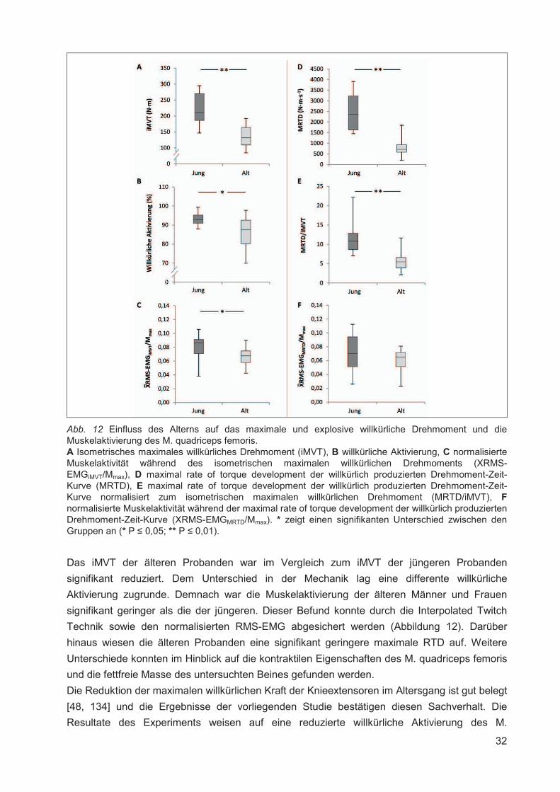
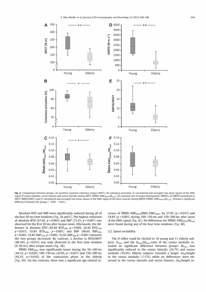
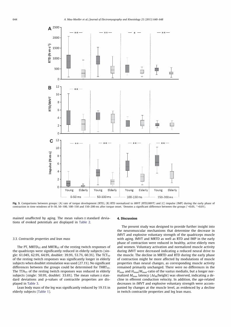
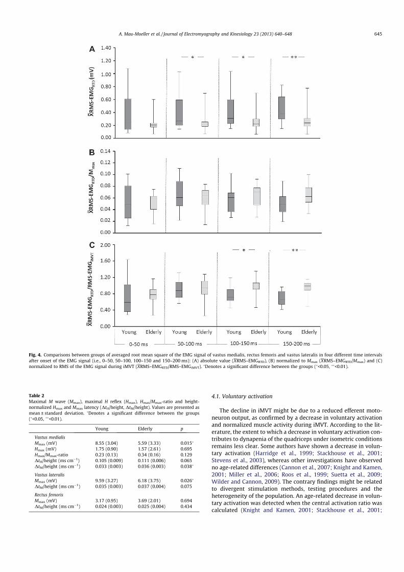
Abb. 12 Einfluss des Alterns auf das maximale und explosive willkürliche Drehmoment und die Muskelaktivierung des M. quadriceps femoris. A Isometrisches maximales willkürliches Drehmoment (iMVT), B willkürliche Aktivierung, C normalisierte Muskelaktivität während des isometrischen maximalen willkürlichen Drehmoments (XRMS-EMGiMVT/Mmax), D maximal rate of torque development der willkürlich produzierten Drehmoment-Zeit-Kurve (MRTD), E maximal rate of torque development der willkürlich produzierten Drehmoment-Zeit-Kurve normalisiert zum isometrischen maximalen willkürlichen Drehmoment (MRTD/iMVT), Fnormalisierte Muskelaktivität während der maximal rate of torque development der willkürlich produzierten Drehmoment-Zeit-Kurve (XRMS-EMGMRTD/Mmax). * zeigt einen signifikanten Unterschied zwischen den Gruppen an (* P � 0,05; ** P � 0,01).
Das iMVT der älteren Probanden war im Vergleich zum iMVT der jüngeren Probanden
signifikant reduziert. Dem Unterschied in der Mechanik lag eine differente willkürliche
Aktivierung zugrunde. Demnach war die Muskelaktivierung der älteren Männer und Frauen
signifikant geringer als die der jüngeren. Dieser Befund konnte durch die Interpolated Twitch
Technik sowie den normalisierten RMS-EMG abgesichert werden (Abbildung 12). Darüber
hinaus wiesen die älteren Probanden eine signifikant geringere maximale RTD auf. Weitere
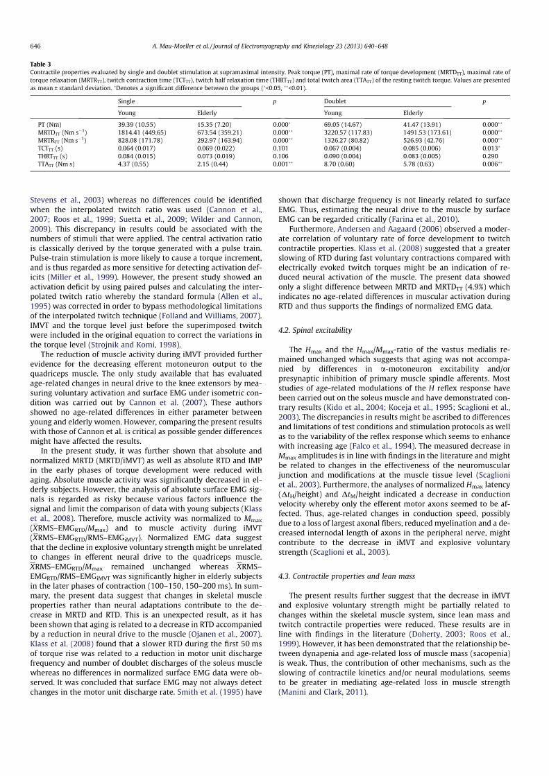
Unterschiede konnten im Hinblick auf die kontraktilen Eigenschaften des M. quadriceps femoris
und die fettfreie Masse des untersuchten Beines gefunden werden.
Die Reduktion der maximalen willkürlichen Kraft der Knieextensoren im Altersgang ist gut belegt
[48, 134] und die Ergebnisse der vorliegenden Studie bestätigen diesen Sachverhalt. Die
Resultate des Experiments weisen auf eine reduzierte willkürliche Aktivierung des M.
33
quadriceps femoris bei den älteren Probanden hin. Demnach scheint ein reduzierter efferenter
Output der �-Motoneuronen mitverantwortlich für die Abnahme des iMVT mit zunehmendem
Alter zu sein. Es existieren Studien, die ein Aktivierungsdefizit bei älteren Menschen
nachweisen konnten [208, 211], während andere Experimente keine altersbedingte Abnahme
der willkürlichen Aktivierung fanden [39, 183, 228]. Die widersprüchlichen Ergebnisse sind
vermutlich auf unterschiedliche elektrische Stimulationsmethoden und heterogene
Probandenpopulationen zurückzuführen. Ein Aktivierungsdefizit wurde zumeist dann
festgestellt, wenn die „Central Activation Ratio“ zur Abschätzung der willkürlichen Aktivierung
verwendet wurde [208, 211]. Wenn die Interpolated Twitch Technik zur Analyse der
Muskelaktivierung herangezogen wurde, konnte größtenteils kein Unterschied in der Aktivierung
der Knieextensoren bei jüngeren und älteren Erwachsenen festgestellt werden [39, 183, 228]. In
der vorliegenden Studie wurden zwei Ansätze zur Messung der willkürlichen Aktivierung des M.
quadriceps femoris verwendet. Zum einen erfolgte die Abschätzung der Muskelaktivierung über
die Berechnung des normalisierten RMS-EMG und zum anderen mittels der Interpolated Twitch
Technik. Zudem wurde für die Berechnung der willkürlichen Aktivierung, die mittels der
Interpolated Twitch Technik erhoben wurde, eine korrigierte Formel verwendet. Diese Formel
berücksichtigt das Drehmomentniveau vor der Applikation der elektrischen Stimuli, welches
aufgrund der Fluktuation der Drehmoment-Zeit-Kurve unter dem iMVT liegen kann.
In der vorliegenden Studie wurden eine schlechtere Kontraktilität der Knieextensoren sowie
eine reduzierte Muskelmasse des untersuchten Beines bei den älteren Probanden gefunden.
Diese Befunde entsprechen den Ergebnissen anderer Studien, die ebenfalls eine altersbedingte
Veränderung der kontraktilen Eigenschaften nachweisen konnten [42, 183, 228]. Im Hinblick auf
die Muskelmasse korrespondieren die Ergebnisse des vorliegenden Experiments ebenfalls mit
der Datenlage anderer Untersuchungen [235, 236]. Als Ursache kann die altersbedingte
Abnahme der Muskelfaseranzahl und Atrophie von Muskelfasern ausgemacht werden. Diese
Prozesse wirken sich stärker auf die Typ II Muskelfasern als auf die Typ I Muskelfasern aus [68,
117, 129]. Als primäre Ursache dafür wird eine reduzierte myofibrilläre Proteinsynthese im Alter
diskutiert [18]. Darüber hinaus wurde eine anabole Resistenz älterer Personen nachgewiesen
[180], die durch eine reduzierte Fähigkeit zur Erhöhung der Muskelproteinsynthese
charakterisiert ist. Demzufolge reagieren jüngere und ältere Menschen unterschiedlich auf
anabole Stimuli, wie z. B. Nahrungsaufnahme und physische Aktivität [53, 180, 229].
34
4. ZUSAMMENFASSUNG
Die Ergebnisse der durchgeführten Studien haben zur Erweiterung des Wissens über akute und
chronische Anpassungen der neuromuskulären Funktion beigetragen. Es konnte gezeigt
werden, dass physische Aktivität sowie der Alterungsprozess, der durch einen generellen
Leistungsverlust aber auch die Zunahme von physischer Inaktivität gekennzeichnet ist, die
neuromuskuläre Funktion der Oberschenkelmuskeln modulieren kann.
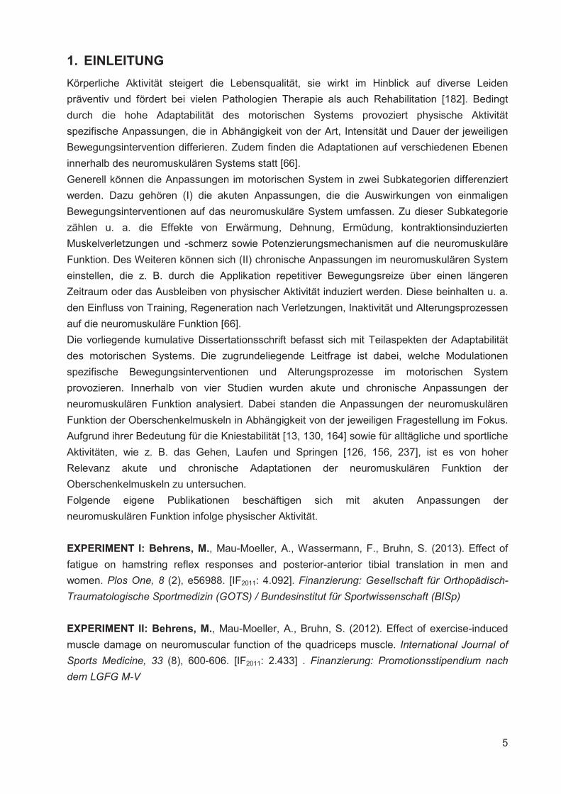
In den Experimenten I und II wurden spezifische akute Anpassungen untersucht und die
Auswirkungen von unterschiedlich belastenden Ermüdungsinterventionen auf die
neuromuskuläre Funktion der Oberschenkelmuskeln analysiert. Es wurde festgestellt, dass die
Leistungsfähigkeit des neuromuskulären Systems ermüdungsbedingt abnimmt und sich
spezifische akute Adaptationen einstellen. So zeigte Experiment I „Effect of fatigue on
hamstring reflex responses and posterior-anterior tibial translation in men and women”, dass die
spezifische Ermüdungsintervention unterschiedliche Auswirkungen auf die anteriore
Tibiatranslation und die Reflexantworten des M. biceps femoris und des M.
semitendinosus/semimembranosus der Männer und Frauen hatte. Auf Grundlage der
Ergebnisse ist es vorstellbar, dass eine reduzierte Reflexaktivierung der ischiocruralen
Muskulatur mit einer korrespondierenden Erhöhung der anterioren Tibiatranslation zur erhöhten
Inzidenz für ACL-Rupturen bei Frauen beitragen könnte. In Experiment II „Effect of exercise-
induced muscle damage on neuromuscular function of the quadriceps muscle” wurde eine
überbelastende Ermüdungsintervention durchgeführt, die zur längerfristigen Modulation der
neuromuskulären Funktion führte. Die Studie zeigte, dass die Abnahme der willkürlichen
Aktivierung und der kontraktilen Funktion des M. quadriceps femoris zum Kraftverlust
unmittelbar nach der Intervention, die eine Muskelverletzung induziert hat, beitrug. Im darauf
folgenden Zeitraum schienen die beeinträchtigten kontraktilen Eigenschaften der
Knieextensoren hauptverantwortlich für die Reduktion des iMVT zu sein. Die Studie zeigte, dass
es keine Korrespondenz zwischen den neurophysiologischen Parametern und dem
Schmerzverlauf gibt.
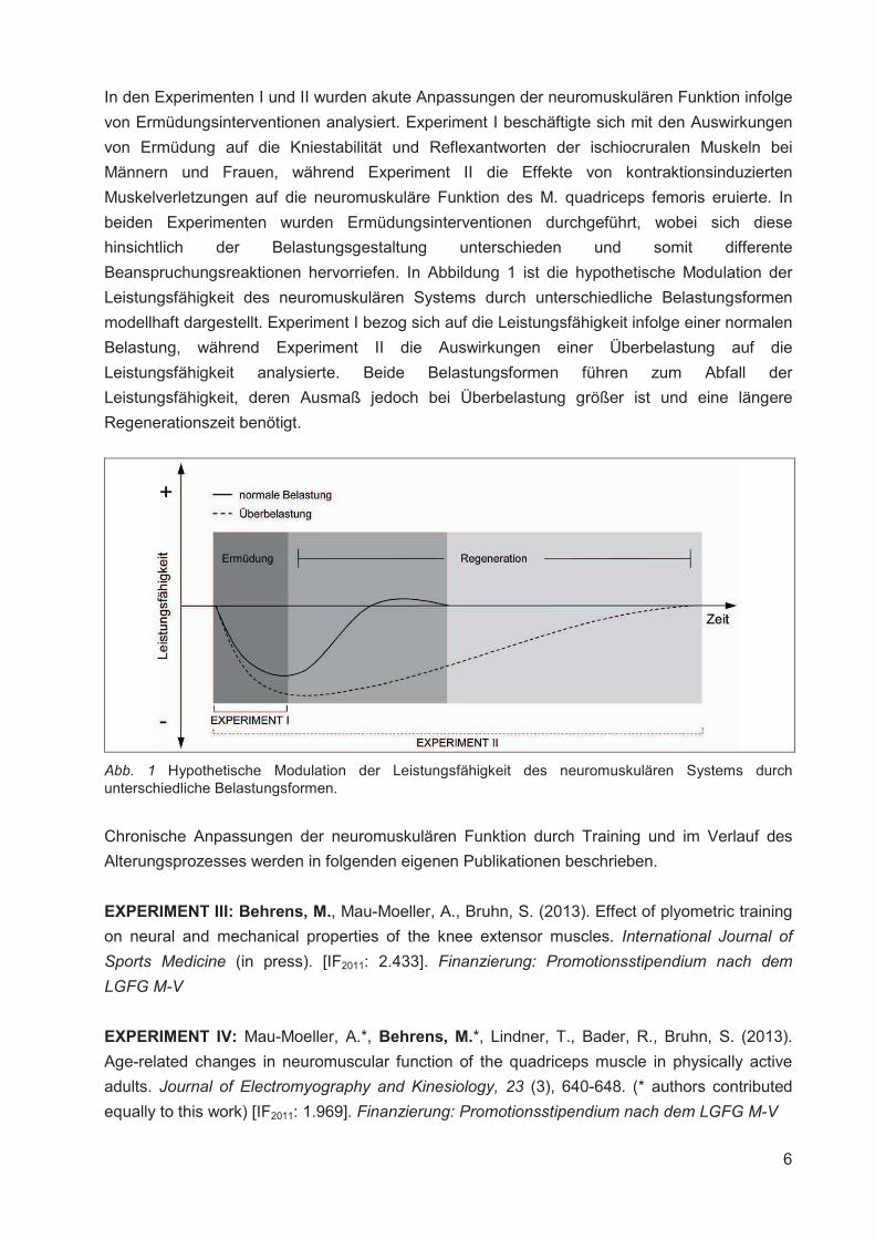
In den Experimenten III und IV wurden spezifische chronische Anpassungen der
neuromuskulären Funktion untersucht. Experiment III beschäftigte sich mit der Modulation der
neuromuskulären Funktion des M. quadriceps femoris durch gezielte repetitive physische
Aktivität, während Experiment IV den Einfluss des Alterns und zunehmender physischer
Inaktivität auf die neuromuskuläre Funktion des M. quadriceps femoris untersuchte. In der
Publikation „Effect of plyometric training on neural and mechanical properties of the knee
extensor muscles”, die aus Experiment III hervorging, wurde dokumentiert, dass das
plyometrische Training primär neuronale Anpassungen provozierte, die wiederum zur
Steigerung des iMVT des M. quadriceps femoris beitrugen. Darüber hinaus konnten
winkelspezifische neuronale Adaptationen nachgewiesen werden, die bis dato lediglich nach
isometrischen Trainingsregimes beobachtet wurden [116, 216]. Das Experiment IV „Age-related
changes in neuromuscular function of the quadriceps muscle in physically active adults” brachte
neue Erkenntnisse im Hinblick auf die Kontribution des Zentralnervensystems zur reduzierten
Kraft der Knieextensoren im Alter. Die Abschätzung der willkürlichen Aktivierung des M.
35
quadriceps femoris mithilfe von zwei differenten Verfahren zeigte eine altersbedingte Reduktion.
Demnach tragen scheinbar Modulationen auf neuronaler und muskulärer Ebene zur Abnahme
10/2009 – 05/2014 Promotion: Adaptabilität des motorischen Systems – Akute und
chronische Anpassung der neuromuskulären Funktion (Betreuer:
Prof. Dr. phil. Sven Bruhn, Prof. Dr. med. Dipl.-Ing. Bader)
10/2002 – 03/2009 Studium an der Universität Rostock
Studium der Fächer Sportwissenschaft und Sozialwissenschaften
(Lehramt Gymnasium)
Preise und Förderungen
2012 Preis für die Forschungsförderung der Gesellschaft für
Orthopädisch-Traumatologische Sportmedizin (GOTS)
2011 2. Platz beim Venture Cup-MV, Kategorie Forscherteam (Joost,
R., Behrens, M., Bruhn, S., Salomon, R.)
2010 Promotionsstipendium nach dem
Landesgraduiertenförderungsgesetz (LGFG M-V)
2010 Hermes-Forschungsförderpreis der Universität Rostock ( Behrens,
M. & Mau-Möller, A.)
54
8. EIGENE PUBLIKATIONEN
I. Behrens, M., Mau-Moeller, A., Wassermann, F., Bruhn, S. (2013). Effect of fatigue on
hamstring reflex responses and posterior-anterior tibial translation in men and women. Plos
One, 8 (2), e56988. [IF2011: 4.092].
II. Behrens, M., Mau-Moeller, A., Bruhn, S. (2012). Effect of exercise-induced muscle damage
on neuromuscular function of the quadriceps muscle. International Journal of Sports Medicine,
33 (8), 600-606. [IF2011: 2.433].
III. Behrens, M., Mau-Moeller, A., Bruhn, S. (2013). Effect of plyometric training on neural and
mechanical properties of the knee extensor muscles. International Journal of Sports Medicine
(in press). [IF2011: 2.433].
IV. Mau-Moeller, A.*, Behrens, M.*, Lindner, T., Bader, R., Bruhn, S. (2013). Age-related
changes in neuromuscular function of the quadriceps muscle in physically active adults. Journal
of Electromyography and Kinesiology, 23 (3), 640-648. (* authors contributed equally to this
work) [IF2011: 1.969].
Effect of Fatigue on Hamstring Reflex Responses andPosterior-Anterior Tibial Translation in Men and Women
Martin Behrens*, Anett Mau-Moeller, Franziska Wassermann, Sven Bruhn
Department of Exercise Science, University of Rostock, Rostock, Germany
Abstract
Anterior cruciate ligament (ACL) rupture ranks among the most common injuries in sports. The incidence of ACL injuries isconsiderably higher in females than in males and the underlying mechanisms are still under debate. Furthermore, it hasbeen suggested that muscle fatigue can be a risk factor for ACL injuries. We investigated gender differences in hamstringreflex responses and posterior-anterior tibial translation (TT) before and after fatiguing exercise. We assessed the isolatedmovement of the tibia relative to the femur in the sagittal plane as a consequence of mechanically induced TT in standingsubjects. The muscle activity of the hamstrings was evaluated. Furthermore, isometric maximum voluntary torque (iMVT)and rate of torque development (RTD) of the hamstrings (H) and quadriceps (Q) were measured and the MVT H/Q as well asthe RTD H/Q ratios were calculated. After fatigue, reflex onset latencies were enhanced in women. A reduction of reflexresponses associated with an increased TT was observed in females. Men showed no differences in these parameters.Correlation analysis revealed no significant associations between parameters for TT and MVT H/Q as well as RTD H/Q. Theresults of the present study revealed that the fatigue protocol used in this study altered the latency and magnitude of reflexresponses of the hamstrings as well as TT in women. These changes were not found in men. Based on our results, it isconceivable that the fatigue-induced decrease in neuromuscular function with a corresponding increase in TT probablycontributes to the higher incidence of ACL injuries in women.
Citation: Behrens M, Mau-Moeller A, Wassermann F, Bruhn S (2013) Effect of Fatigue on Hamstring Reflex Responses and Posterior-Anterior Tibial Translation inMen and Women. PLoS ONE 8(2): e56988. doi:10.1371/journal.pone.0056988
Editor: Junming Yue, The University of Tennessee Health Science Center, United States of America
Received October 2, 2012; Accepted January 16, 2013; Published February 27, 2013
Copyright: ß 2013 Behrens et al. This is an open-access article distributed under the terms of the Creative Commons Attribution License, which permitsunrestricted use, distribution, and reproduction in any medium, provided the original author and source are credited.
Funding: This study was supported by the German-Austrian-Swiss Society for Orthopaedic Traumatologic Sports Medicine (GOTS) and the Bundesinstitut fuerSportwissenschaft (BISp). The funders had no role in study design, data collection and analysis, decision to publish, or preparation of the manuscript.
Competing Interests: The authors have declared that no competing interests exist.
Germany). Subjects were thereby provided with online feedback
about their center of pressure. Furthermore, to avoid the influence
of acoustic signals on the subjects they wore ear protection (Bilsom
Thunder T3). The subjects had no information on the point in
time of the perturbation. A standardized force was applied to the
proximal shank of the dominant leg using a pulley system in order
to induce TT. A device was attached to the tibia to secure two
linear potentiometers (measuring accuracy: ,0.01 mm, linearity:
60.7%, Type CLR13–50; Megatron, Germany) that were placed
on the patella and the tibial tuberosity (Fig. 1). The knee
arthrometer enabled us to measure the translational movement of
the tibia relative to the femur in the sagittal plane. The interface
pressure between the knee arthrometer and the subjects’ tibia was
controlled by an air pressure recorder (Kikuhime, TT MediTrade,
Denmark). The interface pressure was kept constant before and
after the fatiguing exercise. The locations of the subjects’ feet on
the force plate, the stabilizing device and the linear potentiometers
were marked in order to ensure the same positions before and after
the fatigue protocol. The applied force that induced TT was
Figure 1. Schematic drawing of the experimental setup. A: Experimental setup, B: Measurement system | 1: stopper, 2: falling weight, 3: pulley,4: steel rope, 5: force transducer, 6: force plate, 7: visual cover, 8: linear potentiometer. Arrows indicate the direction of the force. Posterior-anteriortibial translation was assessed by two linear potentiometers (8) placed on the patella and the tibial tuberosity. A force transducer (5) was used tomeasure the force transmitted to the shank.doi:10.1371/journal.pone.0056988.g001
Effect of Fatigue on Functional Knee Stability
PLOS ONE | www.plosone.org 2 February 2013 | Volume 8 | Issue 2 | e56988
controlled using a force transducer (measuring range: 0–5000 N,
sensitivity: 23.42 to 3.36 pC?N21, linearity: 60.2–0.3%; Kistler,
Switzerland). The force sensor was placed between the stabilizing
device and the pulley system. TT was elicited 15 times in order to
familiarize the subject with the measurement. Thereafter, further
15 perturbations were applied before and immediately after the
fatigue protocol. The inter stimulus interval was varied between
,8 s and ,14 s to avoid anticipation.
EMG and torque recordingsSurface EMG was recorded using bipolar AmbuH Blue Sensor
N electrodes. The electrodes were attached to the shaved, abraded
and cleaned skin over the biceps femoris (BF) and semitendinosus/
semimembranosus (ST) of the dominant leg (resistance between
electrodes ,5 kV). The electrodes were applied with a center-to-
center distance of 2 cm over the muscle bellies and in line with the
presumed direction of the underlying muscle fibers. The reference
electrode was attached to the patella. Signals were amplified
(2500x), band-pass filtered (10–1300 Hz) and digitized (sampling
frequency: 5 kHz) through an analog-to-digital converter (DAQ
CardTM-6024E, National Instruments, USA).
Torque was measured using a CYBEX NORM dynamometer
(Computer Sports MedicineH, Inc., Stoughton, MA). A hip joint
angle and knee joint angle of 30u, respectively (0u= full extension),
was chosen in order to mimic the posture and therefore the length
of the muscles during the measurement of TT. The axis of the
dynamometer was aligned with the anatomical knee flexion-
extension axis and the lever arm was attached to the anterior
aspect of the shank 2–3 cm above the lateral malleolus. Straps
across the waist and chest prevented excessive movements. The
iMVT was tested by asking the subjects to exert isometric knee
extensions and flexions against the lever arm of the dynamometer
for 3 s. For each trial, subjects were thoroughly instructed to act as
forcefully and as fast as possible. They were motivated by strong
verbal encouragement and online visual feedback of the instan-
taneous dynamometer torque provided on a digital oscilloscope
(HM1508, HAMEG Instruments, Germany). Care was taken that
the iMVT trials were performed without an apparent counter-
movement or pre-tension (change in baseline torque ,0.5 Nm
during 200 ms prior to contraction onset). A rest period of 2 min
was allowed between trials. The maximal attempts were recorded
until the coefficient of variance of five consecutive trials was below
5% [24]. The EMG, linear potentiometer, force and torque signals
were stored on a hard drive for later analysis with custom built
LABVIEWH based software (Imago, Pfisoft, Germany).
Fatigue protocolFatigue was induced by repetitive jumping performed between
,90u and 0u knee flexion (0u= full extension). The fatigue
protocol consisted of consecutive maximal countermovement
jumps [25], each one separated by 4 s according to the sound of
a digital metronome. The jumps were performed until the subjects
reached a fatigued state defined as the inability to reach 50% of
their maximal jump height for 3 consecutive jumps or until the
subjects reached an intolerable state of dyspnea or exhaustion.
Rate of perceived exertion (RPE) was assessed using the Borg 6–20
scale.
Data analysisIn order to analyze the data, the EMG signals of each subject
were averaged. The EMG onset latencies were defined as the time
between onset of TT and onset of significant muscular activity, e.g.
the beginning of EMG deflection (average EMG baseline value
measured over 100 ms 6 3 standard deviations). Muscle activity
was analyzed according to Bruhn et al. [23], that is, the TT signal
indicated the onset of perturbation and muscle activity was
calculated over different time intervals relative to the onset of TT,
i.e. 20–40, 40–60 and 60–95 ms (Fig. 2), using the root mean
square of the EMG signal (RMS-EMG). In order to assess
background activity before and after fatigue, RMS-EMG was
calculated over 50 ms prior to the onset of TT. Consequently,
background activity was subtracted from the reflex responses.
Maximum TT was determined based on the TT curves.
The torque signals were corrected for the effect of gravity and
the three best maximum voluntary contractions were retained for
analysis. The iMVT was defined as the highest peak torque value.
Explosive voluntary muscle strength was determined by analyzing
the average RTD over time intervals of 0–50, 0–100 and 0–
200 ms relative to the onset of contraction. The identification of
torque onset was made manually according to the method of Tillin
et al. [26]. It has been suggested that this is the best method for
detecting signal onsets [27]. MVT H/Q as well as RTD H/Q
(RTD0–50, 0–100, 0–200 H/Q) ratios were calculated as follows [19]:
MVT H/Q=Hamstrings MVT/Quadriceps MVT and RTDx
H/Q=Hamstrings RTDx/Quadriceps RTDx, where x denotes
the analyzed time interval (0–50, 0–100, 0–200 ms).
Statistical analysisData were checked for normal distribution using the Kolmo-
gorov-Smirnov test. Differences between the values before and
after the fatigue protocol were tested for significance by repeated
measures ANOVA. Differences between the groups were tested for
significance by the unpaired Student’s t test. Correlations between
parameters were calculated using Pearson’s correlation coefficient.
In each case the level of significance was established at p#0.05.
SPSS 20.0 (SPSS Inc., Chicago, IL, USA) was used for statistical
analysis. Effect size (ES) was calculated with the statistical software
package G*Power (Version 3.1.5) [28]. The ES characterizes the
effectiveness of an intervention. Furthermore, it is used to
determine whether a statistically significant difference is a
difference of practical importance. ES-values = 0.10 indicate small,
ES= 0.25 medium and ES= 0.40 large effects [29]. Data are
presented as group mean values 6 standard error of the mean in
the figures and as group mean values 6 standard deviation in the
table.
Results
During the fatiguing exercise, men performed 159.8690.9 and
women 150.6662.4 jumps. RPE using the Borg 6–20 scale was
15.262.0 for males and 14.861.6 for females. The force applied
to the proximal shank of the dominant leg remained constant
during the pre- and post-test (Table 1). Reflex onset latencies of BF
and ST were significantly delayed in women at post-test (Table 1).
BF muscle activity decreased after fatigue in women in the time
intervals 20–40 ms (F=4.99, P=0.035, g2=0.166, ES= 0.45)
and 40–60 ms (F=7.22, P=0.013, g2=0.224, ES= 0.54). The
reflex response of BF between 60–95 ms (F=0.455, P=0.506,
g2=0.018, ES= 0.14) was unchanged in women after the fatigue
protocol. The reflex activity of ST was significantly reduced after
fatigue between 20–40 ms in women (F=5.38, P=0.029,
g2=0.183, ES= 0.47), but not between 40–60 ms (F=0.051,
P=0.823, g2=0.002, ES= 0.045) and 60–95 ms (F=3.001,
P=0.096, g2=0.111, ES= 0.35). TT increased significantly after
the fatigue protocol in females (F=11.86, P=0.002, g2=0.322,
ES= 0.69) (Fig. 3).
No statistical differences in the reflex responses of BF between
20–40 ms (F=0.601, P=0.448, g2=0.032, ES= 0.18), 40–60 ms
Effect of Fatigue on Functional Knee Stability
PLOS ONE | www.plosone.org 3 February 2013 | Volume 8 | Issue 2 | e56988
Figure 2. EMG and tibial translation data from one representative subject. EMG activity of biceps femoris and semitendinosus/semimembranosus as well as posterior-anterior tibial translation of one subject. In the figure, EMG data is rectified in order to visualize the differentparts of the hamstring stretch reflex. The vertical bold line indicates the onset of posterior-anterior tibial translation. Three different time intervalswere analyzed (20–40, 40–60 and 60–95 ms).doi:10.1371/journal.pone.0056988.g002
Figure 3. Effect of fatigue on tibial translation (left) and reflex responses (middle and right). Filled bars: Pre, open bars: Post, BF: bicepsfemoris, ST: semitendinosus/semimembranosus. Data are displayed as means 6 standard error of the mean. * denotes a significant differencecompared to the pre-measurement, *P#0.05; **P#0.01.doi:10.1371/journal.pone.0056988.g003
Effect of Fatigue on Functional Knee Stability
PLOS ONE | www.plosone.org 4 February 2013 | Volume 8 | Issue 2 | e56988
(F=4.118, P=0.057, g2=0.186, ES= 0.48) and 60–95 ms
(F=1.429, P=0.247, g2=0.074, ES=0.28) were observed in
men. The reflex activity of ST was not altered in the time intervals
20–40 ms (F=0.008, P=0.930, g2=0.000, ES= 0.00), 40–60 ms
(F=0.806, P=0.382, g2=0.045, ES= 0.22) and 60–95 ms
(F=1.744, P=0.204, g2=0.093, ES=0.32). TT did not change
following the fatigue protocol in males (F=0.932, P=0.346,
g2=0.047, ES= 0.22).
The weight-normalized iMVT of the hamstring and the
quadriceps muscle was significantly higher in men than in women
(P#0.001). Furthermore, weight-normalized RTD0–100 and
RTD0–200 of the hamstrings (P#0.01) but only RTD0–100 of the
quadriceps (P#0.05) were significantly higher in males than in
females. The MVT H/Q ratio and the RTD0–200 H/Q ratio were
significantly different between men and women (P= 0.003 and
P= 0.013, respectively) (Table 1). Correlation analysis for all
participants revealed no significant associations between the
parameters for TT and the MVT H/Q ratio as well as RTD
H/Q ratio (Table 2).
Discussion
The purpose of this study was to elucidate the effect of fatigue,
induced by repetitive jumping, on reflex activity of the hamstrings
and TT in men and women. In addition, we presumed that there
is an association between the extent of TT and the MVT H/Q
ratio as well as the RTD H/Q ratio.
Reflex onset latencies were enhanced in women after the
fatiguing task. The results revealed a fatigue-induced reduction of
reflex responses in women associated with an increased TT. Men
showed no significant differences in the parameters after the
fatigue protocol. Correlation analysis revealed no significant
associations between the parameters for TT and the MVT H/Q
ratio as well as the RTD H/Q ratio.
Tibial translationIt has been assumed that increased joint laxity may contribute
to increased ACL injury risk [30]. Several studies have found
significant increases in anterior knee laxity, for example, after
running [31–33] or a regular workout in volleyball [34]. However,
only Kvist et al. [34] have compared males and females after
fatiguing exercise and found an increase in TT for men.
Nevertheless, these studies measured anterior knee laxity while
subjects were relaxed. In contrast, in the current study TT was
measured in a functional weight-bearing situation. In a situation
such as this, axial loading and forces due to muscle contraction
could reduce rotation and translation compared to the passive
condition [35,36]. The present study found an increase in TT in
women but not in men after the fatiguing exercise. The ES of 0.69
indicates that the fatigue protocol induced a large effect regarding
the parameter TT in females. Studies using a similar methodology
have shown that an isokinetic fatigue protocol performed with a
dynamometer can increase TT [13,16]. However, only the study
by Wojtys et al. [13] has focused on gender-specific differences but
it used a very small sample size (six men and four women). The
authors reported no gender difference in any parameter.
In general, knee ligamentous structures probably undergo some
increase in laxity during exercise, thereby placing athletes at risk
for ligamentous injury [6]. It is assumed that this is due to the fact
that joint structures, particularly the ligaments, exhibit viscoelastic
properties [37]. Therefore, cyclic stress of the ligamentous
structures leads to time-dependent and stress-dependent modifi-
cations and therefore increased ligamentous laxity [6,37]. How-
ever, the muscles that cross the knee joint play a large role in
Table 1. Force applied to the proximal shank of the dominant leg and EMG onset latencies before and after the fatigue protocolfor men and women. In addition, weight-normalized isometric maximum voluntary torque (iMVT), weight-normalized rate oftorque development (RTD0–50, 0–100, 0–200) of the hamstrings (H) and quadriceps (Q), MVT H/Q ratio and RTD H/Q ratios(RTD0–50, 0–100, 0–200 H/Q) for men and women.
Parameter Men Women
Pre Post P Pre Post P
Force (N) 249.73611.48 243.74622.32 NS (0.220) 241.48613.53 240.15612.29 NS (0.468)
0–100 ms 5.7262.06 {{ 10.9364.18 { 3.7561.42 {{ 8.2963.18 {
0–200 ms 4.7060.94 {{ 7.9062.00 3.3260.77 {{ 6.8361.73
H/Q H/Q
MVT H/Q ratio 0.5960.08 {{ 0.5060.10 {{
RTD H/Q ratio
0–50 ms 0.5060.53 0.4660.71
0–100 ms 0.6360.43 0.5160.25
0–200 ms 0.6260.16 { 0.5060.12 {
BF: biceps femoris, ST: semitendinosus/semimembranosus. * denotes a significant difference compared to the pre-measurement, *P#0.05. { indicates a significantdifference between men and women, {P#0.05; {{P#0.01. ES = effect size. Values are means 6 standard deviation.doi:10.1371/journal.pone.0056988.t001
Effect of Fatigue on Functional Knee Stability
PLOS ONE | www.plosone.org 5 February 2013 | Volume 8 | Issue 2 | e56988
maintaining physiological kinematics of the knee. Muscle activity is
able to induce large changes in strains as well as forces experienced
by the ACL [38].
Reflex responsesThe fast activation of muscles by means of reflexes may play a
substantial role in the stabilization of the knee joint [14]. It has
been suggested that the direct reflex arc between the ACL and the
hamstrings makes only a minor contribution to the biphasic reflex
response in the hamstring muscles [39]. Therefore, it has been
suspected that the reflex response is mainly generated by
hamstring stretch reflexes [15]. In the current study, delayed
reflex onset latencies for BF and ST were found in women after
the fatigue protocol. The ES for the parameters (BF=0.57 and
ST=0.62) indicate that the fatiguing exercise had a large effect on
the latencies of both muscles. The onset latencies for BF and ST in
men were not statistically different after fatigue. However, ES of
0.47 and 0.61, respectively, indicate that the fatigue protocol
provoked a large effect for men as well. Similar results were
reported by Melnyk and Gollhofer [16] who found an increased
latency of reflex responses after submaximal fatigue. The authors
have argued that the slowing of reflex responses probably does not
play a substantial role in functional knee stability. Nevertheless, the
authors have not focused on gender-specific differences.
In the current study, the reflex response of the females was
significantly reduced in BF for the time intervals 20-40 and 40–
60 ms (ES= 0.45 and ES= 0.54, respectively). The same was true
for ST from 20–40 ms (ES=0.47). The results of the present study
correspond with the results of Melnyk and Gollhofer [16] who
found significantly decreased iEMG values for the short latency
response and medium latency response of the hamstring stretch
reflex after an isokinetic concentric-eccentric fatigue protocol. As
before, gender-specific differences were not investigated. Moore et
al. [40] have investigated vastus lateralis reflex activity induced by
a standardized tendon tap with a spring-loaded reflex hammer
before and after fatiguing isokinetic contractions. The authors
reported a significant increase in reflex amplitude in men and a
tendency to a reduction in women. They concluded that males
and females might respond differently to fatigue. A reason for this
could be that men and women activate their muscles differently
according to the requirements of the movement task. For example,
Rozzi et al. [41] have observed that women show greater muscle
activity of the lateral hamstring muscle when landing from a jump,
and possess increased knee joint laxity as well as longer time to
detect knee joint motion compared with men. The authors
concluded that the greater EMG peak amplitude and area in
women might be an attempt of the nervous system to compensate
for the greater joint laxity and proprioceptive deficit. Furthermore,
the authors argued that an interruption of this compensatory
mechanism, for example due to fatigue, might increase joint laxity
that may cause ligament injury. The greater muscle activity of the
hamstrings when landing from a jump in females may be an
explanation for the differing results in the present study between
men and women regarding reflex responses and TT. It is
conceivable that the fatigue protocol used in this study, which
consisted of repetitive jumps performed until exhaustion, induced
more fatigue in the hamstring muscles of women and therefore
impaired hamstring reflex responses.
The decrease in muscle activity in distinct time intervals in
women could be explained by different physiological processes: (i)
fatigue-induced changes in intrafusal properties, (ii) presynaptic
inhibition (PSI) of Ia afferents and (iii) changes in intrinsic
properties of motoneurons. Fatigue-induced changes in intrafusal
properties are assumed to occur during sustained MVCs and
probably reduce intrafusal contraction force and thereby the
fusimotor-driven afferent discharge [42]. Furthermore, it has been
suggested that submaximal isometric fatiguing exercise is also able
to change intrafusal properties and, in turn, reflex responses [43].
Another explanation might be the decline in transmission from Ia
afferents to motoneurons due to PSI mediated by group III and IV
afferents [44]. These afferents are sensitive to several parameters
associated with either metabolic fatigue or muscle damage [45]. It
has been found that these afferents have a powerful input to
inhibitory interneurons which induce PSI of Ia afferent terminals
[46]. In addition, the possibility of changes in intrinsic properties
of motoneurons should be taken into account [47]. It has been
found that motoneurons can experience an intrinsic adaptation in
firing frequency to a constant excitatory drive [48]. The decrease
in jump height below 50% during the fatigue protocol and the
RPE indicated that the jumping exercise was exhaustive.
Therefore, it can be assumed that the exercise caused a stressful
metabolic loading and thereby stimulated group III and IV
afferents that probably induced PSI of Ia afferents. Furthermore, it
is conceivable that the reflex inhibition of the motoneuron pool
was accompanied by changes in the intrinsic properties of
motoneurons.
The results of the present study revealed that the fatigue
protocol used in this study altered the latency as well as magnitude
of reflex responses of the hamstring muscles and TT in women.
These changes were not found in men. The authors of various
studies have suggested that the hamstring muscles play an
important role in maintaining knee stability and that they protect
the ACL during movements of the tibia relative to the femur [49–
51]. Therefore, decreased reflex responses of the hamstring
muscles and in turn an increased TT might contribute to the
pathomechanics of knee joint injuries. It has been shown that
female athletes have an increased risk for ACL injuries [2]. Based
on our results it is conceivable that the fatigue-induced decrease in
neuromuscular function with a corresponding increase in TT
probably contributes to the higher incidence of ACL injuries in
women.
Table 2. Correlations between posterior-anterior tibial translation and strength parameters for all subjects.
Parameter MVT H/Q ratio RTD0–50 H/Q ratio RTD0–100 H/Q ratio RTD0–200 H/Q ratio
Tibial translation Pre 20.07 0.10 0.25 0.09
Tibial translation Post 20.24 20.09 0.11 20.03
Tibial translation diff. 20.28 20.30 20.21 20.19
Tibial translation diff. stands for the difference between the tibial translation before and after fatigue.doi:10.1371/journal.pone.0056988.t002
Effect of Fatigue on Functional Knee Stability
PLOS ONE | www.plosone.org 6 February 2013 | Volume 8 | Issue 2 | e56988
Limits of the studyThe measurements used in this study were performed in a
functional weight-bearing situation. Only a few studies have
investigated the effect of fatigue on tibial translation and muscular
responses with a similar methodology [13,16]. Therefore, com-
parisons with studies that used another methodology, e.g.
experiments that measured anterior knee laxity while subjects
were relaxed [31–33], should be viewed with caution. Further-
more, a study by Bruhn et al. [23] has revealed that hamstring
reflex responses are modulated according to the stimulus
characteristics, i.e. stretch velocity vs. stimulus amplitude. It is
possible that men and women respond differentially with regard to
stimulus characteristics. In this respect, it is noteworthy that body
weight may have influenced the response to the constant stimulus
and in turn the observed differences in the neuromuscular
response and TT. Moreover, we assume that the fatigue protocol
used in this study caused a stressful metabolic loading and thereby
modulated the reflex responses. Unfortunately, we have not
measured metabolic data that could support this assumption.
Acknowledgments
The authors would like to thank Detlef Werner for technical support, Rike
Pahnke for drawing of the measurement system and Michael Wolter as well
as Daniel Lexow for their help during data acquisition.
Author Contributions
Conceived and designed the experiments: MB AM SB. Performed the
experiments: MB FW AM. Analyzed the data: MB AM FW. Contributed
reagents/materials/analysis tools: MB AM FW SB. Wrote the paper: MB
AM FW SB.
References
1. Hootman JM, Dick R, Agel J (2007) Epidemiology of collegiate injuries for 15
sports: summary and recommendations for injury prevention initiatives. J Athl
Train 42: 311–319.
2. Hewett TE, Myer GD, Ford KR (2006) Anterior cruciate ligament injuries in
female athletes: Part 1, mechanisms and risk factors. Am J Sports Med 34: 299–
311.
3. Hughes G, Watkins J (2006) A risk-factor model for anterior cruciate ligament
injury. Sports Med 36: 411–428.
4. Miura K, Ishibashi Y, Tsuda E, Okamura Y, Otsuka H, et al. (2004) The effect
of local and general fatigue on knee proprioception. Arthroscopy 20: 414–418.
5. Gribble PA, Hertel J (2004) Effect of lower-extremity muscle fatigue on postural
control. Arch Phys Med Rehabil 85: 589–592.
6. Rozzi SL, Lephart SM, Fu FH (1999) Effects of Muscular Fatigue on Knee Joint
Laxity and Neuromuscular Characteristics of Male and Female Athletes. J Athl
Train 34: 106–114.
7. McLean SG, Fellin RE, Suedekum N, Calabrese G, Passerallo A, et al. (2007)
Impact of fatigue on gender-based high-risk landing strategies. Med Sci Sports
Exerc 39: 502–514.
8. Tsai LC, Sigward SM, Pollard CD, Fletcher MJ, Powers CM (2009) Effects of
Fatigue and Recovery on Knee Mechanics during Side-Step Cutting. Med Sci
44. Duchateau J, Balestra C, Carpentier A, Hainaut K (2002) Reflex regulationduring sustained and intermittent submaximal contractions in humans. J Physiol541: 959–967.
45. Rotto DM, Kaufman MP (1988) Effect of metabolic products of muscularcontraction on discharge of group III and IV afferents. J Appl Physiol 64: 2306–2313.
46. Duchateau J, Hainaut K (1993) Behaviour of short and long latency reflexes infatigued human muscles. J Physiol 471: 787–799.
47. Racinais S, Girard O, Micallef JP, Perrey S (2007) Failed excitability of spinalmotoneurons induced by prolonged running exercise. J Neurophysiol 97: 596–603.
48. Kernell D, Monster AW (1982) Motoneurone properties and motor fatigue. An
intracellular study of gastrocnemius motoneurones of the cat. Exp Brain Res 46:
197–204.
49. Beard DJ, Kyberd PJ, Fergusson CM, Dodd CA (1993) Proprioception after
rupture of the anterior cruciate ligament. An objective indication of the need for
surgery? J Bone Joint Surg Br 75: 311–315.
50. Johansson H, Sjolander P, Sojka P (1990) Activity in receptor afferents from the
anterior cruciate ligament evokes reflex effects on fusimotor neurones. Neurosci
Res 8: 54–59.
51. More RC, Karras BT, Neiman R, Fritschy D, Woo SL, et al. (1993)
Hamstrings--an anterior cruciate ligament protagonist. An in vitro study. Am
J Sports Med 21: 231–237.
Effect of Fatigue on Functional Knee Stability
PLOS ONE | www.plosone.org 8 February 2013 | Volume 8 | Issue 2 | e56988
Physiology & Biochemistry
Behrens M et al. E ect of Muscle Soreness … Int J Sports Med
cates stimulation at H max intensity, the thick arrow
indicates stimulation at supramaximal intensity,
double thick arrow indicates doublet at supramaxi-
mal intensity.
Dow
nlo
aded b
y: U
niv
ers
ität R
osto
ck. C
opyrighte
d m
ate
rial.
Physiology & Biochemistry
Behrens M et al. E ect of Muscle Soreness … Int J Sports Med
twitch responses were evoked using single and doublet stimula-
tion. Voluntary activation was assessed using the interpolated
twitch technique [ 2 ] , i. e., 2 s after torque onset, during the pla-
teau phase, and 2 s after the end of the maximal voluntary con-
traction a transcutaneous supramaximal doublet was given to
the femoral nerve.
EMG and torque recordings To record bipolar surface EMG Ambu ® Blue Sensor N electrodes
(2 cm diameter) were used that were Þ rmly attached to the
shaved, abraded and cleaned skin over vastus medialis, vastus
lateralis and rectus femoris muscle of the right leg. The resist-
ance between electrodes was measured with a digital multime-
ter (MY-68, McVoice, Germany) and kept below 5 k . The
electrodes were applied with a center-to-center distance of 2 cm
over the muscle bellies. The recording electrodes were in line
with the presumed direction of the underlying muscle Þ bres.
The reference electrode was attached to the patella of the ipsilat-
eral leg. Signals were ampliÞ ed (2 500 ×), band-pass Þ ltered (10–
450 Hz) and digitized with a sampling frequency of 5 kHz
through an analogue-to-digital converter (DAQ Card™-6024E,
National Instruments, USA). Both, the EMG and torque signals
were sampled at 5 kHz and stored on a hard drive for later analy-
sis with a custom built LABVIEW ® based program (Imago,
PÞ soft, Germany).
Torque signals were measured using a CYBEX NORM dynamom-
eter (Computer Sports Medicine ® , Inc., Stoughton, MA). The
knee and hip joint angles of 80 ° (0 ° = full extension), respec-
tively, were consistently maintained during the experiments.
The axis of the dynamometer was aligned with the anatomical
knee ß exion-extension axis and the lever arm was attached to
the anterior aspect of the shank 2–3 cm above the lateral malleo-
lus. Straps across the waist and the chest prevented excessive
movements. The iMVT was tested by asking the subjects to exert
isometric knee extensions against the lever arm of the
dynamometer for 3 s. For each trial, subjects were thoroughly
instructed to act as forcefully and as fast as possible. They were
motivated by strong verbal encouragement and online visual
feedback of the instantaneous dynamometer torque provided on
a digital oscilloscope (HM1508, HAMEG Instruments, Germany).
Care was taken that the iMVT trials were performed without an
apparent countermovement or pre-tension (change of baseline
force < 0.3 Nm during the 200 ms prior to contraction onset). A
rest period of 2 min was allowed between the trials. The maxi-
mal attempts were recorded until the coe cient of variance of 5
subsequent trials was below 5 %. The participants performed up
to 10 maximal voluntary contractions.
Muscle soreness assessment The sensation of muscle soreness was assessed using the Likert
scale with 7 items (from 0: no pain to 6: severe pain), the visual
analogue scale of 10 cm (0 cm: no pain, 10 cm: extreme pain)
and the pressure pain threshold. Pressure pain threshold was
measured over the vastus medialis, vastus lateralis and rectus
femoris muscle using a stamp and an air pressure recorder
(Kikuhime, TT MediTrade, Denmark). The stamp was placed on
the relevant muscles in a vertical position and was pressed
down. The air pressure recorder was located between the stamp
and the muscles. The subjects were asked to announce any pain
due to muscle soreness and then the indicating pressure was
recorded as the pressure pain threshold.
Data analysis H max and M max amplitudes were measured peak-to-peak. The
H max /M max -ratio was calculated which can be considered as a
global index of modulations at the spinal level due to alterations
in alpha motoneuron excitability and/or presynaptic inhibition
of primary muscle spindle a erents [ 44 ] . The resting twitch tor-
ques were analyzed regarding their (i) peak torque (PT), i. e. the
highest value of twitch torque signal; (ii) maximal rate of torque
development (MRTD TT ), i. e. the highest value of the Þ rst deriva-
tive of the twitch torque signal (time interval between data
points = 0.2 ms) and (iii) maximal rate of torque relaxation
(MRTR TT ), i. e. the lowest value of the Þ rst derivative of the twitch
torque signal (time interval between data points = 0.2 ms). Vol-
untary activation was calculated with the formula %VA =
(1 superimposed twitch × (T b /iMVT) × control twitch 1 ) × 100
[ 22 , 36 ] , where T b is the torque level immediately before the
superimposed twitch and iMVT the maximum voluntary torque.
This formula counteracts the problem that, in some cases, the
Before
Stimulation
Maximal M wave
3 mV
10 ms
3 mV
10 ms
3 mV
10 ms
3 mV
10 ms
3 mV
10 ms
3 mV
10 ms
Maximal H reflex
artifact
Stimulationartifact
After
24 h
48 h
72 h
7 d
Fig. 2 Evoked potentials of one subject at the dif-
ferent points in time. Each drawing represents the
average of ten maximal H reß exes and 5 maximal
M waves, respectively.
Dow
nlo
aded b
y: U
niv
ers
ität R
osto
ck. C
opyrighte
d m
ate
rial.
Physiology & Biochemistry
Behrens M et al. E ect of Muscle Soreness … Int J Sports Med
instant the superimposed doublet is delivered does not repre-
sent the maximal torque level. In order to bypass this limitation,
Strojnik and Komi [ 36 ] suggested a correction of the original
equation that takes the torque level before the superimposed
twitch and the maximal torque into account. In the corrected
trials T b was 95 % of iMVT. The trials that did not meet this cri-
terion were discarded from the analysis. The best maximal vol-
untary contractions were taken for further analyses, i. e. the
attempt with the highest iMVT and the highest rate of torque
development (RTD), respectively. The following parameters
were calculated: iMVT, RTD (RTD iMVT ), i. e., the average of the dif-
ferentiated torque signal over a time period of 0–200 ms and
RMS of the EMG signal during a 200 ms period at iMVT (RMS-
EMG iMVT ), i. e., 200 ms prior to the electrical stimuli if the maxi-
mal torque level was reached at the instant the superimposed
stimuli were delivered and 100 ms on either side of iMVT if the
maximal torque level was reached before the superimposed
stimuli were administered. In order to normalize muscle activ-
ity, RMS-EMG iMVT of vastus medialis, vastus lateralis and rectus
femoris was divided by their respective M max values (RMS-
EMG iMVT /M max ) [ 39 ] . Furthermore, the average sum of RMS-
EMG iMVT /M max of vastus medialis, vastus lateralis and rectus
femoris (Ø RMS-EMG iMVT /M max ) was calculated.
Statistical analysis Data were checked for normal distribution using the Kolmogorov-
Smirnov test. Di erences between the values before and after the
concentric-eccentric exercise were tested for signiÞ cance by one-
way repeated measures ANOVA. Further, the conÞ dence interval
was adjusted using the Bonferroni correction in order to counter-
act the problem of multiple comparisons. In each case the level of
signiÞ cance was established at p 0.05. SPSS 19 (SPSS Inc., Chi-
cago, IL, USA) was used for statistical analysis. Data are presented
as group mean values ± standard error of the mean in the Þ gures
and as group mean values ± standard deviation in the table.
Results
Isometric maximum voluntary torque, rate of torque development, voluntary activation and contractile properties The iMVT was signiÞ cantly impaired at post ( 40.4 %, p = 0.001),
at 24 h ( 25.2 %, p = 0.005) and at 48 h ( 27.1 %, p = 0.016)
( Fig. 3a ), while voluntary activation and RTD iMVT were only
signiÞ cantly decreased immediately after the intervention
( 16.34 %, p < 0.001; 40.42 %, p < 0.001, respectively) ( Fig. 3b,
c ). Peak torques of the resting twitch responses of the quadri-
ceps, produced by single and doublet stimulation, were signiÞ -
cantly reduced immediately after the maximal voluntary
p = 0.247), respectively, contributed to the persistently decreased
iMVT ( 27.1 %, p = 0.016). Sensation of muscle soreness occurred
24 h, 48 h and 72 h after concentric-eccentric exercise. However,
changes in voluntary activation did not follow this time course.
It has been shown that fatiguing exercise has the ability to
change voluntary activation [ 25 , 34 ] which was the case in the
present study immediately after the concentric-eccentric exer-
cise. Muscle activity expressed by the Ø RMS-EMG iMVT /M max -
ratio, that takes the activity of all investigated muscles into
account, was signiÞ cantly reduced directly after exercise. This
result is in accordance with the decrement of voluntary activa-
tion. The RMS-EMG iMVT /M max -ratio of vastus lateralis, vastus
medialis and rectus femoris, respectively, was not signiÞ cantly
di erent in the time period of seven days but tended toward a
reduction immediately after the concentric-eccentric exercise.
Several studies have shown that eccentric exercise decreases
surface EMG amplitude during isometric maximal voluntary
Dow
nlo
aded b
y: U
niv
ers
ität R
osto
ck. C
opyrighte
d m
ate
rial.
Physiology & Biochemistry
Behrens M et al. E ect of Muscle Soreness … Int J Sports Med
300
250
200
150
100
50
0
0
100
800
700
600
500
400
300
200
100
0
RT
DiM
VT [
Nm
/s]
800
700
600
500
400
300
200
100
0
800
1600
1400
1200
1000
600
400
200
0M
RT
RT
T [
Nm
/s]
MR
TD
TT [
Nm
/s]
95
90
85
80
75
0
10
20
30
40
50
60
70
Before After 24h 48h 72h 7d
Before After 24h 48h 72h 7d
Before After 24h 48h 72h 7d Before After 24h 48h 72h 7d
Before After 24h 48h 72h 7d
Before After 24h 48h 72h 7d
RTR doublet RTR single
RTD doublet RTD single
doublet single
Max
imu
m v
olu
nta
ry t
orq
ue
[Nm
]
Res
tin
g t
wit
ch t
orq
ue
[Nm
]
Vo
lun
tary
act
ivat
ion
[%
]
** *
**
**
*
* *
*
**
**
** **
*
* *
a
b
c
d
e
f
Fig. 3 a Isometric maximum voluntary torque (iMVT), b voluntary activation, c rate of torque development (RTD iMVT ), d resting twitch torque, e maximal
rate of torque development of the resting twitch torque (MRTD TT ), f maximal rate of torque relaxation of the resting twitch torque (MRTR TT ). *denotes a
signiÞ cant di erence to the pre-value (* 0.05; ** 0.01). Data are presented as means ± standard error of the mean.
Table 1 Maximal M wave (M max ), maximal H reß ex (H max ), H max /M max -ratio and RMS-EMG iMVT /M max of vastus medialis (VM) as well as M max and RMS-EMG iMVT /
M max of vastus lateralis (VL) and rectus femoris (RF) at the di erent points in time. Furthermore, the average sum of RMS-EMG iMVT /M max of VM, VL and RF
(Ø RMS-EMG iMVT /M max ) is displayed. Data are means ( ± SD). *denotes a signiÞ cant di erence to the pre-value. (RM ANOVA = one-way repeated measures
ANOVA).
Day of exercise Recovery RM ANOVA
Before After 24 h 48 h 72 h 7 days
Vastus medialis
M max (mV) 9.28 (3.10) 8.74 (2.87) 9.04 (2.45) 8.86 (3.49) 8.23 (3.05) 8.94 (2.78) NS
H max (mV) 2.12 (1.75) 1.90 (1.77) 1.73 (1.70) 1.84 (1.94) 1.71 (1.63) 1.88 (1.69) NS
H max /M max -ratio 0.24 (0.14) 0.21 (0.12) 0.19 (0.14) 0.19 (0.13) 0.21 (0.14) 0.21 (0.14) NS
References1 Allen D G . Skeletal muscle function: role of ionic changes in fatigue,
damage and disease . Clin Exp Pharmacol Physiol 2004 ; 31 : 485 – 493 2 Allen G M , Gandevia S C , McKenzie D K . Reliability of measurements of
muscle strength and voluntary activation using twitch interpolation . Muscle Nerve 1995 ; 18 : 593 – 600
3 Avela J , Finni J , Komi P V . Excitability of the soleus reß ex arc during intensive stretch-shortening cycle exercise in two power-trained ath-lete groups . Eur J Appl Physiol 2006 ; 97 : 486 – 493
4 Bakhtiary A H , Safavi-Farokhi Z , Aminian-Far A . Inß uence of vibration on delayed onset of muscle soreness following eccentric exercise . Br J Sports Med 2007 ; 41 : 145 – 148
5 Balnave C D , Allen D G . Intracellular calcium and force in single mouse muscle Þ bres following repeated contractions with stretch . J Physiol 1995 ; 488 : 25 – 36
6 Balnave C D , Davey D F , Allen D G . Distribution of sarcomere length and intracellular calcium in mouse skeletal muscle following stretch-induced injury . J Physiol 1997 ; 502 : 649 – 659
7 Byrne C , Twist C , Eston R . Neuromuscular function after exercise-induced muscle damage: theoretical and applied implications . Sports Med 2004 ; 34 : 49 – 69
8 Dideriksen J L , Enoka R M , Farina D . Neuromuscular adjustments that constrain submaximal EMG amplitude at task failure of sustained iso-metric contractions . J Appl Physiol 2011 ; 111 : 485 – 494
9 Dideriksen J L , Farina D , Enoka R M . Inß uence of fatigue on the simu-lated relation between the amplitude of the surface electromyogram and muscle force . Philos Transact A Math Phys Eng Sci 2010 ; 368 : 2765 – 2781
10 Duchateau J , Balestra C , Carpentier A , Hainaut K . Reß ex regulation dur-ing sustained and intermittent submaximal contractions in humans . J Physiol 2002 ; 541 : 959 – 967
11 Farina D , Holobar A , Merletti R , Enoka R M . Decoding the neural drive to muscles from the surface electromyogram . Clin Neurophysiol 2010 ; 121 : 1616 – 1623
12 Farina D , Merletti R , Enoka R M . The extraction of neural strategies from the surface EMG . J Appl Physiol 2004 ; 96 : 1486 – 1495
13 Folland J P , Wakamatsu T , Fimland M S . The inß uence of maximal iso-metric activity on twitch and H-reß ex potentiation, and quadriceps femoris performance . Eur J Appl Physiol 2008 ; 104 : 739 – 748
14 Folland J P , Williams A G . Methodological issues with the interpolated twitch technique . J Electromyogr Kinesiol 2007 ; 17 : 317 – 327
15 Fuchs D P , Sanghvi N , Wieser J , Schindler-Ivens S . Pedaling alters the excitability and modulation of vastus medialis H-reß exes after stroke . Clin Neurophysiol 2011 ; 122 : 2036 – 2043
16 Gandevia S C . Neural control in human muscle fatigue: changes in mus-cle a erents, motoneurones and motor cortical drive . Acta Physiol Scand 1998 ; 162 : 275 – 283
17 Gandevia S C . Spinal and supraspinal factors in human muscle fatigue . Physiol Rev 2001 ; 81 : 1725 – 1789
18 Harriss D J , Atkinson G . Update – Ethical standards in sport and exercise science research . Int J Sports Med 2011 ; 32 : 819 – 821
19 Hedayatpour N , Falla D , Arendt-Nielsen L , Farina D . E ect of delayed-onset muscle soreness on muscle recovery after a fatiguing isometric contraction . Scand J Med Sci Sports 2010 ; 20 : 145 – 153
20 Jones C , Allen T , Talbot J , Morgan D L , Proske U . Changes in the mechani-cal properties of human and amphibian muscle after eccentric exer-cise . Eur J Appl Physiol 1997 ; 76 : 21 – 31
21 Martin P G , Weerakkody N , Gandevia S C , Taylor J L . Group III and IV muscle a erents di erentially a ect the motor cortex and motoneu-rones in humans . J Physiol 2008 ; 586 : 1277 – 1289
22 Martin V , Millet G Y , Lattier G , Perrod L . E ects of recovery modes after knee extensor muscles eccentric contractions . Med Sci Sports Exerc 2004 ; 36 : 1907 – 1915
23 Michaut A , Pousson M , Babault N , Van Hoecke J . Is eccentric exercise-induced torque decrease contraction type dependent? Med Sci Sports Exerc 2002 ; 34 : 1003 – 1008
24 Millet G Y , Lepers R . Alterations of neuromuscular function after pro-longed running, cycling and skiing exercises . Sports Med 2004 ; 34 : 105 – 116
25 Millet G Y , Martin V , Lattier G , Ballay Y . Mechanisms contributing to knee extensor strength loss after prolonged running exercise . J Appl Physiol 2003 ; 94 : 193 – 198
26 Morton J P , Atkinson G , MacLaren D P , Cable N T , Gilbert G , Broome C , McArdle A , Drust B . Reliability of maximal muscle force and voluntary activation as markers of exercise-induced muscle damage . Eur J Appl Physiol 2005 ; 94 : 541 – 548
27 O’Connor P J , Cook D B . Exercise and pain: the neurobiology, measure-ment, and laboratory study of pain in relation to exercise in humans . Exerc Sport Sci Rev 1999 ; 27 : 119 – 166
28 Prasartwuth O , Allen T J , Butler J E , Gandevia S C , Taylor J L . Length-dependent changes in voluntary activation, maximum voluntary torque and twitch responses after eccentric damage in humans . J Physiol 2006 ; 571 : 243 – 252
29 Prasartwuth O , Taylor J L , Gandevia S C . Maximal force, voluntary acti-vation and muscle soreness after eccentric damage to human elbow ß exor muscles . J Physiol 2005 ; 567 : 337 – 348
30 Proske U , Morgan D L . Muscle damage from eccentric exercise: mecha-nism, mechanical signs, adaptation and clinical applications . J Physiol 2001 ; 537 : 333 – 345
31 Racinais S , Bringard A , Puchaux K , Noakes T D , Perrey S . Modulation in voluntary neural drive in relation to muscle soreness . Eur J Appl Physiol 2008 ; 102 : 439 – 446
32 Schieppati M . The Ho mann reß ex: a means of assessing spinal reß ex excitability and its descending control in man . Prog Neurobiol 1987 ; 28 : 345 – 376
33 Semmler J G , Tucker K J , Allen T J , Proske U . Eccentric exercise increases EMG amplitude and force ß uctuations during submaximal contrac-tions of elbow ß exor muscles . J Appl Physiol 2007 ; 103 : 979 – 989
34 Sidhu S K , Bentley D J , Carroll T J . Locomotor exercise induces long-lasting impairments in the capacity of the human motor cortex to voluntarily activate knee extensor muscles . J Appl Physiol 2009 ; 106 : 556 – 565
35 Skurvydas A , Brazaitis M , Kamandulis S . Muscle-damaging exercise a ects isokinetic torque more at short muscle length . J Strength Cond Res 2011 ; 25 : 1400 – 1406
36 Strojnik V , Komi P V . Neuromuscular fatigue after maximal stretch-shortening cycle exercise . J Appl Physiol 1998 ; 84 : 344 – 350
37 Takekura H , Fujinami N , Nishizawa T , Ogasawara H , Kasuga N . Eccentric exercise-induced morphological changes in the membrane systems involved in excitation-contraction coupling in rat skeletal muscle . J Physiol 2001 ; 533 : 571 – 583
38 Taylor J L . Point: the interpolated twitch does/does not provide a valid measure of the voluntary activation of muscle . J Appl Physiol 2009 ; 107 : 354 – 355
39 Tillin N A , Pain M T , Folland J P . Short-term unilateral resistance train-ing a ects the agonist-antagonist but not the force-agonist activation relationship . Muscle Nerve 2011 ; 43 : 375 – 384
40 Turner T S , Tucker K J , Rogasch N C , Semmler J G . Impaired neuromuscular function during isometric, shortening, and lengthening contractions after exercise-induced damage to elbow ß exor muscles . J Appl Physiol 2008 ; 105 : 502 – 509
41 Vila-Cha C , Hassanlouei H , Farina D , Falla D . Eccentric exercise and delayed onset muscle soreness of the quadriceps induce adjustments in agonist-antagonist activity, which are dependent on the motor task . Exp Brain Res 2011 , doi: 10.1007/s00221-011-2942-2
42 Warren G L , Ingalls C P , Lowe D A , Armstrong R B . Excitation-contraction uncoupling: major role in contraction-induced muscle injury . Exerc Sport Sci Rev 2001 ; 29 : 82 – 87
43 Yeung S S , Yeung E W . Shift of peak torque angle after eccentric exercise . Int J Sports Med 2008 ; 29 : 251 – 256
44 Zehr P E . Considerations for use of the Ho mann reß ex in exercise studies . Eur J Appl Physiol 2002 ; 86 : 455 – 468
Dow
nlo
aded b
y: U
niv
ers
ität R
osto
ck. C
opyrighte
d m
ate
rial.
Training & Testing
Behrens M et al. E ect of Plyometric Training … Int J Sports Med
tion at rest at both knee angles was carried out prior to the vol-
untary contractions. Jump height in CMJ and SJ was estimated
using a force plate (Kistler ® type 9290AD, Winterthur, Switzer-
land). Before the jump tests, all subjects underwent a standard-
ized warm up on a cycle ergometer for 5 min at 60 W. The
measurement of neuromuscular function of the quadriceps and
jump performance took place on di erent days separated by at
least 24 h.
Electrical stimulation Transcutaneous electrical femoral nerve stimulation was used to
produce the stimulus-response curve at the 2 knee angles, i. e.,
80 ° and 45 ° (0 ° = full extension). Prior to attaching the stimula-
tion electrodes, the skin was prepared by shaving and cleaning
the relevant area. A hand-held stimulation probe was used to
locate the optimum site of stimulation. Consequently, the femo-
ral nerve was stimulated using a cathode ball electrode which
was Þ xed to the subject’s femoral triangle, 3–5 cm below the
inguinal ligament. The anode was a self-adhesive electrode
(35 × 45 mm, Spes Medica, Italy) placed over the greater tro-
chanter. The percutaneous electrical stimuli were single (1 ms
duration, 400 V maximal voltage) and paired rectangular pulses
(1 ms duration, 10 ms apart, 400 V maximal voltage) delivered by
a constant-current stimulator (Digitimer ® DS7A, Hertfordshire,
UK). Constant inter stimulus intervals (ISI) were provided by a
Digitimer ® train/delay generator (DG2A, Hertfordshire, UK). The
testing procedure included random stimulation (ISI 7 s) with dif-
ferent current intensities, resulting in a recruitment curve, until
identiÞ cation of peak-to-peak maximal H reß ex (H max ) and
maximal M wave (M max ) of the vastus medialis muscle. The 5–10
current intensities around H max were then repeated with 2 stim-
uli given at each current. Afterwards H max and M max were elic-
ited and recorded 10 and 5 times, respectively [ 4 , 5 ] . A stimulation
intensity of 40 % greater than that needed for maximal twitch
response and concomitant M max was used for evaluating con-
tractile properties and voluntary activation. Resting twitch
responses were evoked using single and doublet stimulation.
Voluntary activation was assessed using the interpolated twitch
technique [ 2 ] , i. e., 2 s after torque onset, during the plateau
phase, and 2 s after the end of the maximum voluntary contrac-
tion a transcutaneous supramaximal doublet was given to the
femoral nerve.
EMG and torque recordings Surface EMG was recorded using bipolar EMG Ambu ® Blue Sen-
sor N electrodes (2 cm diameter). The electrodes were Þ rmly
attached to the shaved, abraded and cleaned skin over vastus
medialis, vastus lateralis and rectus femoris muscle of the right
leg. The resistance between electrodes was measured with a
digital multimeter (MY-68, McVoice, Germany) and was kept
below 5 k . The electrodes were applied with a center-to-center
distance of 2 cm over the middle of the muscle bellies. The
recording electrodes were in line with the presumed direction of
the underlying muscle Þ bers. The reference electrode was
attached to the patella of the ipsilateral leg. Signals were ampli-
Þ ed (2 500 ×), band-pass Þ ltered (10–450 Hz) and digitized with
a sampling frequency of 5 kHz through an analog-to-digital con-
verter (DAQ Card™-6024E, National Instruments, USA). Both the
EMG and torque signals were sampled at 5 kHz and stored on a
hard drive for later analysis with a custom-built LABVIEW ®
based program (Imago, PÞ soft, Germany).
Torque signals were measured using a CYBEX NORM dynamom-
eter (Computer Sports Medicine ® , Inc., Stoughton, MA). The indi-
vidual positioning for each participant was similar before and
after the training intervention. The hip joint angle of 80 ° was
consistently maintained during the experiments, while the
measurements were performed at 80 ° and 45 ° of knee ß exion
(0 ° = full extension), respectively. The axis of the dynamometer
was aligned with the anatomical knee ß exion-extension axis,
and the lever arm was attached to the anterior aspect of the
shank 2–3 cm above the lateral malleolus. The shin cushion was
removed to avoid artifacts in the torque signal. A shin guard
ensured that subjects could exert maximal forces without dis-
comfort. Straps across the waist and the chest prevented exces-
sive movements. The iMVT was tested by asking the subjects to
exert isometric knee extensions against the lever arm of the
dynamometer for 3 s. For each trial, subjects were thoroughly
instructed to act as forcefully and as fast as possible. They were
motivated by strong verbal encouragement and online visual
feedback of the instantaneous dynamometer torque provided on
a digital oscilloscope (HM1508, HAMEG Instruments, Germany).
Care was taken to ensure that the iMVT trials were performed
Fig. 1 An overview of the procedures carried out during neuromuscular testing. The neuromuscular tests were performed at 2 knee angles, i. e., 80 ° and
45 ° knee ß exion (0 ° = full extension). Submaximal and supramaximal electrical stimulation at rest at both knee angles was done prior to the voluntary
contractions. The thin arrow indicates stimulation at H max intensity, the thick arrow indicates stimulation at supramaximal intensity, double thick arrow indi-
cates doublet at supramaximal intensity. H max : maximal H reß ex, M max : maximal M wave, iMVT: isometric maximum voluntary torque, RTD: rate of torque
development, IMP: impulse, VA: voluntary activation, RMS-EMG: root mean square of the EMG signal.
Behrens M et al. E ect of Plyometric Training … Int J Sports Med
without an apparent countermovement or pre-tension (change
of baseline torque < 0.5 Nm during the 200 ms prior to contrac-
tion onset). A rest period of 2 min was allowed between the tri-
als. The maximal attempts were recorded until the coe cient of
variance of 5 subsequent trials was below 5 %.
Jump height The participants performed maximal vertical CMJs and SJs on a
one-dimensional force plate (Kistler ® type 9290AD, Winterthur,
Switzerland). The vertical ground reaction force was sampled at
500 Hz. The SJs were performed from ~80 ° knee ß exion (0 ° = full
extension) with hands akimbo and the trunk as straight as pos-
sible. During the CMJs, subjects stood in an upright position
with hands on the hips and were instructed to begin the jump
with a downward movement, which was immediately followed
by an upward movement. As warm up, subjects completed up to
6 CMJs and SJs, respectively, with immediate feedback about
their jump height. During the test, participants performed 4
maximal CMJs and SJs, respectively, with a resting period of at
least 30 s between jumps. The subjects were instructed to per-
form the jumps with maximal e ort in order to achieve explo-
sive force production and maximal jump height. The best trial in
terms of maximal jumping height was taken for further analysis.
Data analysis The resting twitch torques were analyzed regarding their
(i) peak torque (PT), the highest value of twitch torque signal,
(ii) maximal rate of torque development (MRTD TT ) – the highest
value of the Þ rst derivative of the twitch torque signal and
(iii) maximal rate of torque relaxation (MRTR TT ) – the lowest
value of the Þ rst derivative of the twitch torque signal. Voluntary
activation was calculated with the formula %VA = (1 – superim-
posed twitch × (T b /iMVT) × control twitch 1 ) × 100 [ 27 , 35 ] ,
where T b is the torque level immediately before the superim-
posed twitch and iMVT the maximum voluntary torque. This
formula counteracts the problem that, in some cases, the instant
the superimposed doublet is delivered does not represent the
maximal torque level. In the analyzed trials T b was 95 % of
iMVT, and the trials that did not meet this criterion were dis-
carded from the analysis. The torque signals were corrected for
the e ect of gravity and the 3 best maximum voluntary contrac-
tions were retained for analysis. Explosive voluntary muscle
strength was determined by analyzing the average RTD and the
impulse (IMP) over time intervals of 0–50 ms, 50–100 ms, 100–
150 ms and 150–200 ms relative to the onset of contraction. The
muscle activation during the early phase of contraction was ana-
lyzed by calculating the RMS-EMG over time intervals of
0–50 ms, 50–100 ms, 100–150 ms and 150–200 ms relative to
the onset of the EMG signals. RMS-EMG during iMVT (RMS-
EMG iMVT ) was calculated over a 200 ms period at iMVT, i. e.,
200 ms prior to the electrical stimuli if the maximal torque level
was reached at the instant the superimposed stimuli were deliv-
ered and 100 ms on either side of iMVT if the maximal torque
level was reached before the superimposed stimuli were admin-
istered. In order to normalize muscle activity, RMS-EMG of vas-
tus medialis, vastus lateralis and rectus femoris was divided by
their respective M max values (RMS-EMG/M max ). Furthermore,
RMS-EMG/M max was averaged across the 3 muscles to calculate
quadriceps activation during the early phase of contraction and
at iMVT (RMS-EMG RTD /M max and RMS-EMG iMVT /M max , respec-
tively). The identiÞ cation of torque and EMG onsets was per-
formed manually according to the method of Tillin et al. [ 38 ] . It
has been suggested that this is the best method for detecting
signal onsets [ 39 ] . H max and M max amplitudes were measured
peak-to-peak. The H max /M max -ratio was calculated, which can be
considered as a global index of modulations at the spinal level
due to alterations in -motoneuron excitability and/or presyn-
aptic inhibition of primary muscle spindle a erents [ 45 ] . This
data should be viewed with caution, because the H reß ex of the
vastus medialis muscle could be elicited in only 8 subjects of the
intervention group and 3 subjects of the control group.
Statistical analysis We had access to 23 subjects. Unfortunately, 3 subjects dropped
out. However, none of the subjects withdrew from the study for
a reason related to the study treatment. Therefore, a total of 20
participants completed the study. The data of these subjects
were collected successfully. Data were checked for normal distri-
bution using the Kolmogorov-Smirnov test. Di erences between
groups at pre-test were tested with independent t-tests. Fur-
thermore, the statistical analysis consisted of ANCOVA with
baseline measurements and gender entered as covariates [ 40 ] .
The level of signiÞ cance was established at p 0.05. SPSS 20.0
(SPSS Inc., Chicago, IL, USA) was used for statistical analysis. Data
are presented as group mean values ± standard deviation for the
pre-test and adjusted means ± standard deviation for the post-
test in the Þ gures and in the table. In the “Results” section, data
are presented as di erence between means (95 % conÞ dence
interval).
Results
The subjects’ characteristics and values of the analyzed param-
eters were not found to be di erent between the groups at pre-
test ( Table 1 and Fig. 2 – 4 ). Jump height in SJ and CMJ was
4.5 cm (1.4–7.6 cm, P = 0.007) and 4.6 cm (0.4–8.8 cm, P = 0.035)
higher at post-test, respectively, for the intervention group com-
pared with controls ( Fig. 2 ). With regard to the neuromuscu-
lar tests, all parameters at 45 ° knee ß exion did not di er
between groups after the training. SigniÞ cant di erences of sev-
eral parameters at post-test were observed only at 80 ° knee
ß exion as detailed below. The iMVT was 23.1 N · m (0.1–46.1 N · m,
P = 0.049) higher at post-test for the intervention group com-
pared with the control group. Voluntary activation and RMS-
EMG iMVT /M max were signiÞ cantly di erent between both groups
after the training [4.6 % (0.1–9.1 %, P = 0.049) and 0.021 (0.001–
0.042, P = 0.039), respectively] ( Fig. 3a–c ).
Furthermore, the slope of the moment-time curve in the time
interval 0–50 ms was 308.7 N · m · s 1 (28.8–588.6 N · m · s 1 ,
P = 0.033) steeper for the intervention group compared with con-
trols. Accordingly, there was a di erence between groups at post-
test regarding the parameter IMP in the time interval 0–50 ms
[0.32 N · m · s (0.05–0.60 N · m · s, P = 0.026)] ( Fig. 4a, b ). In the
initial phase of quadriceps contraction, i. e., 0–50 ms, the nor-
malized muscle activity was 0.027 ( 0.003–0.057, P = 0.075)
higher at post-test for the intervention group compared with the
control group, although this di erence did not reach statistical
signiÞ cance. RMS-EMG RTD /M max in the time interval 50–100 ms
was signiÞ cantly di erent between groups after the interven-
tion [0.027 (0.000–0.054, P = 0.048)] ( Fig. 4c ).
No statistical di erences between groups in the twitch mechan-
ical parameters and evoked potentials at 80 ° and 45 ° knee ß ex-
Dow
nlo
aded b
y: U
niv
ers
ität R
osto
ck. C
opyrighte
d m
ate
rial.
Training & Testing
Behrens M et al. E ect of Plyometric Training … Int J Sports Med
ion were observed after the training ( Table 1 ; M max of vastus
lateralis and rectus femoris muscle is not displayed).
Discussion
The present study was designed to elucidate the neuromuscular
adaptations of the knee extensors after plyometric training as
well as the e ect of the training regime on jump height in CMJ
and SJ. The results demonstrate a knee angle-speciÞ c adaptation
of neuromuscular function, i. e., a modulation at 80 ° knee ß ex-
ion but not at 45 ° knee ß exion. It was found that iMVT as well as
RTD and IMP in the time interval 0–50 ms were signiÞ cantly dif-
ferent between groups after the intervention. The increased
mechanical output of the intervention group was accompanied
by an enhanced neural drive to the quadriceps muscle, as evi-
denced by increases in voluntary activation as well as normal-
ized muscle activity during iMVT and the initial phase of
contraction (RMS-EMG iMVT /M max and RMS-EMG RTD /M max ,
respectively). The intervention did not a ect H max , M max and the
H max /M max -ratio of the vastus medialis muscle. Furthermore,
adaptations at the muscle level seem unlikely because twitch
mechanical parameters did not change. However, hypertrophic
changes cannot be ruled out, because muscle volume was not
measured. The strength gains at 80 ° knee ß exion probably con-
tributed to the enhanced jump height in SJ and CMJ.
The iMVT at 80 ° knee ß exion was signiÞ cantly higher for the
intervention group compared with controls after the training.
This is in line with results of other studies that reported
increased maximal voluntary strength after plyometric training
[ 15 , 21 , 30 , 34 ] . The strength enhancement was probably due to
increases in e erent motoneuron output, as evidenced by
Table 1 Twitch mechanical parameters and evoked potentials at 80 ° and 45 ° knee ß exion (0 ° = full extension) before and after the training for the intervention
(INT) and control (CON) group. Di . (95 % CI): di erence between means (95 % conÞ dence interval), PTT: peak twitch torque, MRTD TT : maximal rate of torque
development, MRTR TT : maximal rate of torque relaxation, H max : maximal H reß ex, M max : maximal M wave, VM: vastus medialis. Data are means ± SD for the pre-
test and adjusted means ± SD for the post-test.
Parameter Pre Post
INT CON Di . P INT CON Di . (95 % CI) P
PTT (N · m)
Single at 80 ° 32.0 ± 18.4 31.2 ± 9.5 0.8 0.926 27.6 ± 8.1 25.3 ± 11.1 2.3 ( 5.9 to 10.4) 0.563
which have the ability to modulate the innervation of muscle
spindles [ 29 ] .
The results of the present study indicate an angular speciÞ city of
training. Several studies have shown that training e ects could
depend on angular position selected for training [ 18 , 37 ] . It has
been suggested that this angular speciÞ city could be attributed
to neural adaptations rather than to hypertrophic changes [ 18 ] .
This assumption is supported by the results of Thepaut-Mathieu
et al. [ 37 ] , who have found that the neural drive to muscles was
greater at the joint angles trained. In contrast, it has been pro-
posed that hypertrophic changes tend to enhance force at all
joint angles [ 18 , 31 ] . A study of Voigt et al. [ 41 ] has revealed that
the soleus tendon reß ex at rest as well as the H reß ex during the
stance phase of a jumping task were enhanced after 4 weeks of
hopping training. The authors hypothesized that the sensitivity
of the muscle spindles as well as the extent of presynaptic inhi-
bition of Ia a erents might have changed. In the present study, it
is conceivable that the angle-speciÞ c adaptation is the result of
the characteristics of the training regime, because the training
consisted to a large extent of SJ and CMJ exercises that were per-
formed between ~80 ° and 0 ° knee ß exion (0 ° = full extension).
As mentioned before, in a ß exed position, i. e. ~80 knee ß exion,
the muscle spindle length is increased, leading to an enhanced
excitation of quadriceps motoneurons via Ia a erents, which is
not the same at 45 ° knee ß exion [ 3 , 20 ] . It might be that the
training modulated spindle sensitivity only at 80 ° knee ß exion,
which, in turn, led to an increment in Ia a erent input to the
quadriceps motoneuron pool at this knee angle. This could have
increased the total excitatory drive and, therefore, voluntary
activation exclusively at 80 ° knee ß exion. The H reß ex of vastus
medialis evoked at rest did not change indicating that
-motoneuron excitability and/or presynaptic inhibition of Ia
a erents was not altered at rest. However, we should take into
account that H reß exes evoked during voluntary contraction [ 1 ]
and/or during performance [ 41 ] probably represent a more
functional assessment of training-induced adaptations at the
Fig. 4 Rate of torque development a , impulse b
and normalized muscle activity during the initial
phase of contraction (RMS-EMG RTD /M max , c ) at
80 ° knee ß exion before and after the plyometric
training. The parameters were calculated in time
intervals of 0–50 ms, 50–100 ms, 100–150 ms
and 150–200 ms from the onset of the respective
signals. INT: intervention group, CON: control
group. * denotes a signiÞ cant di erence between
groups, * P 0.05.
1800a
b
1600
1400
1200
Rat
e o
f to
rqu
ed
evel
op
men
t (N
. m. s
–1)
Imp
uls
e (N
. m. s
)R
MS-
EMG
RT
D/M
max
1000
800
600
400
200
0
2 6
5
4
3
2
1
0
109876543210
1
0
0.16
0.14
0.12
0.10
0.08
0.06
0.04
0.02
0.00
Pre Post
0–50 ms
Pre Post Pre Post Pre Post
50–100ms 100–150ms 150–200ms
Pre Post
0–50ms
Pre Post Pre Post Pre Post
50–100ms 100–150ms 150–200ms
Pre Post
0–50ms
INT CON
Pre Post Pre Post Pre Post
50–100ms 100–150ms 150–200ms
*
*
*
c
Dow
nlo
aded b
y: U
niv
ers
ität R
osto
ck. C
opyrighte
d m
ate
rial.
Training & Testing
Behrens M et al. E ect of Plyometric Training … Int J Sports Med
spinal level. Thus, a reduction of inhibitory inputs to the quadri-
ceps motoneuron pool could be an explanation as well.
Jump height in SJ and CMJ was signiÞ cantly higher at post-test
for the intervention group compared with controls. This is in
line with the results of several studies that reported increased
jump height after plyometric training [ 9 , 25 ] . The increased
maximal voluntary strength and explosive voluntary strength at
80 ° knee ß exion probably contributed to the enhanced jump
height in SJ and CMJ. It has been shown that jump height is sen-
sitive to muscle strength and speed, i. e., stronger and faster
muscles are important factors to achieve greater jump heights.
These factors enable the subject to exert more force on the
ground [ 44 ] .
In summary, the present study revealed that neural adaptations
were mainly responsible for the enhancement of maximum vol-
untary strength and explosive voluntary strength at 80 ° of knee
ß exion, as evidenced by increases in voluntary activation as well
as normalized muscle activity during iMVT and the initial phase
of contraction. Adaptations at the muscle level seem unlikely
because twitch mechanical parameters and evoked potentials
induced by supramaximal electrical stimulation did not change.
Interestingly, data suggest that the neural adaptations were spe-
ciÞ cally related to the knee angle. Until now, this angular spe-
ciÞ city of adaptation was primarily shown for isometric training
regimes [ 18 , 37 ] . The Þ ndings of the present study indicate that
an angular speciÞ city may also appear after plyometric training
that contains primarily eccentric and concentric muscle actions.
Acknowledgements
The authors would like to thank Detlef Werner for the technical
support and Benjamin Pohlenz for his help during data acquisi-
tion. This study was supported by a grant of LGF M-V (Landes-
graduiertenfoerderung Mecklenburg Vorpommern).
References1 Aagaard P , Simonsen E B , Andersen J L , Magnusson P , Dyhre-Poulsen P .
Neural adaptation to resistance training: changes in evoked V-wave and H-reß ex responses . J Appl Physiol 2002 ; 92 : 2309 – 2318
2 Allen G M , Gandevia S C , McKenzie D K . Reliability of measurements of muscle strength and voluntary activation using twitch interpolation . Muscle Nerve 1995 ; 18 : 593 – 600
3 Becker R , Awiszus F . Physiological alterations of maximal voluntary quadriceps activation by changes of knee joint angle . Muscle Nerve 2001 ; 24 : 667 – 672
4 Behrens M , Mau-Moeller A , Bruhn S . E ect of exercise-induced muscle damage on neuromuscular function of the quadriceps muscle . Int J Sports Med 2012 ; 33 : 600 – 606
5 Behrens M , Mau-Moeller A , Zschorlich V , Bruhn S . Repetitive peripheral magnetic stimulation (15 Hz RPMS) of the human soleus muscle did not a ect spinal excitability . Journal of Sports Science and Medicine 2011 ; 10 : 39 – 44
6 Brown M E , Mayhew J L , Boleach L W . E ect of plyometric training on vertical jump performance in high school basketball players . J Sports Med Phys Fitness 1986 ; 26 : 1 – 4
7 Chimera N J , Swanik K A , Swanik C B , Straub S J . E ects of plyometric training on muscle-activation strategies and performance in female athletes . J Athl Train 2004 ; 39 : 24 – 31
8 Cornu C , Almeida Silveira M I , Goubel F . Inß uence of plyometric training on the mechanical impedance of the human ankle joint . Eur J Appl Physiol 1997 ; 76 : 282 – 288
9 de Villarreal E S , Kellis E , Kraemer W J , Izquierdo M . Determining vari-ables of plyometric training for improving vertical jump height per-formance: a meta-analysis . J Strength Cond Res 2009 ; 23 : 495 – 506
10 Duchateau J , Semmler J G , Enoka R M . Training adaptations in the behav-ior of human motor units . J Appl Physiol 2006 ; 101 : 1766 – 1775
11 Folland J P , Wakamatsu T , Fimland M S . The inß uence of maximal iso-metric activity on twitch and H-reß ex potentiation, and quadriceps femoris performance . Eur J Appl Physiol 2008 ; 104 : 739 – 748
12 Fuchs D P , Sanghvi N , Wieser J , Schindler-Ivens S . Pedaling alters the excitability and modulation of vastus medialis H-reß exes after stroke . Clin Neurophysiol 2011 ; 122 : 2036 – 2043
13 Grosset J F , Piscione J , Lambertz D , Perot C . Paired changes in electro-mechanical delay and musculo-tendinous sti ness after endurance or plyometric training . Eur J Appl Physiol 2009 ; 105 : 131 – 139
14 Häkkinen K , Komi P V , Tesch P A . E ect of combined concentric and eccentric strength training and detraining on force-time, muscle Þ ber and metabolic characteristics of leg extensor muscles . Scand J Sports Sci 1981 ; 3 : 50 – 58
15 Häkkinen K , Pakarinen A , Kyrolainen H , Cheng S , Kim D H , Komi P V . Neuromuscular adaptations and serum hormones in females during prolonged power training . Int J Sports Med 1990 ; 11 : 91 – 98
16 Harriss D J , Atkinson G . Ethical standards in sport and exercise science research . Int J Sports Med 2011 ; 32 : 819 – 821
17 Kato T , Terashima T , Yamashita T , Hatanaka Y , Honda A , Umemura Y . E ect of low-repetition jump training on bone mineral density in young women . J Appl Physiol 2006 ; 100 : 839 – 843
18 Kitai T A , Sale D G . SpeciÞ city of joint angle in isometric training . Eur J Appl Physiol 1989 ; 58 : 744 – 748
19 Kubo K , Morimoto M , Komuro T , Yata H , Tsunoda N , Kanehisa H , Fuku-naga T . E ects of plyometric and weight training on muscle-tendon complex and jump performance . Med Sci Sports Exerc 2007 ; 39 : 1801 – 1810
20 Kubo K , Tsunoda N , Kanehisa H , Fukunaga T . Activation of agonist and antagonist muscles at di erent joint angles during maximal isometric e orts . Eur J Appl Physiol 2004 ; 91 : 349 – 352
21 Kyrolainen H , Avela J , McBride J M , Koskinen S , Andersen J L , Sipila S , Takala T E , Komi P V . E ects of power training on muscle structure and neuromuscular performance . Scand J Med Sci Sports 2005 ; 15 : 58 – 64
22 Malisoux L , Francaux M , Nielens H , Renard P , Lebacq J , Theisen D . Cal-cium sensitivity of human single muscle Þ bers following plyometric training . Med Sci Sports Exerc 2006 ; 38 : 1901 – 1908
23 Malisoux L , Francaux M , Nielens H , Theisen D . Stretch-shortening cycle exercises: an e ective training paradigm to enhance power output of human single muscle Þ bers . J Appl Physiol 2006 ; 100 : 771 – 779
24 Markolf K L , Gorek J F , Kabo J M , Shapiro M S . Direct measurement of resultant forces in the anterior cruciate ligament. An in vitro study performed with a new experimental technique . J Bone Joint Surg Am 1990 ; 72 : 557 – 567
25 Markovic G . Does plyometric training improve vertical jump height? A meta-analytical review . Br J Sports Med 2007 ; 41 : 349 – 355
26 Markovic G , Mikulic P . Neuro-musculoskeletal and performance adap-tations to lower-extremity plyometric training . Sports Med 2010 ; 40 : 859 – 895
27 Martin V , Millet G Y , Lattier G , Perrod L . E ects of recovery modes after knee extensor muscles eccentric contractions . Med Sci Sports Exerc 2004 ; 36 : 1907 – 1915
28 Moritani T , deVries H A . Neural factors versus hypertrophy in the time course of muscle strength gain . Am J Phys Med 1979 ; 58 : 115 – 130
29 Rice D A , McNair P J . Quadriceps arthrogenic muscle inhibition: neu-ral mechanisms and treatment perspectives . Semin Arthritis Rheum 2010 ; 40 : 250 – 266
30 Saez-Saez de Villarreal E , Requena B , Newton R U . Does plyometric train-ing improve strength performance? A meta-analysis . J Sci Med Sport 2010 ; 13 : 513 – 522
31 Sale D , MacDougall D . SpeciÞ city in strength training: a review for the coach and athlete . Can J Appl Sport Sci 1981 ; 6 : 87 – 92
32 Salonikidis K , Zafeiridis A . The e ects of plyometric, tennis-drills, and combined training on reaction, lateral and linear speed, power, and strength in novice tennis players . J Strength Cond Res 2008 ; 22 : 182 – 191
33 Saunders P U , Telford R D , Pyne D B , Peltola E M , Cunningham R B , Gore C J , Hawley J A . Short-term plyometric training improves running economy in highly trained middle and long distance runners . J Strength Cond Res 2006 ; 20 : 947 – 954
34 Spurrs R W , Murphy A J , Watsford M L . The e ect of plyometric training on distance running performance . Eur J Appl Physiol 2003 ; 89 : 1 – 7
35 Strojnik V , Komi P V . Neuromuscular fatigue after maximal stretch-shortening cycle exercise . J Appl Physiol 1998 ; 84 : 344 – 350
36 Taube W , Leukel C , Lauber B , Gollhofer A . The drop height determines neuromuscular adaptations and changes in jump performance in stretch-shortening cycle training . Scand J Med Sci Sports 2011 ; 22 : 671 – 683
Dow
nlo
aded b
y: U
niv
ers
ität R
osto
ck. C
opyrighte
d m
ate
rial.
Training & Testing
Behrens M et al. E ect of Plyometric Training … Int J Sports Med
41 Voigt M , Chelli F , Frigo C . Changes in the excitability of soleus muscle short latency stretch reß exes during human hopping after 4 weeks of hopping training . Eur J Appl Physiol 1998 ; 78 : 522 – 532
42 Witzke K A , Snow C M . E ects of plyometric jump training on bone mass in adolescent girls . Med Sci Sports Exerc 2000 ; 32 : 1051 – 1057
43 Wu Y K , Lien Y H , Lin K H , Shih T T , Wang T G , Wang H K . Relationships between three potentiation e ects of plyometric training and per-formance . Scand J Med Sci Sports 2010 ; 20 : e80 – e86
44 Zajac F E . Muscle coordination of movement: a perspective . J Biomech 1993 ; 26 (Suppl 1 ): 109 – 124
45 Zehr P E . Considerations for use of the Ho mann reß ex in exercise studies . Eur J Appl Physiol 2002 ; 86 : 455 – 468
37 Thepaut-Mathieu C , Van Hoecke J , Maton B . Myoelectrical and mechan-ical changes linked to length speciÞ city during isometric training . J Appl Physiol 1988 ; 64 : 1500 – 1505
38 Tillin N A , Jimenez-Reyes P , Pain M T , Folland J P . Neuromuscular per-formance of explosive power athletes versus untrained individuals . Med Sci Sports Exerc 2010 ; 42 : 781 – 790
39 Tillin N A , Pain M T , Folland J P . Short-term unilateral resistance train-ing a ects the agonist-antagonist but not the force-agonist activation relationship . Muscle Nerve 2011 ; 43 : 375 – 384
40 Vickers A J , Altman D G . Statistics notes: Analysing controlled trials with baseline and follow up measurements . BMJ 2001 ; 323 : 1123 – 1124
Dow
nlo
aded b
y: U
niv
ers
ität R
osto
ck. C
opyrighte
d m
ate
rial.
Age-related changes in neuromuscular function of the quadriceps muscle
in physically active adults
Anett Mau-Moeller a,⇑,1, Martin Behrens a,1, Tobias Lindner b, Rainer Bader b, Sven Bruhn a
aDepartment of Exercise Science, University of Rostock, Ulmenstrasse 69, 18057 Rostock, GermanybDepartment of Orthopaedics, University Medicine Rostock, Doberaner Str. 142, 18057 Rostock, Germany
a r t i c l e i n f o
Article history:
Received 5 July 2012
Received in revised form 10 December 2012
Accepted 10 January 2013
Keywords:
Aging
Quadriceps muscle
Dynapenia
Voluntary activation
Interpolated twitch technique
Electromyography
Spinal excitability
H reflex
M wave
Contractile properties
Twitch contraction
a b s t r a c t
Substantial evidence exists for the age-related decline in maximal strength and strength development.
Despite the importance of knee extensor strength for physical function andmobility in the elderly, studies
focusing on the underlying neuromuscular mechanisms of the quadriceps muscle weakness are limited.
The aim of this study was to investigate the contributions of age-related neural andmuscular changes in
the quadricepsmuscle to decreases in isometricmaximal voluntary torque (iMVT) and explosive voluntary
strength. The interpolated twitch technique andnormalized surface electromyography (EMG) signal during
iMVTwere analyzed to assess changes in neural drive to themuscles of 15 young and 15 elderly volunteers.
Themaximal rate of torque development as well as rate of torque development, impulse and neuromuscu-
lar activation in the early phase of contractionwere determined. Spinal excitabilitywas estimated using the
H reflex technique. Changes at themuscle level were evaluated by analyzing the contractile properties and
lean mass.
The age-related decrease in iMVT was accompanied by a decline in voluntary activation and normalized
surface EMG amplitude. Mechanical parameters of explosive voluntary strength were reduced while the
correspondingmuscle activation remainedprimarily unchanged. The spinal excitability of the vastusmedi-
alis was not differentwhileMwave latencywas longer. Contractile properties and leanmasswere reduced.
In conclusion, the age-related decline in iMVT of the quadricepsmusclemight be due to a reduced neural
drive and changes in skeletalmuscle properties. The decrease in explosive voluntary strength seemed to be
more affected by muscular than by neural changes.
Crown Copyright Ó 2013 Published by Elsevier Ltd. All rights reserved.
1. Introduction
The age-related decrease in muscle strength (dynapenia) and
strength development is well-documented and might be caused
by neural modulations and alterations of skeletal muscle proper-
ties (Clark and Taylor, 2011; Manini and Clark, 2011). However,
the results differ in part between the investigated muscle groups
possibly due to differences in the function and physiological profile
of the muscles (Clark and Taylor, 2011).
Numerous studies have described the modulations of voluntary
strength and morphology of the quadriceps muscle with aging
(Porter et al., 1995; Roos et al., 1999). Due to its functional impor-
tance for gait (Murdock and Hubley-Kozey, 2012) and maintaining
posture (Barbeau et al., 2000), it is surprising that studies focusing
on the underlying neuromuscular mechanisms of the knee exten-
sor weakness are limited. Moreover, the reported results are par-
tially conflicting, particularly with regard to changes in neural
drive to the quadriceps muscle (Harridge et al., 1999; Roos et al.,
1999; Stevens et al., 2003; Wilder and Cannon, 2009). The ability
to voluntarily activate the knee extensors during isometric con-
tractions has been estimated using either the interpolated twitch
technique or the central activation ratio. The inconsistent findings
may be primarily related to methodological limitations and differ-
ences of these two techniques (Klass et al., 2007). Thus, the simul-
taneous application of two different methods may provide greater
insight into modulations of neural drive to the muscle. Only one
study has considered a twofold approach to detecting age-related
changes in efferent motoneuron output to the knee extensors (Can-
non et al., 2007). Besides the interpolated twitch technique, Can-
non et al. have used normalized muscle activity during isometric
maximal strength to estimate the neural drive. Both parameters
were not changed indicating no differences in efferent motoneuron
output to the quadriceps muscle. However, the authors used the
standard formula for estimating voluntary activation. It has been
demonstrated that the interpolated twitch technique has its limita-
tions when the standard formula is applied (Folland and Williams,
2007). The moment when the stimulus is delivered is not always
the time point of maximal strength. Thus, the use of a corrected
1050-6411/$ - see front matter Crown Copyright Ó 2013 Published by Elsevier Ltd. All rights reserved.
http://dx.doi.org/10.1016/j.jelekin.2013.01.009
⇑ Corresponding author. Tel.: +49 3814982760.
E-mail address: [email protected] (A. Mau-Moeller).1 Authors contributed equally to this work.
Journal of Electromyography and Kinesiology 23 (2013) 640–648
Contents lists available at SciVerse ScienceDirect
Journal of Electromyography and Kinesiology
journal homepage: www.elsevier .com/locate / je lek in
formula which includes the variations of the torque level has been
recommended (Strojnik and Komi, 1998). Only Suetta et al. (2009)
applied this corrected formula and also demonstrated no age-re-
lated change in voluntary activation under isometric conditions.
However, these authors did not use a second method to assess
the neural drive to the muscle in order to confirm their results.
It is further noteworthy that age-related changes in neural drive
to the quadriceps during strength development have been less fre-
quently investigated. Dynapenia of the quadriceps is a determinant
of fall risk (Lord et al., 1994) and mortality (Newman et al., 2006).
Considering the fact that the capacity to activate the muscle
quickly may be more essential for maintaining balance and pre-
venting falls than the maximal strength (Schultz, 1995), neuro-
muscular modulations of the quadriceps muscle during strength
development should be investigated more closely.
In summary, there is some lack of knowledge about the neuro-
muscular mechanisms behind the modulations of maximal
strength and strength development of the quadriceps muscle with
advancing age. The aim of the present study was to investigate the
contributions of age-related neural and muscular changes to de-
creases in isometric maximal voluntary torque (iMVT) and explo-
sive voluntary strength. Explosive voluntary strength was
analyzed by calculating the maximal rate of torque development
(MRTD) as well as the rate of torque development (RTD) and im-
pulse (IMP) in the early phase of contraction in time intervals of
0–50 ms, 50–100 ms, 100–150 ms and 150–200 ms after torque
onset. The neural drive to the muscle during iMVT was estimated
using two approaches: the interpolated twitch technique (cor-
rected formula) and the root mean square of the EMG signal
(RMS–EMG) normalized to maximal M wave (Mmax). The neural
drive to the muscle during MRTD and RTD was assessed using
the RMS–EMG normalized to the Mmax and normalized to the
RMS–EMG during iMVT. Age-related changes in a-motoneuron
excitability via Ia afferents were determined with the H reflex tech-
nique. Modulations at the skeletal muscle level were evaluated by
analyzing the twitch torque signal induced by supramaximal elec-
trical stimulation and by determining the lean mass of the leg.
It was hypothesized that iMVT, MRTD, RTD and IMP were re-
duced with aging accompanied by changes in neural drive to the
quadriceps muscle. It was further assumed that aging would affect
spinal excitability, contractile properties and skeletal muscle mass.
2. Methods
2.1. Subjects
Thirty physically active and healthy subjects with no history of
neurological and musculoskeletal disorders or injuries volunteered
to participate in this study. Physically active and healthy subjects
were chosen in order to take account of the problem of lack of
motivation and disuse atrophy. The subjects were assigned to
two groups: (1) young, (2) elderly, each with 15 participants who
refrained from consuming caffeine or alcohol and performing
strenuous leg exercise 24 h and 48 h preceding the experiment.
Prior to participation written informed consent was obtained from
all subjects. The study was conducted according to the declaration
of Helsinki and approved by the local ethics committee (A 2009
52). Subject characteristics are displayed in Table 1.
2.2. Experimental procedure
The subjects participated in three experimental sessions. The
first session included the measurement of body composition using
dual-energy X-ray absorptiometry (DXA) followed by two sessions
of neuromuscular tests: (1) familiarization, (2) experiment, sepa-
rated by one week. Subjects were seated in a standardized position
on a CYBEX NORM dynamometer. Prior to the experiment subjects
sat passively for 15 min without any warm-up in order to avoid
potentiation effects (Folland et al., 2008). The neuromuscular tests
were carried out on the quadriceps muscle of the dominant leg. A
schematic illustration of the neuromuscular tests is displayed in
Fig. 1.
2.3. Torque and EMG recordings
Torque signals were measured using a CYBEX NORM dynamom-
eter (Computer Sports MedicineÒ, Inc., Stoughton, MA). The tests
were performed in a sitting position with a knee angle of 75° and
a hip angle of 50° (0° = full extension). The iMVT was tested by ask-
ing the subjects to exert isometric knee extensions as forcefully
and as fast as possible against the lever arm of the dynamometer
for 3 s. The maximal attempts were recorded until the coefficient
of variance of five subsequent trials was below 5%. A rest period
of 2 min was allowed between the trials.
The surface EMG was recorded using bipolar EMG AmbuÒ Blue
Sensor N electrodes (2 cm diameter). The electrodes were applied
to the shaved, abraded and cleaned skin over the middle of the
muscle bellies of the vastus medialis, vastus lateralis and rectus
femoris. Signals were amplified (2500�), band-pass filtered (10–
450 Hz) and digitized with a sampling frequency of 5 kHz through
an analog-to-digital converter (DAQ Card™-6024E, National
Instruments, USA). The EMG and torque signals were analyzed
using a custom built LABVIEWÒ based program (Imago, Pfisoft,
Germany). A detailed description of EMG and torque measure-
ments has been given previously (Behrens et al., 2012).
2.4. Transcutaneuous electrical stimulation
Transcutaneuous electrical stimulation was used to assess vol-
untary activation, spinal excitability and contractile properties of
the quadriceps muscle. The anode (self-adhesive electrode,
35 � 45 mm, Spes Medica, Italy) was placed over the greater tro-
chanter. The cathode (ball electrode, 1 cm diameter) was fixed to
the subject’s femoral triangle, 3–5 cm below the inguinal ligament.
Table 1
Subject characteristics and lean mass of the leg. �Denotes a significant difference
between the groups (�<0.05, ��<0.01).
Young
(n = 15)
Elderly
(n = 15)
p
Men, n (%) 8 (53.3) 8 (53.3)
Age, yrs, Mean (SD) 25.3 (3.6) 69.6 (3.1) 0.000��
Weight, kg, Mean (SD) 71.3 (11.6) 72.6 (9.9) 0.750
Height, m, Mean (SD) 1.75 (0.9) 1.69 (0.9) 0.058
Physical activity, h/week, Mean
(SD)
4.6 (3.6) 3.7 (3.3) 0.134
Lean mass, kg, Mean (SD) 9.27 (1.65) 7.79 (1.72) 0.028�
Fig. 1. Schematic illustration of neuromuscular tests. The thin arrow indicates
stimulation at Hmax intensity, the thick arrow indicates stimulation at supramaximal
intensity, double thick arrow indicates doublet at supramaximal intensity.
A. Mau-Moeller et al. / Journal of Electromyography and Kinesiology 23 (2013) 640–648 641
One millisecond rectangular pulses (400 V) were applied to the fem-
oral nerve by a constant-current stimulator (DigitimerÒ DS7A, Hert-
fordshire, UK). The inter stimulus intervals (ISI 10 s, i.e. 0.1 Hz) were
provided by a DigitimerÒ train/delay generator (DG2A, Hertford-
shire, UK). Different current intensities were randomly delivered
to the femoral nerve to evoke the maximal H reflex (Hmax) of the
vastus medialis and Mmax of the vastus medialis, rectus femoris
and vastus lateralis. The Hmax and Mmax were elicited and recorded
5 and 10 times, respectively. The evaluation of contractile properties
and voluntary activation was realized at a stimulation intensity of
40% above the level that was needed for eliciting a maximal twitch
response and concomitant Mmax. Resting twitch responses were
evoked using single and doublet (ISI 10 ms, i.e. 100 Hz) stimulation.
Voluntary activation was assessed using the interpolated twitch
technique (Allen et al., 1995). A transcutaneous supramaximal dou-
blet was given 2 s after torque onset, during the plateau phase. The
second doublet was applied 2 s after the contraction.
2.5. Lean body mass
Leg lean body mass was assessed using DXA. A Lunar Prodigy
densitometer (General Electric (GE) Medical System Lunar, Madi-
son, WI, USA) measured the attenuation of X-rays (76 kV,
0.15 mA, 0.4 lGy) while a total body scan was performed in the su-
pine position with arms at the side. The scan time took 6–7 min,
using the standard speedmode (irradiation time 274 s). The quality
assurance procedure was performed before each test using a cuboid
100, whereby Tb represents the torque level just before the super-
imposed twitch (Strojnik and Komi, 1998).
2.6.2. Spinal excitability
The amplitudes of Hmax and Mmax were measured peak-to-peak.
Hmax was normalized to Mmax (Hmax/Mmax-ratio) in order to assess
alterations in a-motoneuron excitability and/or presynaptic inhibi-
tion of primary muscle spindle afferents (Zehr, 2002). Furthermore,
the latencies of H andM waves were calculated by determining the
time interval between the stimulus artefact and the first deflection
of the amplitudes. The latencies of H and M waves were measured
to detect modulations in the signal conduction speed through the
reflex arc and the direct motor pathway. The latencies were nor-
malised to the height of the subjects (DtH/height,DtM/height) since
the length of the reflex pathway is related to the subjects’ height
(Guiheneuc and Bathien, 1976).
2.6.3. Contractile properties and lean mass
The resting twitch torques were analyzed with regard (1) peak
torque (PT), the highest value of the twitch torque signal, (2) max-
imal rate of torque development (MRTDTT), the highest value of the
first derivative of the twitch toque signal, (3) maximal rate of tor-
que relaxation (MRTRTT), the lowest value of the first derivative of
the twitch toque signal, (4) twitch contraction time (TCTTT), the
time from the onset to the maximal twitch torque, (5) twitch
half-relaxation time (THRTTT), the time from the maximal twitch
torque to one-half of its peak value and (6) total twitch area
(TTATT), the area under the torque-time curve.
Lean mass of the dominant leg was calculated by the software
Lunar enCORE™ 2007 (version 11.40.004). It has been shown that
the reliability of regional ALST is high when machine-made regions
of interest are manual adjusted (Lohman et al., 2009). In order to
avoid measurement errors, the calculated regions were checked
manually to make corrective adjustments if necessary.
2.7. Statistical analysis
Data were checked for normal distribution using a Shapiro–
Wilk W test. Age-related differences between the groups were
tested for significance by the unpaired Student’s t test or Mann–
Whitney U test. All data were analysed using the SPSS statistical
package 19.0 (SPSS Inc., Chicago, IL, USA). The level of significance
was established at p < 0.05. Data are presented as mean ± standard
deviation in the tables and displayed as boxplots in the figures.
3. Results
3.1. IMVT, explosive voluntary strength and voluntary activation
IMVT and voluntary activation were decreased by 37.9%
(p < 0.001) and 6.1% (p = 0.034), respectively, in the elderly sub-
jects. XRMS–EMGiMVT/Mmax was significantly reduced by 16.4%
(p = 0.036) (Fig. 2A–C).
Absolute MRTD and MRTD/iMVT were significantly lower
(66.1%, p < 0.001 and 48.0%, p < 0.001) in the elderly compared
with young subjects (Fig. 2D and E). No differences were found
for normalized muscle activity during MRTD (Fig. 2F).
642 A. Mau-Moeller et al. / Journal of Electromyography and Kinesiology 23 (2013) 640–648
Absolute RTD and IMP were significantly reduced during all of
the four 50 ms time windows (Fig. 3A and C). The highest reduction
of absolute RTD (67.6%, p = 0.003) and IMP (73.2%, p = 0.007) was
observed for the first 50 ms after torque onset. Afterwards, the dif-
ference in absolute RTD (45.8% RTD100, p = 0.002, 34.4% RTD150,
p = 0.015, 35.8% RTD200, p = 0.001) and IMP (60.6% IMP100,
p < 0.001, 53.8% IMP150, p < 0.001, 52.6% IMP200 p < 0.001) between
the two groups decreased. By contrast, a decline in RTD/iMVT
(48.18%, p = 0.015) was only observed in the first time window
(0–50 ms) after torque onset (Fig. 3B).
XRMS–EMGRTD was significantly lower during the 50–100 ms
(44.1%, p = 0.030), 100–150 ms (43.0%, p = 0.027) and 150–200 ms
(43.3%, p = 0.010) of the contraction phase in the elderly
(Fig. 4A). On the contrary, there was a significant age-related in-
crease of XRMS–EMGRTD/RMS–EMGiMVT by 27.0% (p = 0.015) and
34.8% (p = 0.001) during 100–150 ms and 150–200 ms after onset
of the EMG signal (Fig. 4C). No differences for XRMS–EMGRTD/Mmax
were found during any of the four time windows (Fig. 4B).
3.2. Spinal excitability
The H reflex could be elicited in 10 young and 11 elderly sub-
jects. Hmax and the Hmax/Mmax-ratio of the vastus medialis re-
vealed no significant difference between groups. Mmax was
significantly reduced in the vastus lateralis (24.7%) and vastus
medialis (35.6%). Elderly subjects revealed a longer DtM/height
in the vastus medialis (7.17%) while no differences were ob-
served in the vastus lateralis and rectus femoris. DtH/height re-
Fig. 2. Comparisons between groups. (A) isometric maximal voluntary torque (iMVT), (B) voluntary activation, (C) normalized and averaged root mean square of the EMG
signal of vastus medialis, rectus femoris and vastus lateralis during iMVT (XRMS–EMGiMVT/Mmax), (D) maximal rate of torque development (MRTD), (E) MRTD normalized to
iMVT (MRTD/iMVT) and (F) normalized and averaged root mean square of the EMG signal of the three muscles during MRTD (XRMS–EMGMRTD/Mmax).�Denotes a significant
difference between the groups (� <0.05, ��<0.01).
A. Mau-Moeller et al. / Journal of Electromyography and Kinesiology 23 (2013) 640–648 643
mained unaffected by aging. The mean values ± standard devia-
tions of evoked potentials are displayed in Table 2.
3.3. Contractile properties and lean mass
The PT, MRTDTT and MRTRTT of the resting twitch responses of
the quadriceps were significantly reduced in elderly subjects (sin-
gle: 61.04%, 62.9%, 64.9%, doublet: 39.9%, 53.7%, 60.3%). The TCTTTof the resting twitch responses was significantly longer in elderly
subjects when doublet stimulation was used (27.1%). No significant
differences between the groups could be determined for THRTTT.
The TTATT of the resting twitch responses was reduced in elderly
subjects (single: 50.9%, doublet: 33.6%). The mean values ± stan-
dard deviations and p-values of contractile properties are dis-
played in Table 3.
Lean body mass of the leg was significantly reduced by 19.1% in
elderly subjects (Table 1).
4. Discussion
The present study was designed to provide further insight into
the neuromuscular mechanisms that determine the decrease in
iMVT and explosive voluntary strength of the quadriceps muscle
with aging. IMVT and MRTD as well as RTD and IMP in the early
phase of contraction were reduced in healthy, active elderly men
and women. Voluntary activation and normalized muscle activity
during iMVT were decreased indicating a reduced neural drive to
the muscle. The decline in MRTD and RTD during the early phase
of contraction might be more affected by modulations of muscle
properties than neural changes, as corresponding muscle activity
remained primarily unchanged. There were no differences in the
Hmax and Hmax/Mmax-ratio of the vastus medialis, but a longer nor-
malized Mmax latency (DtM/height) was observed, indicating a de-
cline in efferent conduction velocity. In addition, the age-related
decreases in iMVT and explosive voluntary strength were accom-
panied by changes at the muscle level, as evidenced by a decline
in twitch contractile properties and leg lean mass.
Fig. 3. Comparisons between groups: (A) rate of torque development (RTD), (B) RTD normalized to iMVT (RTD/iMVT) and (C) impulse (IMP) during the early phase of
contraction in time windows of 0–50, 50–100, 100–150 and 150–200 ms after torque onset. �Denotes a significant difference between the groups (�<0.05, ��<0.01).
644 A. Mau-Moeller et al. / Journal of Electromyography and Kinesiology 23 (2013) 640–648
4.1. Voluntary activation
The decline in iMVT might be due to a reduced efferent moto-
neuron output, as confirmed by a decrease in voluntary activation
and normalized muscle activity during iMVT. According to the lit-
erature, the extent to which a decrease in voluntary activation con-
tributes to dynapenia of the quadriceps under isometric conditions
remains less clear. Some authors have shown a decrease in volun-
tary activation (Harridge et al., 1999; Stackhouse et al., 2001;
Stevens et al., 2003), whereas other investigations have observed
no age-related differences (Cannon et al., 2007; Knight and Kamen,
2001; Miller et al., 2006; Roos et al., 1999; Suetta et al., 2009;
Wilder and Cannon, 2009). The contrary findings might be related
to divergent stimulation methods, testing procedures and the
heterogeneity of the population. An age-related decrease in volun-
tary activation was detected when the central activation ratio was
calculated (Knight and Kamen, 2001; Stackhouse et al., 2001;
Fig. 4. Comparisons between groups of averaged root mean square of the EMG signal of vastus medialis, rectus femoris and vastus lateralis in four different time intervals
after onset of the EMG signal (i.e., 0–50, 50–100, 100–150 and 150–200 ms): (A) absolute value (XRMS–EMGRTD), (B) normalized to Mmax (XRMS–EMGRTD/Mmax) and (C)
normalized to RMS of the EMG signal during iMVT (XRMS–EMGRTD/RMS–EMGiMVT).�Denotes a significant difference between the groups (�<0.05, ��<0.01).
Table 2
Maximal M wave (Mmax), maximal H reflex (Hmax), Hmax/Mmax-ratio and height-
normalized Hmax and Mmax latency (DtH/height, DtM/height). Values are presented as
mean ± standard deviation. �Denotes a significant difference between the groups
A. Mau-Moeller et al. / Journal of Electromyography and Kinesiology 23 (2013) 640–648 645
Stevens et al., 2003) whereas no differences could be identified
when the interpolated twitch ratio was used (Cannon et al.,
2007; Roos et al., 1999; Suetta et al., 2009; Wilder and Cannon,
2009). This discrepancy in results could be associated with the
numbers of stimuli that were applied. The central activation ratio
is classically derived by the torque generated with a pulse train.
Pulse-train stimulation is more likely to cause a torque increment,
and is thus regarded as more sensitive for detecting activation def-
icits (Miller et al., 1999). However, the present study showed an
activation deficit by using paired pulses and calculating the inter-
polated twitch ratio whereby the standard formula (Allen et al.,
1995) was corrected in order to bypass methodological limitations
of the interpolated twitch technique (Folland and Williams, 2007).
IMVT and the torque level just before the superimposed twitch
were included in the original equation to correct the variations in
the torque level (Strojnik and Komi, 1998).
The reduction of muscle activity during iMVT provided further
evidence for the decreasing efferent motoneuron output to the
quadriceps muscle. The only study available that has evaluated
age-related changes in neural drive to the knee extensors by mea-
suring voluntary activation and surface EMG under isometric con-
dition was carried out by Cannon et al. (2007). These authors
showed no age-related differences in either parameter between
young and elderly women. However, comparing the present results
with those of Cannon et al. is critical as possible gender differences
might have affected the results.
In the present study, it was further shown that absolute and
normalized MRTD (MRTD/iMVT) as well as absolute RTD and IMP
in the early phases of torque development were reduced with
aging. Absolute muscle activity was significantly decreased in el-
derly subjects. However, the analysis of absolute surface EMG sig-
nals is regarded as risky because various factors influence the
signal and limit the comparison of data with young subjects (Klass
et al., 2008). Therefore, muscle activity was normalized to Mmax
(XRMS–EMGRTD/Mmax) and to muscle activity during iMVT
(XRMS–EMGRTD/RMS–EMGiMVT). Normalized EMG data suggest
that the decline in explosive voluntary strength might be unrelated
to changes in efferent neural drive to the quadriceps muscle.
XRMS–EMGRTD/Mmax remained unchanged whereas XRMS–
EMGRTD/RMS–EMGiMVT was significantly higher in elderly subjects
in the later phases of contraction (100–150, 150–200 ms). In sum-
mary, the present data suggest that changes in skeletal muscle
properties rather than neural adaptations contribute to the de-
crease in MRTD and RTD. This is an unexpected result, as it has
been shown that aging is related to a decrease in RTD accompanied
by a reduction in neural drive to the muscle (Ojanen et al., 2007).
Klass et al. (2008) found that a slower RTD during the first 50 ms
of torque rise was related to a reduction in motor unit discharge
frequency and number of doublet discharges of the soleus muscle
whereas no differences in normalized surface EMG data were ob-
served. It was concluded that surface EMG may not always detect
changes in the motor unit discharge rate. Smith et al. (1995) have
shown that discharge frequency is not linearly related to surface
EMG. Thus, estimating the neural drive to the muscle by surface
EMG can be regarded critically (Farina et al., 2010).
Furthermore, Andersen and Aagaard (2006) observed a moder-
ate correlation of voluntary rate of force development to twitch
contractile properties. Klass et al. (2008) suggested that a greater
slowing of RTD during fast voluntary contractions compared with
electrically evoked twitch torques might be an indication of re-
duced neural activation of the muscle. The present data showed
only a slight difference between MRTD and MRTDTT (4.9%) which
indicates no age-related differences in muscular activation during
RTD and thus supports the findings of normalized EMG data.
4.2. Spinal excitability
The Hmax and the Hmax/Mmax-ratio of the vastus medialis re-
mained unchanged which suggests that aging was not accompa-
nied by differences in a-motoneuron excitability and/or
presynaptic inhibition of primary muscle spindle afferents. Most
studies of age-related modulations of the H reflex response have
been carried out on the soleus muscle and have demonstrated con-
trary results (Kido et al., 2004; Koceja et al., 1995; Scaglioni et al.,
2003). The discrepancies in results might be ascribed to differences
and limitations of test conditions and stimulation protocols as well
as to the variability of the reflex response which seems to enhance
with increasing age (Falco et al., 1994). The measured decrease in
Mmax amplitudes is in line with findings in the literature and might
be related to changes in the effectiveness of the neuromuscular
junction and modifications at the muscle tissue level (Scaglioni
et al., 2003). Furthermore, the analyses of normalized Hmax latency
(DtH/height) and DtM/height indicated a decrease in conduction
velocity whereby only the efferent motor axons seemed to be af-
fected. Thus, age-related changes in conduction speed, possibly
due to a loss of largest axonal fibers, reduced myelination and a de-
creased internodal length of axons in the peripheral nerve, might
contribute to the decrease in iMVT and explosive voluntary
strength (Scaglioni et al., 2003).
4.3. Contractile properties and lean mass
The present results further suggest that the decrease in iMVT
and explosive voluntary strength might be partially related to
changes within the skeletal muscle system, since lean mass and
twitch contractile properties were reduced. These results are in
line with findings in the literature (Doherty, 2003; Roos et al.,
1999). However, it has been demonstrated that the relationship be-
tween dynapenia and age-related loss of muscle mass (sacopenia)
is weak. Thus, the contribution of other mechanisms, such as the
slowing of contractile kinetics and/or neural modulations, seems
to be greater in mediating age-related loss in muscle strength
(Manini and Clark, 2011).
Table 3
Contractile properties evaluated by single and doublet stimulation at supramaximal intensity. Peak torque (PT), maximal rate of torque development (MRTDTT), maximal rate of
torque relaxation (MRTRTT), twitch contraction time (TCTTT), twitch half relaxation time (THRTTT) and total twitch area (TTATT) of the resting twitch torque. Values are presented
as mean ± standard deviation. �Denotes a significant difference between the groups (�<0.05, ��<0.01).
646 A. Mau-Moeller et al. / Journal of Electromyography and Kinesiology 23 (2013) 640–648
In conclusion, the data from this study confirm that the age-re-
lated decline in iMVT of the quadriceps muscle might be due to
modulations of the nervous system and changes in skeletal muscle
properties. Surprisingly, the reduction in MRTD as well as RTD and
IMP during the early contraction phases might be more affected by
modulations at the muscle level than by neural changes.
Acknowledgement
This study was supported by a scholarship of the doctorate pro-
gram ‘‘Aging of individuals and society’’ of the Interdisciplinary
Faculty, University of Rostock.
References
Allen GM, Gandevia SC, McKenzie DK. Reliability of measurements of musclestrength and voluntary activation using twitch interpolation. Muscle Nerve1995;18(6):593–600.
Andersen LL, Aagaard P. Influence of maximal muscle strength and intrinsic musclecontractile properties on contractile rate of force development. Eur J ApplPhysiol 2006;96(1):46–52.
Barbeau H, Marchand-Pauvert V, Meunier S, Nicolas G, Pierrot-Deseilligny E.Posture-related changes in heteronymous recurrent inhibition from quadricepsto ankle muscles in humans. Exp Brain Res 2000;130(3):345–61.
Behrens M, Mau-Moeller A, Bruhn S. Effect of exercise-induced muscle damage onneuromuscular function of the quadriceps muscle. Int J Sports Med2012;33(8):600–6.
Cannon J, Kay D, Tarpenning KM, Marino FE. Comparative effects of resistancetraining on peak isometric torque, muscle hypertrophy, voluntary activationand surface EMG between young and elderly women. Clin Physiol FunctImaging 2007;27(2):91–100.
Clark BC, Taylor JL. Age-related changes in motor cortical properties and voluntaryactivation of skeletal muscle. Curr Aging Sci 2011;4(3):192–9.
Doherty TJ. Invited review: aging and sarcopenia. J Appl Physiol2003;95(4):1717–27.
Falco FJ, Hennessey WJ, Goldberg G, Braddom RL. H reflex latency in the healthyelderly. Muscle Nerve 1994;17(2):161–7.
Farina D, Holobar A, Merletti R, Enoka RM. Decoding the neural drive to musclesfrom the surface electromyogram. Clin Neurophysiol 2010;121(10):1616–23.
Folland JP, Williams AG. Methodological issues with the interpolated twitchtechnique. J Electromyogr Kinesiol 2007;17(3):317–27.
Folland JP, Wakamatsu T, Fimland MS. The influence of maximal isometric activityon twitch and H-reflex potentiation, and quadriceps femoris performance. Eur JAppl Physiol 2008;104(4):739–48.
Guiheneuc P, Bathien N. Two patterns of results in polyneuropathies investigatedwith the H reflex. correlation between proximal and distal conductionvelocities. J Neurol Sci 1976;30(1):83–94.
Harridge SD, Kryger A, Stensgaard A. Knee extensor strength, activation, and size invery elderly people following strength training. Muscle Nerve1999;22(7):831–9.
Kido A, Tanaka N, Stein RB. Spinal excitation and inhibition decrease as humans age.Can J Physiol Pharmacol 2004;82(4):238–48.
Kim J, Wang Z, Heymsfield SB, Baumgartner RN, Gallagher D. Total-body skeletalmuscle mass: estimation by a new dual-energy X-ray absorptiometry method.Am J Clin Nutr 2002;76(2):378–83.
Klass M, Baudry S, Duchateau J. Voluntary activation during maximal contractionwith advancing age: a brief review. Eur J Appl Physiol 2007;100(5):543–51.
Klass M, Baudry S, Duchateau J. Age-related decline in rate of torque development isaccompanied by lower maximal motor unit discharge frequency during fastcontractions. J Appl Physiol 2008;104(3):739–46.
Knight CA, Kamen G. Adaptations in muscular activation of the knee extensormuscles with strength training in young and older adults. J ElectromyogrKinesiol 2001;11(6):405–12.
Koceja DM, Markus CA, Trimble MH. Postural modulation of the soleus H reflex inyoung and old subjects. Electroencephalogr Clin Neurophysiol1995;97(6):387–93.
Levine JA, Abboud L, Barry M, Reed JE, Sheedy PF, Jensen MD. Measuring leg muscleand fat mass in humans: comparison of CT and dual-energy X-rayabsorptiometry. J Appl Physiol 2000;88(2):452–6.
Lohman M, Tallroth K, Kettunen JA, Marttinen MT. Reproducibility of dual-energyX-ray absorptiometry total and regional body composition measurements usingdifferent scanning positions and definitions of regions. Metabolism2009;58(11):1663–8.
Lord SR, Ward JA, Williams P, Anstey KJ. Physiological factors associated with falls inolder community-dwelling women. J Am Geriatr Soc 1994;42(10):1110–7.
Manini TM, Clark BC. Dynapenia and aging: an update. J Gerontol: Biol Sci Med Sci2011;67(1):28–40.
Miller M, Downham D, Lexell J. Superimposed single impulse and pulse trainelectrical stimulation: a quantitative assessment during submaximal isometricknee extension in young, healthy men. Muscle Nerve 1999;22(8):1038–46.
Miller M, Flansbjer UB, Downham D, Lexell J. Superimposed electrical stimulation:assessment of voluntary activation and perceived discomfort in healthy,moderately active older and younger women and men. Am J Phys MedRehabil 2006;85(12):945–50.
Murdock GH, Hubley-Kozey CL. Effect of a high intensity quadriceps fatigueprotocol on knee joint mechanics and muscle activation during gait in youngadults. Eur J Appl Physiol 2012;112(2):439–49.
Newman AB, Kupelian V, Visser M, Simonsick EM, Goodpaster BH, Kritchevsky SB,et al. Strength, but not muscle mass, is associated with mortality in the health,aging and body composition study cohort. J Gerontol: Biol Sci Med Sci2006;61(1):72–7.
Ojanen T, Rauhala T, Hakkinen K. Strength and power profiles of the lower andupper extremities in master throwers at different ages. J Strength Cond Res2007;21(1):216–22.
Porter MM, Myint A, Kramer JF, Vandervoort AA. Concentric and eccentric kneeextension strength in older and younger men and women. Can J Appl Physiol1995;20(4):429–39.
Roos MR, Rice CL, Connelly DM, Vandervoort AA. Quadriceps muscle strength,contractile properties, and motor unit firing rates in young and old men. MuscleNerve 1999;22(8):1094–103.
Scaglioni G, Narici MV, Maffiuletti NA, Pensini M, Martin A. Effect of ageing on theelectrical and mechanical properties of human soleus motor units activated bythe H reflex and M wave. J Physiol 2003;548(Pt 2):649–61.
Schultz AB. Muscle function and mobility biomechanics in the elderly: an overviewof some recent research. J Gerontol: Biol Sci Med Sci 1995;50 [Spec No60-3].
Smith L, Zhong T, Bawa P. Nonlinear behaviour of human motoneurons. Can JPhysiol Pharmacol 1995;73(1):113–23.
Stackhouse SK, Stevens JE, Lee SC, Pearce KM, Snyder-Mackler L, Binder-Macleod SA.Maximum voluntary activation in nonfatigued and fatigued muscle of youngand elderly individuals. Phys Ther 2001;81(5):1102–9.
Stevens JE, Stackhouse SK, Binder-Macleod SA, Snyder-Mackler L. Are voluntarymuscle activation deficits in older adults meaningful? Muscle Nerve2003;27(1):99–101.
Suetta C, Aagaard P, Magnusson SP, Andersen LL, Sipila S, Rosted A, et al. Musclesize, neuromuscular activation, and rapid force characteristics in elderly menand women: effects of unilateral long-term disuse due to hip-osteoarthritis. JAppl Physiol 2007;102(3):942–8.
Suetta C, Hvid LG, Justesen L, Christensen U, Neergaard K, Simonsen L, et al. Effectsof aging on human skeletal muscle after immobilization and retraining. J ApplPhysiol 2009;107(4):1172–80.
Wilder MR, Cannon J. Effect of age on muscle activation and twitch propertiesduring static and dynamic actions. Muscle Nerve 2009;39(5):683–91.
Zehr PE. Considerations for use of the Hoffmann reflex in exercise studies. Eur J ApplPhysiol 2002;86(6):455–68.
Anett Mau-Moeller received a Master’s degree in Sports
Science at the University of Rostock. She is currently a
Ph.D. candidate in the doctorate program Aging of
Individuals and Society of the Interdisciplinary Faculty
at the same university. Her research interests focuses on
neuromuscular adaptations of lower limb muscles, with
a particular interest in changes with aging and after
musculoskeletal disorders. Research projects are cen-
tered around the effects of exercise for rehabilitations
purposes.
Martin Behrens received his 1st state exam in Sport
Science and Social Sciences at the University of Rostock.
He is currently a Ph.D. candidate. His research interests
include function and adaptation of the neuromuscular
system.
A. Mau-Moeller et al. / Journal of Electromyography and Kinesiology 23 (2013) 640–648 647
Tobias Lindner, Dipl.-Ing., graduated from University of
Applied Sciences Zwickau (Germany) in 2005 with a
diploma as a Biomedical Engineer. He is currently
employed as a Research associate at Implant Technol-
ogy Research Laboratory (FORBIOMIT) at the Depart-
ment of Orthopaedics at the University Medicine
Rostock. His research interests include biomechanics,
technical orthopaedics, gait analysis and assistive
devices for neuromuscular and orthopaedic disorders.
Rainer Bader, MD and Dipl.-Ing, is Professor for Biome-
chanics and Implant Technology in the Department of
Orthopaedics at theUniversityofRostock.Hegraduatedat
the University of Ulm, the University of Applied Science
Ulm and the Technical University of Munich as a Medical
Doctor and with a diploma as a Biomedical Engineer. He
was post-doc at the Department of Orthopaedics at the
Technical University of Munich and the Department of
Orthopaedics at the University of Rostock. Currently, his
research activities are focused on biomechanics, bone and
cartilage regeneration as well as technical orthopaedics
and assistive devices for neuromuscular disorders.
Sven Bruhn, Ph.D., is a Professor in the Department of
Sport Science and in the Department of Aging of Indi-
viduals and Society at the University of Rostock. He
received his Ph.D. in 1999 in Sport Science at the Uni-
versity of Stuttgart and was a post-doc at the Albert-
Ludwigs-University of Freiburg from 1999 to 2002. His
research, which combines neurophysiological and bio-
mechanical techniques in humans, addresses the neural
control of movement with a particular focus on muscle
strength, joint stability and postural control during
voluntary, automated and reflectory movements.
648 A. Mau-Moeller et al. / Journal of Electromyography and Kinesiology 23 (2013) 640–648