9 Anhang 62 9 Anhang: Der Dissertationsschrift zu Grunde liegende Veröffentlichungen Anhang 9 A Marx, C., Hertel, T. und Pietzsch, M. (2007). “Soluble expression of a pro-transglutaminase from Streptomyces mobaraensis in Escherichia coli.” Enzyme and Microbial Technology, 40, 1543 - 1550. Anhang 9 B Marx, C. K., Hertel, T. C. and Pietzsch, M. (2008). “Purification and activation of a recombinant histidine-tagged pro-transglutaminase after soluble expression in E. coli and partial characterization of the active enzyme.”, Enzyme and Microbial Technology, 42, 568- 575. Anhang 9 C Marx, C. K., Hertel, T. C. and Pietzsch, M. (2008). “Random mutagenesis of a recombinant microbial transglutaminase for the generation of thermostable and heat sensitive variants.”, Journal of Biotechnology, 136, 156-162
Transcript
9 Anhang 62
9 Anhang: Der Dissertationsschrift zu Grunde
liegende Veröffentlichungen
Anhang 9 A
Marx, C., Hertel, T. und Pietzsch, M. (2007). “Soluble expression of a pro-transglutaminase
from Streptomyces mobaraensis in Escherichia coli.” Enzyme and Microbial Technology, 40,
1543 - 1550.
Anhang 9 B
Marx, C. K., Hertel, T. C. and Pietzsch, M. (2008). “Purification and activation of a
recombinant histidine-tagged pro-transglutaminase after soluble expression in E. coli and
partial characterization of the active enzyme.”, Enzyme and Microbial Technology, 42, 568-
575.
Anhang 9 C
Marx, C. K., Hertel, T. C. and Pietzsch, M. (2008). “Random mutagenesis of a recombinant
microbial transglutaminase for the generation of thermostable and heat sensitive variants.”,
Journal of Biotechnology, 136, 156-162
Enzyme and Microbial Technology 40 (2007) 1543–1550
Soluble expression of a pro-transglutaminase fromStreptomyces mobaraensis in Escherichia coli
Christian K. Marx, Thomas C. Hertel, Markus Pietzsch ∗
Institute of Pharmaceutical Technology and Biopharmacy, MartinLutherUniversitat HalleWittenberg,
06099 Halle (Saale), Germany
Received 24 July 2006; received in revised form 26 October 2006; accepted 30 October 2006
Abstract
Microbial pro-transglutaminase (MTG) from Streptomyces mobaraensis was cloned and expressed for the first time as a soluble protein at high
levels in Escherichia coli. According to SDS-PAGE, more than 90% of the transglutaminase was produced in soluble form. This high solubility was
obtained either by using a lactose auto-induction medium at a cultivation temperature of 28 ◦C or a temperature shift strategy comprising of growth
at 37 ◦C and a post-induction temperature of 24 ◦C in a conventional LB medium and IPTG as the inductor. Using the auto-induction procedure
the specific activity of MTG was as high as 627U g−1 CDM after activation by cleaving off the pro-sequence. Using shake flask cultivation, yields
of 1460U l−1 bioreactor volume were obtained. This amount of soluble MTG is sufficient to enable high-throughput screening of mutant libraries
without the restraint of refolding. Preceding optimization experiments of the transglutaminase expression included variation of the inductor type,
temperature after expression, the localization as well as the supply of rare t-RNAs. Almost exclusively insoluble inclusion bodies were formed by
IPTG induction at 37 ◦C. Transport to the periplasm using a pelB leader sequence failed and the supply of rare t-RNAs by E. coli Rosetta(DE3) did
not enhance the soluble fraction of the protein. Thirty percent soluble transglutaminase were obtained by lowering the growth temperature from
37 to 24 ◦C after induction with IPTG when a pelB leader sequence was used.
1ml genomic DNA, 1ml forward primer (12.5 pmolml−1), 1ml reverse primer
(12.5 pmolml−1), 0.5ml Pfu polymerase (2.5Uml−1) and water to 50ml.
C.K. Marx et al. / Enzyme and Microbial Technology 40 (2007) 1543–1550 1545
The forward primer (5′-CATG CCATG GAC AAT GGC GCG GGG GAA
G-3′) contained a NcoI recognition sequence, the reverse primer (5′-CCG CTC-
GAG CGG CCA GCC CTG CTT TAC C-3′) contained a XhoI recognition
sequence.
The reaction was performed in a Whatman Biometra (Gottingen, Germany)
Thermocycler with 10min at 95 ◦C, addition of Pfu polymerase, 30 cycles of
1min at 94 ◦C, 30 s at 65 ◦C, 2.5min at 72 ◦C and a final elongation step of
10min at 72 ◦C.
2.5. Construction of plasmid pCM28
Ten microlitre of pET20b (Novagen, about 20 ngml−1) were digested with
0.5ml NcoI (10Uml−1) and XhoI (10Uml−1) with 6ml Y+ buffer (Fermentas)
and water to 20ml. The PCR product was digested accordingly (10ml, about
8 ngml−1). Incubation was carried out for 14 h at 37 ◦C followed by a heat
inactivation of the restriction enzymes for 20min at 80 ◦C.
The digested DNA was purified by preparative gel electrophoresis and the
gel bands were purified with the MinElute Gel Extraction Kit (Qiagen, Hilden,
Germany) and eluted with 10ml 10mM Tris–HCl pH 8.5.
About 30 ng of plasmidwere ligatedwith about 27 ng of PCRproduct (equiv-
alent to a molar ratio of 1:3) with 2ml of T4 DNA ligase (2Uml−1, Fermentas)
in 2ml ligation buffer and water to 20ml. The sample was incubated for 14 h at
14 ◦C followed by an inactivation of ligase for 10min at 65 ◦C.
The resulting plasmid is called pCM2-8.
2.6. Preparation of competent cells and transformation
Preparation of electrocompetent cells of E. coli BL21Gold(DE3)
(Stratagene) and E. coli Rosetta(DE3) (Novagen) and transformation via elec-
troporation were carried out following the protocol in the Qiagen Bench guide
[31].
One microlitre of the ligated product was introduced into 40ml of compe-
tent BL21 cells (approximately 2× 10¹¹ cellsml−1). Cells were grown on LB
mediumcontaining ampicillin and clones containing the insertwere identified by
colony PCR. Positive clones were identified by sequencing (MWGBiotech AG,
Ebersberg, Germany) after plasmid preparation using the “QIAprep Miniprep
Kit” (Qiagen). One microlitre of a plasmid with the correct sequence was trans-
formed into 40ml of competent Rosetta cells. Positive clones were analyzed for
activity.
2.7. Construction of plasmid pDJ13
The pro-MTG gene was cloned into pET20b employing the steps discussed
above. The only difference here, was that the ligation was carried out such that
the pelB signal sequence was removed. Primers used were the same as before,
except for the forward primer containing an NdeI instead of an NcoI cleavage
site.
2.8. Fermentation and induction
BL21(DE3) cells carrying pCM2-8 or pDJ1-3 were grown for up to 12 h
in 5ml LB medium containing 100mgml−1 Ampicillin at 37 ◦C and shaken at
120 rpm (Multitron2, Infors, Basel, Switzerland). Three millilitre of this culture
were added to 100ml LB medium containing the same amount of antibiotics
and grown at 37 ◦C. At an OD600 of 0.5, cells were induced by addition of
400ml 100mM stock solution of IPTG (400mM final concentration). Growth
was continued at 37 or 24 ◦C for up to 7 h.
For auto-induction, 3ml of a pre-culture of BL21(DE3) or Rosetta(DE3)
cells carrying pCM2-8 or pDJ1-3 were added to 100ml LB medium containing
100mgml−1 ampicillin (plus 34mgml−1 chloramphenicol for Rosetta) and the
solutions of the Overnight Express Autoinduction System 1 (Novagen) prepared
according to the manufacturers manual [32] and incubated for up to 24 h at
28 ◦C and 120 rpm. Alternatively, LB medium containing 0.45 g l−1 glucose,
1.2 g l−1 lactose, 4.5 g l−1 glycerol, 22.5mM KH2PO4, 17.3mM Na2HPO4,
1.8mM MgSO4 was used, modified according to Studier [33].
After 0, 1, 2, 3 and 4 h of induction samples were taken. In order to compare
the expression, samples were analyzed for biomass using UV spectroscopy.
1/OD samples of equal cell mass were prepared. The volume of sample was
calculated using the equation Vsample = 1/OD600.
For the SDS-PAGEof the total cell protein, the 1/OD samplewas centrifuged
and 100ml SDS sample buffer were added to the pellet. Ten microlitre were
applied to the lane.
2.9. Activation of the proTG
The 1/OD samples were centrifuged and the pellets resuspended in 130ml
50mM Tris–HCl pH 8. Cells were lysed by five alternating freeze-thaw-steps
(1 h at−25 ◦C, 20min at 20 ◦C, whole cell extract) and centrifuged for 5min at
16,100× g. Activation of the pro-MTG to a constant activity levelwas performed
by adding 10ml dispase (0.5 caseinUml−1, ICN Pharmaceuticals, Frankfurt,
Germany) or 10ml trypsin (12,000BAEEUml−1) to the supernatant followed
by incubation for 30min at 30 ◦C according to Pasternack et al. [21].
2.10. Enzyme assay
The activity ofMTGwas assayed according to the colorimetric hydroxamate
procedure [34].
A calibration curve was measured using commercially available l-glutamic
acid g-monohydroxamate in the range from 0 to 5mM. One unit of MTG is
defined as the formation of 1mmol l-glutamic acid g-monohydroxamate per
min at 37 ◦C.
2.11. Periplasm preparation
The periplasm fraction was prepared according to the method of Kang and
Yoon [35].
Measurement of the activities of the periplasmic enzyme alkaline phos-
phatase and the cytosolic enzyme b-galactosidase was used to confirm the
separation of periplasm from cytoplasm.
The activity of alkaline phosphatase was determined according to King and
Delory [36]. The activity ofb-galactosidase was determined according to Leder-
berg [37]. In the case of thewhole cell extract the experiments have been repeated
three times and for the periplasm fraction eight times.
2.12. Preparation of soluble and insoluble proMTG fractions
1/OD samples were dissolved in 130ml cell disintegration buffer (50mM
Tris–HCl pH 8.0) and lysed by five repeated freeze/thaw steps as described
above. Insoluble matter (IBs, intact cells and cell debris) was removed by cen-
trifuging for 5min at 16,100× g. Twenty-sixmicrolitre of the supernatant,which
represents the soluble fraction, were transferred to a fresh tube and the proteins
were precipitated with 50ml of ice cold acetone. After incubation for 5min and
centrifugation for 10min at 16,100× g, 20ml of SDS sample buffer were added
to the pellet and 10ml were applied on the SDS PAGE.
The pellet from the first centrifugation step containing the insoluble frac-
tion of pro-MTG was resuspended in 1ml 50mM Tris–HCl pH 8.0. Seventy
microlitre 90mM MgCl2 and 10ml benzonase (2.5Uml−1, Merck, Darmstadt,
Germany) were added and the resuspended cells were incubated for 45min at
25 ◦C. After centrifugation (5min, 16,100× g) the pellet was washed with 1ml
50mM Tris–HCl pH 8.0 and centrifuged again. The supernatant was discarded
and 100ml of SDS sample buffer was added to the pellet and 10ml were applied
on the SDS page.
2.13. SDSPAGE
Polyacrylamide gel electrophoresis (PAGE) was performed according to the
method of Laemmli [38] using a Mighty Small apparatus from Hoefer (Amer-
shamBiosciences, Freiburg, Germany). Gels were stainedwith Coomassie Blue.
After staining, the gels were dried between two cellophane foils fixed in a frame.
Quantitative analysis of the stained gels was carried out using a Gene Genius
device (Syngene, Cambridge, United Kingdom) and the GeneSnap and Gene-
Tools software. The amount of insoluble and soluble pro-MTG was determined
1546 C.K. Marx et al. / Enzyme and Microbial Technology 40 (2007) 1543–1550
by integration of the peak areas of the corresponding bands by choosing “lowest
slope” as the integration algorithm provided by the software.
2.14. Purification of (pelB)proMTGHis6
BL21(DE3) cells carrying pCM2-8 were cultivated in 100ml medium as
described above, using IPTG induction at 24 ◦C. The cells were harvested by
centrifugation for 20min at 10,000× g. Cells were lysed by freeze-thaw-steps
as described above. The lysed cells were centrifuged for 20min at 20,000× g
and 1 volume of 2x concentrated binding buffer (final concentrations 0.5M
NaCl, 5mM imidazole, 20mM sodium phosphate, pH 7.4) was added to the
supernatant. The crude extract was applied to a “His Trap HP 1ml” column
(GE Healthcare, Freiburg, Germany). After washing with two column volumes
of binding buffer, elution was performed using a linear gradient of 5–500mM
imidazole. The purest fraction was applied on the SDS-PAGE.
3. Results and discussion
Due to the ease of transformation, cloning and high cell
density cultivation, E. coli is by far the most widely used
microorganism for the production of recombinant proteins and
enzymes. Solubility of the expressed enzymes is important if
a screening for optimized properties is desired and if large
libraries generated by directed evolution, for example have to
be searched. Unfortunately, so far all attempts to express and
overproduce microbial TG in soluble form in E. coli have failed.
Therefore, as already stated forMTG, a very time consuming
process would have been necessary to refold inclusion bodies of
a potential mutant library [15].
3.1. Cloning of the proMTG gene
In order to amplify the MTG gene with its natural pro
sequence primers were designed according to Pasternack et al.
[21] and Kanaji et al. [39]. The pro-MTG expression plasmid
pCM2-8 was constructed by cloning the gene for pro-MTG into
the pET20b plasmid. A pelB signal sequence was added at the
N-terminus of the pro-MTG in order to target the pre-pro-MTG
to the periplasm and to possibly overcome the problem with
insolubleMTG.At the C-terminus aHis6-tagwas added to facil-
itate protein purification. The correct sequence was verified by
sequencing. The final gene product is called pelB-pro-MTG-
His6 in the following.
3.2. Expression of pelBproMTGHis6 in E. coli
In order to overexpress pelB-pro-MTG-His6 we transformed
the strain E. coli BL21Gold(DE3) with the expression plasmid
pCM2-8. The expression strain was cultivated in a 100ml shak-
ing flask containing LBmedium and ampicillin at 37 ◦C. Protein
expressionwas induced by IPTG either at 37 ◦C, i.e. maintaining
cultivation temperature or after cooling to 24 ◦C. Samples were
taken up to 4 h after induction andMTG activity was determined
after activation. The latter temperature shift strategy combines
the advantages of fast growth with reduced expression rates
which may lead to reduced IB formation [25,26].
To determine the amount of soluble and insoluble pelB-pro-
TG, 1/OD samples of both experiments were collected and lysed
by freeze-thaw-steps. After centrifugation, the pellet represents
the insoluble fraction and the supernatant represents the soluble
fraction of pelB-pro-MTG-His6, which was precipitated with
cold acetone. Ten microlitre of each fraction were analyzed by
SDS-PAGE (Fig. 1).
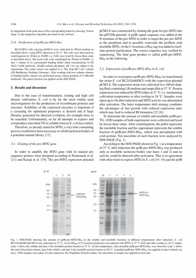
According to the SDS-PAGE shown in Fig. 1 at a temperature
of 37 ◦C after induction the pelB-pro-MTG-His6 was produced
only as insoluble inclusion bodies (see lanes 1 and 2) and no
activity could be detected after activation. This is in agreement
with other trials to express MTG in E. coli [16–18] and the pelB
Fig. 1. SDS-PAGE showing the amount of pelB-pro-MTG-His6 in the soluble and insoluble fractions at different temperatures after induction. E. coli
BL21Gold(DE3)pCM2-8 was cultivated at 37 ◦C. At an OD600 of 0.5 protein production was induced with IPTG at 37◦C (left) and after cooling to 24 ◦C (right).
Lane 1 shows the soluble and lane 2 the insoluble protein fraction at 37 ◦C. At this temperature, only insoluble pelB-pro-MTG-His6 was observed. Lane 3 shows
the total cell protein content, lane 4 the soluble, and lane 5 the insoluble protein fraction at 28 ◦C. A purified pelB-pro-MTG-His6 was applied in lane 6 (details see
text). 1/OD samples were taken 3 h after induction. M=PageRuler Protein Ladder. Ten microlitre of sample was applied in each lane.
C.K. Marx et al. / Enzyme and Microbial Technology 40 (2007) 1543–1550 1547
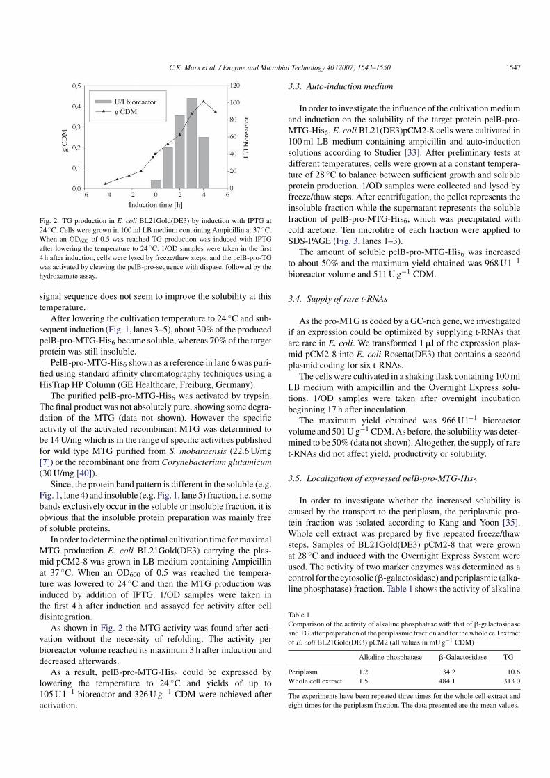
Fig. 2. TG production in E. coli BL21Gold(DE3) by induction with IPTG at
24 ◦C. Cells were grown in 100ml LB medium containing Ampicillin at 37 ◦C.
When an OD600 of 0.5 was reached TG production was induced with IPTG
after lowering the temperature to 24 ◦C. 1/OD samples were taken in the first
4 h after induction, cells were lysed by freeze/thaw steps, and the pelB-pro-TG
was activated by cleaving the pelB-pro-sequence with dispase, followed by the
hydroxamate assay.
signal sequence does not seem to improve the solubility at this
temperature.
After lowering the cultivation temperature to 24 ◦C and sub-
sequent induction (Fig. 1, lanes 3–5), about 30% of the produced
pelB-pro-MTG-His6 became soluble, whereas 70% of the target
protein was still insoluble.
PelB-pro-MTG-His6 shown as a reference in lane 6 was puri-
fied using standard affinity chromatography techniques using a
HisTrap HP Column (GE Healthcare, Freiburg, Germany).
The purified pelB-pro-MTG-His6 was activated by trypsin.
The final product was not absolutely pure, showing some degra-
dation of the MTG (data not shown). However the specific
activity of the activated recombinant MTG was determined to
be 14U/mg which is in the range of specific activities published
for wild type MTG purified from S. mobaraensis (22.6U/mg
[7]) or the recombinant one from Corynebacterium glutamicum
(30U/mg [40]).
Since, the protein band pattern is different in the soluble (e.g.
Fig. 1, lane 4) and insoluble (e.g. Fig. 1, lane 5) fraction, i.e. some
bands exclusively occur in the soluble or insoluble fraction, it is
obvious that the insoluble protein preparation was mainly free
of soluble proteins.
In order to determine the optimal cultivation time formaximal
MTG production E. coli BL21Gold(DE3) carrying the plas-
mid pCM2-8 was grown in LB medium containing Ampicillin
at 37 ◦C. When an OD600 of 0.5 was reached the tempera-
ture was lowered to 24 ◦C and then the MTG production was
induced by addition of IPTG. 1/OD samples were taken in
the first 4 h after induction and assayed for activity after cell
disintegration.
As shown in Fig. 2 the MTG activity was found after acti-
vation without the necessity of refolding. The activity per
bioreactor volume reached its maximum 3 h after induction and
decreased afterwards.
As a result, pelB-pro-MTG-His6 could be expressed by
lowering the temperature to 24 ◦C and yields of up to
105U l−1 bioreactor and 326U g−1 CDM were achieved after
activation.
3.3. Autoinduction medium
In order to investigate the influence of the cultivationmedium
and induction on the solubility of the target protein pelB-pro-
MTG-His6, E. coli BL21(DE3)pCM2-8 cells were cultivated in
100ml LB medium containing ampicillin and auto-induction
solutions according to Studier [33]. After preliminary tests at
different temperatures, cells were grown at a constant tempera-
ture of 28 ◦C to balance between sufficient growth and soluble
protein production. 1/OD samples were collected and lysed by
freeze/thaw steps. After centrifugation, the pellet represents the
insoluble fraction while the supernatant represents the soluble
fraction of pelB-pro-MTG-His6, which was precipitated with
cold acetone. Ten microlitre of each fraction were applied to
SDS-PAGE (Fig. 3, lanes 1–3).
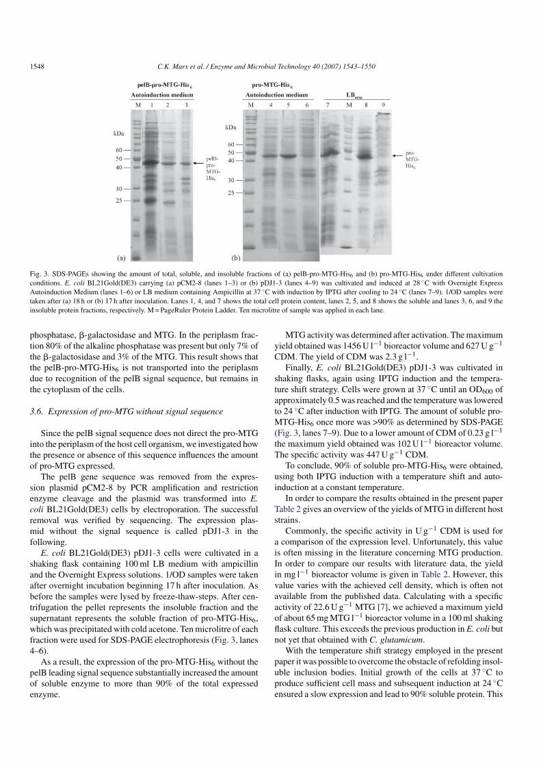
The amount of soluble pelB-pro-MTG-His6 was increased
to about 50% and the maximum yield obtained was 968U l−1
bioreactor volume and 511U g−1 CDM.
3.4. Supply of rare tRNAs
As the pro-MTG is coded by a GC-rich gene, we investigated
if an expression could be optimized by supplying t-RNAs that
are rare in E. coli. We transformed 1ml of the expression plas-
mid pCM2-8 into E. coli Rosetta(DE3) that contains a second
plasmid coding for six t-RNAs.
The cells were cultivated in a shaking flask containing 100ml
LB medium with ampicillin and the Overnight Express solu-
tions. 1/OD samples were taken after overnight incubation
beginning 17 h after inoculation.
The maximum yield obtained was 966U l−1 bioreactor
volume and 501U g−1 CDM.As before, the solubilitywas deter-
mined to be 50% (data not shown). Altogether, the supply of rare
t-RNAs did not affect yield, productivity or solubility.
3.5. Localization of expressed pelBproMTGHis6
In order to investigate whether the increased solubility is
caused by the transport to the periplasm, the periplasmic pro-
tein fraction was isolated according to Kang and Yoon [35].
Whole cell extract was prepared by five repeated freeze/thaw
steps. Samples of BL21Gold(DE3) pCM2-8 that were grown
at 28 ◦C and induced with the Overnight Express System were
used. The activity of two marker enzymes was determined as a
control for the cytosolic (b-galactosidase) andperiplasmic (alka-
line phosphatase) fraction. Table 1 shows the activity of alkaline
Table 1
Comparison of the activity of alkaline phosphatase with that of b-galactosidase
andTGafter preparation of the periplasmic fraction and for thewhole cell extract
of E. coli BL21Gold(DE3) pCM2 (all values in mUg−1 CDM)
Alkaline phosphatase b-Galactosidase TG
Periplasm 1.2 34.2 10.6
Whole cell extract 1.5 484.1 313.0
The experiments have been repeated three times for the whole cell extract and
eight times for the periplasm fraction. The data presented are the mean values.
1548 C.K. Marx et al. / Enzyme and Microbial Technology 40 (2007) 1543–1550
Fig. 3. SDS-PAGEs showing the amount of total, soluble, and insoluble fractions of (a) pelB-pro-MTG-His6 and (b) pro-MTG-His6 under different cultivation
conditions. E. coli BL21Gold(DE3) carrying (a) pCM2-8 (lanes 1–3) or (b) pDJ1-3 (lanes 4–9) was cultivated and induced at 28 ◦C with Overnight Express
Autoinduction Medium (lanes 1–6) or LB medium containing Ampicillin at 37 ◦C with induction by IPTG after cooling to 24 ◦C (lanes 7–9). 1/OD samples were
taken after (a) 18 h or (b) 17 h after inoculation. Lanes 1, 4, and 7 shows the total cell protein content, lanes 2, 5, and 8 shows the soluble and lanes 3, 6, and 9 the
insoluble protein fractions, respectively. M=PageRuler Protein Ladder. Ten microlitre of sample was applied in each lane.
phosphatase, b-galactosidase and MTG. In the periplasm frac-
tion 80% of the alkaline phosphatase was present but only 7% of
the b-galactosidase and 3% of the MTG. This result shows that
the pelB-pro-MTG-His6 is not transported into the periplasm
due to recognition of the pelB signal sequence, but remains in
the cytoplasm of the cells.
3.6. Expression of proMTG without signal sequence
Since the pelB signal sequence does not direct the pro-MTG
into the periplasm of the host cell organism, we investigated how
the presence or absence of this sequence influences the amount
of pro-MTG expressed.
The pelB gene sequence was removed from the expres-
sion plasmid pCM2-8 by PCR amplification and restriction
enzyme cleavage and the plasmid was transformed into E.
coli BL21Gold(DE3) cells by electroporation. The successful
removal was verified by sequencing. The expression plas-
mid without the signal sequence is called pDJ1-3 in the
following.
E. coli BL21Gold(DE3) pDJ1-3 cells were cultivated in a
shaking flask containing 100ml LB medium with ampicillin
and the Overnight Express solutions. 1/OD samples were taken
after overnight incubation beginning 17 h after inoculation. As
before the samples were lysed by freeze-thaw-steps. After cen-
trifugation the pellet represents the insoluble fraction and the
supernatant represents the soluble fraction of pro-MTG-His6,
whichwas precipitated with cold acetone. Tenmicrolitre of each
fraction were used for SDS-PAGE electrophoresis (Fig. 3, lanes
4–6).
As a result, the expression of the pro-MTG-His6 without the
pelB leading signal sequence substantially increased the amount
of soluble enzyme to more than 90% of the total expressed
enzyme.
MTGactivitywas determined after activation. Themaximum
yield obtained was 1456U l−1 bioreactor volume and 627U g−1
CDM. The yield of CDM was 2.3 g l−1.
Finally, E. coli BL21Gold(DE3) pDJ1-3 was cultivated in
shaking flasks, again using IPTG induction and the tempera-
ture shift strategy. Cells were grown at 37 ◦C until an OD600 of
approximately 0.5was reached and the temperaturewas lowered
to 24 ◦C after induction with IPTG. The amount of soluble pro-
MTG-His6 once more was >90% as determined by SDS-PAGE
(Fig. 3, lanes 7–9). Due to a lower amount of CDM of 0.23 g l−1
the maximum yield obtained was 102U l−1 bioreactor volume.
The specific activity was 447U g−1 CDM.
To conclude, 90% of soluble pro-MTG-His6 were obtained,
using both IPTG induction with a temperature shift and auto-
induction at a constant temperature.
In order to compare the results obtained in the present paper
Table 2 gives an overview of the yields of MTG in different host
strains.
Commonly, the specific activity in U g−1 CDM is used for
a comparison of the expression level. Unfortunately, this value
is often missing in the literature concerning MTG production.
In order to compare our results with literature data, the yield
in mg l−1 bioreactor volume is given in Table 2. However, this
value varies with the achieved cell density, which is often not
available from the published data. Calculating with a specific
activity of 22.6U g−1 MTG [7], we achieved a maximum yield
of about 65mgMTG l−1 bioreactor volume in a 100ml shaking
flask culture. This exceeds the previous production in E. coli but
not yet that obtained with C. glutamicum.
With the temperature shift strategy employed in the present
paper it was possible to overcome the obstacle of refolding insol-
uble inclusion bodies. Initial growth of the cells at 37 ◦C to
produce sufficient cell mass and subsequent induction at 24 ◦C
ensured a slow expression and lead to 90% soluble protein. This
C.K. Marx et al. / Enzyme and Microbial Technology 40 (2007) 1543–1550 1549
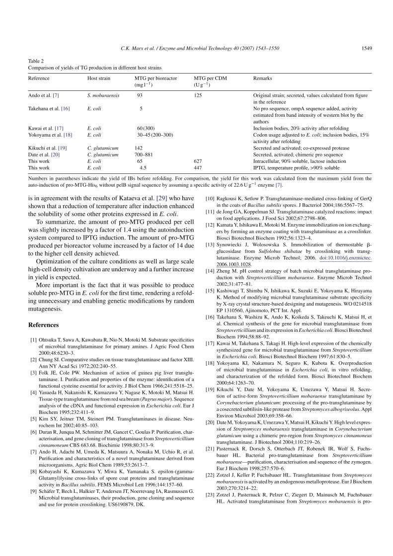
Table 2
Comparison of yields of TG production in different host strains
Reference Host strain MTG per bioreactor
(mg l−1)
MTG per CDM
(Ug−1)
Remarks
Ando et al. [7] S. mobaraensis 93 125 Original strain; secreted, values calculated from figure
in the reference
Takehana et al. [16] E. coli 5 No pro sequence, ompA sequence added, activity
estimated from band intensity of western blot by the
authors
Kawai et al. [17] E. coli 60 (300) Inclusion bodies, 20% activity after refolding
Yokoyama et al. [18] E. coli 30–45 (200–300) Codon usage adjusted to E. coli; inclusion bodies, 15%
activity after refolding
Kikuchi et al. [19] C. glutamicum 142 Secreted and activated; co-expressed protease
Date et al. [20] C. glutamicum 700–881 Secreted, activated; chimeric pro sequence
This work E. coli 65 627 Intracellular, 90% soluble, lactose induction
This work E. coli 4.5 447 IPTG, temperature profile, >90% soluble
Numbers in parentheses indicate the yield of IBs before refolding. For comparison, the yield for this work was calculated from the maximum yield from the
auto-induction of pro-MTG-His6 without pelB signal sequence by assuming a specific activity of 22.6U g−1 enzyme [7].
is in agreement with the results of Kataeva et al. [29] who have
shown that a reduction of temperature after induction enhanced
the solubility of some other proteins expressed in E. coli.
To summarize, the amount of pro-MTG produced per cell
was slightly increased by a factor of 1.4 using the autoinduction
system compared to IPTG induction. The amount of pro-MTG
produced per bioreactor volume increased by a factor of 14 due
to the higher cell density achieved.
Optimization of the culture conditions as well as large scale
high-cell density cultivation are underway and a further increase
in yield is expected.
More important is the fact that it was possible to produce
soluble pro-MTG in E. coli for the first time, rendering a refold-
ing unnecessary and enabling genetic modifications by random
mutagenesis.
References
[1] Ohtsuka T, Sawa A, Kawabata R, Nio N, Motoki M. Substrate specificities
of microbial transglutaminase for primary amines. J Agric Food Chem
2000;48:6230–3.
[2] Chung SI. Comparative studies on tissue transglutaminase and factor XIII.
Ann NY Acad Sci 1972;202:240–55.
[3] Folk JE, Cole PW. Mechanism of action of guinea pig liver transglu-
taminase. I. Purification and properties of the enzyme: identification of a
functional cysteine essential for activity. J Biol Chem 1966;241:5518–25.
[4] Yasueda H, Nakanishi K, Kumazawa Y, Nagase K, Motoki M, Matsui H.
Tissue-type transglutaminase from red sea bream (Pagrus major). Sequence
analysis of the cDNA and functional expression in Escherichia coli. Eur J
Biochem 1995;232:411–9.
[5] Kim SY, Jeitner TM, Steinert PM. Transglutaminases in disease. Neu-
rochem Int 2002;40:85–103.
[6] Duran R, Junqua M, Schmitter JM, Gancet C, Goulas P. Purification, char-
acterisation, and gene cloning of transglutaminase from Streptoverticillium
cinnamoneum CBS 683.68. Biochimie 1998;80:313–9.
[7] Ando H, Adachi M, Umeda K, Matsuura A, Nonaka M, Uchio R, et al.
Purification and characteristics of a novel transglutaminase derived from
microorganisms. Agric Biol Chem 1989;53:2613–7.
[8] Kobayashi K, Kumazawa Y, Miwa K, Yamanaka S. epsilon-(gamma-
Glutamyl)lysine cross-links of spore coat proteins and transglutaminase
activity in Bacillus subtilis. FEMS Microbiol Lett 1996;144:157–60.
[31] Qiagen. Competent cells and transformation. Bench guide; 2001:2–3.
[32] Novagen. Overnight express autoinduction system manual; 2005.
[33] Studier FW. Protein production by auto-induction in high density shaking
cultures. Protein Exp Purif 2005;41:207–34.
[34] Folk JE, Cole PW. Transglutaminase: mechanistic features of the active
site as determined by kinetic and inhibitor studies. Biochim Biophys Acta
1966;122:244–64.
[35] Kang Y, Yoon J-W. Effect of modification of connecting peptide of proin-
sulin on its export. J Biotechnol 1994;36:45–54.
[36] King EJ, Delory GE. The rates of enzymic hydrolysis of phosphoric esters.
Biochem J 1939;33:1185–90.
[37] Lederberg J. The b-d-galactosidase of Escherichia coli, strain K-12. J
Bacteriol 1950;60:381–92.
[38] Laemmli UK. Cleavage of structural proteins during the assembly of the
head of bacteriophage T4. Nature 1970;227:680–5.
[39] Kanaji T, Ozaki H, Takao T, Kawajiri H, Ide H, Motoki M, et al. Primary
structure of microbial transglutaminase from Streptoverticillium sp. strain
s-8112. J Biol Chem 1993;268:11565–72.
[40] Date M, Yokoyama K, Umezawa Y, Matsui H, Kikuchi Y. Produc-
tion of native-type Streptoverticillium mobaraense transglutaminase in
Corynebacterium glutamicum. Appl Environ Microbiol 2003;69:3011–4.
Enzyme and Microbial Technology 42 (2008) 568–575
Contents lists available at ScienceDirect
Enzyme and Microbial Technology
journa l homepage: www.e lsev ier .com/ locate /emt
Purification and activation of a recombinant histidinetaggedprotransglutaminase after soluble expression in Escherichia coli and partialcharacterization of the active enzyme
Christian K. Marx, Thomas C. Hertel, Markus Pietzsch ∗
Department of Downstream Processing, Faculty of Natural Sciences I, Institute of Pharmacy, MartinLutherUniversitat HalleWittenberg,
Weinbergweg 22, 06099 Halle (Saale), Germany
a r t i c l e i n f o
Article history:
Received 7 September 2007Received in revised form 11 January 2008Accepted 7 March 2008
Protransglutaminase from Streptomyces mobaraensis was expressed in Escherichia coli as a fusion protein carrying a Cterminal histidine tag (proMTGHis6). The recombinant organism was cultivated in 15 Lbioreactor scale and proMTGHis6 was purified by immobilized metal affinity chromatography. Activationof the inactive proenzyme using trypsin resulted in an unexpected degradation of the transglutaminaseand a concomitant loss of activity. Therefore, a set of commercially available proteases was investigatedfor their activation potential without destroying the target enzyme. Besides trypsin, chymotrypsin andproteinase K were found to activate but hydrolyze the (proMTGHis6). Cathepsin B, dispase I, and thrombin were shown to specifically hydrolyze proMTGHis6 without deactivation. TAMEP, the endogeneousprotease from S. mobaraensis was purified for comparison and also found to activate the recombinanthistidinetagged transglutaminase without degradation. The TAMEP activated MTGHis6 was purifiedand characterized. The specific activity (23 U/mg) of the recombinant histidinetagged transglutaminase,the temperature optimum (50 ◦C), and the temperature stability (t1/2 at 60 ◦C = 1.7 min) were comparable to the wildtype enzyme. A Cterminal peptide tag did neither affect the activity nor the stabilitybut facilitated the purification. The purification of the histidinetagged protein is possible before or afteractivation.
Transglutaminases (proteinglutamine gglutamyltransferase,EC 2.3.2.13, TG) are enzymes that catalyze an acyl transfer reactionbetween a gcarboxyamide group of a glutamine residue of a peptide and the «amino group of a lysine residue. This reaction leads tothe formation of an «(gglutamyl) lysinebond. Apart from lysineresidues, several other primary amines can serve as substrates [1].
Transglutaminases have been detected in eukaryotes (including amongst others the human blood coagulation factor XIIIa, thetissueTG [2], TG from the liver of guinea pig [3] and fish [4]), andprokaryotes (MTGs were found in Streptomyces [5,6], Bacillus strains[7] and in various other organisms [8]). In the food industry, micro
bial TG (MTG) from Streptomyces mobaraensis is widely used, e.g.for the crosslinking of proteins in meat and fish and to supportgelling of yogurt and cheese [9].
In vivo, toxic enzymes are often produced as inactive proenzymes. For a biotechnological application of the respectiveenzyme an activation is required, e.g. by proteolytic cleavage of theprosequence. Up to now, MTG is produced by conventional cultivation of the wildtype S. mobaraensis strain which is of GRAS statusand subsequent isolation of the secreted enzyme. Activation of thesecreted inactive preproenzyme is performed simultaneously bytwo endogenous proteases [6,10]. Recently, a Streptomyces proMTGcarrying an histidine tag (proMTGHis6) was overproduced in soluble form in Escherichia coli for the first time [11].
The first report on the activation of a (eukaryotic) protransglutaminase using a commercial protease was published in1975. Trypsin was used for the activation of Factor XIII [12].Thrombin and Factor Xa where also effective in activation whilechymotrypsin failed. Since that time several proteases have beeninvestigated for the activation of various eukaryotic transglutaminases. Dispase, a neutral protease from Bacillus polymyxa, is apowerful fibronectinase and type IV collagenase [13] and was used
C.K. Marx et al. / Enzyme and Microbial Technology 42 (2008) 568–575 569
for the first time in 1988 for the activation of skin transglutaminaseactivity of newborn mice [14]. Later, dispase, Proteinase K, trypsin,and thrombin were shown to activate protransglutaminase E fromguinea pig skin whereas chymotrypsin and Endoproteinase GluCresulted in almost no activity but showed a considerable degradation of the zymogen [15]. Several of the proteases used beforefor the activation of mammalian protransglutaminases were alsoinvestigated for the activation of microbial protransglutaminase(proMTG).
Trypsin and dispase [16], and immobilized trypsin and chymotrypsin, and free dispase I [17] were previously used for theactivation of proMTG isolated from S. mobaraensis. In contrast,trypsin and chymotrypsin were not able to activate proMTG fromStreptomyces cinnamoneum [18].
Besides the commercially available proteases, several investigations have been carried out on endogenous proteases fromStreptomyces species. Transglutaminase activating metalloprotease (TAMEP) was purified from S. mobaraensis DSM 40587and characterized [17]. This protease is activating the endogenous transglutaminase of the same microbial strain and leavinga tetrapeptide (FRAP) on the Nterminus of the MTG. This extension does not affect the specific activity of the transglutaminase.In cultures of S. mobaraensis DSM 40847, FRAPMTGase producedby TAMEP is further hydrolyzed by a tripeptidyl aminopeptidase(SMTAP) resulting in the mature wildtype transglutaminase [19].
The three proteases from Streptomyces albogriseolus were investigated for the activation of the proMTG of this strain. SAMP20showed the highest activity, but caused a gradual decrease of theMTG activity. SAMP26 did not completely process the proMTGand SAMP45 showed only half of the activity of SAMP20.
In order to optimize the production of MTG, coexpressionand cosecretion of the protease SAMP45 from S. albogriseolus
together with the proMTG from S. mobaraensis was performed inCorynebacterium glutamicum [20]. However, the protease SAMP45was degrading the MTG after prolonged times of cultivation [21,22].
In our ongoing experiments for the isolation and productionof large amounts of highly pure and active MTG we scaledupthe cultivation scale to 15 L using a bioreactor. Then, we purifiedour histidinetagged recombinant proMTGHis6 after expressionin E. coli by means of immobilized metal affinity chromatography (IMAC). From the enzymes published for the activation ofeukaryotic and/or prokaryotic MTGs we selected trypsin becauseit is commercially available in a stabilized form, which preventsautoproteolysis. So far, unwanted sidereactions by trypsin havenot been reported. Surprisingly, after applying trypsin we foundsevere hydrolysis of the (pro)MTGHis6 with concomitant decreasein activity. Since, this result was completely unexpected we carried out a screening of commercially available proteases previouslyinvestigated for their ability to activate eukaryotic or prokaryoticprotransglutaminases as well as the endogenous protease from S.
mobaraensis (TAMEP). The activated MTGHis6 was characterized.
2. Materials and methods
2.1. Materials
Unless otherwise stated all chemicals were of analytical grade and were purchased from Sigma–Aldrich, Taufkirchen, Germany. The protein marker (ProteinMolecular Weight Marker) used for SDS–PAGE was purchased from Fermentas (St.LeonRot, Germany). Material and equipment (AKTAexplorer 100) for chromatography was purchased from GE Healthcare, Freiburg, Germany.
The following commercial enzymes were used: Trypsin, TPCK treated(Sigma–Aldrich, Taufkirchen, Germany, order No T1426, lot No 026K7770),chymotrypsin (Sigma–Aldrich, Taufkirchen, Germany, order No C4129, lot No105K7670), dispase I (ICN Biomedicals, Eschwege, Germany, order No 195022, lotNo 6735E, 6.8 U/mg), Cathepsin B (Sigma–Aldrich, Taufkirchen, Germany, order NoC6286, lot No 025K7670), Thrombin (Sigma–Aldrich, Taufkirchen, Germany, orderNo T7513, lot No 115K7589 and proteinase K (Fermentas, St. LeonRot, Germany,
order No EO0491, lot No 00019680). Casein was purchased from Sigma–Aldrich(Taufkirchen, Germany, order No 22080, lot No S33356 426).
The dialysis tube had a cutoff of 25 000 Da and was purchased from Roth (SpectraPor 7, Karlsruhe, Germany). Silicone oil was purchased from Roth (Karlsruhe,Germany, order No 0865.2). Deionized water was used throughout the experiments.
2.2. Bacterial strains
S. mobaraensis DSM 40847 was purchased from the German Collectionof Microorganisms and Cell Cultures (DSMZ, Braunschweig, Germany). E. coli
BL21Gold(DE3) was purchased from Stratagene (Amsterdam, The Netherlands).
2.3. Bioreactor cultivation
The cultivation of E. coli BL21(DE3) pDJ13 was carried out in a batch processusing the autoinduction method as described previously [11].
Five milliliters of preculture I (LB medium) were inoculated by a single colonyof E. coli BL21(DE3) pDJ13. After growth for 18 h at 37 ◦C at 200 rpm, 100 mL LBmedium were inoculated with 3 mL preculture I (starting OD = 0.04) and grown for7 h at 37 ◦C and 120 rpm (rotary shaker, preculture II). Five hundred milliliters ofpreculture III (LB medium) were inoculated with preculture II resulting in a startingOD600 of 0.02 and grown overnight at 37 ◦C and 80 rpm to an OD600 of 2.8. 14.73 Lof fermentation medium (see below) were inoculated with 270 mL preculture III(starting OD = 0.05). Bioreactor cultivation was carried out using a 20 L Biostat C at atemperature of 28 ◦C, a stirrer speed of 300 rpm and an air flow rate of 4.0 standardliters per minute (slpm). During the fermentation, the pH was measured but notcontrolled. Silicone oil was used as an antifoaming agent.
After 19 h, the cells were harvested by centrifugation (4 ◦C, 6000× g, EppendorfZK 630, Hamburg, Germany). The biomass was resuspended in 0.9% NaCl solution,centrifuged again, and stored as sheets of approximately 0.5 cm thickness at−80 ◦C.From 15 L of cultivation volume, 238× g wet cell mass were harvested.
2.3.1. Cultivation medium
For the preparation of 15 L autoinduction medium 135 g trypton (final concentration 9 g/L), 67.5 g yeast extract (4.5 g/L), and 135 g NaCl (9 g/L) were dissolved in13.5 L deionized water in a 20 L Biostat C bioreactor (Braun Melsungen, Germany)and sterilized. Solutions of 7.5 g glucose, 75 g glycerol, 30 g lactose in 300 mL water,51 g KH2PO4 , 53.25 g Na2HPO4 in 750 mL water, 7.40 g MgSO4·7 H2O in 15 mL water,and 1.5 g ampicillin in 30 mL water were prepared. After separate sterilization, thesesolutions were added to the bioreactor (final concentrations were 0.5 g/L glucose,5 g/L glycerol, 2 g/L lactose, 25 mM KH2PO4 , 25 mM Na2HPO4 , 2 mM MgSO4 , and100 mg/mL ampicillin). The pH was adjusted to 7.5 using NaOH. Before inoculation,the volume was filled up with sterile water to 14.73 L.
2.4. Purification of proMTGHis6
2.4.1. Cell disintegration
Ten grams of frozen cells (E. coli BL21(DE3) pDJ13) were resuspended in 100 mLof 50 mM Tris/HCl buffer, 300 mM NaCl, 20 mM imidazole, pH 8.0, tempered to 4 ◦Cand disintegrated by three passages at >10 000 psi using a high pressure homogenizer (EmulsiFlexC5, Avestin Europe, Mannheim, Germany). After each passage, thesuspension was allowed to cool to 4 ◦C before starting the next disintegration cycle.The suspension was then centrifuged for 20 min at 10 000× g in order to get a clearsolution. The clear supernatant (crude extract, 100 mL) was applied directly to anIMAC column.
2.4.2. Affinity purification
Histidinetagged proMTG was purified by means of IMAC using the following conditions. Support: Streamline Chelating, NiNTA; column: XK 16/20; CV:30 mL; flow rate for sample application: 3 mL/min; flow rate for washing and elution:3 mL/min; binding buffer: 50 mM Tris/HCl buffer with 300 mM NaCl, 20 mM imidazole, pH 8.0; elution buffer: 50 mM Tris/HCl buffer with 300 mM NaCl, 500 mMimidazole, pH 8.0; sample: clarified lysate from E. coli containing proMTGHis6
fusion protein after high pressure homogenization and centrifugation (crudeextract); sample volume: 100 mL; fraction volume: 5 mL. After loading and washingthe column with 3 CV of binding buffer, step elution was carried out at 500 mM imidazole using the elution buffer. ProMTGHis6 containing fractions were analyzedby SDS–PAGE. Fractions were stored at 4 ◦C.
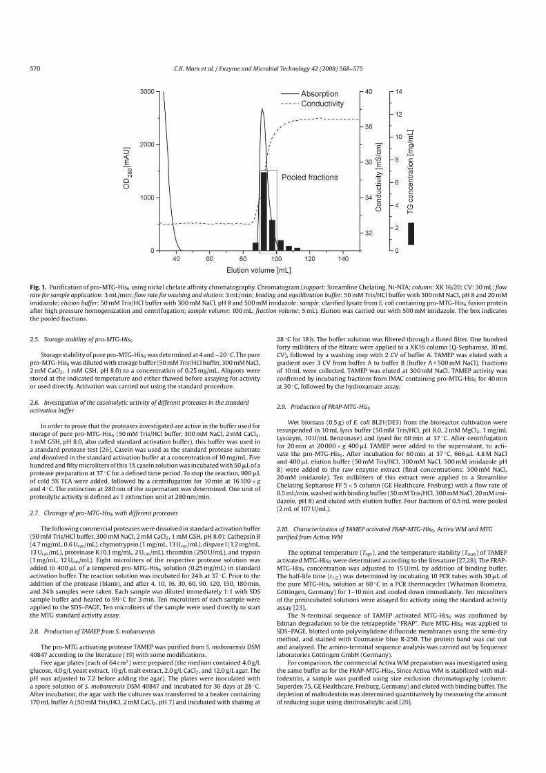
ProMTGHis6 containing fractions were pooled (9.5 mL, see Fig. 1 for details)and dialyzed twice against 1000 mL of 50 mM Tris/HCl buffer, 300 mM NaCl, 2 mMCaCl2 , pH 8.0 for 1 h each at room temperature. Thirtytwo microliters of a 300 mMGSH stock solution were added to 9.5 mL of dialysate (final concentration 1 mM).GSH was used in order to prevent oxidation of the enzyme [23]. CaCl2 was usedsince it is frequently used in order to stabilize the proteases investigated (see, e.g.trypsin [24], Proteinase K [25], TAMEP and also (pro) MTG [17]).
The dialysate (pure proMTGHis6) was stored in aliquots of 2 mL at 4 ◦C for amaximum of 4 days. The protein concentration was determined to be 4.56 g/L.
570 C.K. Marx et al. / Enzyme and Microbial Technology 42 (2008) 568–575
rate for sample application: 3 mL/min; flow rate for washing and elution: 3 mL/min; binding and equilibration buffer: 50 mM Tris/HCl buffer with 300 mM NaCl, pH 8 and 20 mMimidazole; elution buffer: 50 mM Tris/HCl buffer with 300 mM NaCl, pH 8 and 500 mM imidazole; sample: clarified lysate from E. coli containing proMTGHis6 fusion proteinafter high pressure homogenization and centrifugation; sample volume: 100 mL; fraction volume: 5 mL). Elution was carried out with 500 mM imidazole. The box indicatesthe pooled fractions.
2.5. Storage stability of proMTGHis6
Storage stability of pure proMTGHis6 was determined at 4 and−20 ◦C. The pureproMTGHis6 was diluted with storage buffer (50 mM Tris/HCl buffer, 300 mM NaCl,2 mM CaCl2 , 1 mM GSH, pH 8.0) to a concentration of 0.25 mg/mL. Aliquots werestored at the indicated temperature and either thawed before assaying for activityor used directly. Activation was carried out using the standard procedure.
2.6. Investigation of the caseinolytic activity of different proteases in the standard
activation buffer
In order to prove that the proteases investigated are active in the buffer used forstorage of pure proMTGHis6 (50 mM Tris/HCl buffer, 300 mM NaCl, 2 mM CaCl2 ,1 mM GSH, pH 8.0, also called standard activation buffer), this buffer was used ina standard protease test [26]. Casein was used as the standard protease substrateand dissolved in the standard activation buffer at a concentration of 10 mg/mL. Fivehundred and fifty microliters of this 1% casein solution was incubated with 50 mL of aprotease preparation at 37 ◦C for a defined time period. To stop the reaction, 900 mLof cold 5% TCA were added, followed by a centrifugation for 10 min at 16 100× g
and 4 ◦C. The extinction at 280 nm of the supernatant was determined. One unit ofproteolytic activity is defined as 1 extinction unit at 280 nm/min.
2.7. Cleavage of proMTGHis6 with different proteases
The following commercial proteases were dissolved in standard activation buffer(50 mM Tris/HCl buffer, 300 mM NaCl, 2 mM CaCl2 , 1 mM GSH, pH 8.0): Cathepsin B(4.7 mg/mL, 0.6 Ucas/mL), chymotrypsin (1 mg/mL, 13 Ucas/mL), dispase I (1.2 mg/mL,13 Ucas/mL), proteinase K (0.1 mg/mL, 2 Ucas/mL), thrombin (250 U/mL), and trypsin(1 mg/mL, 12 Ucas/mL). Eight microliters of the respective protease solution wasadded to 400 mL of a tempered proMTGHis6 solution (0.25 mg/mL) in standardactivation buffer. The reaction solution was incubated for 24 h at 37 ◦C. Prior to theaddition of the protease (blank), and after 4, 10, 16, 30, 60, 90, 120, 150, 180 min,and 24 h samples were taken. Each sample was diluted immediately 1:1 with SDSsample buffer and heated to 99 ◦C for 3 min. Ten microliters of each sample wereapplied to the SDS–PAGE. Ten microliters of the sample were used directly to startthe MTG standard activity assay.
2.8. Production of TAMEP from S. mobaraensis
The proMTG activating protease TAMEP was purified from S. mobaraensis DSM40847 according to the literature [19] with some modifications.
Five agar plates (each of 64 cm2) were prepared (the medium contained 4.0 g/Lglucose, 4.0 g/L yeast extract, 10 g/L malt extract, 2.0 g/L CaCl2 , and 12.0 g/L agar. ThepH was adjusted to 7.2 before adding the agar). The plates were inoculated witha spore solution of S. mobaraensis DSM 40847 and incubated for 36 days at 28 ◦C.After incubation, the agar with the cultures was transferred to a beaker containing170 mL buffer A (50 mM Tris/HCl, 2 mM CaCl2 , pH 7) and incubated with shaking at
28 ◦C for 18 h. The buffer solution was filtered through a fluted filter. One hundredforty milliliters of the filtrate were applied to a XK16 column (QSepharose, 30 mLCV), followed by a washing step with 2 CV of buffer A. TAMEP was eluted with agradient over 3 CV from buffer A to buffer B (buffer A + 500 mM NaCl). Fractionsof 10 mL were collected. TAMEP was eluted at 300 mM NaCl. TAMEP activity wasconfirmed by incubating fractions from IMAC containing proMTGHis6 for 40 minat 30 ◦C, followed by the hydroxamate assay.
2.9. Production of FRAPMTGHis6
Wet biomass (0.5 g) of E. coli BL21(DE3) from the bioreactor cultivation wereresuspended in 10 mL lysis buffer (50 mM Tris/HCl, pH 8.0, 2 mM MgCl2 , 1 mg/mLLysozym, 10 U/mL Benzonase) and lysed for 60 min at 37 ◦C. After centrifugationfor 20 min at 20 000× g 400 mL TAMEP were added to the supernatant, to activate the proMTGHis6 . After incubation for 60 min at 37 ◦C, 666 mL 4.8 M NaCland 400 mL elution buffer (50 mM Tris/HCl, 300 mM NaCl, 500 mM imidazole pH8) were added to the raw enzyme extract (final concentrations: 300 mM NaCl,20 mM imidazole). Ten milliliters of this extract were applied to a StreamlineChelating Sepharose FF 5×5 column (GE Healthcare, Freiburg) with a flow rate of0.5 mL/min, washed with binding buffer (50 mM Tris/HCl, 300 mM NaCl, 20 mM imidazole, pH 8) and eluted with elution buffer. Four fractions of 0.5 mL were pooled(2 mL of 107 U/mL).
2.10. Characterization of TAMEP activated FRAPMTGHis6 , Activa WM and MTG
purified from Activa WM
The optimal temperature (Topt), and the temperature stability (Tstab) of TAMEPactivated MTGHis6 were determined according to the literature [27,28]. The FRAPMTGHis6 concentration was adjusted to 15 U/mL by addition of binding buffer.The halflife time (t1/2) was determined by incubating 10 PCR tubes with 30 mL ofthe pure MTGHis6 solution at 60 ◦C in a PCR thermocycler (Whatman Biometra,Gottingen, Germany) for 1–10 min and cooled down immediately. Ten microlitersof the preincubated solutions were assayed for activity using the standard activityassay [23].
The Nterminal sequence of TAMEP activated MTGHis6 was confirmed byEdman degradation to be the tetrapeptide “FRAP”. Pure MTGHis6 was applied toSDS–PAGE, blotted onto polyvinylidene difluoride membranes using the semidrymethod, and stained with Coumassie blue R250. The protein band was cut outand analyzed. The aminoterminal sequence analysis was carried out by Sequencelaboratories Gottingen GmbH (Germany).
For comparison, the commercial Activa WM preparation was investigated usingthe same buffer as for the FRAPMTGHis6 . Since Activa WM is stabilized with maltodextrin, a sample was purified using size exclusion chromatography (column:Superdex 75, GE Healthcare, Freiburg, Germany) and eluted with binding buffer. Thedepletion of maltodextrin was determined quantitatively by measuring the amountof reducing sugar using dinitrosalicylic acid [29].
C.K. Marx et al. / Enzyme and Microbial Technology 42 (2008) 568–575 571
The amount of proMTGHis6 in the crude extract was calculated from the band intensity of Coomassie stained SDS–PAGE by integration of the peak area of the correspondingband and comparison with a concentration marker of purified proMTGHis6 . The concentration of the pooled fractions and of dialyzed proMTGHis6 was calculated bymeasuring the absorption at 280 nm.
For determination of the temperature optimum, FRAPMTGHis6 was dilutedwith binding buffer to a concentration of 5 U/mL. The activity was determined usingthe standard activity assay at different temperatures.
2.11. Analytical methods
The activity of MTG(His6) was assayed according to the colorimetric hydroxamate procedure [23] (MTG standard assay). Usually, the enzyme sample volumewas 10 mL and the volume of the substrate solution 140 mL. A calibration curve wasmeasured using commercially available lglutamic acid gmonohydroxamate in therange from 0 to 5 mM. One unit of MTG(His6) is defined as the formation of 1 mmol lglutamic acid gmonohydroxamate per min at 37 ◦C. The absorption was measuredusing a microtiter plate reader (Fluostar Galaxy, BMG Labtech GmbH, Offenburg,Germany).
Polyacrylamide gel electrophoresis (PAGE) was performed according to themethod of Laemmli [30] using a Mighty Small apparatus from Hoefer (AmershamBiosciences, Freiburg, Germany). 12.5% gels were stained with Coomassie Blue. Afterdestaining, the gels were dried between two cellophane foils fixed in a frame.The amount of proMTGHis6 in the crude extract was calculated from the bandintensity of Coomassie stained SDS–PAGE by integration of the peak area of thecorresponding band and comparison with a concentration marker of purified proMTGHis6 .
The protein concentration of the purified proMTGHis6 was determined bymeasuring the UVabsorption at 280 nm (ε1%
280 nm= 17.21) [31].
3. Results and discussion
3.1. Production and purification of proMTGHis6
E. coli biomass containing recombinant histidinetaggedprotransglutaminase (proMTGHis6) was produced using theovernight expression system as described previously [11].
The biomass was disintegrated by highpressure homogenization, as described in material and methods. After centrifugation, thesupernatant was applied to a Streamline NiNTA affinity columnand eluted by an linear gradient of imidazole. The elution profile isshown in Fig. 1.
ProMTGHis6 fractions containing the purified protein werepooled, dialyzed and used for the activation experiments.
The results of the purification procedure are summarized inTable 1. Some 43 mg of proMTGHis6 could be isolated in highlypure form from 100 mL crude extract prepared from 10 g cell wetmass. The yield was calculated from the band intensity of the targetprotein compared to the initial amount to be 43%.
3.2. Storage stability of proMTGHis6
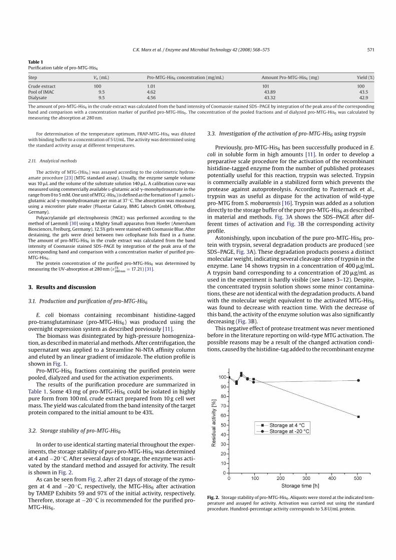
In order to use identical starting material throughout the experiments, the storage stability of pure proMTGHis6 was determinedat 4 and−20 ◦C. After several days of storage, the enzyme was activated by the standard method and assayed for activity. The resultis shown in Fig. 2.
As can be seen from Fig. 2, after 21 days of storage of the zymogen at 4 and −20 ◦C, respectively, the MTGHis6 after activationby TAMEP Exhibits 59 and 97% of the initial activity, respectively.Therefore, storage at −20 ◦C is recommended for the purified proMTGHis6.
3.3. Investigation of the activation of proMTGHis6 using trypsin
Previously, proMTGHis6 has been successfully produced in E.
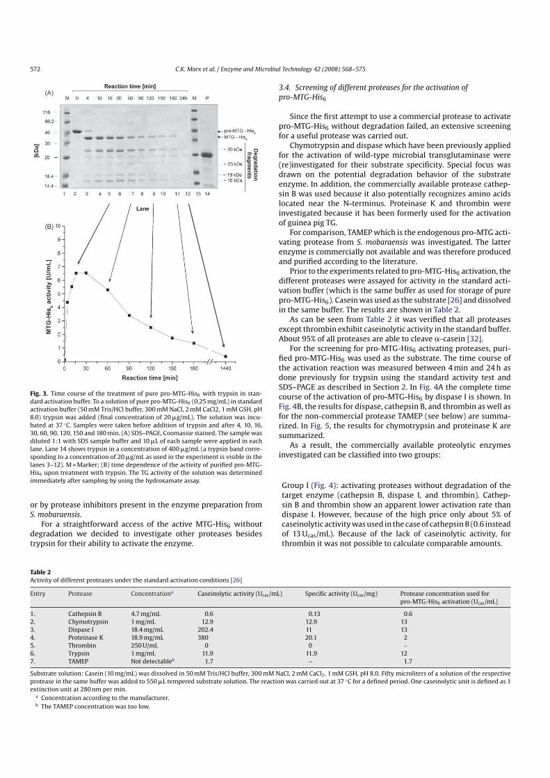
coli in soluble form in high amounts [11]. In order to develop apreparative scale procedure for the activation of the recombinanthistidinetagged enzyme from the number of published proteasespotentially useful for this reaction, trypsin was selected. Trypsinis commercially available in a stabilized form which prevents theprotease against autoproteolysis. According to Pasternack et al.,trypsin was as useful as dispase for the activation of wildtypeproMTG from S. mobaraensis [16]. Trypsin was added as a solutiondirectly to the storage buffer of the pure proMTGHis6 as describedin material and methods. Fig. 3A shows the SDS–PAGE after different times of activation and Fig. 3B the corresponding activityprofile.
Astonishingly, upon incubation of the pure proMTGHis6 protein with trypsin, several degradation products are produced (seeSDS–PAGE, Fig. 3A). These degradation products possess a distinctmolecular weight, indicating several cleavage sites of trypsin in theenzyme. Lane 14 shows trypsin in a concentration of 400 mg/mL.A trypsin band corresponding to a concentration of 20 mg/mL asused in the experiment is hardly visible (see lanes 3–12). Despite,the concentrated trypsin solution shows some minor contaminations, these are not identical with the degradation products. A bandwith the molecular weight equivalent to the activated MTGHis6
was found to decrease with reaction time. With the decrease ofthis band, the activity of the enzyme solution was also significantlydecreasing (Fig. 3B).
This negative effect of protease treatment was never mentionedbefore in the literature reporting on wildtype MTG activation. Thepossible reasons may be a result of the changed activation conditions, caused by the histidinetag added to the recombinant enzyme
Fig. 2. Storage stability of proMTGHis6 . Aliquots were stored at the indicated temperature and assayed for activity. Activation was carried out using the standardprocedure. Hundredpercentage activity corresponds to 5.8 U/mL protein.
572 C.K. Marx et al. / Enzyme and Microbial Technology 42 (2008) 568–575
Fig. 3. Time course of the treatment of pure proMTGHis6 with trypsin in standard activation buffer. To a solution of pure proMTGHis6 (0.25 mg/mL) in standardactivation buffer (50 mM Tris/HCl buffer, 300 mM NaCl, 2 mM CaCl2, 1 mM GSH, pH8.0) trypsin was added (final concentration of 20 mg/mL). The solution was incubated at 37 ◦C. Samples were taken before addition of trypsin and after 4, 10, 16,30, 60, 90, 120, 150 and 180 min. (A) SDS–PAGE, Coomassie stained. The sample wasdiluted 1:1 with SDS sample buffer and 10 mL of each sample were applied in eachlane. Lane 14 shows trypsin in a concentration of 400 mg/mL (a trypsin band corresponding to a concentration of 20 mg/mL as used in the experiment is visible in thelanes 3–12). M = Marker; (B) time dependence of the activity of purified proMTGHis6 upon treatment with trypsin. The TG activity of the solution was determinedimmediately after sampling by using the hydroxamate assay.
or by protease inhibitors present in the enzyme preparation fromS. mobaraensis.
For a straightforward access of the active MTGHis6 withoutdegradation we decided to investigate other proteases besidestrypsin for their ability to activate the enzyme.
3.4. Screening of different proteases for the activation of
proMTGHis6
Since the first attempt to use a commercial protease to activateproMTGHis6 without degradation failed, an extensive screeningfor a useful protease was carried out.
Chymotrypsin and dispase which have been previously appliedfor the activation of wildtype microbial transglutaminase were(re)investigated for their substrate specificity. Special focus wasdrawn on the potential degradation behavior of the substrateenzyme. In addition, the commercially available protease cathepsin B was used because it also potentially recognizes amino acidslocated near the Nterminus. Proteinase K and thrombin wereinvestigated because it has been formerly used for the activationof guinea pig TG.
For comparison, TAMEP which is the endogenous proMTG activating protease from S. mobaraensis was investigated. The latterenzyme is commercially not available and was therefore producedand purified according to the literature.
Prior to the experiments related to proMTGHis6 activation, thedifferent proteases were assayed for activity in the standard activation buffer (which is the same buffer as used for storage of pureproMTGHis6). Casein was used as the substrate [26] and dissolvedin the same buffer. The results are shown in Table 2.
As can be seen from Table 2 it was verified that all proteasesexcept thrombin exhibit caseinolytic activity in the standard buffer.About 95% of all proteases are able to cleave acasein [32].
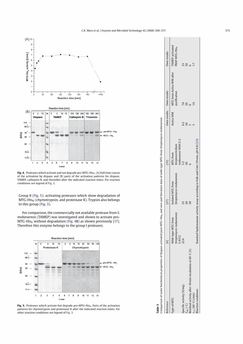
For the screening for proMTGHis6 activating proteases, purified proMTGHis6 was used as the substrate. The time course ofthe activation reaction was measured between 4 min and 24 h asdone previously for trypsin using the standard activity test andSDS–PAGE as described in Section 2. In Fig. 4A the complete timecourse of the activation of proMTGHis6 by dispase I is shown. InFig. 4B, the results for dispase, cathepsin B, and thrombin as well asfor the noncommercial protease TAMEP (see below) are summarized. In Fig. 5, the results for chymotrypsin and proteinase K aresummarized.
As a result, the commercially available proteolytic enzymesinvestigated can be classified into two groups:
Group I (Fig. 4): activating proteases without degradation of thetarget enzyme (cathepsin B, dispase I, and thrombin). Cathepsin B and thrombin show an apparent lower activation rate thandispase I. However, because of the high price only about 5% ofcaseinolytic activity was used in the case of cathepsin B (0.6 insteadof 13 Ucas/mL). Because of the lack of caseinolytic activity, forthrombin it was not possible to calculate comparable amounts.
Table 2
Activity of different proteases under the standard activation conditions [26]
Entry Protease Concentrationa Caseinolytic activity (Ucas/mL) Specific activity (Ucas/mg) Protease concentration used forproMTGHis6 activation (Ucas/mL)
Substrate solution: Casein (10 mg/mL) was dissolved in 50 mM Tris/HCl buffer, 300 mM NaCl, 2 mM CaCl2 , 1 mM GSH, pH 8.0. Fifty microliters of a solution of the respectiveprotease in the same buffer was added to 550 mL tempered substrate solution. The reaction was carried out at 37 ◦C for a defined period. One caseinolytic unit is defined as 1extinction unit at 280 nm per min.
a Concentration according to the manufacturer.b The TAMEP concentration was too low.
C.K. Marx et al. / Enzyme and Microbial Technology 42 (2008) 568–575 573
Fig. 4. Proteases which activate and not degrade proMTGHis6 . (A) Full time courseof the activation by dispase and (B) parts of the activation patterns for dispase,TAMEP, cathepsin B, and thrombin after the indicated reaction times. For reactionconditions see legend of Fig. 3.
Group II (Fig. 5): activating proteases which show degradation ofMTGHis6 (chymotrypsin, and proteinase K). Trypsin also belongsto this group (Fig. 3).
For comparison, the commercially not available protease from S.
mobaraensis (TAMEP) was investigated and shown to activate proMTGHis6 without degradation (Fig. 4B) as shown previously [17].Therefore this enzyme belongs to the group I proteases.
Fig. 5. Proteases which activate but degrade proMTGHis6 . Parts of the activationpatterns for chymotrypsin and proteinase K after the indicated reaction times. Forother reaction conditions see legend of Fig. 3. T
ab
le3
Co
mp
aris
on
of
som
eb
ioch
emic
alp
rop
erti
eso
fd
isp
ase
acti
vate
dp
ure
MT
GH
is6
and
ow
nan
dli
tera
ture
dat
ao
fw
ild
ty
pe
MT
Gfr
om
Stre
pto
myce
sm
ob
ara
ensi
s
Ref
eren
ce[6
][2
7]
[33
]O
wn
resu
lts
Ow
nre
sult
sO
wn
resu
lts
Ty
pe
of
MT
GW
ild
ty
pe
MT
Gfr
om
Stre
pto
myce
sm
ob
ara
ensi
s
S8
112
Iso
late
dM
TG
fro
mSt
rep
tom
yce
sm
ob
ara
ensi
s
MT
Gfr
om
Stre
pto
ver
tici
liu
m
mob
ara
ense
WSH
Z2
Act
iva
WM
MT
Gfr
om
Act
iva
WM
afte
rp
uri
fica
tio
nTA
ME
Pa
ctiv
ated
FRA
PM
TG
His
6
Spec
ific
acti
vit
y(U
/mg
)2
2.6
25
5.2
6.2
7.6
23
To
pt
(◦C
)5
05
05
05
05
0R
esid
ual
acti
vit
yaf
ter
10m
inin
cub
atio
nat
60◦C
(%)
50
65
06
t 1/2
(60◦C
)(m
in)
31.
61.
7R
eact
ion
con
dit
ion
sSt
and
ard
hy
dro
xam
ate
acti
vit
yas
say
acco
rdin
gto
Folk
and
Co
le(1
0m
in,p
H6
.0)
[23
]
574 C.K. Marx et al. / Enzyme and Microbial Technology 42 (2008) 568–575
Dispase and TAMEP activated MTGHis6 did not lose activity for aperiod of at least 3 h. After elevated reaction times (24 h) the activityis reduced significantly, either because of thermal deactivation ofMTGHis6 or because of insterile conditions.
Group II proteases show a specific pattern of truncated transglutaminase in SDS–PAGE. Three distinct hydrolysis bands are foundfor chymotrypsin, four for trypsin, and at least four for proteinaseK. For all proteases, the overall band intensity decreases withhydrolysis time, indicating that there is a further breakdown tosmall molecular weight peptides not visible on the SDS–PAGE. Theactivity of the transglutaminase is almost entirely depleted afterelevated reaction times.
In conclusion, trypsin cannot be used to activate the proformof the recombinant, histagged microbial transglutaminase from S.
mobaraensis. A distinct degradation pattern was found upon incubation with trypsin, as well as with chymotrypsin. Proteinase K alsoactivates but degrades the proMTGHis6 and should not be usedfor activation.
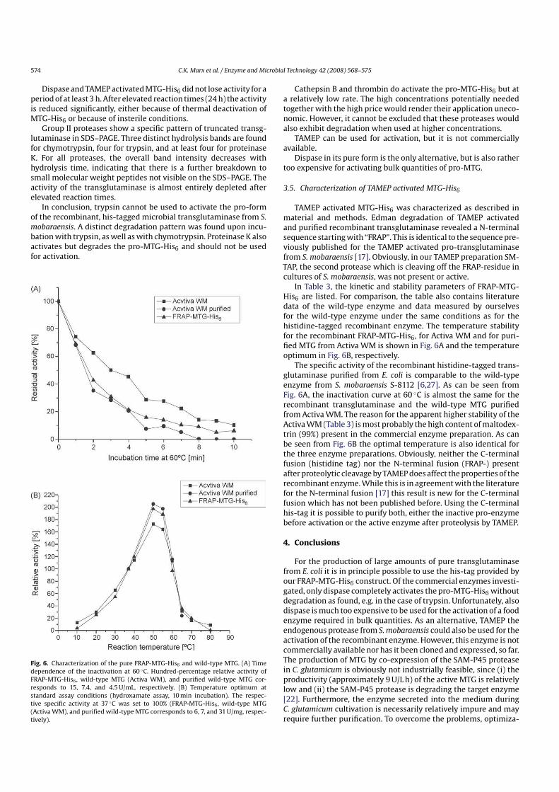
Fig. 6. Characterization of the pure FRAPMTGHis6 and wildtype MTG. (A) Timedependence of the inactivation at 60 ◦C. Hundredpercentage relative activity ofFRAPMTGHis6 , wildtype MTG (Activa WM), and purified wildtype MTG corresponds to 15, 7.4, and 4.5 U/mL, respectively. (B) Temperature optimum atstandard assay conditions (hydroxamate assay, 10 min incubation). The respective specific activity at 37 ◦C was set to 100% (FRAPMTGHis6 , wildtype MTG(Activa WM), and purified wildtype MTG corresponds to 6, 7, and 31 U/mg, respectively).
Cathepsin B and thrombin do activate the proMTGHis6 but ata relatively low rate. The high concentrations potentially neededtogether with the high price would render their application uneconomic. However, it cannot be excluded that these proteases wouldalso exhibit degradation when used at higher concentrations.
TAMEP can be used for activation, but it is not commerciallyavailable.
Dispase in its pure form is the only alternative, but is also rathertoo expensive for activating bulk quantities of proMTG.
3.5. Characterization of TAMEP activated MTGHis6
TAMEP activated MTGHis6 was characterized as described inmaterial and methods. Edman degradation of TAMEP activatedand purified recombinant transglutaminase revealed a Nterminalsequence starting with “FRAP”. This is identical to the sequence previously published for the TAMEP activated protransglutaminasefrom S. mobaraensis [17]. Obviously, in our TAMEP preparation SMTAP, the second protease which is cleaving off the FRAPresidue incultures of S. mobaraensis, was not present or active.
In Table 3, the kinetic and stability parameters of FRAPMTGHis6 are listed. For comparison, the table also contains literaturedata of the wildtype enzyme and data measured by ourselvesfor the wildtype enzyme under the same conditions as for thehistidinetagged recombinant enzyme. The temperature stabilityfor the recombinant FRAPMTGHis6, for Activa WM and for purified MTG from Activa WM is shown in Fig. 6A and the temperatureoptimum in Fig. 6B, respectively.
The specific activity of the recombinant histidinetagged transglutaminase purified from E. coli is comparable to the wildtypeenzyme from S. mobaraensis S8112 [6,27]. As can be seen fromFig. 6A, the inactivation curve at 60 ◦C is almost the same for therecombinant transglutaminase and the wildtype MTG purifiedfrom Activa WM. The reason for the apparent higher stability of theActiva WM (Table 3) is most probably the high content of maltodextrin (99%) present in the commercial enzyme preparation. As canbe seen from Fig. 6B the optimal temperature is also identical forthe three enzyme preparations. Obviously, neither the Cterminalfusion (histidine tag) nor the Nterminal fusion (FRAP) presentafter proteolytic cleavage by TAMEP does affect the properties of therecombinant enzyme. While this is in agreement with the literaturefor the Nterminal fusion [17] this result is new for the Cterminalfusion which has not been published before. Using the Cterminalhistag it is possible to purify both, either the inactive proenzymebefore activation or the active enzyme after proteolysis by TAMEP.
4. Conclusions
For the production of large amounts of pure transglutaminasefrom E. coli it is in principle possible to use the histag provided byour FRAPMTGHis6 construct. Of the commercial enzymes investigated, only dispase completely activates the proMTGHis6 withoutdegradation as found, e.g. in the case of trypsin. Unfortunately, alsodispase is much too expensive to be used for the activation of a foodenzyme required in bulk quantities. As an alternative, TAMEP theendogenous protease from S. mobaraensis could also be used for theactivation of the recombinant enzyme. However, this enzyme is notcommercially available nor has it been cloned and expressed, so far.The production of MTG by coexpression of the SAMP45 proteasein C. glutamicum is obviously not industrially feasible, since (i) theproductivity (approximately 9 U/L h) of the active MTG is relativelylow and (ii) the SAMP45 protease is degrading the target enzyme[22]. Furthermore, the enzyme secreted into the medium duringC. glutamicum cultivation is necessarily relatively impure and mayrequire further purification. To overcome the problems, optimiza
C.K. Marx et al. / Enzyme and Microbial Technology 42 (2008) 568–575 575
tion of the MTG sequence by mutating the protease cleavage sitesrecognized by trypsin or optimization of the Nterminal cleavagesite for the recognition by a highly specific protease are possible.
As a consequence, today the proteolytic activation of bulk quantities of protransglutaminase remains an open task for industrialbiotechnology.
Appendix A. Supplementary data
Supplementary data associated with this article can be found,in the online version, at 10.1016/j.enzmictec.2008.03.003.
References
[1] Ohtsuka T, Sawa A, Kawabata R, Nio N, Motoki M. Substrate specificities of microbial transglutaminase for primary amines. J Agric Food Chem2000;48:6230–3.
[2] Chung SI. Comparative studies on tissue transglutaminase and factor XIII. AnnNY Acad Sci 1972;202:240–55.
[3] Folk JE, Cole PW. Mechanism of action of guinea pig liver transglutaminase. I.Purification and properties of the enzyme: identification of a functional cysteine essential for activity. J Biol Chem 1966;241:5518–25.
[4] Yasueda H, Nakanishi K, Kumazawa Y, Nagase K, Motoki M, Matsui H. Tissuetype transglutaminase from red sea bream (Pagrus major). Sequence analysisof the cDNA and functional expression in Escherichia coli. Eur J Biochem1995;232:411–9.
[5] Duran R, Junqua M, Schmitter JM, Gancet C, Goulas P. Purification, characterisation, and gene cloning of transglutaminase from Streptoverticilliumcinnamoneum CBS 683.68. Biochimie 1998;80:313–9.
[6] Ando H, Adachi M, Umeda K, Matsuura A, Nonaka M, Uchio R, et al. Purificationand characteristics of a novel transglutaminase derived from microorganisms.Agric Biol Chem 1989;53:2613–7.
[7] Kobayashi K, Kumazawa Y, Miwa K, Yamanaka S. Epsilon(gammaGlutamyl)lysine crosslinks of spore coat proteins and transglutaminaseactivity in Bacillus subtilis. FEMS Microbiol Lett 1996;144:157–60.
[8] T Schafer, L Bech, T Halkier, JT Andersen, IA Noerrevang, G Rasmussen, Microbialtransglutaminases, their production, gene cloning and sequence and use forprotein crosslinking. 2001; US6190879, DK.
[9] de Jong GA, Koppelman SJ. Transglutaminase catalyzed reactions: impact onfood applications. J Food Sci 2002;67:2798–806.
[10] Zheng M. pH control strategy of batch microbial transglutaminase productionwith Streptoverticillium mobaraense. Enzyme Microb Technol 2002;31:477–81.
[11] Marx C, Hertel T, Pietzsch M. Soluble expression of a protransglutaminasefrom Streptomyces mobaraensis in Escherichia coli. Enzyme Microb Technol2007;40:1543–50.
[12] McDonagh J, McDonagh RP. Alternative pathways for the activation of factorXIII. Br J Haematol 1975;30:465–77.
[13] Stenn KS, Link R, Moellmann G, Madri J, Kuklinska E. Dispase, a neutral proteasefrom Bacillus polymyxa, is a powerful fibronectinase and type IV collagenase. JInvest Dermatol 1989;93:287–90.
[14] Martinet N, Kim HC, Girard JE, Nigra TP, Strong DH, Chung SI, et al. Epidermaland hair follicle transglutaminases. Partial characterization of soluble enzymesin newborn mouse skin. J Biol Chem 1988;263:4236–41.
[15] Kim HC, Lewis MS, Gorman JJ, Park SC, Girard JE, Folk JE, et al. Protransglutaminase E from guinea pig skin. Isolation and partial characterization. J Biol Chem1990;265:21971–8.
[16] Pasternack R, Dorsch S, Otterbach JT, Robenek IR, Wolf S, Fuchsbauer HL. Bacterial protransglutaminase from Streptoverticillium mobaraense—purification,characterisation and sequence of the zymogen. Eur J Biochem 1998;257:570–6.
[17] Zotzel J, Keller P, Fuchsbauer HL. Transglutaminase from Streptomycesmobaraensis is activated by an endogenous metalloprotease. Eur J Biochem2003;270:3214–22.
[18] Taguchi S, Arakawa K, Yokoyama K, Takehana S, Takagi H, Momose H. Overexpression and purification of microbial protransglutaminase from Streptomycescinnamoneum and in vitro processing by Streptomyces albogriseolus proteases.J Biosci Bioeng 2002;94:478–81.
[19] Zotzel J, Pasternack R, Pelzer C, Ziegert D, Mainusch M, Fuchsbauer HL. Activatedtransglutaminase from Streptomyces mobaraensis is processed by a tripeptidylaminopeptidase in the final step. Eur J Biochem 2003;270:4149–55.
[20] Date M, Yokoyama K, Umezawa Y, Matsui H, Kikuchi Y. High level expressionof Streptomyces mobaraensis transglutaminase in Corynebacterium glutamicumusing a chimeric proregion from Streptomyces cinnamoneus transglutaminase.J Biotechnol 2004;110:219–26.
[21] Date M, Yokoyama K, Umezawa Y, Matsui H, Kikuchi Y. Production of nativetypeStreptoverticillium mobaraense transglutaminase in Corynebacterium glutamicum. Appl Environ Microbiol 2003;69:3011–4.
[22] Kikuchi Y, Date M, Yokoyama K, Umezawa Y, Matsui H. Secretion ofactiveform Streptoverticillium mobaraense transglutaminase by Corynebacterium glutamicum: processing of the protransglutaminase by a cosecretedsubtilisinlike protease from Streptomyces albogriseolus. Appl Environ Microbiol2003;69:358–66.
[23] Folk JE, Cole PW. Transglutaminase: mechanistic features of the activesite as determined by kinetic and inhibitor studies. Biochim Biophys Acta1966;122:244–64.
[24] Schwert GW, Takenaka Y. Spectrophotometric determination of trypsin andchymotrypsin. Biochim Biophys Acta 1955;16:570–5.
[25] Bajorath J, Hinrichs W, Saenger W. The enzymic activity of proteinase K iscontrolled by calcium. Eur J Biochem 1988;176:441–7.
[26] Quiroga E, Priolo N, Marchese J, Barberis S. Behavior of araujiain, a new cysteinephytoprotease, in organic media with low water content. Electron J Biotechnol2006;9:18–25.
[27] Umezawa Y, Ohtsuka T, Yokoyama K, Nio N. Comparison of enzymatic properties of microbial transglutaminase from Streptomyces sp. Food Sci Technol Res2002;8:113–8.
[28] Lu S, Zhou N, Tian Y, Li H, Chen J. Purification and properties of transglutaminasefrom Streptoverticillium mobaraense. J Food Biochem 2002;27:109–25.
[29] Miller G. Use of dinitrosalicylic acid reagent for determination of reducingsugars. Anal Chem 1959;31:426–8.
[30] Laemmli UK. Cleavage of structural proteins during the assembly of the headof bacteriophage T4. Nature 1970;227:680–5.
[31] Pace CN, Schmid FX. How to determine the molar absorption coefficient of aprotein. Oxford: IRL Press; 1997.
[32] Kim JH, Shin HJ, Cho H, Kwak SM, Cho H, Kim TS, et al. A microfluidic proteaseactivity assay based on the detection of fluorescence polarization. Anal ChimActa 2006;577:171–7.
[33] Lu S, Zhou N, Tian Y, Li H, Chen J. Purification and properties of transglutaminasefrom Streptoverticillium mobaraense. J Food Biochem 2003;27:109–25.
Received 16 January 2008Received in revised form 9 June 2008Accepted 17 June 2008
Keywords:
Random mutagenesisRecombinant microbial transglutaminaseproMTGHis6
Streptomyces mobaraensis
High throughput screeningThermostability
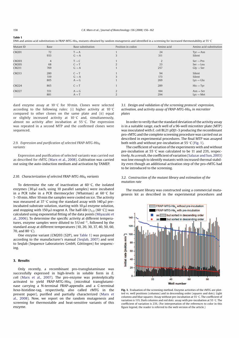
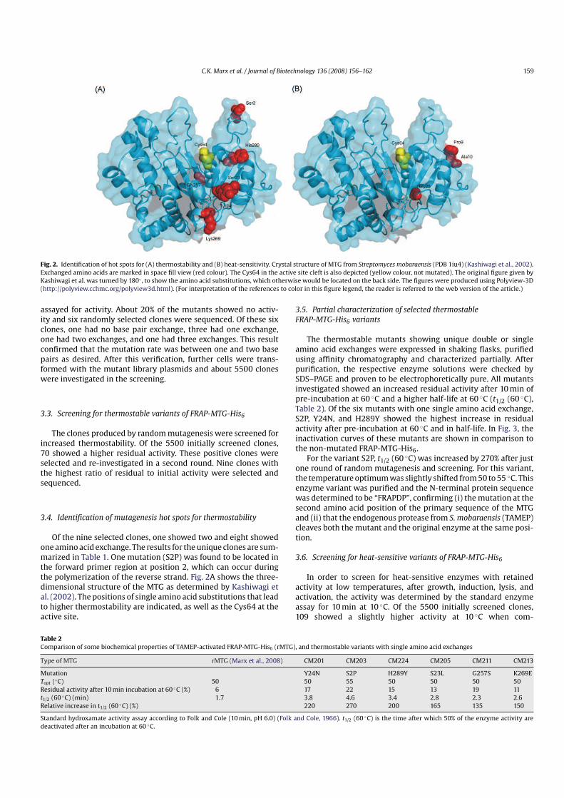
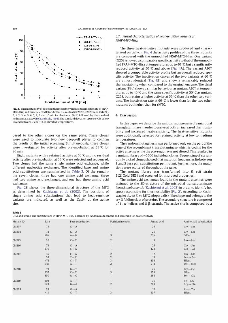
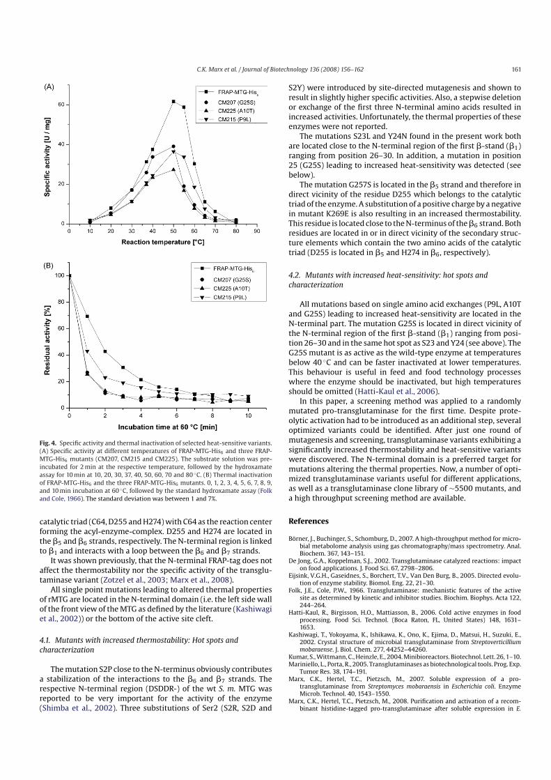
a b s t r a c t
Recombinant microbial transglutaminase (rMTG), an enzyme useful for the crosslinking or the posttranslational modification of (therapeutic) proteins, was optimized by random mutagenesis for the first time. Ascreening method was developed which, in addition to stateoftheart procedures, includes a proteolyticactivation step of the expressed soluble proenzyme. The library of 5,500 clones was screened for variantswith increased thermostability and heatsensitivity, respectively. Mutant enzymes were overproduced,isolated and characterized. After just one round of mutagenesis, nine variants with a single amino acidexchange showed a remarkably increased thermostability at 60 ◦C. The exchange of a serine residue closeto the Nterminus against proline resulted in an rMTG mutant (S2P) with 270% increased halflife. Sevenvariants exhibited an increased heatsensitivity at 60 ◦C of which one mutant (G25S) retained its specific activity between 10 and 40 ◦C. The mutations responsible for the increased thermostability and theheatsensitivity were identified and assigned to the threedimensional (3D) structure. All single pointmutations related to changed thermal properties of rMTG are located in the Nterminal domain (i.e. theleft side wall of the active site cleft of the front view of the MTG as defined by the literature [Kashiwagi,T., Yokoyama, K., Ishikawa, K., Ono, K., Ejima, D., Matsui, H., Suzuki, E., 2002. Crystal structure of microbialtransglutaminase from Streptoverticillium mobaraense. J. Biol. Chem. 277, 44252–44260] showing theimportance of this part of the protein.
Transglutaminases (proteinglutamine gglutamyltransferase,EC 2.3.2.13, TG) are enzymes that catalyze an acyl transfer reaction between a gcarboxyamide group of a glutamine residue of apeptide and the «amino group of a lysine residue.
In the food industry, microbial TG from Streptomyces mobaraen
sis (MTG) is widely used, e.g., for the crosslinking of proteins inmeat and fish and to support gelling of yogurt and cheese (DeJong and Koppelman, 2002). Other potential applications are thesitedirected covalent modification of therapeutic proteins, e.g.,the PEGylation of recombinant human interleukin 2 (Sato, 2002)and the production of proteinbased materials, for example, films(Mariniello and Porta, 2005).
In vivo, toxic enzymes as, e.g., the protein crosslinking transglutaminase are usually produced as inactive proenzymes. Forany application of the respective enzyme, activation by proteolyticcleavage of the prosequence is required.
For some applications of MTG, it would be advantageous tocarry out the crosslinking reaction at a higher temperature andwith increased activity. For this purpose, a thermostable transglutaminase is required. On the other hand, for food processing, theapplication of enzymes at low temperatures provides various benefits, including lower energy requirements, protection of substratesand products from degradation and inactivation of the modifyingenzyme by a mild increase of the temperature (HattiKaul et al.,2006). For this purpose, a heatsensitive MTG with retained activityat low temperatures is desired.
Several enzymes belonging to different enzyme classes havebeen successfully optimized with regard to thermostability (Eijsinket al., 2005).
In recent years, microtiter plates have been successfully used as“minibioreactors” for growth of clonal libraries in Escherichia coli
(Kumar et al., 2004). A consistent growth and expression is necessary to minimize welltowell variations of the enzyme level.Small variations of the initial biomass may lead to different growthbehaviour, making it difficult to fix the optimal time for induction. By using an autoinduction medium, cells are not induced atthe same time, but at the same phase of growth (Studier, 2005),thus compensating for deviations of the initial biomass. Recently,
C.K. Marx et al. / Journal of Biotechnology 136 (2008) 156–162 157
a proMTG carrying a histidine tag (proMTGHis6) was overproduced in soluble form in E. coli for the first time (Marx et al.,2007). This, together with the application of a suitable activatingprotease (Marx et al., 2008) was the basis for the development ofa microtiter plate based screening method of MTG variants produced by random mutagenesis. The aim of the present work was toprovide new transglutaminases with increased thermostability orincreased heatsensitivity.
2. Material and methods
2.1. General
Unless otherwise stated, all chemicals were of analytical gradeand were purchased from Sigma Aldrich (Taufkirchen). The protein marker (Protein Molecular Weight Marker) used for SDS–PAGEwas purchased from Fermentas (St. LeonRot). Material and equipment (Äkta Purifier 100) for chromatography was purchased fromGE Healthcare (Freiburg). The following commercial enzymes wereused: Lysozyme (order no. 8259.2, lot no. 2357473, Carl Roth GmbH& Co. KG, Karlsruhe), benzonase (order no. 1.01695.0001, lot no.K35986795 617, Merck KGaA, Darmstadt). Deionized water wasused throughout the experiments.
2.2. Bacterial strains
E. coli BL21Gold(DE3) was purchased from Stratagene (Amsterdam, The Netherlands).
2.3. Random mutagenesis of MTG
Random mutagenesis of the MTG gene was carried out usingthe GeneMorph II EZCLone Domain Mutagenesis Kit (Stratagene)according to the manufacturers manual (Stratagene, 2005). Fivehundred nanograms template DNA (MTG gene in plasmid pDJ13(Marx et al., 2007), GenBankTM accession number: EU301664) wereapplied in the errorprone PCR reaction, the annealing temperature was 60 ◦C and the cycle number was 25 (Whatman BiometraThermocycler, Göttingen). The PCR primers were derived fromsequences at the beginning and at the end of the MTG gene withoutprosequence (forwardprimer: 5′GACTCCGACGACAGGGTCACC3′, ReversePrimer: 5′GAGCGGCCAGCCCTGCTTTAC3′). The PCRproduct was purified using the MinElute Gel Extraction Kit (Qiagen,Hilden). For the EZClone reaction, 50 ng template plasmid pDJ13and 250 ng of mutated MTG gene as megaprimer were used. Afterdigestion with DpnI, the resulting plasmids containing the mutatedMTG gene were transformed into E. coli BL21Gold(DE3) (Stratagene)using electroporation as described previously (Marx et al., 2007).The transformed cells were plated on 30 agar plates (LB ampicillin,LBamp) with approximately 200 colonies each. Sequencing was carried out by MWGBiotech AG (Ebersberg).
2.4. Cultivation in microtiter plates and expression of
proMTGHis6 and its mutants
Deepwell microtiter plates (MTP, no. 0030502.310, Eppendorf)were filled with 500 ml LBamp medium (10 g l−1 tryptone/peptone,10 g l−1 NaCl, 5 g l−1 yeast extract, pH 7.5, 100 mg ml−1 ampicillin)per well. Each well was inoculated with a single colony of transformed E. coli cells using a sterile toothpick. The MTP was sealedwith two different sterile foils, first with BreathSeal (no. 676050,Greiner BioOne, Solingen), then additionally with gas permeableadhesive seal (no. AB0718, Abgene Germany, Hamburg). A combination of two foils was previously shown to be a good compromise
between low water evaporation and good growth of bacterial cultures (Börner et al., 2007). The MTP were incubated in a shakingincubator (Innova 4230, New Brunswick Scientific, Edison, USA)at 400 rpm, 37 ◦C and 2.5 cm shaking radius for 16–18 h. For protein expression, 20 ml of the culture were used to inoculate a newdeepwell MTP, containing 480 ml autoinduction medium according to Studier (Studier, 2005). This MTP was sealed as describedabove and incubated in a shaking incubator (Multitron, Infors HT,Bottmingen, Switzerland) at 300 rpm, 28 ◦C and 5 cm shaking radiusfor 24 h, followed by centrifugation (4000 rpm, 20 min. Centrifuge5403, Eppendorf). The MTP containing the pellet was stored at−80 ◦C until cell lysis.
2.5. Cell lysis and activation of proMTGHis6 and its mutants
with dispase
To lyse the cells, 300 ml of lysis buffer (50 mM Tris–HClbuffer,2 mM MgCl2, pH 8,0, 1 mg ml−1 lysozyme, and 10 U ml−1 benzonase) were added per well and the pellet was resuspended.The MTP was sealed (sealing mat, order no. 0030 127.579, Eppendorf) and incubated at 650 rpm and 37 ◦C for 1 h in a thermomixer(Eppendorf). The cell debris was sedimented by centrifugation at4000 rpm and 180 ml of the supernatant were transferred to a newMTP (no. 267245, Nunc). The prorMTG was activated with 20 mldispase (final concentration: 1 U ml−1, no. 354235, BD Biosciences,Heidelberg) at 37 ◦C for 20 min in a thermomixer.
2.6. Activity test of FRAPMTGHis6 and its mutants in MTP
The activity of rMTG was assayed according to the colorimetric hydroxamate procedure (Folk and Cole, 1966) and wasadapted to MTP format. To 50 ml of enzyme solution 90 mlsubstrate solution (final concentrations: 0.2 M Tris–HClbuffer,100 mM hydroxylamine, 10 mM reduced glutathione, 30 mM ZGlnGly, pH 6.0) were added. After incubation at 37 ◦C for10 min in a thermomixer, the reaction was stopped with 160 mlreagent A (1 vol. 3 M HCl, 1 vol. 12% trichloroacetic acid, 1 vol.5% FeCl3·6H2O (in 0.1 M HCl)). An aliquot of 200 ml was transferred to a transparent MTP (no. 701304, Brand GmbH & Co. KG,Wertheim) and the extinction was measured at 525 nm usinga microtiter plate reader (FluoStar, BMG Labtech GmbH, Offenburg). One unit of TG activity is defined as the formation of1 mmol lglutamic acid gmonohydroxamate per min at 37 ◦C. Thecoefficient of variation (CV) in the screening validation experiment was calculated by CV = standard deviation/mean×100%.Standard deviation = sqrt (sum (x−mean)2 (n−1)−1) (Salazar andSun, 2003).
2.7. Screening for thermostable FRAPMTGHis6 variants
To screen for thermostable variants of FRAPMTGHis6, one 96well MTP containing 50 ml of the activated cell supernatant wassealed with a sealing mat (order no. 276002, Nunc) and incubated at55 ◦C using a water bath. After 30 min, the MTP was cooled down inanother water bath to 20 ◦C, and afterwards the enzyme activity wasdetermined. Mutants with high residual activity were selected andused to inoculate another microtiter plate and assayed for activitywith and without preincubation at 55 ◦C. The ratio between theactivities with preincubation to the one without preincubationwas used to identify the best mutants.
2.8. Screening for heatsensitive FRAPMTGHis6 variants
To screen for heatsensitive variants of FRAPMTGHis6, 50 mlof the activated cell supernatant were assayed using the stan
158 C.K. Marx et al. / Journal of Biotechnology 136 (2008) 156–162
Table 1
DNA and amino acid substitutions in FRAPMTGHis6 mutants obtained by random mutagenesis and identified in a screening for increased thermostability at 55 ◦C
Mutant ID Base Base substitution Position in codon Amino acid Amino acid substitution
dard enzyme assay at 10 ◦C for 10 min. Clones were selectedaccording to the following rules: (i) higher activity at 10 ◦Ccompared to other clones on the same plate and (ii) equalor slightly increased activity at 10 ◦C and, simultaneously,almost no activity after incubation at 55 ◦C. The expressionwas repeated in a second MTP and the confirmed clones weresequenced.
2.9. Expression and purification of selected FRAPMTGHis6
variants
Expression and purification of selected variants was carried outas described for rMTG (Marx et al., 2008). Cultivation was carriedout using the autoinduction medium and activation by TAMEP.
2.10. Characterization of selected FRAPMTGHis6 variants
To determine the rate of inactivation at 60 ◦C, the isolatedenzymes (30 ml each, using 10 parallel samples) were incubatedin a PCR tube in a PCR thermocycler (Whatman) at 60 ◦C for1–10 min. After 10 min the samples were cooled on ice. The activitywas measured at 37 ◦C using the standard assay with 140 ml preincubated substrate solution, starting with 10 ml enzyme solution,and stopping with 150 ml reagent A. The halflife (t1/2 (60 ◦C)) wascalculated using exponential fitting of the data points (Miyazaki etal., 2006). To determine the specific activity at different temperatures, enzyme samples were diluted to 5 U ml−1, followed by thestandard assay at different temperatures (10, 20, 30, 37, 40, 50, 60,70, and 80 ◦C).
One enzyme variant (CM203 (S2P), see Table 1) was preparedaccording to the manufacturer’s manual (Seqlab, 2007) and sentto Seqlab (Sequence Laboratories GmbH, Göttingen) for sequencing.
3. Results
Only recently, a recombinant protransglutaminase wassuccessfully expressed in highlevels in soluble form in E.
coli (Marx et al., 2007). The proenzyme was proteolyticallyactivated to yield FRAPMTGHis6 (microbial transglutaminase carrying a Nterminal FRAPappendix and a Cterminalhexahistidinetag, respectively, also called rMTG in thepresent paper), purified and partially characterized (Marx etal., 2008). Now, we report on the random mutagenesis andscreening for thermostable and heatsensitive variants of thisenzyme.
3.1. Design and validation of the screening protocol: expression,
activation, and activity assay of FRAPMTGHis6 in microtiter
plates
In order to verify that the standard deviation of the activity assayis in a suitable range, each well of a 96well microtiter plate (MTP)was inoculated with E. coli BL21 pDJ13 producing the recombinantprorMTG and the complete screening procedure was carried out asdescribed in experimental procedures. The final MTP was assayedboth with and without preincubation at 55 ◦C (Fig. 1).
The coefficient of variation of the experiments with and withoutpreincubation at 55 ◦C was calculated to be 11 and 23%, respectively. As a result, the coefficient of variation (Salazar and Sun, 2003)was low enough to identify mutants with increased thermal stability even though an additional activation step of the prorMTG hadto be introduced to the screening.
3.2. Construction of the mutant library and estimation of the
mutation rate
The mutant library was constructed using a commercial mutagenesis kit as described in the experimental procedures and
Fig. 1. Evaluation of the screening method. Enzyme activities of the rMTG are plotted vs. well positions (columns) and in descending order (squares and dots). Lightcolumns and blue squares: Assay without preincubation at 55 ◦C. The coefficient ofvariation is 11%. Dark columns and red dots: assay with preincubation at 55 ◦C. Thecoefficient of variation is 23%. (For interpretation of the references to color in thisfigure legend, the reader is referred to the web version of the article.)
C.K. Marx et al. / Journal of Biotechnology 136 (2008) 156–162 159
Fig. 2. Identification of hot spots for (A) thermostability and (B) heatsensitivity. Crystal structure of MTG from Streptomyces mobaraensis (PDB 1iu4) (Kashiwagi et al., 2002).Exchanged amino acids are marked in space fill view (red colour). The Cys64 in the active site cleft is also depicted (yellow colour, not mutated). The original figure given byKashiwagi et al. was turned by 180◦ , to show the amino acid substitutions, which otherwise would be located on the back side. The figures were produced using Polyview3D(http://polyview.cchmc.org/polyview3d.html). (For interpretation of the references to color in this figure legend, the reader is referred to the web version of the article.)
assayed for activity. About 20% of the mutants showed no activity and six randomly selected clones were sequenced. Of these sixclones, one had no base pair exchange, three had one exchange,one had two exchanges, and one had three exchanges. This resultconfirmed that the mutation rate was between one and two basepairs as desired. After this verification, further cells were transformed with the mutant library plasmids and about 5500 cloneswere investigated in the screening.
3.3. Screening for thermostable variants of FRAPMTGHis6
The clones produced by random mutagenesis were screened forincreased thermostability. Of the 5500 initially screened clones,70 showed a higher residual activity. These positive clones wereselected and reinvestigated in a second round. Nine clones withthe highest ratio of residual to initial activity were selected andsequenced.
3.4. Identification of mutagenesis hot spots for thermostability
Of the nine selected clones, one showed two and eight showedone amino acid exchange. The results for the unique clones are summarized in Table 1. One mutation (S2P) was found to be located inthe forward primer region at position 2, which can occur duringthe polymerization of the reverse strand. Fig. 2A shows the threedimensional structure of the MTG as determined by Kashiwagi etal. (2002). The positions of single amino acid substitutions that leadto higher thermostability are indicated, as well as the Cys64 at theactive site.
3.5. Partial characterization of selected thermostable
FRAPMTGHis6 variants
The thermostable mutants showing unique double or singleamino acid exchanges were expressed in shaking flasks, purifiedusing affinity chromatography and characterized partially. Afterpurification, the respective enzyme solutions were checked bySDS–PAGE and proven to be electrophoretically pure. All mutantsinvestigated showed an increased residual activity after 10 min ofpreincubation at 60 ◦C and a higher halflife at 60 ◦C (t1/2 (60 ◦C),Table 2). Of the six mutants with one single amino acid exchange,S2P, Y24N, and H289Y showed the highest increase in residualactivity after preincubation at 60 ◦C and in halflife. In Fig. 3, theinactivation curves of these mutants are shown in comparison tothe nonmutated FRAPMTGHis6.
For the variant S2P, t1/2 (60 ◦C) was increased by 270% after justone round of random mutagenesis and screening. For this variant,the temperature optimum was slightly shifted from 50 to 55 ◦C. Thisenzyme variant was purified and the Nterminal protein sequencewas determined to be “FRAPDP”, confirming (i) the mutation at thesecond amino acid position of the primary sequence of the MTGand (ii) that the endogenous protease from S. mobaraensis (TAMEP)cleaves both the mutant and the original enzyme at the same position.
3.6. Screening for heatsensitive variants of FRAPMTGHis6
In order to screen for heatsensitive enzymes with retainedactivity at low temperatures, after growth, induction, lysis, andactivation, the activity was determined by the standard enzymeassay for 10 min at 10 ◦C. Of the 5500 initially screened clones,109 showed a slightly higher activity at 10 ◦C when com
Table 2
Comparison of some biochemical properties of TAMEPactivated FRAPMTGHis6 (rMTG), and thermostable variants with single amino acid exchanges
Type of MTG rMTG (Marx et al., 2008) CM201 CM203 CM224 CM205 CM211 CM213
Standard hydroxamate activity assay according to Folk and Cole (10 min, pH 6.0) (Folk and Cole, 1966). t1/2 (60 ◦C) is the time after which 50% of the enzyme activity aredeactivated after an incubation at 60 ◦C.
160 C.K. Marx et al. / Journal of Biotechnology 136 (2008) 156–162
Fig. 3. Thermostability of selected thermostable variants: thermostability of FRAPMTGHis6 and three selected FRAPMTGHis6 mutants (CM201, CM203 and CM224).0, 1, 2, 3, 4, 5, 6, 7, 8, 9 and 10 min incubation at 60 ◦C, followed by the standardhydroxamate assay (Folk and Cole, 1966). The standard deviation up to 60 ◦C is below6% and between 7 and 11% at elevated temperatures.
pared to the other clones on the same plate. These cloneswere used to inoculate two new deepwell plates to confirmthe results of the initial screening. Simultaneously, these cloneswere investigated for activity after preincubation at 55 ◦C for30 min.
Eight mutants with a retained activity at 10 ◦C and no residualactivity after preincubation at 55 ◦C were selected and sequenced.Two clones had the same single amino acid exchange, whiledifferent nucleotide exchanges. The identified base and aminoacid substitutions are summarized in Table 3. Of the remaining seven clones, three had one amino acid exchange, threehad two amino acid exchanges, and one had three amino acidexchanges.
Fig. 2B shows the threedimensional structure of the MTGas determined by Kashiwagi et al. (2002). The positions ofsingle amino acid substitutions that lead to heatsensitivevariants are indicated, as well as the Cys64 at the activesite.
3.7. Partial characterization of heatsensitive variants of
FRAPMTGHis6
The three heatsensitive mutants were produced and characterized partially. In Fig. 4 the activity profiles of the three mutantsare compared with the unmodified FRAPMTGHis6. One variant(G25S) showed a comparable specific activity to that of the unmodified FRAPMTGHis6 at temperatures up to 40 ◦C, but a significantlyreduced activity at 50 ◦C and above (Fig. 4A). The variant A10Tshowed a comparable activity profile but an overall reduced specific activity. The inactivation curves of the two variants at 60 ◦Care almost identical (Fig. 4B) and show a remarkably reducedthermostability when compared to the original enzyme. The thirdvariant (P9L) shows a similar behaviour as mutant A10T at temperatures up to 40 ◦C and the same specific activity at 50 ◦C as mutantG25S, but retains a higher activity at 55 ◦C than the other two variants. The inactivation rate at 60 ◦C is lower than for the two othermutants but higher than for rMTG.
4. Discussion
In this paper, we describe the random mutagenesis of a microbialtransglutaminase in order to arrive at both an increased thermostability and increased heatsensitivity. The heatsensitive mutantswere additionally selected for retained activity at low to mediumtemperatures.
The random mutagenesis was performed only on the part of thegene of the recombinant transglutaminase which is coding for theactive enzyme while the proregion was not altered. This resulted ina mutant library of∼5500 individual clones. Sequencing of six randomly picked clones showed that mutation frequencies lie between1 and 3 base pair substitutions per mutant. Furthermore, the mutations were scattered throughout the gene.
The mutant library was transformed into E. coli strainBL21Gold(DE3) and screened for improved properties.
The amino acid exchanges found in the mutant enzymes wereassigned to the 3Dstructure of the microbial transglutaminasefrom S. mobaraensis (Kashiwagi et al., 2002) in order to identify hotspots responsible for thermostability (Fig. 2). According to Kashiwagi et al., wt S. m. MTG adopts a disklike shape and belongs to thea + b folding class of proteins. The secondary structure is composedof 11 ahelices and 8 bstrands. The active site is composed by a
Table 3
DNA and amino acid substitutions in FRAPMTGHis6 obtained by random mutagenesis and screening for heat sensitivity
Mutant ID Base Base substitution Position in codon Amino acid Amino acid substitution
C.K. Marx et al. / Journal of Biotechnology 136 (2008) 156–162 161
Fig. 4. Specific activity and thermal inactivation of selected heatsensitive variants.(A) Specific activity at different temperatures of FRAPMTGHis6 and three FRAPMTGHis6 mutants (CM207, CM215 and CM225). The substrate solution was preincubated for 2 min at the respective temperature, followed by the hydroxamateassay for 10 min at 10, 20, 30, 37, 40, 50, 60, 70 and 80 ◦C. (B) Thermal inactivationof FRAPMTGHis6 and the three FRAPMTGHis6 mutants. 0, 1, 2, 3, 4, 5, 6, 7, 8, 9,and 10 min incubation at 60 ◦C, followed by the standard hydroxamate assay (Folkand Cole, 1966). The standard deviation was between 1 and 7%.
catalytic triad (C64, D255 and H274) with C64 as the reaction centerforming the acylenzymecomplex. D255 and H274 are located inthe b5 and b6 strands, respectively. The Nterminal region is linkedto b1 and interacts with a loop between the b6 and b7 strands.
It was shown previously, that the Nterminal FRAPtag does notaffect the thermostability nor the specific activity of the transglutaminase variant (Zotzel et al., 2003; Marx et al., 2008).
All single point mutations leading to altered thermal propertiesof rMTG are located in the Nterminal domain (i.e. the left side wallof the front view of the MTG as defined by the literature (Kashiwagiet al., 2002)) or the bottom of the active site cleft.
4.1. Mutants with increased thermostability: Hot spots and
characterization
The mutation S2P close to the Nterminus obviously contributesa stabilization of the interactions to the b6 and b7 strands. Therespective Nterminal region (DSDDR) of the wt S. m. MTG wasreported to be very important for the activity of the enzyme(Shimba et al., 2002). Three substitutions of Ser2 (S2R, S2D and
S2Y) were introduced by sitedirected mutagenesis and shown toresult in slightly higher specific activities. Also, a stepwise deletionor exchange of the first three Nterminal amino acids resulted inincreased activities. Unfortunately, the thermal properties of theseenzymes were not reported.
The mutations S23L and Y24N found in the present work bothare located close to the Nterminal region of the first bstand (b1)ranging from position 26–30. In addition, a mutation in position25 (G25S) leading to increased heatsensitivity was detected (seebelow).
The mutation G257S is located in the b5 strand and therefore indirect vicinity of the residue D255 which belongs to the catalytictriad of the enzyme. A substitution of a positive charge by a negativein mutant K269E is also resulting in an increased thermostability.This residue is located close to the Nterminus of the b6 strand. Bothresidues are located in or in direct vicinity of the secondary structure elements which contain the two amino acids of the catalytictriad (D255 is located in b5 and H274 in b6, respectively).
4.2. Mutants with increased heatsensitivity: hot spots and
characterization
All mutations based on single amino acid exchanges (P9L, A10Tand G25S) leading to increased heatsensitivity are located in theNterminal part. The mutation G25S is located in direct vicinity ofthe Nterminal region of the first bstand (b1) ranging from position 26–30 and in the same hot spot as S23 and Y24 (see above). TheG25S mutant is as active as the wildtype enzyme at temperaturesbelow 40 ◦C and can be faster inactivated at lower temperatures.This behaviour is useful in feed and food technology processeswhere the enzyme should be inactivated, but high temperaturesshould be omitted (HattiKaul et al., 2006).
In this paper, a screening method was applied to a randomlymutated protransglutaminase for the first time. Despite proteolytic activation had to be introduced as an additional step, severaloptimized variants could be identified. After just one round ofmutagenesis and screening, transglutaminase variants exhibiting asignificantly increased thermostability and heatsensitive variantswere discovered. The Nterminal domain is a preferred target formutations altering the thermal properties. Now, a number of optimized transglutaminase variants useful for different applications,as well as a transglutaminase clone library of ∼5500 mutants, anda high throughput screening method are available.
References
Börner, J., Buchinger, S., Schomburg, D., 2007. A highthroughput method for microbial metabolome analysis using gas chromatography/mass spectrometry. Anal.Biochem. 367, 143–151.
De Jong, G.A., Koppelman, S.J., 2002. Transglutaminase catalyzed reactions: impacton food applications. J. Food Sci. 67, 2798–2806.
Eijsink, V.G.H., Gaseidnes, S., Borchert, T.V., Van Den Burg, B., 2005. Directed evolution of enzyme stability. Biomol. Eng. 22, 21–30.
Folk, J.E., Cole, P.W., 1966. Transglutaminase: mechanistic features of the activesite as determined by kinetic and inhibitor studies. Biochim. Biophys. Acta 122,244–264.
HattiKaul, R., Birgisson, H.O., Mattiasson, B., 2006. Cold active enzymes in foodprocessing. Food Sci. Technol. (Boca Raton, FL, United States) 148, 1631–1653.
Kashiwagi, T., Yokoyama, K., Ishikawa, K., Ono, K., Ejima, D., Matsui, H., Suzuki, E.,2002. Crystal structure of microbial transglutaminase from Streptoverticillium
mobaraense. J. Biol. Chem. 277, 44252–44260.Kumar, S., Wittmann, C., Heinzle, E., 2004. Minibioreactors. Biotechnol. Lett. 26, 1–10.Mariniello, L., Porta, R., 2005. Transglutaminases as biotechnological tools. Prog. Exp.
Tumor Res. 38, 174–191.Marx, C.K., Hertel, T.C., Pietzsch, M., 2007. Soluble expression of a pro
transglutaminase from Streptomyces mobaraensis in Escherichia coli. EnzymeMicrob. Technol. 40, 1543–1550.
Marx, C.K., Hertel, T.C., Pietzsch, M., 2008. Purification and activation of a recombinant histidinetagged protransglutaminase after soluble expression in E.
162 C.K. Marx et al. / Journal of Biotechnology 136 (2008) 156–162
coli and characterization of the active enzyme. Enzyme Microb. Technol. 42,568–575.
Miyazaki, K., Takenouchi, M., Kondo, H., Noro, N., Suzuki, M., Tsuda, S., 2006. Thermalstabilization of Bacillus subtilis family11 xylanase by directed evolution. J. Biol.Chem. 281, 10236–10242.
Salazar, O., Sun, L., 2003. Evaluating a screen and analysis of mutant libraries. Methods Mol. Biol. 230, 85–97.
Sato, H., 2002. Enzymatic procedure for sitespecific pegylation of proteins. Adv.Drug Deliv. Rev. 54, 487–504.
Seqlab, 2007. Preparation of samples for Nterminal protein sequencing. Manufacturer’s Manual, www.seqlab.de.
Shimba, N., Shinohara, M., Yokoyama, K., Kashiwagi, T., Ishikawa, K., Ejima, D., Suzuki,E., 2002. Enhancement of transglutaminase activity by NMR identification of itsflexible residues affecting the active site. FEBS Lett. 517, 175–179.
Stratagene, 2005. GeneMorph II EZClone Domain Mutagenesis Kit. Manufacturer’sManual.
Studier, F.W., 2005. Protein production by autoinduction in high density shakingcultures. Protein Expr. Purif. 41, 207–234.
Zotzel, J., Pasternack, R., Pelzer, C., Ziegert, D., Mainusch, M., Fuchsbauer, H.L.,2003. Activated transglutaminase from Streptomyces mobaraensis is processedby a tripeptidyl aminopeptidase in the final step. Eur. J. Biochem. 270, 4149–4155.